- prescription · rubin and dixon: modern ankle-foot orthoses prescription procedures for...
TRANSCRIPT

THE MODERN ANKLE-FOOT ORTHOSES (AFO'S)
Gustav Rubin, M.D., F.A.C.S. Consultant in Orthopedics
Senior Physician, Veterans Administration
Malcolm Dixon, M.A., R.P.T. Research Physical Therapist
Veterans Administration Prosthetics Center 252 Seventh Avenue, New York, N. Y. 10001
INTRODUCTION
There is a strong trend away from the use of metal in the fabrication of orthoses. Advanced concepts and the availability of plastics have frequently resulted in wholly new designs of lower-limb orthoses rather than part-for-part substitution of plastic components for metal compo- nents. Many centers, including our own Veterans Administration Pros- thetics Center (VAPC), are replacing the ugly, noisy, and bulky double- bar metal braces with light, cosmetic plastic orthoses (Fig. 1).
The versatility of plastics allows for sufficient functional variety to be incorporated in the construction of the orthoses, so that each type of orthosis can be assigned a fairly clearly demarcated area of clinical appli- cation (see Appendix). In this article each of the ankle-foot orthoses (AFO's) most frequently used at the VAPC will be individualJy-.d' cussed and its use in clinical practice outlined. %a
THE VAPC SHOE-CLASP ORTHOSIS
This is a posterior leaf spring dropfoot orthosis which clips on to the counter of a shoe (1) and is therefore interchangeable from shoe to shoe (Fig. 2). The upright is fabricated from a fiber glass-epoxy material which provides adequate spring resistance on plantar flexion of the foot: The ankle plantar flexes on heel strike and is returned to the neutral position for swing through. This orthosis has a very efficient toe pick-up action. The proximal end of the upright slides beneath a keeper attached to a polypropylene cuff. The piston action, which might other- wise be transferred to the skin, is harmlessly expended in this manner. A single .pin joins the distal end of the upright to the clip. The distal upright fits into a shallow well, and the pin allows the upright to swivel

Rubin and Dixon: Modern Ankle-Foot Orthoses
PRESCRIPTION PROCEDURES FOR AFO's
ETIOLOGY I PAlHOLOGY MODIFYIN; FACTORS PRESCRIPTION - VAPC SHOE CUSP I . LOUCR MOTOR NEURON t FUCCID PES EQUINUS
DEFECT (PERONUL N . )
FUCCIO PES EQUINUS STABLE* - VAPC SHOE CUSP (WITH CALF MUSCLE
mm-) < UNSTABLE* 4 ~ ~ ~ ~ ~ o p m E N E
2 . LOUER MOTOR NEURON IRW SPIRIL ORTHOSIS-IF
DEFECT (SCIATIC N.) BIUIERIL INVOLVEWEM. IHEN-
FLACCID PES EQUINO- STABILITY NOT A CALCNtEUS (UITHOUI FACTOR SINCE IEUFEL OR POLYETWENE
CALF MUSCLE c ~ R I C T w ) ~ :E;iB:: ~~~~~~s POLYPROPYLENE FABRICATED
- TO RESIST LORSIFLEXION * N O PUNTAR FLEXION
MIL- t ;gC SHOE CUSP
3. UPPER MOTOR NEURON SPASTIC PES EQUINUS MOD.m TEUFEL-IF NOT AOEQWE, THEN-
DEFECT POLIPROPYLENE
SEVERE- -k ~ E Q m $ ~ ~ ~ ~ ~ l ~ :gEw (SHOE AITACWNI') BRICE
EDQU OF FCO-ANKLE VMC SINCLE MR ROTATIMI OITHOSIS (FUCCIO)
MUBLE BAR BRACE IF
(REQUIRING T-STW) SWECI IS WEWEICUI OR n a y m l v r
r . P A I N F ~ omnucrxv r ('On- PAIN MI AP OR ra + P O L W ~ ~ I Y E LITHOSIS
DISUSE OF ANKLE TRIUUTIC. INFECTIOUS, MODIFIED TO RESTRICT M S I - 1wrmToP.r . mc.) FLEXION AND PMAR FLEXION
6. .)STRUCTmL IN- a)NON-LWICN OR DEIAYED TISSUE IENUTH THE ADEQUACY DISTAL TO THE KNEE
ATINC THE PRESSURES OF PARTIAL mEICMINOi FOR EUPLE, SENSAIIMI
MI UElCUI BURINC
+ s t a b i l i t y i s : a. cva lu t ed during rci.1 of a stock brace (VMC shoe elasp, Tcufel. Polnropylane) on the pat ient by rhe Cllnic Team, or , b. can be ass-d by the nature of the Lerrain the subject tm). walk upon (field., golf courses, crc.).
++ ~ v l y p.timts with s d a ~ i c new. i n ju r i e s develop calf conrr.crure. suff icient t o st.bilire tha ankle .r about 90". In the r e i f i t bearing position. mesa o ~ f i m r s need only corcsction for the f laccid pea quinu.. - curins the c l i n i c re- cvallurion of ortho.=., the degree of .PasLic~t7< *- % + .=late6 ro rhe "Lrig&erinl" of spast ic q u i n u s (or quino-varus) by the stock brace. tested dlrecrly on the pa r imr a. part of the cv.llufion proeedur.. lor e lmple . i f the 'rock shoe elasp c r i ~ ~ r s the foot Into sp.sLic . ~ u i n u s , one m s c fv the stock Teufel or f i na l ly rhe stock ~ o ~ y p r o p y ~ m m . ~f the foot defonm within rhe'~ol).propyle:e. external (.hoe .rr.chmr) br.cin8 1. required. very severe spas t i c i t y cannot be control l rd by a brace.
mr Most such patients rill tolerare a prop-rly f i r r ed shoe in se r t brace. or a shoe clasp. Ihosc .+to develop .re*. of i r r i t a t i o n should be chmled to external bracing with individumlized shoe modifications, i f indicated.
FIGURE 1.-VAPC prescription procedures for AFO's. This chart was prepared by the authors in association with Mr. Anthony Staros, Director, VA Prosthetics Center. Functional Electrical Stimulation (FES) is not in general use and should still be considered experimental.
medially and laterally about 5 deg. The walls of the well act as a stop. This arrangement introduces subtalar motion of a sufficient degree to approximate the physiological range used during normal walking (6 deg.) (2). As the extremity moves into the swing phase of gait, the heel drops into slight varus (6 deg.) which is eliminated in stance at foot-flat. The

Bulletin of Prosthetics Research-Spring 1973
FIGURE 2.-a. The VAPC shoe-clasp orthosis. Note the keeper, which retains the sliding proximal end of the upright, the distal pin, and the shoe clasp. Note also that the heel of the shoe shows wear on its posterior-lateral aspect where heel strike should normally occur. b. Note the varus during swing-through. Following heel strike the foot will move into the foot-flat position, eliminating the varus.
normal foot strikes the ground, in slight varus, on the postero-lateral border of the heel, and then moves into the foot-flat position as th%e,nter of gravity is carried forward. The retention of this motion by the orthosis contributes to a more normal gait pattern than is the case when this movement is restricted.
In the VAPC Clinic Team facilities, we have available several stock shoe-clasp orthoses. I t is a part of our procedure to test a stock orthosis on a patient before issuing a definitive prescription. With this tool we can evaluate the usefulness of the orthosis for a specific patient at the time of the initial examination. We have found the simple and inex- pensive shoe-clasp orthosis to be extremely useful, not only for the patient with a flaccid dropfoot, but for the patient with mild spastic equinus, and even for certain patients with complete sciatic nerve paralysis. Most of the latter group of patients develop calf contractures which are sufficient to prevent ankle dorsiflexion on weight-bearing. The shoe-clasp orthosis will bring these feet to the neutral position (with the slight physiological varus referred to above during swing-through) (1). If, occasionally, the shoe-clasp orthosis cannot be used on a patient

Rubin and Dixon: Modern Ankle-Foot Orthoses
because of uncorrectable heel irritation, then the Teufel polyethylene (Ortholen) a orthosis would be the second choice. Also, if ankle instabil- ity or "triggered" clonus are manifested when the patient is observed by the clinic team during the preliminary evaluation with a stock shoe- clasp orthosis, then another type of AFO must be employed.
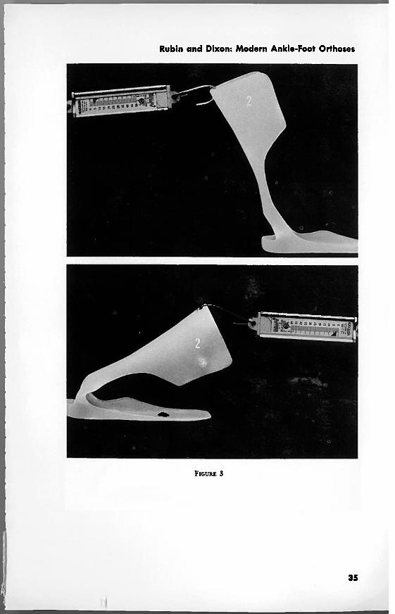
THE TEUFEL "ORTHOLEN8' ORTHOSIS
The Teufel brace (Fig. 3) is a shoe-insert orthosis which should be worn with a laced oxford shoe or chukka boot. This is a dropfoot brace which provides greater ankle stability than does the shoe-clasp orthosis. I t is commercially available in three oversized blanks of Ortholen, a thermoplastic material which can also be cold-molded with an ordinary hammer. Subtalar motion is not permitted upon heel strike. Plantar flexion is allowed by virtue of the flexibility of the upright material (Fig. 4), and dorsiflexion occurs as the foot passes into mid-stance and beyond. The flexibility of the Ortholen contributes to push-off. This orthosis is cosmetic, simple, and efficient. We use it for patients who have mild ankle instability with peroneal nerve lesions and flaccid dropfoot, who have sciatic nerve lesions with plantar flexion contrac- tures as described above, or who have hemiplegias with mild-to-moderate spasticity. I t should be noted that the choice of the Teufel over the shoe-clasp orthosis would be dictated by 1. ankle instability of mild degree or 2. mild-temoderate ankle spasticity with "triggering" into spastic equinus by the shoe-clasp orthosis during the clinic trial (see Fig. 1). If either situation is observed during trial with the shoe-clasp orthosis, our clinic team will then fit a stock Teufel orthosis to the patient. If the spasticity is of such a degree that it overcomes the resist- ance to plantar flexion of the Teufel orthosis, or if the s$rrn@esistance of the Teufel orthosis "triggers" ankle spasticity, then the next choice must be an AFO with a plantar flexion stop.
THE POLYPROPYLENE ORTHOSIS
The polypropylene orthosis is another in the series of plastic shoe- insert AFO's. The shoe insert and upright are fabricated of polypropy- lene (Fig. 5). The orthosis can be constructed to provide stop resistance to plantar flexion. This is accomplished by retaining adequate width of the X-crossing of the upright, and by closing the posterior opening at the heel. If the lower portions of the X are narrowed and the posterior heel opening retained, the orthosis will permit plantar flexion (of less range than the Teufel orthosis), and dorsiflexion. Since plantar flexion
Manufactured by Wilh. J. Teufel. Stuttgart, West Germany.

Bulletin of Prosthetics Research-Spring 1973
FIGURE 3.-The Teufel "Ortholen" or- FIGURE (.-Note the flexibility of the thosis. Teufel orthosis. T h e stability (and,
therefore, the flexibility of the upright) can be varied for the individual patient and is dependent upon the amount of ortholen material removed by the or- thotist.
is somewhat limited,b a patient who takes a long stride should not be given this orthosis. Under such circumstances, if the X-crossing of the upright is not sturdy enough it will bend at that point (Fig. 6), intro- ducing an area of potential irritation.
At our clinic, our primary indication for this orthosis is a moderate spastic equino-varus deformity not controlled by the Teufel orthosis. If there is diminution of foot sensation, or foot deformity, an 'iniie&le or other lining can be placed on the shoe insert portion of the orthosis (Fig. 7).
We also use the polypropylene orthosis for ankle immobilization as, for example, in traumatic arthritis of the ankle. This is accomplished by closing the posterior opening of the polypropylene orthosis and ex- tending the polypropylene anterior to the malleoli. This modification is used to restrict stresses on the ankle joint.
The orthotist's ability to control the degree of flexibility of this orthosis allows for a wide range of application. At the VAPC we use the polypropylene orthosis, with a toe filler, as a Chopart amputation prosthesis (Fig. 8) (3).
Plantar flexion range can be increased at the expense of stability by progressive narrowing of the lower portions of the X-crossing. The authors prefer to avoid this.
24

Rubin and Dixon: Modern Ankle-Foot Orthoses
When the orthosis and foot are placed in the shoe, there will be a snug fit of heel to orthosis. I t is important to flare out the borders of the posterior heel opening to prevent irritation. If the posterior opening is closed and the lower portions of the X-crossing are broad- ened to extend to the malleoli, this or- thosis will immobilize the ankle. If the lower portions of the X-crossing remain as shown in the illustration but are narrowed to a greater degree, the range of plantar flexion can be increased.
FIGURE 6.-Note the effect of excessive thinning of the polypropylene at the X-crossing in the case of the long-striding patient. Such a patient will develop an area of ini- tation at the point of angulation.

Bulletin of Prosthetics Research-Spring 1973
FIGURE 7.-Manner of fitting the poiy- propylene orthosis to the foot of the FIGURE 8.-The polypropylene orthosis patient who requires an innermold for fabricate to restrict ankle motion. This shortening of the extremity and plantar illustration alsd shows its use for a foot problems. Chopart amputation.
When used for the spastic foot, care must be taken to flare out the leading edges of the polypropylene in the region of the ankle to avoid marginal pressure if the foot should deform at times within the brace.
When the patient has edema of the foot which could produce pressure areas against the rigid margins of a shoe-insert orthosis, or when the patient has a degree of spasticity which is not controlled by the poly- propylene orthosis, then the shoe insert approach to bracing mutg,be bypassed.
THE VAPC SINGLE-BAR BRACE
If the patient has foot edema and is not overweight or unusually active, a single-bar orthosis should be used. This orthosis may also be used for the patient who has greater than a moderate degree of spasticity, and whose foot deforms within the shoe,^ but is able to ambulate. When very severe spasticity is present no AFO will maintain correction.
In the case of the patient with spasticity the rotation element of the single-bar orthosis should be eliminated. When rotation is retained, as in the flaccid paralytic, the rotatory movement of the upright should be limited to 10 deg., since the concomitant range of subtalar motion is
'If a varus or valgus correction strap is required, a bar will be needed as an at- tachment point.
26

Rubin and Dixon: Modern Ankle-Foot Orthoses
FIGURE 9.-The VAPC single-bar rotation brace. Markers have been fixed to the brace to illustrate the occurrence of rotation.
6 deg.; i.e., on subtalar joint inversion of 6 deg. the tibia externally r e tates 10 deg. (4) (Fig. 9 a and b). Because the orthosis is attached to the shoe stirrup, the subtalar motion occurs, to a limited degree, within the shoe at the shoe-foot "false joint." The VAPC single-bar rotation brace allows for the retention of some of this normal motion. Overweight or excessively active patients could subject this orthosis to stresses that a single-bar orthosis cannot tolerate; under such circumstances another orthosis may be substituted, such as the double-bar orthosis.
THE IRM (INSTITUTE OF REHABILITATION MEDICINE) SPIRAL ORTHOSIS %.
This AFO (Fig. lo), like the Teufel orthosis, provides spring resistance to plantar flexion and dorsiflexion. I t is fabricated of an acrylic-nylon material. This interesting orthosis was originally conceived as permitting tibial rotation. However, this is a shoe-insert orthosis with the insert component attached to an upright, and, since the corollary of tibial rotation is subtalar motion, it was recognized that the design of the orthosis necessarily limits subtalar motion (and the associated tibial rotation). This AFO is useful for the patient who has a totally flaccid foot which can be freely dorsiflexed and plantar-flexed beyond the neutral position. We do not use the IRM Spiral Orthosis if the patient has a plantar-flexion contracture, limiting dorsiflexion to the neutral or 5 deg. beyond the neutral position. With the knee fully extended, most peripheral-nerve-injured patients have just such a contracture and can use the simpler, less expensive, shoe-clasp, Teufel, or polypropylene

Bulletin of Prosthetics Research-Spring 1973
FIGURE 10.-The IRM spiral orthosis. Note that the orthotist fabricates this orthosis to avoid tibial crest pressure.
orthoses. Because of the increased frequency of breakage at the ankle- joint level, the spiral brace is not indicated for the bilaterally involved patient. The VAPC procedure of pre-prescription clinic team trial with a stock shoe clasp, a stock Teufel, or a stock polypropylene orthosis, is very helpful in determining the final prescription. Our clinical use of the IRM Spiral Orthosis is, in fact, very infrequent.
THE VAPC PTB ORTHOSlS
If pain or structural inadequacy on weight-bearing exists .am& the vertical load must be reduced, the VAPC PTB orthosis is the AFO of choice. This has been fabricated so as to make use of the weight-bearing areas employed by the below-knee amputee to carry weight from the knee to the floor, thereby bypassing the anatomical structures below the knee. The prototype of this orthosis was designed by the senior author (5) (Fig. 1 l), and the first VAPC PTB orthosis was fabricated in 1958 (6). I t was subsequently used for a variety of below-knee unweighting prob- lems, such as non-union of tibial fractures, disease of the foot and ankle, painful arthritis of the ankle, and severe deformities of the foot and ankle which interfere significantly with weight-bearing (7).
A major advance in the principles introduced by the PTB orthosis was proposed and developed by Sarmiento (8). I-Ie was the first to use these principles for the acute fracture, whereas PTB unweighting had been used for old, established tibial non-union prior to his contribution. In 1963, a brief 5 years after the initial VAPC report, Sarmiento began

Rubin and Dixon: Modem Ankle-Foot Orthosec
FIGURE 11.-a. The VAPC PTB orthosis. This patient had a fracture dislocation of the. ankle with secondary traumatic arthritis. b. X-ray of ankle of PTB orthosis wearer.
to apply this procedure to the acute fracture and this approach is now widely accepted.
I t should be clarified that the term "PTB" is an over-simplification. No patient, amputee or brace wearer, carries his superincumbent weight solely on the patellar tendon. Weight is transmitted to the tissues, in varying degrees, throughout the entire PTB cuff contact area. The major portion of the weight is carried on those areas "wh;clFbhave the greatest tolerance for pressure. A significant one of these areas is the patellar tendon, and, therefore, the designation "PTB" was employed.
SUMMARY
An AFO should interfere with existing normal functions to the least possible extent, while providing maximum correction of the presenting gait abnormality. T o accomplish this purpose it is desirable to retain as much joint motion as possible, not only in the ankle but also in the subtalar joint, without, at the same time, permitting the incursion of undesirable features such as instability.
In most simple peroneal nerve paralyses, this goal is ideally reached by the VAPC Shoe-Clasp Orthosis, and to a lesser extent by the VAPC Single-Bar spring-loaded rotation orthosis, which permit plantar flexion and dorsiflexion as well as varying degrees of subtalar motion.

Bulletin of Prosthetics Research-Spring 1973
Anterior-posterior instability, when present, can be controlled by other available orthoses with plantar flexion or dorsiflexion stops or fixed ankles, as in the case of the double-bar and single-bar AFO's and the modified polypropylene orthosis; and by resistance to both dorsi- flexion and plantar flexion, as in the case of the IRM spiral orthosis. Many patients with flail ankles develop calf contractures sufficient to stabilize the foot on weight-bearing, so that it is only necessary to add a dropfoot orthosis to enable such a patient to walk.
Medio-lateral instability can be partially controlled by the Teufel orthosis, and more rigidly controlled by the polypropylene, the IRM Spiral, and the single- and double-bar orthoses.
CONCLUSION
The authors have presented their approach to the problem of routine ankle-foot bracing as used at the Veterans Administration Prosthetics Center. Unusual, individual problems will necessarily require individual- ized solutions.
REFERENCES
1. Greenbaum. Werner: Draft Manual, VAPC Equinus-Control Ankle Foot Shoe- Clasp Orthosis, 1971.
2. Peizer, Edward, Donald W. Wright, and Carl Mason: Human Locomotion. Bull. Prosthetics Res., BPR 10-12:48-105, Fall 1969.
3. Rubin, Gustav and Michael Danisi: A Functional Partial-Foot Prosthesis. ISPO Bull., 35-7, July 1972.
4. Rubin, Gustav: Tibial Rotation. Bull. Prosthetics Res., BPR 10-15:95-101, Spring 1971. " 'I*
5. Staros, Anthony and Edward Peizer: Application of the Veterans ~dmin~strai ion Prosthetics Center Below-Knee Weight-Bearing Brace to a Bilateral Case. Artif. Limbs, 9(1):3544, 1965 (footnote 4, p. 35).
6. McIlmurray, William J. and Werner Greenbaum: A Below-Knee Weight-Bearing Brace. Orthop. & Pros. Appl. J., 12(2):81-82, June 1958.
7. Kay, Hector and Heidi Vorchheimer: A Survey of Eight Wearers of the Veterans Administration Prosthetics Center Patellar Tendon-Bearing Brace. Prosthetic and Orthotic Studies Research Division, School of Engineering and Science, New York University, New York, July 1965.
8. Sarmiento, Augusto and William Sinclair: Tibial and Femoral Fractures, Bracing Management. University of Miami, School of Medicine, Department of Ortho- paedics and Rehabilitation, Miami, Florida 33152.

Rubin and Dixon: Modern Ankle-Foot Orthoses
APPENDIX
MODIFICATION OF POLYETHYLENE AND POLYPROPYLENE ORTHOSES IN RELATION TO FUNCTION (FIG. $1)
FIGURE 1.-Modified polyethylene and polypropylene orthoses.
31

Bulletin of Prosthetics Research-Spring 1973
Polypropylene Orthosis No. 1 (Fig. 2)
This orthosis is fabricated without heel cut-out, and leading edges are trimmed posterior to the malleoli. Does not allow as great a range of plantar flexion-dorsiflexion as Polypropylene Orthosis No. 2 (compare scale recordings). Application: Indicated for the same pathology as in Polyethylene
(Teufel-Ortholen) Orthosis No. 3, below.

Rubin and Dixon: Modern Ankle-Foot Orthoses

Bulletin of Prosthetics Research-Spring 1973
Polypropylene Orthosis No. 2 (Fig. 3)
This orthosis is fabricated with the heel cut-out, and leading edges are carried to just behind the malleoli. Allows plantar flexiondorsiflexion proportional to the degree of thinning of the lower bands, while, at the same time, provides medio-lateral ankle stability.
Afiplication: Indicated for moderately unstable flaccid pes equinus.
Rubin and Dixon: Modern Ankle-Foot Orthoses

Bulletin of Prosthetics Research-.-Spring 1973
Polyethylene (Teufel-Ortholen) Orthosir No. 3 (Fig. 4)
This orthosis permits plantar flexion-dorsiflexion proportional to the degree of thinning of the posterior upright. When inserted in a shoe, it provides support for the mildly unstable ankle. Because of the thickness of the ortholen, an ortho-inlay shoe prescribed with the orthosis will simplify fitting. The inlay on the orthosis side is removed, an adequate heel buildup is added, and the need for mismated shoe sizes is eliminated.
Application: Flaccid pes equinus manifesting mild medio-lateral ankle instability.

Rubin and Dixon: Modern Ankle-Foot Orthoses

Bulletin of Prosthetics Research-Spring '1979
Polyethylene (Teufel-Ortholen) Orthosis No. 4 (Fig. 5)
Retention of maximum thickness of the posterior upright increases resistance of this orthosis to plantar flexiondorsiflexion. (Note the scale recordings for orthoses no. 3 and no. 4, Fig. 4 and 5.)
Application: a. Moderately unstable spastic pes equinus b. Flaccid pes equino-calcaneus (see Rx Procedures Chart)

Rubin and Dixon: Modern Ankle-Foot Orthoses

Bulletin of Prosthetics Research-Spring '1973
Polypropylene Orthosis No. 5 (Fig. 6)
In this orthosis the leading edges are brought forward to the malleoli, and the posterior opening has been eliminated. This orthosis completely restricts plantar flexion-dorsiflexion and medio-lateral motion.
Application: a. Flaccid pes equino-calcaneus b. Painful destructive disease of the ankle. The applications listed above should be coordinated with the "Pre-
scription Procedures for AFO's" chart (Fig. 1 in the main body of this article). The scale measurements are all gross and only approximately comparable bnt, nevertheless, are sufficient for clinical considerations.

Rubin and Dixon: Modern Ankle-Foot Orthoses