monitor respond process constantly scanning internal and external conditions it scans the big...
TRANSCRIPT

Nervous System
Nervous System Functions Monitor
Respond
Process
Constantly scanning internal and external conditions
It scans the big picture but only pays attention to the unusual or potentially harmful stimuli.
It compares the internal to its normal standard and responds only when needed to regain homeostasis
Monitoring Brain

Integration - the process of evaluating and interpreting sensory input and deciding on the response.
Information is channeled through the brain stem where it is evaluated based on prior experience and emotion.
The frontal lobe gathers information from other areas of the brain to make a decision on the appropriate response.
Assessing Brain
Acting on the information generated by the stimulus the brain stimulates a motor response or glands to cause organs to regain homeostasis.
Neurotransmitters cause muscles to move or glands to release hormones.
Responsive Brain


Two Structural Divisions of the Nervous System
Central Nervous System (CNS)
The brain and Spinal Cord
Peripheral Nervous System (PNS)
Nerves carrying messages from the CNS to organs.

Afferent or Sensory Division carries information into the brain
Efferent or Motor division carries messages from the CNS to effector organs, muscles, and glands. It has two divisions.
a The somatic nervous system provides control of our muscles. It is often called the voluntary nervous system.
b. The autonomic nervous system regulates smooth and cardiac muscles and glands. It is sometimes called the involuntary nervous system.
It has two divisions – 1. the sympathetic and 2. the parasympathetic nervous
system.
Two Functional Divisions of the Peripheral Nervous System

.

Nervous Tissue – Two Groups
Nerves
Sensory fibers from skin, muscles, and joints are called somatic sensory nerves.
Sensory fibers from the visceral organs are referred to as visceral sensory fibers.
Neuroglia
Support, protect, and insulate nerves.
Astrocytes – anchor nerves in place and are a barrier to protect them from harmful substances
Microglial – Clean up dead or damaged cells
Ependymal – circulate spinal fluid
Oligodendrocytes- Schwann cells, wrap and insulate the nerve with fat


Cell body Contain extraordinary amounts of mitochondria because
the brain uses more energy than any other part of the body.
In the CNS they are called nucleiIn the PNS they are called ganglia
Schwann Cells- Omega 3 Fatty AcidsAxon
It sends nerve impulses away from the cell body. Axons branch into 100's of axonal terminals at the end.Bundles in the CNS are called tractsBundles in the PNS are called nerves
Dendrites Each neuron may have 100's of dendrites.They carry nerve impulses toward the cell body.
White Matter – Myelinated tractsGrey Matter – Unmyelinated nerve fibers
Nerve Structure


Neurons Also called nerves
send and receive impulses
Length varies from microscopic up to four feet (from the spine to the big toe)
Excitable – respond to stimuli
Conductive – transmit impulses

Most nerve fibers are covered by a fatty insulation known as myelin.
a. Myelin protects, insulates,
increases the rate of nerve transmission.
b. In the PNS, myelin is produced by Schwann cells.
These cells wrap around the axons and enclose them in several layers of myelin.
The cell membranes of the Schwann cells is called the neurilemma.
c. Tiny gaps between Schwann cells are called Nodes of Ranvier.

Three Functional Nerve Groups Afferent Neurons carry sensory impulses from
the body to the CNS.The cell bodies are always found in ganglia outside the CNS.Their dendrite endings are usually associated with special
receptors that react to certain changes. a. Ex. pain receptors, proprioceptors
Visceral(internal) and Somatic (to the environment) Efferent Neurons carry motor impulses from
the CNS to the body. Their nerve cell bodies are always in the CNS
Association/Interneuron provide connections between afferent and efferent neurons.

1. Reflexes are rapid, predictable, and involuntary responses to stimuli. 2. The nerve pathways that produce reflexes are called reflex
pathways.
3. There are two types of reflexes in the body - autonomic and somatic a. Autonomic reflexes regulate the activity of smooth muscles,
glands, and the heart. b. Somatic reflexes stimulate skeletal muscles.
Reflex Arc


http://ed.ted.com/lessons/how-do-nerves-work

Steps in Nerve Impulse Transmission 1. Stimulus makes cell membrane permeable to Na+ ions. 2. Na+ ions diffuse into the neuron changing the membrane potential 3. Cell membrane is depolarized and an action potential is generated. 4. Cell membrane becomes impermeable to Na+ ions. 5. Cell membrane becomes permeable to K+ ions. 6. K+ ions diffuse out of the cell. 7. Cell membrane is repolarized. 8. Normal concentration of Na+ ions and K+ ions reestablished by Na-K pump.


Arteries of the Brain
The human brain requires a constant supply of oxygen. A lack of oxygen of just a few minutes results in irreversible damage to the brain.

The Cerebrum
The largest portion of the brain is the cerebrum. It consists of two hemispheres that are connected together at the corpus callosum.
The cerebrum is often divided into five lobes that are responsible for different brain functions.
Corpus callosum

Lobes of the Cerebrum
Parietal Lobe
Temporal Lobe
Frontal Lobe
Limbic Lobe
Occipital Lobe

Frontal LobeThe frontal lobe is the area of the brain responsible for higher cognitive functions.
These include:
• Problem solving• Spontaneity• Memory• Language• Motivation• Judgment• Impulse control• Social and sexual behavior.

Temporal LobeThe temporal lobe plays a role in emotions, and is also responsible for smelling, tasting, perception, memory, understanding music, aggressiveness, and sexual behavior.
The temporal lobe also contains the language area of the brain.
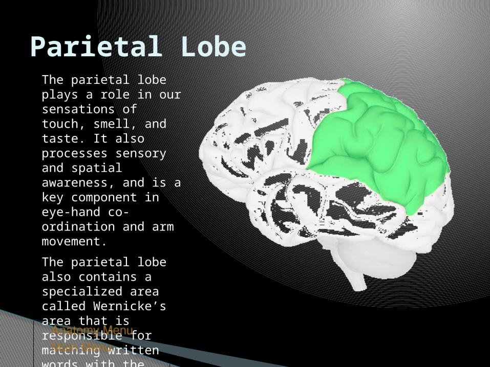
Parietal LobeThe parietal lobe plays a role in our sensations of touch, smell, and taste. It also processes sensory and spatial awareness, and is a key component in eye-hand co-ordination and arm movement.
The parietal lobe also contains a specialized area called Wernicke’s area that is responsible for matching written words with the sound of spoken speech.

Occipital LobeThe occipital lobe is at the rear of the brain and controls vision and recognition.

The Limbic System
A. Cingulate gyrusB. FornixC. Anterior thalamic
nucleiD. HypothalamusE. Amygdaloid nucleusF. Hippocampus
The limbic system is the area of the brain that regulates emotion and memory. It directly connects the lower and higher brain functions.

CerebellumThe cerebellum is connected to the brainstem, and is the center for body movement and balance.
Click image to play or pause video

ThalamusThalamus means “inner room” in Greek, as it sits deep in the brain at the top of the brainstem.
The thalamus is called the gateway to the cerebral cortex, as nearly all sensory inputs pass through it to the higher levels of the brain.

HypothalamusThe hypothalamus sits under the thalamus at the top of the brainstem. Although the hypothalamus is small, it controls many critical bodily functions:
• Controls autonomic nervous system
• Center for emotional response and behavior
• Regulates body temperature
• Regulates food intake
• Regulates water balance and thirst
• Controls sleep-wake cycles
• Controls endocrine system
The hypothalamus is shaded blue. The pituitary gland extends from the hypothalamus.

The Medulla OblongataThe medulla oblongata merges seamlessly with the spinal cord and creates the base of the brainstem.
The medulla is primarily a control center for vital involuntary reflexes such as swallowing, vomiting, sneezing, coughing, and regulation of cardiovascular and respiratory activity.
The medulla is also the origin of many cranial nerves.

The PonsThe pons is the rounded brainstem region between the midbrain and the medulla oblongata. In fact, pons means “bridge” in Latin.
The main function of the pons is to connect the cerebellum to the rest of the brain and to modify the respiratory output of the medulla.
The pons is the origin of several cranial nerves.

The Ventricles
Click image to play or pause video
The ventricles are a complex series of spaces and tunnels through the center of the brain.
The ventricles secrete cerebrospinal fluid, which suspends the brain in the skull.
The ventricles also provide a route for chemical messengers that are widely distributed through the central nervous system.

Cerebrospinal FluidCerebrospinal fluid is a colorless liquid that bathes the brain and spine.
It is formed within the ventricles of the brain, and it circulates throughout the central nervous system.
Cerebrospinal fluid fills the ventricles and meninges, allowing the brain to “float” within the skull.
Click image to play or pause video

The BrainstemThe brainstem is the most primitive part of the brain and controls the basic functions of life: breathing, heart rate, swallowing, reflexes to sight or sound, sweating, blood pressure, sleep, and balance.
The brainstem can be divided into three major sections.
Detailed brainstem anatomy.
Click image to play or pause video
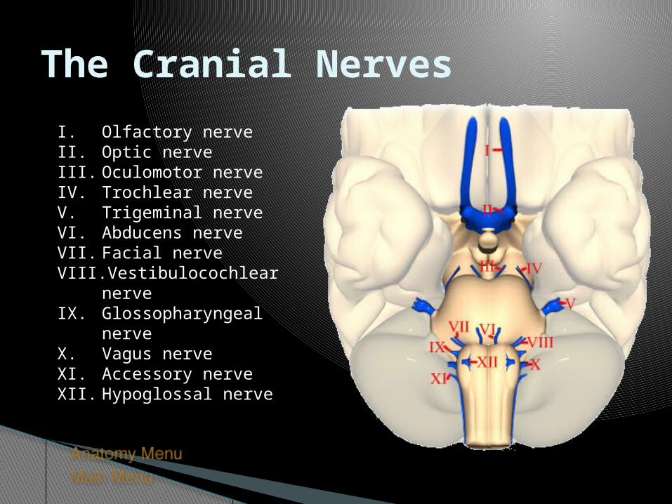
The Cranial Nerves
I. Olfactory nerveII. Optic nerveIII. Oculomotor nerveIV. Trochlear nerveV. Trigeminal nerveVI. Abducens nerveVII. Facial nerveVIII. Vestibulocochlear nerveIX. Glossopharyngeal nerveX. Vagus nerveXI. Accessory nerveXII. Hypoglossal nerve

Injury MechanismsThe brain is a complex and delicate organ, and one that is vulnerable to injury from a variety of different traumas. These include:
Frontal Lobe Injury
Occipital Lobe Injury
Temporal Lobe Injury
Side Impact Injury
Coup/Contre-coup Injury
Diffuse Axonal Injury
Epidural Hematoma
Subdural Hematoma

Frontal Lobe Injury
Click image to play or pause video
The frontal lobe of the brain can be injured from direct impact on the front of the head.
During impact, the brain tissue is accelerated forward into the bony skull. This can cause bruising of the brain tissue and tearing of blood vessels.
Frontal lobe injuries can cause changes in personality, as well as many different kinds of disturbances in cognition and memory.

Occipital Lobe Injury
Click image to play or pause video
Occipital lobe injuries occur from blows to the back of the head.
This can cause bruising of the brain tissue and tearing of blood vessels.
These injuries can result in vision problems or even blindness.

Temporal Lobe Injury
Click image to play or pause video
The temporal lobe of the brain is vulnerable to injury from impacts of the front of the head.
The temporal lobe lies upon the bony ridges of the inside of the skull, and rapid acceleration can cause the brain tissue to smash into the bone, causing tissue damage or bleeding.

Side Impact Injury
Click image to play or pause video
Injuries to the right or left side of the brain can occur from injuries to the side of the head.
Injuries to this part of the brain can result in language or speech difficulties, and sensory or motor problems.

Coup/Contre-coup Injury
Click image to play or pause video
A French phrase that describes bruises that occur at two sites in the brain.
When the head is struck, the impact causes the brain to bump the opposite side of the skull. Damage occurs at the area of impact and on the opposite side of the brain.

Diffuse Axonal InjuryBrain injury does not require a direct head impact. During rapid acceleration of the head, some parts of the brain can move separately from other parts. This type of motion creates shear forces that can destroy axons necessary for brain functioning.
These shear forces can stretch the nerve bundles of the brain.
More on diffuse axonal injury.Click image to play or pause video

Diffuse Axonal InjuryThe brain is a complex network of interconnections. Critical nerve tracts can be sheared and stressed during an acceleration-type of injury.
Diffuse axonal injury is a very serious injury, as it directly impacts the major pathways of the brain.

Epidural Hematoma
Click image to play or pause video
An epidural hematoma is a blood clot that forms between the skull and the top lining of the brain (dura).
This blood clot can cause fast changes in the pressure inside the brain.
When the brain tissue is compressed, it can quickly result in compromised blood flow and neuron damage.

Subdural Hematoma
Click image to play or pause video
A subdural hematoma is a blood clot that forms between the dura and the brain tissue.
The clot may cause increased pressure and may need to be removed surgically.
When the brain tissue is compressed, it can quickly result in compromised blood flow and tissue damage.
Brain Functions• Vision• Taste• Cognition• Emotion• Speech• Language• Hearing• Motor Cortex• Sensory Cortex• Autonomic Functions

VisionThe visual cortex resides in the occipital lobe of the brain.
Sensory impulses travel from the eyes via the optic nerve to the visual cortex.
Damage to the visual cortex can result in blindness.
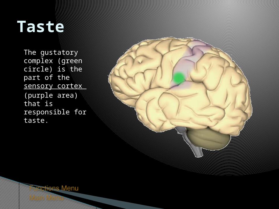
TasteThe gustatory complex (green circle) is the part of the sensory cortex (purple area) that is responsible for taste.
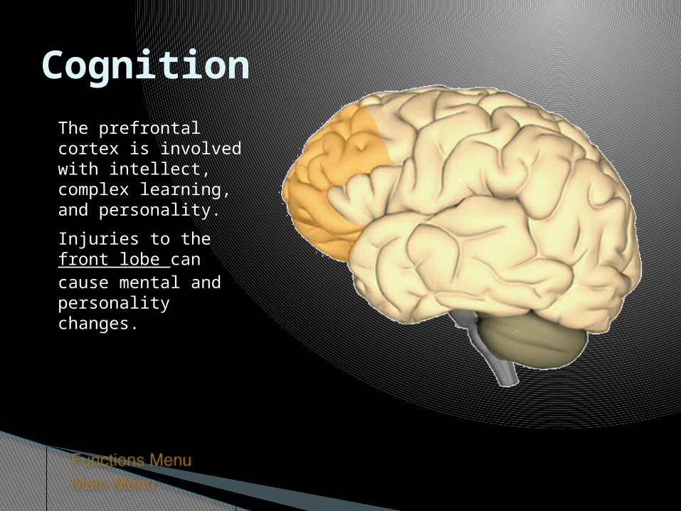
CognitionThe prefrontal cortex is involved with intellect, complex learning, and personality.
Injuries to the front lobe can cause mental and personality changes.

EmotionEmotions are an extremely complex brain function. The emotional core of the brain is the limbic system. This is where senses and awareness are first processed in the brain.
Mood and personality are mediated through the prefrontal cortex. This part of the brain is the center of higher cognitive and emotional functions.
Prefrontal cortex
Limbic system

SpeechBroca’s area is where we formulate speech and the area of the brain that sends motor instructions to the motor cortex.
Injury to Broca’s area can cause difficulty in speaking. The individual may know what words he or she wishes to speak, but will be unable to do so.
Broca’s Area

LanguageWernicke’s area is a specialized portion of the parietal lobe that recognizes and understands written and spoken language.
Wernicke’s area surrounds the auditory association area.
Damage to this part of the brain can result in someone hearing speech, but not understanding it. Wernicke’s Area
Auditory Association Area

HearingThere are two auditory areas of the brain:
• The primary auditory area (brown circle) is what detects sounds that are transmitted from the ear. It is located in the sensory cortex.
• The auditory association area (purple circle) is the part of the brain that is used to recognize the sounds as speech, music, or noise.

Motor CortexThe motor portion of the cerebrum is illustrated here. The light red area is the premotor cortex, which is responsible for repetitive motions of learned motor skills. The dark red area is the primary motor area, and is responsible for control of skeletal muscles.
Different areas of the brain are associated with different parts of the body.
Injury to the motor cortex can result in motor disturbance in the associated body part.

Sensory CortexThe sensory portion of the cerebrum is illustrated here.
Different areas of the brain are associated with different parts of the body, as can be seen below.
Injury to the sensory cortex can result in sensory disturbance in the associated body part.

Autonomic FunctionsThe brainstem controls the basic functions of life. Damage to these areas of the brain are usually fatal:
•The pons plays a critical role in respiration.
•The medulla oblongata is responsible for respiration and cardiovascular functions.
Pons
Medulla Oblongata

BibliographyThe following are excellent resources and were the basis of the anatomical and functional components of this presentation:
• The Human Brain: An Introduction to Its Functional Anatomy, Fifth Edition. John Nolte, Mosby, 2002. ISBN: 0-323-01320-1 Purchase Here
• Coping with Mild Traumatic Brain Injury. Dr. Diane Stoler, Avery Penguin Putnam, 1998. ISBN: 0895297914 Purchase Here
• Human Anatomy and Physiology, Fifth Edition. Elaine N. Marieb, Benjamin/Cummings, 2000. ISBN: 0805349898. Purchase Here

K. Traumatic Brain Injuries 1. Concussion - slight brain injury. A patient may lose consciousness
briefly or may be dizzy. There is no permanent brain injury but there could be swelling in the braincase (edema).
2. Contussion - significant destruction of tissue. Brain stem
contussions always result in coma lasting from hours to a lifetime due to injury to the RAS.
3. Cerebral edema may result from any brain injury. The brain swells
as a response to the injury. The swelling causes compression of brain tissue and resulting loss of function.
a. A patient who is alert initially after a brain injury but then deteriorates likely is developing cerebral edema.b. Intracranial bleeding will cause a similar set of symptoms.

L. Cerebrovascular accident - a stroke 1. CVA's occur when the blood flow to an area of the brain is
nterrupted by a blood clot or a ruptured blood vessel.a. When blood flow is interrupted, brain tissue dies. b. The patient shows symptoms related to the areas of brain affected.
2. Transient ischemic attack (TSA) - also called a mini-stroke. short
term episodes of restricted blood flow that last usually from 5 to 50 minutes. Symptoms include numbness, temporary paralysis and impaired speech. These attacks are short lived but warm that more serious CVA's are possible.