β-glucosidase from the cellulolytic system of alternaria alternata autolyzed cultures
TRANSCRIPT

FEMS Microbiology Letters 55 (1988) 263-268 263 Published by Elsevier
FEM 03314
fl-Glucosidase from the cellulolytic system of A lternaria alternata autolyzed cultures
Maria Jesfis Mar t inez 1, Covadonga V ~ q u e z 2, Francisco Gui l l rn 1 and F u e n s a n t a Reyes
1 Centro de Investigaciones Biolbgicas, Unidad de Microbiologla Aplicada, C.S.L C., Madria~ and 2 Facultad de Ciencias Biolbgicas, Departamento de Microbiologla I, Universidad Complutense, Madria~ Spain
Received 20 May 1988 Revision received 20 June 1988
Accepted 24 June 1988
Key words: fl-glucosidase; Alternaria alternata; Autolysis
1. SUMMARY
A fl-glucosidase from centrifugated autolyzed cultures of Alternaria alternata has been purified 71 times by Sephadex G-200, CM-Biogel A and DEAE-Biogel A successively. The enzyme is a glycoprotein with 16% sugar and a M r of 160000, formed by two subunits of 60 000 and 80 000. The enzyme has optimum pH of 5 units and optimum reaction temperature of 50 ° C, being stable in a pH range of 3-8 and 0 to 60°C. The enzyme hydrolyzes different substrates showing maximum affinity and maximum hydrolysis velocity on cel- lobiose. The fl-glucosidase is inhibited by gluco- nolactone but not by 10 mM glucose.
2. INTRODUCTION
tem the fl-glucosidase (EC 3.2.1.21) is important principally for hydrolyzing cellobiose, a potent inhibitor of cellulolytic enzymes [1-3] and also because the low fl-glucosidase activity in cellulo- lytic systems appears to be the rate-limiting factor if glucose is the desired product [4]. There is considerable interest in the production of fl-gluco- sidases from a variety of sources, principally fungi [5,6].
fl-Glucosidase is released into the medium from fungal cell walls during autolysis [7]. The cellulo- lytic system produced during A. alternata autoly- sis could be induced during this process when crystalline cellulose (Avicel) was present in the culture medium [8]. In this work a fl-glucosidase from this system has been purified and its proper- ties investigated.
The hydrolysis of native cellulose to glucose is carried out by a multienzyme system. In this sys-
3. MATERIALS AND METHODS
Correspondence to: Fuensanta Reyes, Centro de Investigaciones Biol6gicas, Unidad de Microbiologla Aplicada, C.S.I.C., Velfizquez 144, 28006 Madrid, Spain.
3.1. Organism and culture conditions Alternaria alternata (Fr.) Keissler, 105.24 strain,
was obtained from Centraalburean voor Schim- melcultures, Baarn. It was kept on malt-agar slant. Inocula were prepared by harvesting 8-day-old
0378-1097/88/$03.50 © 1988 Federation of European Microbiological Societies
264
conidia in sterile distilled water at a final con- centration of 108 conidia m1-1.
The fungus was grown at 25°C in shaking culture (150 rpm) in a medium containing micro- crystalline cellulose (Avicel, Merck) [8].
3.2. Production of cellulolytic enzymes Lytic enzymes were precipitated from the su-
pernatant of 28-day-old autolyzed cultures using tannic acid [9]. The dried precipitate was stored at 4 °C and the fl-glucosidase activity was stable for at least one year.
3.3. Enzyme assay fl-Glucosidase was assayed at 50°C using p-
nitrophenyl-fl-D-glucoside as substrate [10] in 50 mM acetate buffer pH 5.0. Unit of enzyme activ- ity was defined as ffmol of product formed in 1 min under the assay conditions. Protein was determined according to Lowry et al. [11]. Endo- and exo-cellulase activities were determined as described by Guillen et al. [8] using carbo- xymethyl cellulose (CMC) and Avicel as sub- strates respectively.
3.4. Preparative techniques Gel filtration was carried out on a column of
Sephadex G-200 (26 x 700 mm) equilibrated with 50 mM acetate buffer pH 5.0. After application of 3 ml of solubilized proteins (20 mg), the column was eluted at a flow rate of 15 ml h -1 at room temperature.
Ion exchange chromatographies were carried out on columns of DEAE and CM Bio-gel A (15 x 80 mm). DEAE Bio-gel A was equilibrated with 10 mM sodium phosphate buffer pH 7 and the column was eluted with 100 ml of a 0 to 0.5 M NaC1 gradient in the same buffer. CM Bio-gel A was equilibrated in 10 mM acetate buffer pH 4.
3.5. Analytical techniques Slab gel electrophoresis was made according to
Laemmli [12] in 7% polyacrylamide (PAGE) and under denaturing conditions (sodium dodecyl sulphate polyacrylamide gel electrophoresis, SDS- PAGE) in 17% polyacrylamide. Gels were stained by means of the silver technique [13].
Isoelectric focusing was made in polyacryla-
mide gels with a thickness of 0.1 mm and pH range 3.5 to 10 (LKB Ampholine). The anode and cathode solutions were 0.33 M phosphoric acid and 1 M NaOH respectively. Gels were stained with Coomassie Brillant Blue R-250.
In comparison the purified enzyme was also treated with 6 M urea in 0.034 M octylglucoside for one hour at room temperature [14,15], prior to isoelectric focusing.
g m and Vma ~ were determined by Lineweaver- Burk plots using p-nitrophenyl-fl-D-glucoside, p- nitrophenyl-fl-D-galactoside, p-nitrophenyl-a-D- glucoside, laminarin, salicin and cellobiose as sub- strates. Estimates of optimal temperature and pH as well as thermal and pH stability were assayed using p-nitrophenyl-fl-D-glucoside as substrate.
Sugar composition was performed hydrolyzing the material with 0.5 M H2SO 4 (24 h and 100°C) and the degradation products were identified and quantified as alditol acetates by gas-liquid chro- matography [16].
4. RESULTS
The cellulolytic system present in the enzymic complex obtained from autolyzed cultures of AI- ternaria alternata has been described [8]. From this complex two fl-glucosidases activities have been separated by Sephadex G-200 (Fig. 1). The activity fl-glucosidase collected in the fractions 10 to 20 was free of other cellulolytic activities, but in fractions 43 to 56 there were also activities on CMC and Avicel. The fl-glucosidase free of other cellulolytic activities was chosen for further purifi- cation and study.
The fractions 13 to 20 from Sephadex G-200 were pooled (24 ml), dialyzed against distilled water for 24 h, concentrated to 2 ml and passed through a CM Bio-gel A column equilibrated with 10 mM acetate buffer pH 4.0. The fl-glucosidase was not adsorbed by the gel in these conditions, being collected in 10 ml. This eluate was neutral- ized with 0.1 M N a O H to pH 7.0 and then was passed through the DEAE Bio gel A colunm equi- librated with 10 mM phosphate buffer pH 7.0, being eluted by a 0.30 to 0.36 M NaC1 gradient.
The enzyme was purified 71 times from the

265
,ooo "/i 750 oJ
32
, , ,.I~.'.~ ~_ ," ~-~.~.,,.,,.~.o-¢~-*'.,/~, ~" "~'~,~, ...% .o.- ~ -.-..0-- ~-,, o:~O
I0 30 SO 70
Frac t ion number
Fig. 1. Separation by Sephadex G-200 of /3-glucosidase (o), endocellulase (zx) and exocelhilase (13) activities present in the cellulolytic system from Alternaria alternata autolyzed cul- tures. Protein profile (@) at 280 nm.
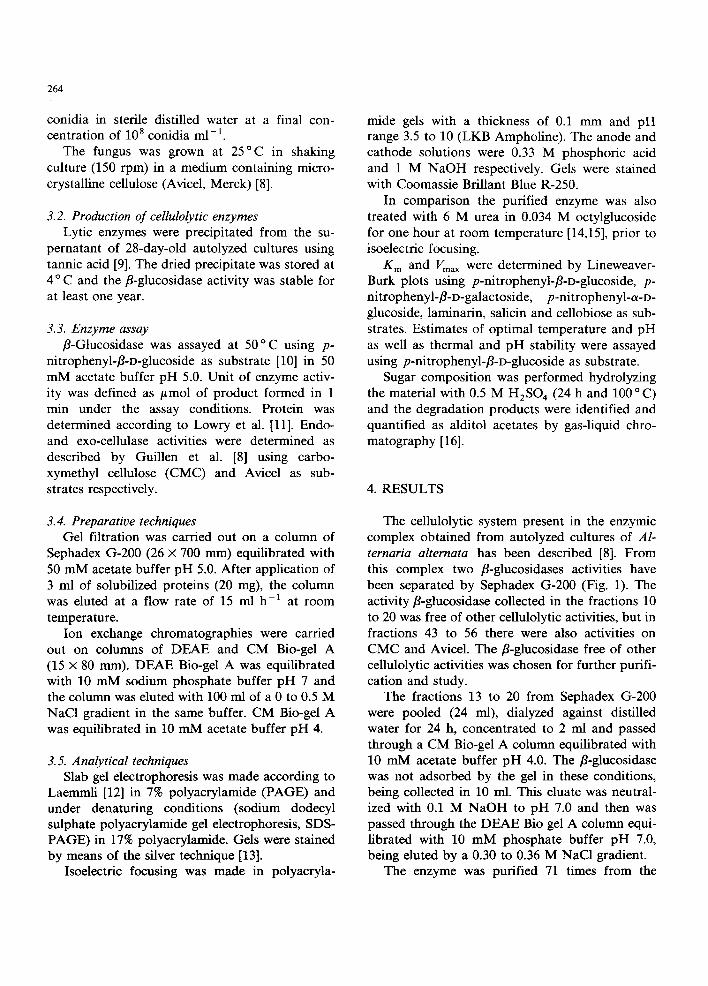
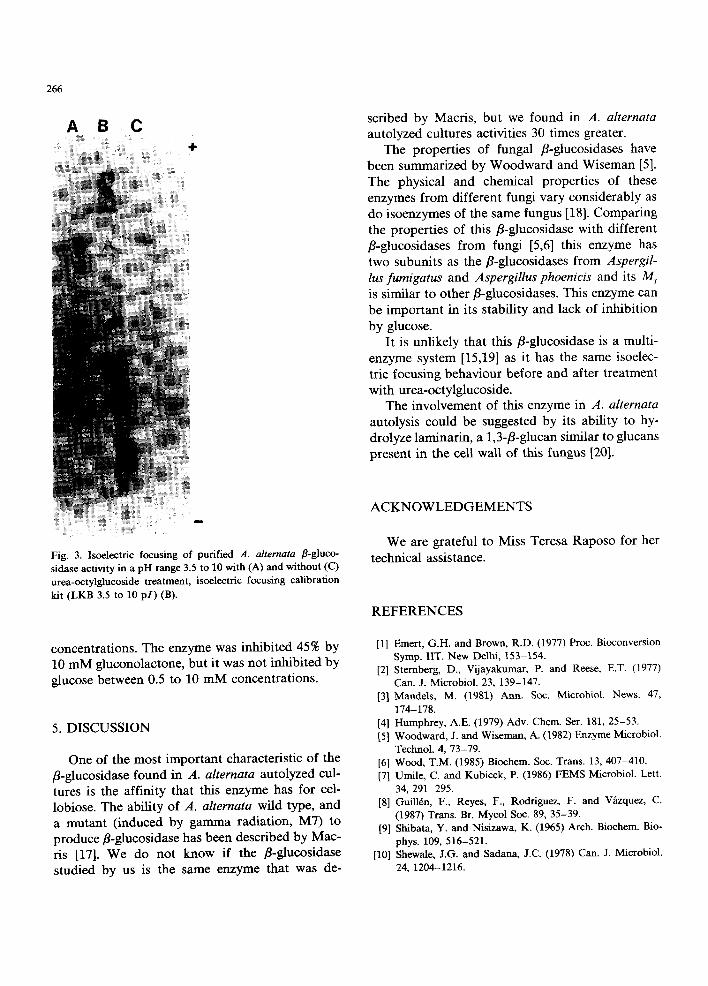
enzymic complex of A. a l t e rna ta au to lyzed cul- tures (Table 1), be ing homogeneous in a b a n d in na t ive P A G E (Fig. 2C) and two b a n d s unde r dena tu r ing condi t ions ( S D S - P A G E ) (Fig. 2A). The enzyme before and after t r ea tmen t wi th urea-oc- ty lg lucos ide showed one b a n d on isoelectr ic focus- ing, wi th a p I of 4.8 (Fig. 3).
The enzyme had a M r of 160000, as de- t e rmined in Sephadex G-200 and the M r of the subuni t s were 80 000 and 60 000 as de t e rmined in S D S - P A G E (Fig. 2A, B). The o p t i m u m p H was 5.0. The o p t i m u m tempera tu re of enzyme reac t ion was 50 ° C. The enzyme was s table in a p H range of 3 to 8 and in a t empera tu re range of 0 to 60 ° C for 60 min. The enzyme was a g lycopro te in wi th 11% galactose, 4% mannose and 1% glucose. I t b o u n d concanava l in A Sepharose . The aff in i ty of
Fig. 2. Polyacrylamide gel electrophoresis of purified A. alter- nata fl-glucosidase in SDS-PAGE showing two subunits (Fig. 2A), electrophoresis calibration kit (Pharmacia, 14400 to 94000 Mr) in the same conditions (Fig. 2B) and native PAGE of this enzyme showing a single band (Fig. 2C).
/~-glucosidase for d i f fe ren t subs t ra tes is shown in T a b l e 2.
The hydro lys i s of p - n i t r o p h e n y l - f l - D - g l u c o s i d e
b y the enzyme was no t af fec ted b y the presence of N a ÷, K +, F e 2+, Z n 2+, Co 2+, M g 2+, and Ca 2÷ at
concen t ra t ions 5 and 10 mM. This hydro lys i s was inh ib i t ed b y 81% and 100% b y H g 2+ at these
Table 1
Purification of/~-glucosidase from autolyzed cultures of Alter- naria a#ernata
Stage Total Specific Total Yield Puff- protein activity activity % fica- (rag) (mU (mU) tion
rag- 2) (fold)
Tannic acid ppt 20.00 200 4000 100 1 Sephadex G-200 0.47 6 832 3 000 75 32 CM-Biogel A 0.20 11180 2 236 56 56 DEAE-Biogel A 0 . 1 4 1 4 2 8 5 2000 50 71
Table 2
Affinity of the enzyme ~-glucosidase and maximum velocity of hydrolysis for different substrates
Subs t ra t e K m Vma x (raM) (mU mg- 1)
PNP-l~-D-glucoside 1.1 50.0 PNPfl-galactoside 16.6 10.0 PNP-a-D-glucoside 20.0 5.0 laminarin 50.0 200.0 salicin 5.0 8.0 cellobiose 1.0 370.0
Results are the mean of five replicates. PNP = p-nitrophenyl.
266
A B C 4.
scribed by Macris, but we found in A. alternata autolyzed cultures activities 30 times greater.
The properties of fungal fl-glucosidases have been summarized by Woodward and Wiseman [5]. The physical and chemical properties of these enzymes from different fungi vary considerably as do isoenzymes of the same fungus [18]. Comparing the properties of this fl-glucosidase with different fl-glucosidases from fungi [5,6] this enzyme has two subunits as the fl-glucosidases from Aspergil- lus fumigatus and Aspergillus phoenicis and its M r is similar to other fl-glucosidases. This enzyme can be important in its stability and lack of inhibition by glucose.
It is unlikely that this fl-glucosidase is a multi- enzyme system [15,19] as it has the same isoelec- tric focusing behaviour before and after treatment with urea-octylglucoside.
The involvement of this enzyme in A. alternata autolysis could be suggested by its ability to hy- drolyze laminarin, a 1,3-fl-glucan similar to glucans present in the cell wall of this fungus [20].
Fig. 3. Isoelectric focusing of purified A. alternata fl-gluco- sidase activity in a pH range 3.5 to 10 with (A) and without (C) urea-octylglucoside treatment, isoelectric focusing calibration kit (LKB 3.5 to 10 p I ) (B).
concentrations. The enzyme was inhibited 45% by 10 mM gluconolactone, but it was not inhibited by glucose between 0.5 to 10 mM concentrations.
5. DISCUSSION
One of the most important characteristic of the fl-glucosidase found in A. alternata autolyzed cul- tures is the affinity that this enzyme has for cel- lobiose. The ability of A. alternata wild type, and a mutant (induced by gamma radiation, MT) to produce fl-glucosidase has been described by Mac- ris [17]. We do not know if the fl-glucosidase studied by us is the same enzyme that was de-
ACKNOWLEDGEMENTS
We are grateful to Miss Teresa Raposo for her technical assistance.
REFERENCES
[1] Emert, G.H. and Brown, R.D. (1977) Proc. Bioconversion Symp. liT. New Delhi, 153-154.
[2] Sternberg, D., Vijayakumar, P. and Reese, E.T. (1977) Can. J. Microbiol. 23, 139-147.
[3] Mandels, M. (1981) Ann. Soc. Microbiol. News. 47, 174-178.
[4] Humphrey, A.E. (1979) Adv. Chem. Ser. 181, 25-53. [5] Woodward, J. and Wiseman, A. (1982) Enzyme Microbiol.
Technol. 4, 73-79. [6] Wood, T.M. (1985) Biochem. Soc. Trans. 13, 407-410. [7] Umile, C. and Kubicek, P. (1986) FEMS Microbiol. Lett.
34, 291-295. [8] Guillrn, F., Reyes, F., Rodriguez, F. and Vfizquez, C.
(1987) Trans. Br. Mycol Soc. 89, 35-39. [9] Shibata, Y. and Nisizawa, K. (1965) Arch. Biochem. Bio-
phys. 109, 516-521. [10] Shewale, J.G. and Sadana, J.C. (1978) Can. J. Microbiol.
24, 1204-1216.
[11] Lowry, O.H., Rosebrough, M.J., Far, A.L. and Randall, R.J. (1951) J. Biol. Chem. 193, 265-275.
[12] Laemmli, U.K. (1970) Nature 227, 680-685. [13] Morrisey, J.M. (1981) Anal. Bioehem. 117, 307-310. [14] Baron, C. and Thomson, T.E. (1975) Biochim. Biophys.
Acta 382, 276-285. [15] Sprey, B. and Lambert, C. (1983) FEMS Microbiol. Lett.
18, 217-222.
267
[16] Laine, R.A., Esselman, W.J. and Sweeley, C.C. (1972) in Methods in Enzymology (Ginsburg, V., Ed.), 28, pp. 159-167.
[17] Macris, B.J. (1984) Biotechnol. Bioeng. 26, 194-196. [18] Shewale, J.G. (1982) J. Biochem. 14, 435-443. [19] Sprey, B. (1987) FEMS Microbiol. Lett. 43, 25-32. [20] Bartnicki-Garcia, S. (1968) Ann. Rev. Microbiol. 22,
87-107.