dr. mirzaei. one of the most common surgical emergencies highest incidence in the second and third...
TRANSCRIPT

Acute Appendicitis
Dr. Mirzaei

One of the most common surgical emergencies
Highest incidence in the second and third decades
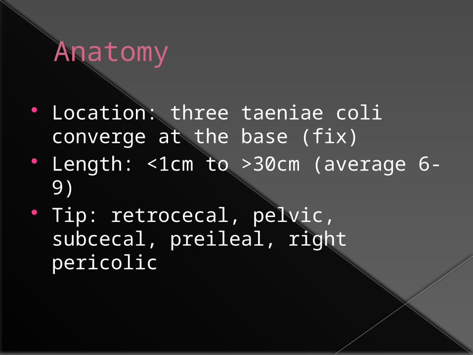
Anatomy
Location: three taeniae coli converge at the base (fix)
Length: <1cm to >30cm (average 6-9) Tip: retrocecal, pelvic, subcecal,
preileal, right pericolic

Function?
Secretion of immunoglobulin A
Appendectomy and U.C protection?
As a reservoir to recolonize the colon with healthy bacteria

Pathogenesis
Obstruction - Fecalith - Hypertrophy of lymphoid - Inspissated barium - Tumors - Vegetable and fruit - Intestinal parasites

Pathogenesis
Normal luminal capacity 0.1 ml Proximal obstruction => closed-loop
obstrucation Normal secretion of mucosa =>
distention 0.5 ml secretion => intraluminal
pressure 60cm H2O

Pathogenesis
Secretion + rapid multiplication of bacteria => venous pressure increased => occlusion of capillaries
Arteriolar inflow continue => vascular congestion

Pathogenesis
Impairment of blood supply => mucosal integrity compromised => bacterial invasion
Infarction in antimesentric border => perforation

Appendiceal Rupture
Overall Rate: 25.8% Children < 5 years: 45% Patients > 65: 51%

Appendiceal Rupture
Walling-off process -> Phlegmon: Adherence of bowel loops to the inflamed appendix or a periappendiceal abscess.
Mass in exam:2-6%
Duration: At least 5-7 days


symptoms
Distention => visceral nerve endings stimulation => vague, dull, diffuse pain in the mid abdomen or lower epigastrium
Distention => reflex nausea &vomiting
Inflammation of serosa & parietal peritoneum => shift in pain to the right lower quadrant
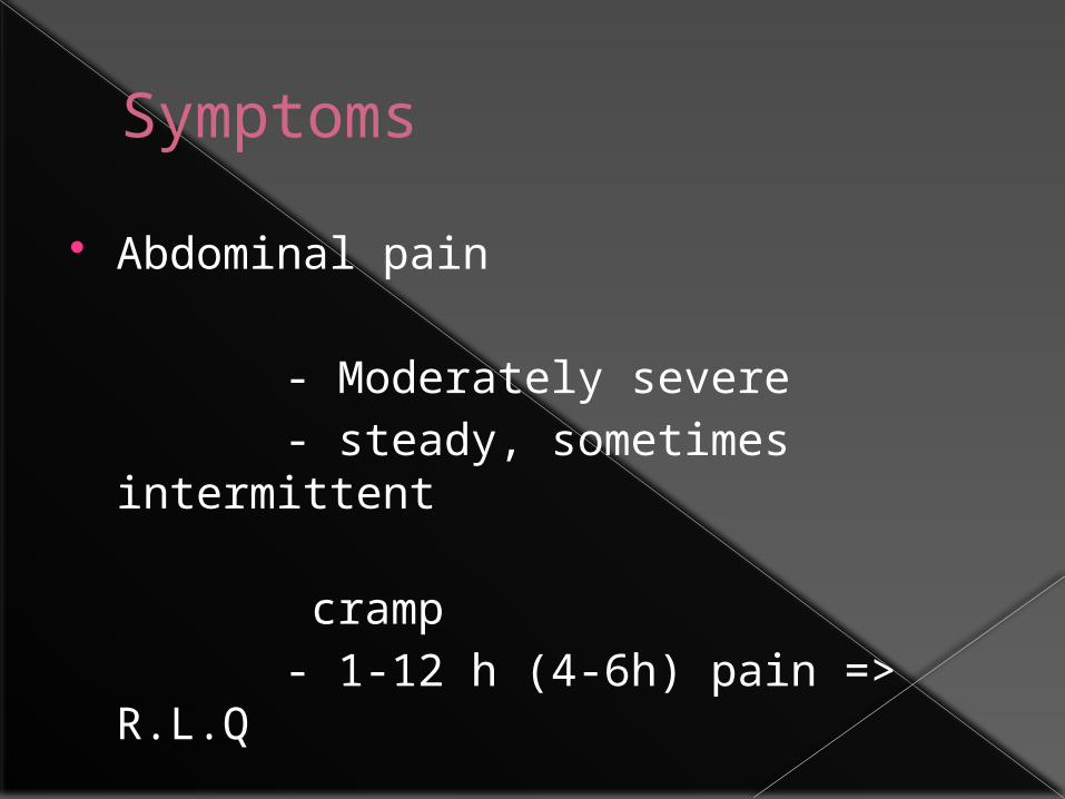
Symptoms
Abdominal pain - Moderately severe - steady, sometimes intermittent
cramp - 1-12 h (4-6h) pain => R.L.Q

Symptoms
Pain variation - Begins in the R.L.Q - Shift to the L.L.Q (tip in the L.L.Q) - Retrocecal => flank or back pain - Pelvic => suprapubic - Retroileal => testicular pain
(irritation of the spermatic artery & ureter)

Symptoms
Intenstinal malrotation
-Visceral: normal location - Somatic: where the cecum has been
arrested

Symptoms
Anorexia (nearly always) - loss of anorexia: diagnosis should
be questioned
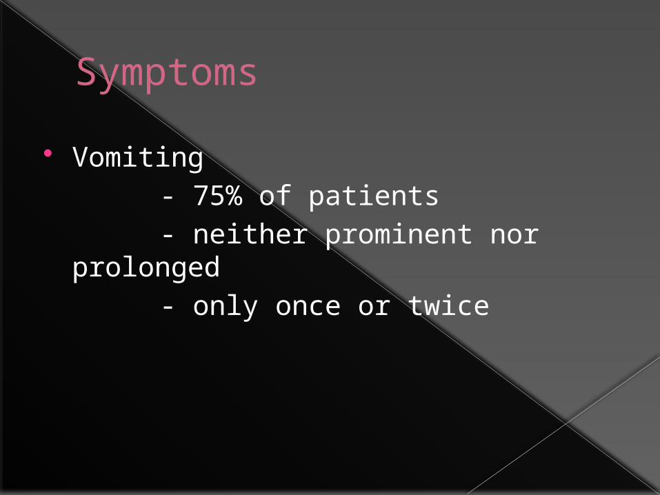
Symptoms
Vomiting - 75% of patients - neither prominent nor prolonged - only once or twice

Sequence of Symptoms
95% anorexia – pain - vomiting
Vomiting – pain: diagnosis should be questioned

Signs
Temperature : rarely > 1ºC
PR: normal or slightly elevated
More change: complication?

Signs
Lie supine
Right thigh drawn up
Any motion increases pain
Move slowly with caution
Signs
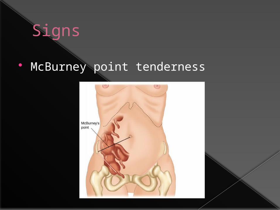
McBurney point tenderness

Signs
Local tenderness
rebound tenderness Voluntary guarding True reflex (involuntary) rigidity
(irritation progress)

Signs
Flank tenderness Local tenderness in rectal exam
(pelvic) psoas sign Obturator sign Rovsings sign

Lab test
W.B.C 10,000 – 18,000 Moderate P.M.N predominance W.B.C > 18,000 => possibility of
complication CRP U/A: several W.B.C or R.B.C (ureteral or
bladder irritation) Bacteriuria generally not seen

Imaging
Plain film (rarely helpful) - abnormal bowel gas pattern - fecalith (highly suggestive)
C.X.R (R/O right lower lobe pneumonia)


Imaging
Sonography (inexpensive, rapid, no contrast medium, even in pregnancy
Noncompressible appendix > 6mm Appendicolith Thickening of appendiceal wall &
periappendiceal fluid Remainder of abdominal cavity

Imaging - Sono
Exclude Gyn pathology
Effective in children & pregnancy

Imaging - sono
Limitations - user dependent - false – positive: dilated fallopian
tube, inspissated stool can mimic
appendicolith, obesity, - false – negative: appendicitis in tip, retrocecal, markedly enlarged, perforation

Imaging – C.T
Dilated appendix>5mm + wall thickening
thickened mesoappendix Phlegmon Periappendiceal fat stranding Free fluid Other inflammatory processes

Imaging – C.T
Expensive, exposes to radiation, cannot be used during pregnancy, allergy to contrast, intolerance of oral contast

Laparoscopy
Most useful in females (30 – 40% normal appendix)
Differentiating acute Gyn pathology

Misdiagnosis
highest rate: child-bearing women,very young,very old
Accuracy of preoperative diagnosis should be: 85%
Accuracy>90%: Missed some patients Depends on: anatomic location of the
appendix, simple or ruptured, age, sex

Alvarado scale

Alvarado scale
7-10 high likelihood
4-6 consider further imaging
1-3 low likelihood

Differential diagnosis

Acute Mesenteric Adenitis
Most in Children Upper respiratory tract infection is
present or has recently subsided. Pain is diffuse Tenderness is not sharply localized Guarding sometimes present

Acute Mesenteric Adenitis
True rigidity is rare Generalized lymphadenopathy (may) Relative lymphocytosis suggestive Self limited May need immediate exploration

Gynecologic Disorders
Pelvic Inflammatory Disease- Usually bilateral - Nausea & Vomiting: 50%- Tenderness Usually lower- Motion of cervix is painful- Diplococci on smear of purulent vaginal
discharge- Higher during early phase of cycle

Gynecologic Disorders
Ruptured Graafian Follicle- Spillage of follicular fluid - Pain and tenderness diffuse- Leukocytosis & fever: minimal - Midcycle: Mittelschmerz

Gynecologic Disorders
Twisted Ovarian Cyst- Sudden pain - CT & Sono (transvaginal)- Need emergent operation - Leakage of ovarian cyst: Treated
nonoperatively


Gynecologic Disorders
Ectopic pregnancy Abnormal menses Missing one or two periods or only slight
vaginal bleeding Elevated level of human chorionic
gonadotropin(B-HCG) Hct level falls Vaginal exam:cervical motion tenderness culdocentesis

Acute Gastroenteritis
Diarrhea, nausea, vomiting Abdominal Cramps Soft Abdomen between cramps No localizing sign Vomiting - Pain

Cecum or sigmoid Diverticulitis Meckel’s Diverticulitis Perforating Carcinoma of the cecum Epiploic appendagitis Pleuritis of the right lower chest Acute Cholecystitis Acute Pancreatitis Hematoma of the abdominal wall Epididymitis, Testicular torsion, U. T. I,
Ureteral Stone