dentine is the most effective protection for the pulp, due to its excellent insulation and capacity...
TRANSCRIPT

DENTINOGENETIC CAPACITY OF DENTAL PULP: A HISTOLOGICAL STUDY
Alexandra Stoica, Marius Ursu, Szabo Anamaria
(Assoc. Prof. Monica Monea, Lecturer Cosmin Moldovan)

Background - Dentin
Dentine is the most effective protection for the pulp, due to its excellent insulation and capacity to reduce diffusion of chemicals from cavity floor to the pulp.

Background - Dental pulp
Trans-dentinal stimulation of tertiary dentin after diffusion of biomaterials (MTA, Ca(OH)2) leads to the development of a hard tissue barrier, that prevents irreversible inflammatory reactions in the pulp tissue.

Historical debate on treatment options
The concepts behind treatment of deep carious lesions are an area of debate and constant change. In many cases the diagnosis is difficult, which makes the treatment more complicated.
”It is better that a layer of discolored dentin should be allowed to remain for the protection of the pulp, rather than run the risk of sacrificing the tooth” (Tomes J. 1885)

Historical debate on treatment options
A thorough knowledge of the histopathology of deep lesions is, therefore, a prerequisite for studies of treatment outcome of dental caries.
”It is better to expose the pulp of a tooth thanto leave it covered onlywith softened dentin”
( Black G.V. 1908)

Pulp capping
Pulp capping is a conservative dental treatment due to the regenerative nature of dentine – pulp complex and its ability to produce tertiary dentine.
Aim of the study
We conducted a randomized clinical study to assess the histological aspects of tertiary dentine formation induced by calcium hydroxide -Ca(OH)2 and Mineral Trioxide Aggregate -MTA in indirect pulp capping procedures.

Material and method
Study group: 23 third molars scheduled for extraction due to orthodontic reasons, with occlusal or approximal simple carious lesions, in 18 patients of 18-24 years of age, who agreed to join the research group. 18
years, 5
20 years, 2
21 years, 4
22 years, 5
24 years; 7
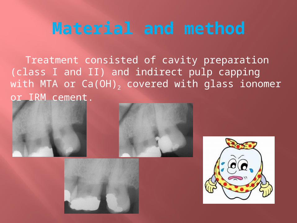
Material and method
Treatment consisted of cavity preparation (class I and II) and indirect pulp capping with MTA or Ca(OH)2 covered with glass ionomer or IRM cement.

Material and method
The extractions were performed after 2-8 weeks and all teeth were processed in order to be studied by optical microscopy.
The specimens were processed for routine histological examination, serial sections were cut and stained with Hematoxylin – Eosin.
Material and method
Histologic evaluation was done by the same person, in a double-blind manner. The dentine bridge of tertiary dentine that was deposited during this period of time was assessed.

ResultsCa(OH)2 - 2 weeks
, Presence of the odontoblastic layer in contact with predentin, many blood vessels in the pulp, which are indicator of an intense metabolic activity.
Tertiary dentin formation.
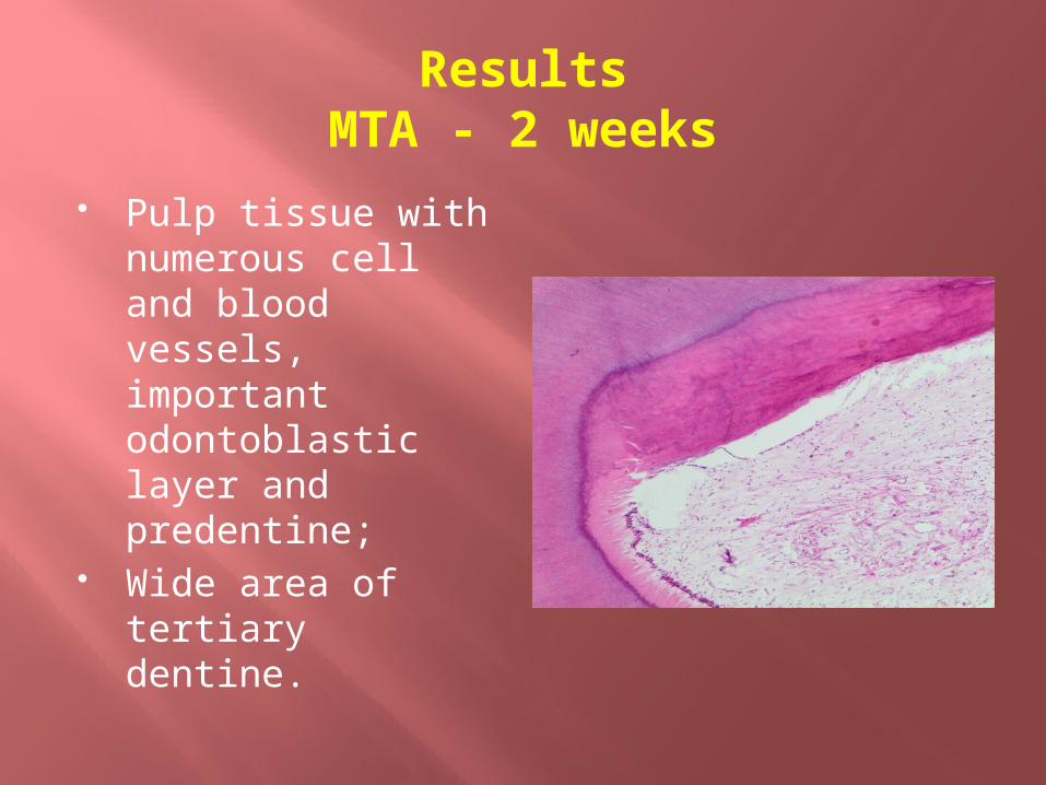
ResultsMTA - 2 weeks
Pulp tissue with numerous cell and blood vessels, important odontoblastic layer and predentine;
Wide area of tertiary dentine.

ResultsCa(OH)2 - 4 weeks
High density of pulp cells, intense circulation, odontoblasts exhibiting intense metabolic reactions.
Tertiary dentine with tubules, similar with secondary dentine.
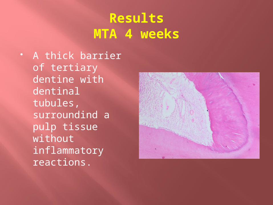
ResultsMTA 4 weeks
A thick barrier of tertiary dentine with dentinal tubules, surroundind a pulp tissue without inflammatory reactions.

ResultsMTA and Ca(OH)2 6 weeks

ResultsCa(OH)2 - 8 weeks
Calcifications in the pulp chamber, near de dentine wall, were observed in specimens treated with calcium hidroxyde.
Discussion
Tertiary dentine induced by mild stimulation is the result of primary odontoblasts and is characterized by the presence of dentinal tubules, sometimes in connection with those from secondary dentine.
Severe stimulations triggers formation of new odontoblasts from pulpal stem cells; the new dentine will be amorphous, with no tubules.
Discussion
The available literature suggests that MTA is more efficient at inducing reparative dentinogenesis in vivo, compared to calcium hidroxyde. Our study showed that this was true for 2-4 weeks; after longer periods of time there is no difference in new dentine formation.
However, both materials share several biologic properties that contribute to the induction of reparative dentinogenesis. Mostly, this is due to the fact that set MTA acts as a calcium hidroxyde releasing material.

Conclusions
Indirect pulp capping is a reliable treatment option for deep carious lesions, even in the presence of demineralised dentine, so please avoid dental pulp exposure in young patients!
Golden standard for success - the placement of a coronal filling that will avoid the marginal leakage.
Continues research is required to achieve a comprehensive picture of pulp tissue response to biomaterials used in pulp capping procedures, in order to obtain optimum repair and regeneration.
Selective refferences
Patel U, Hughes J. Preserving pulp vitality. Dental Health 2013, Vol. 52 (2): 27-29.
Huang YH, Yang JK, Wang CW, Lee SY. Dental stem cells and tooth banking for regenerative medicine. J Exp Clin Med 2010; 2 (3): 111-117.
Kitamura C, Nishihara T, Terashita M. Local regeneration of dentin – pulp Complex using controlled release of FGF – 2. Int J of Dentistry, 2012; ID 190561.
Oliveira DA, Biffi JCG, Maura CG, Pascon EA. A histological assessment of dentine after the clinical removal of caries in extracted human teeth. Dental Press Endod 2011, 1 (3); 79-87.
Accorinte MLR, Reis E. Response of human dental pulp capped with MTA and calcium hidroxyde. Operative Dentistry 2008, 33; 5: 488-495.

”The pulp lives for the dentin and the dentin lives by the grace of the pulp. Few marriages in nature are marked by a greater interrelationship”
John I. Ingle