β-arrestin- but not g protein-mediated signaling by the ... · β-arrestin- but not g...
TRANSCRIPT

β-arrestin- but not G protein-mediated signalingby the “decoy” receptor CXCR7Sudarshan Rajagopala, Jihee Kima, Seungkirl Ahna, Stewart Craigb,c, Christopher M. Lama, Norma P. Gerardb,c, CraigGerardb,c,1, and Robert J. Lefkowitza,d,e,1
aDepartment of Medicine, Duke University Medical Center, Durham, NC 27710; bPulmonary Division, Department of Pediatrics, Children’s Hospital,Boston, MA 02115; cDepartment of Medicine, Harvard Medical School, Boston, MA 02115; dDepartment of Biochemistry, Duke University Medical Center,Durham, NC 27710; and eHoward Hughes Medical Institute, Duke University Medical Center, Durham, NC 27710
Contributed by Robert J. Lefkowitz, November 9, 2009 (sent for review October 1, 2009)
Ubiquitously expressed seven-transmembrane receptors (7TMRs)classically signal through heterotrimeric G proteins and are com-monly referred to as G protein-coupled receptors. It is now recog-nized that 7TMRs also signal through β-arrestins, which act asversatile adapters controlling receptor signaling, desensitization,and trafficking. Most endogenous receptors appear to signal ina balanced fashion using both β-arrestin and G protein-mediatedpathways. Some 7TMRs are thought to be nonsignaling “decoys”because of their inability to activate typical G protein signalingpathways; it has been proposed that these receptors act to scav-enge ligands or function as coreceptors. Here we demonstrate thatligand binding to the decoy receptor CXCR7 does not result inactivation of signaling pathways typical of G proteins but does ac-tivate MAP kinases through β-arrestins in transiently transfectedcells. Furthermore, we observe that vascular smooth muscle cellsthat endogenously express CXCR7 migrate to its ligand interferon-inducible T-cell alpha chemoattractant (ITAC), an effect that is sig-nificantly attenuated by treatment with either a CXCR7 antagonistor β-arrestin depletion by siRNA. This example of an endogenous“β-arrestin-biased” 7TMR that signals through β-arrestin in theabsence of G protein activation demonstrates that some 7TMRsencoded in the genome have evolved to signal through β-arrestinexclusively and suggests that other receptors that are currentlythought to be orphans or decoys may also signal through suchnonclassical pathways.
biased receptor ∣ chemokine ∣ G protein-coupled receptors ∣seven-transmembrane receptor
Seven-transmembrane receptors (7TMRs) are the most com-mon class of receptors in the genome and the most common
target for medical therapeutics (1). Classically, these receptorswere thought to signal exclusively through heterotrimeric G pro-teins and be desensitized by β-arrestins; therefore, they are fre-quently referred to as G protein-coupled receptors. It is nowappreciated that β-arrestins act as multifunctional adapter pro-teins that mediate signaling in their own right and also controlreceptor desensitization and trafficking (2). Most endogenous7TMRs are thought to signal through G protein- and β-arrestin-mediated pathways in a balanced fashion (3). However, somereceptors can be made to signal in a biased fashion towardβ-arrestin or G protein signaling by ligand modifications (4),so-called biased ligands (5), or by mutation of key receptor resi-dues involved in coupling to G proteins or β-arrestins, resulting inG protein- or β-arrestin-biased receptors (6). To date, no endog-enously expressed biased 7TMRs have been described. Wespeculated that candidates for such receptors are the “decoyreceptors,” which do not appear to signal through G proteinsand are thought to scavenge or internalize ligands or act ascoreceptors for other chemokine receptors (7).
Seven-transmembrane decoy receptors that have been demon-strated to be unable to activate typical G protein-mediatedsignaling pathways include C5L2, Duffy antigen receptor forchemokines, D6, and CXCR7 (7). Some of these receptors con-
tain variants of highly conserved residues that are thought to playan important role in signaling to G proteins (8). The decoy re-ceptor CXCR7 (9, 10), which binds the ligands (Stromal-derivedfactor) SDF-1α/CXCL12 and ITAC/CXCL11, does not activatetypical Gαi pathways of a chemokine receptor that would resultin GTP hydrolysis or calcium mobilization (9, 11–13). It is cur-rently thought that CXCR7 acts primarily to modulate signalingof CXCR4, an alternate receptor for SDF-1α. A number ofmechanisms underlying this function have been proposed. Somepropose that CXCR7 may scavenge or sequester SDF-1α, therebygenerating gradients of SDF-1α that lead to differential signalingby CXCR4 (12, 14). It has also been proposed that CXCR7 actsas a coreceptor for CXCR4 (11, 13), as the two receptors formheterodimers in the context of overexpression in transiently trans-fected cells. It has also been proposed that ligand binding toCXCR7 may result in crosstalk with CXCR4 mediated by intra-cellular signaling molecules (15). More recently, it has been dem-onstrated that CXCR7 interacts with β-arrestin in a ligand-dependent manner (15–17), resulting in ligand internalization,but without any assessment of changes in signaling. Given thesefindings we set out to test whether CXCR7 can signal throughβ-arrestins to determine whether this receptor acts as an endog-enous β-arrestin-biased receptor.
ResultsCXCR7 Recruits β-Arrestin in a Ligand-Dependent Fashion. Previousstudies have demonstrated the inability of CXCR7 to generatetypical G protein responses such as an increase in intracellularcalcium after treatment with either SDF-1α or ITAC (9, 11–13).However, we did observe recruitment of β-arrestin to the plasmamembrane upon treatment with both ligands in HEK293 cellstransiently transfected with CXCR7 and GFP-labeled β-arrestin2(Fig. 1A), consistent with recent reports of ligand-dependentinteractions between these two molecules (15, 16). Notably, wedid not observe such recruitment in mock transfected HEK293cells, which express endogenous CXCR4, an alternate receptorfor SDF-1α (Fig. 1B). Recruitment of β-arrestin was rapid withtranslocation to the plasma membrane by 2 min, but at late timepoints (30 min) there was a distinct difference in the pattern ofrecruitment between those cells treated with SDF-1α or ITAC.In those cells treated with SDF-1α for more than 10 min, β-arrest-in2 was localized to cytoplasmic vesicles, referred to as a “class B”pattern, whereas in those cells treated with ITAC, β-arrestin2continued to localize to the plasma membrane, referred to as a“class A” pattern (Fig. 1A) (18). These patterns of β-arrestin
Author contributions: S.R., J.K., S.A., N.P.G., C.G., andR.J.L. designed research; S.R., J.K., S.C.,C.M.L., and N.P.G. performed research; S.R., J.K., S.C., and N.P.G. analyzed data; S.R., C.G.,and R.J.L. wrote the paper.
The authors declare no conflict of interest.1To whom correspondence may be addressed. E-mail: [email protected] [email protected].
This article contains supporting information online at www.pnas.org/cgi/content/full/0912852107/DCSupplemental.
628–632 ∣ PNAS ∣ January 12, 2010 ∣ vol. 107 ∣ no. 2 www.pnas.org/cgi/doi/10.1073/pnas.0912852107
Dow
nloa
ded
by g
uest
on
Dec
embe
r 25
, 201
9
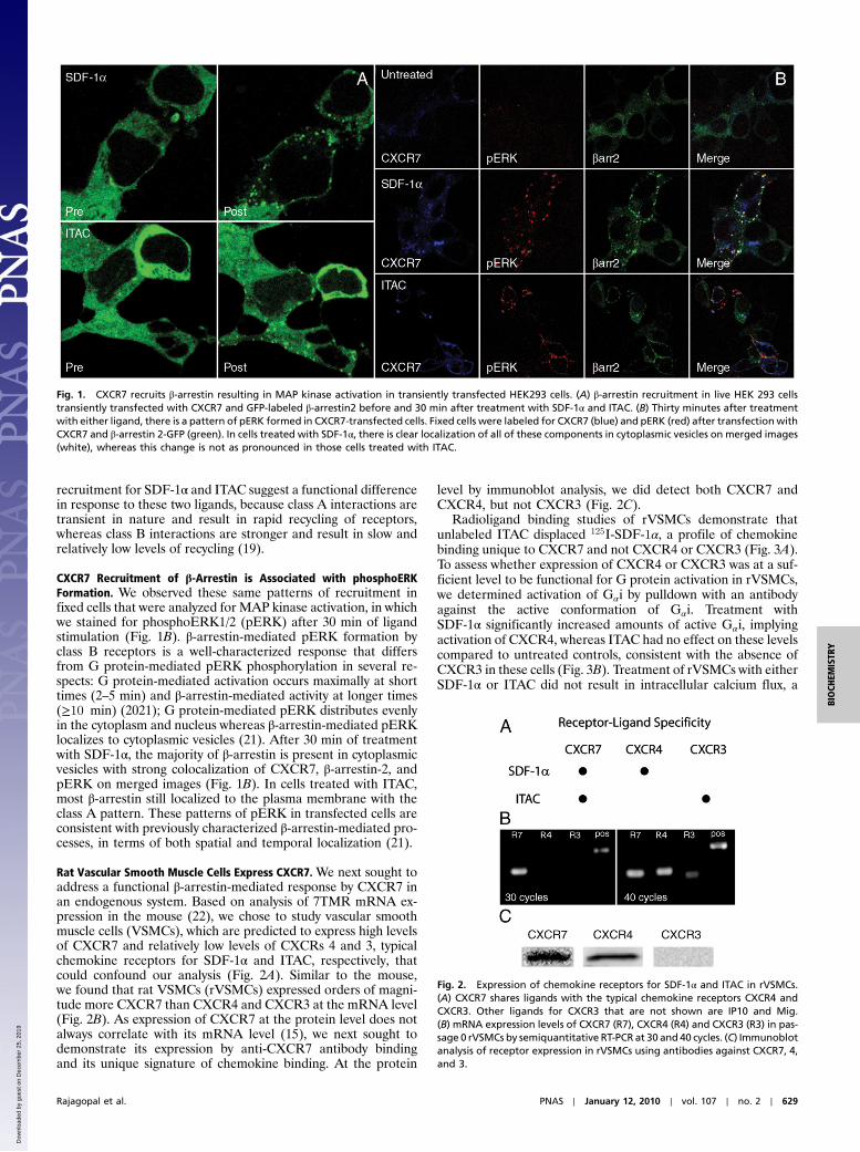
recruitment for SDF-1α and ITAC suggest a functional differencein response to these two ligands, because class A interactions aretransient in nature and result in rapid recycling of receptors,whereas class B interactions are stronger and result in slow andrelatively low levels of recycling (19).
CXCR7 Recruitment of β-Arrestin is Associated with phosphoERKFormation. We observed these same patterns of recruitment infixed cells that were analyzed for MAP kinase activation, in whichwe stained for phosphoERK1/2 (pERK) after 30 min of ligandstimulation (Fig. 1B). β-arrestin-mediated pERK formation byclass B receptors is a well-characterized response that differsfrom G protein-mediated pERK phosphorylation in several re-spects: G protein-mediated activation occurs maximally at shorttimes (2–5 min) and β-arrestin-mediated activity at longer times(≥10 min) (2021); G protein-mediated pERK distributes evenlyin the cytoplasm and nucleus whereas β-arrestin-mediated pERKlocalizes to cytoplasmic vesicles (21). After 30 min of treatmentwith SDF-1α, the majority of β-arrestin is present in cytoplasmicvesicles with strong colocalization of CXCR7, β-arrestin-2, andpERK on merged images (Fig. 1B). In cells treated with ITAC,most β-arrestin still localized to the plasma membrane with theclass A pattern. These patterns of pERK in transfected cells areconsistent with previously characterized β-arrestin-mediated pro-cesses, in terms of both spatial and temporal localization (21).
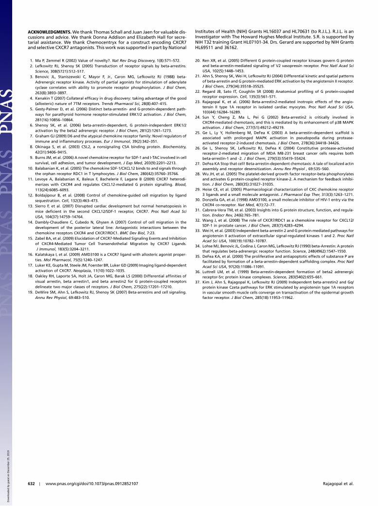
Rat Vascular Smooth Muscle Cells Express CXCR7. We next sought toaddress a functional β-arrestin-mediated response by CXCR7 inan endogenous system. Based on analysis of 7TMR mRNA ex-pression in the mouse (22), we chose to study vascular smoothmuscle cells (VSMCs), which are predicted to express high levelsof CXCR7 and relatively low levels of CXCRs 4 and 3, typicalchemokine receptors for SDF-1α and ITAC, respectively, thatcould confound our analysis (Fig. 2A). Similar to the mouse,we found that rat VSMCs (rVSMCs) expressed orders of magni-tude more CXCR7 than CXCR4 and CXCR3 at the mRNA level(Fig. 2B). As expression of CXCR7 at the protein level does notalways correlate with its mRNA level (15), we next sought todemonstrate its expression by anti-CXCR7 antibody bindingand its unique signature of chemokine binding. At the protein
level by immunoblot analysis, we did detect both CXCR7 andCXCR4, but not CXCR3 (Fig. 2C).
Radioligand binding studies of rVSMCs demonstrate thatunlabeled ITAC displaced 125I-SDF-1α, a profile of chemokinebinding unique to CXCR7 and not CXCR4 or CXCR3 (Fig. 3A).To assess whether expression of CXCR4 or CXCR3 was at a suf-ficient level to be functional for G protein activation in rVSMCs,we determined activation of Gαi by pulldown with an antibodyagainst the active conformation of Gαi. Treatment withSDF-1α significantly increased amounts of active Gαi, implyingactivation of CXCR4, whereas ITAC had no effect on these levelscompared to untreated controls, consistent with the absence ofCXCR3 in these cells (Fig. 3B). Treatment of rVSMCs with eitherSDF-1α or ITAC did not result in intracellular calcium flux, a
Fig. 1. CXCR7 recruits β-arrestin resulting in MAP kinase activation in transiently transfected HEK293 cells. (A) β-arrestin recruitment in live HEK 293 cellstransiently transfected with CXCR7 and GFP-labeled β-arrestin2 before and 30 min after treatment with SDF-1α and ITAC. (B) Thirty minutes after treatmentwith either ligand, there is a pattern of pERK formed in CXCR7-transfected cells. Fixed cells were labeled for CXCR7 (blue) and pERK (red) after transfectionwithCXCR7 and β-arrestin 2-GFP (green). In cells treated with SDF-1α, there is clear localization of all of these components in cytoplasmic vesicles on merged images(white), whereas this change is not as pronounced in those cells treated with ITAC.
Fig. 2. Expression of chemokine receptors for SDF-1α and ITAC in rVSMCs.(A) CXCR7 shares ligands with the typical chemokine receptors CXCR4 andCXCR3. Other ligands for CXCR3 that are not shown are IP10 and Mig.(B) mRNA expression levels of CXCR7 (R7), CXCR4 (R4) and CXCR3 (R3) in pas-sage 0 rVSMCs by semiquantitative RT-PCR at 30 and 40 cycles. (C) Immunoblotanalysis of receptor expression in rVSMCs using antibodies against CXCR7, 4,and 3.
Rajagopal et al. PNAS ∣ January 12, 2010 ∣ vol. 107 ∣ no. 2 ∣ 629
BIOCH
EMISTR
Y
Dow
nloa
ded
by g
uest
on
Dec
embe
r 25
, 201
9

response that would be expected for activation of either CXCR4or CXCR3 (Fig. 3C) (compared to a positive control of angioten-sin, a well-characterized Gαq-coupled receptor) (23). Thus, weconclude that ITAC binds CXCR7 alone without any activationof G proteins, and SDF-1α binds to both CXCR4 and CXCR7,resulting in Gαi activation, albeit with minimal signaling througha typical Gαi-coupled pathway.
Rat VSMCs Migrate to ITAC in a CXCR7- and β-Arrestin-MediatedProcess. We next addressed whether activation of endogenousCXCR7 in the rVSMCs could result in a functional β-arrestin-mediated response. We first assessed for MAP kinase activationthat we had observed in the transiently transfected HEK293 cells,but we were unable to detect significant increases in pERK upontreatment with either SDF-1α or ITAC over the background highbasal activation in the rVSMCs even after more than 48 h ofserum starvation (Fig. S1). We also did not observe a mitogenicresponse to these ligands (Fig. S1), suggesting that CXCR7couples to different signaling pathways in rVSMCs than it doesin the context of overexpression in HEK293 cells. We next as-sessed rVSMC migration to these ligands as β-arrestin-mediatedchemotaxis is a well-described phenomenon in a number of
7TMR systems (24–27). We found that SDF-1α and ITAC re-sulted in significant rVSMC migration across a range of concen-trations tested (Fig. 4A), with a response similar to that of otherligands known to induce rVSMC chemotaxis, such as PDGF (28).
To avoid possible confounding with CXCR4-mediatedprocesses, we then further characterized the response of rVSMCsto ITAC alone. To assess whether this migration response wasmediated by CXCR7, cells were treated with selective chemokinereceptor antagonists. Treatment with the CXCR3 antagonistT487 (29) or the CXCR4 antagonist AMD3100 (30) had nosignificant effect on migration, however, the CXCR7 antagonistCCX733 had a significant inhibitory effect on migration, whereasa structurally related compound which does not bind CXCR7,CCX704, had none (Fig. 4B). Therefore, we conclude that themajority of the migration response to ITAC is mediated byCXCR7 and not the typical chemokine receptors CXCR3 andCXCR4. To determine whether this CXCR7-mediated migrationwas β-arrestin-dependent, we depleted β-arrestins by siRNAknockdown. Although depletion of β-arrestin1 had no statisticallysignificant effect on migration, depletion of β-arrestin2 resultedin a marked attenuation of rVSMC migration in response toITAC compared to control siRNA-transfected cells (Fig. 4C).Although it is impossible to exclude signaling by the dozens ofdifferent Gα and Gβγ subunits (31), we did find that this responsewas not sensitive to treatment with pertussis toxin and thus inde-pendent of Gαi (Fig. S2). The specificity of CXCR7 antagonismand effect of β-arrestin depletion demonstrate that CXCR7-mediated rVSMC migration is a β-arrestin-mediated processand that CXCR7 acts as a β-arrestin-biased receptor.
DiscussionAlthough virtually all reports in the literature have agreed thatCXCR7 does not couple to G proteins, functional roles forthe receptor that belie a signaling event suggest a nonclassicalpositive signaling role for this receptor. In the initial character-ization of CXCR7, its expression increased the adhesion of can-cer cells to endothelia and enhanced their survival in vitro andin vivo (9). In the zebrafish, polarized expression of CXCR4and CXCR7 is essential for primordial germ cell migration(12, 14). These reports suggested that CXCR7 may scavengethe CXCL12 in the stationary, nonmigrating cells. Prostate can-cer cells expressing CXCR7 suggest a role for this receptor inadhesion, invasiveness, and survival, where activation of Akt islinked to CXCR7 and results in the regulation of a number ofgenes associated with the NF-κB and MAP kinase pathways(32). Our current data, in which CXCR7 is an active receptorcapable of signaling through β-arrestin, provide a mechanisticbasis for understanding these earlier studies.
For the vast majority of 7TMR signaling, it is thought thatthere is a balance between G protein- and β-arrestin-mediatedpathways (Fig. 5A). Agonist binding typically results in signaling
Fig. 3. Chemokine receptor expression and lack of G protein activation byITAC in rVSMCs. (A) Treatment with unlabeled ITAC or SDF competes offradiolabeled SDF-1α from rVSMCs. (B) Activation of Gαi in SDF-1α-treatedrVSMCs (SDF) but not in untreated (UT) or ITAC-treated (ITAC) samples.The negative control was incubated with GDP (GDP) and the positive controlincubated with GTP (GTP) for 90 min prior to immunoprecipitation. Datashown are mean� SEM from three independent experiments. (C) Absenceof calcium influx as assessed by change in Fura-2 fluorescence emission ratioupon stimulation of rVSMCs with SDF-1α (blue) or ITAC (red) compared toAngiotensin II (black) from a representative experiment from three indepen-dent experiments.
Fig. 4. rVSMCmigration in response to ITAC is a CXCR7- and β-arrestin-dependent process. (A) Migration of rVSMCs to 1–100 nM of SDF-1α and ITAC comparedto PDGF positive control. Shown is representative data from at least three independent experiments. (B) Effects of CCX704 (inactive control), CCX733 (CXCR7antagonist), T497 (CXCR3 antagonist), and AMD3100 (CXCR4 antagonist) on ITAC-stimulated migration. Only treatment with CCX733 resulted in a significantinhibitory effect (�p < 0.05). (C) Significant inhibition of ITAC-stimulated rVSMC migration by β-arrestin2 depletion (�p < 0.05) but not β-arrestin1 depletion.Data shown are mean� SEM from at least four independent experiments.
630 ∣ www.pnas.org/cgi/doi/10.1073/pnas.0912852107 Rajagopal et al.
Dow
nloa
ded
by g
uest
on
Dec
embe
r 25
, 201
9

mediated by G proteins and β-arrestins accompanied by receptordesensitization and internalization mediated by β-arrestins. Thisis in contrast to a system with biased signaling, where signaling ismediated selectively through only one of these two pathways. Twolimit cases of biased signaling can be considered. In the case of abiased ligand, treatment of a balanced receptor with a biased ago-nist results in a biased response (Fig. 5B), whereas, in the case of abiased receptor, binding of a balanced agonist results in a biasedresponse (Fig. 5C). Before the present study, receptors thathave been demonstrated to be biased have been geneticallyengineered from balanced receptors by mutation of key residuesinvolved in G protein coupling, such as mutations of the highlyconserved DRY motif of the AT1AR to generate the variantAT1ARðDRY∕AAYÞ (33) or mutations of three highly conservedresidues in the β2 adrenergic receptor to generate the β-arrestin-biased β2AR(TYY) (6). Surprisingly, similar mutations areobserved in decoy receptors such as CXCR7, which lacks a typicalsequence surrounding the DRY motif (7), suggesting that biasedreceptors have evolved in our genome.
β-arrestins were initially discovered as negative regulators ofG protein-mediated signaling by 7TMRs (34), but it was subse-quently discovered that they themselves were capable of positivelyregulating cell signaling (35, 36). The discovery of an endogenousβ-arrestin-biased receptor has important implications for our basicunderstanding of 7TMR biology. It inverts the classical paradigmof signaling from the view that all signaling by 7TMRs is mediatedby heterotrimeric G proteins, to one where receptors mayonly signal through non-G protein-mediated mechanisms. Thissuggests that other receptors that have been considered to be“nonsignaling” may also signal through such nonclassical path-ways. Many drug discovery and deorphanization strategies arebased solely on assays for G protein-mediated actions and maynot recognize β-arrestin-mediated processes. In the future, a morecomplete understanding of 7TMR pharmacology can only begained by looking at G protein and β-arrestin activity in concert.
Materials and MethodsMaterials. A plasmid encoding CXCR7 in a modified version of pcDNA3(pFIRES) was a gift from Chemocentryx (9). Recombinant chemokines wereobtained from R&D Systems. Anti-CXCR7 monoclonal antibodies were ob-tained from R&D systems, anti-rat CXCR4 and anti-rat CXCR3 antibodies fromAbcam, antiphosphoERK antibody from Cell Signaling Technologies, andantitotalERK antibody from Upstate Biotechnology. Chemokine inhibitorsCCX704 and CCX733 were obtained from Chemocentryx. T487 was obtainedfrom Tularik, and AMD3100 was purchased from Sigma.
Transient Transfection. HEK-293 cells were maintained in MEM media supple-mented with 10% FBS and 1% penicillin/streptomycin. For DNA transfection,FuGene transfection reagent (Roche Applied Science) was added at a ratio of
5-μl to 1-μg plasmid DNA to a solution of plasmid DNA in 500-μl serum-freeMEM. This transfection mixture was incubated for 30–45 min prior to addi-tion to HEK293 cells of 50% confluence. For a 10-cm cell culture dish, 5 μg ofplasmid DNA encoding CXCR7 was used. Cells were split and used for assays48 h after transfection. For rVSMCs, 80–90% confluent early passage (<3)VSMCs were transfected with siRNA using Lipofectamine 2000 transfectionreagent (Invitrogen) according to the modified manufacturer’s instructions(37). The siRNA sequence targeting β arrestin1 was 5-AGCCUUCUGUGCUGA-GAAC-3, corresponding to position 431–459 relative to the start codon. ThesiRNA sequences targeting β arrestin2 were 5-GGACCGCAAAGUGUUUGUG-3and 5-CCAACCTCATTGAATTCGA-3, corresponding to positions 150–168 and1115–1133 relative to the start codon.
Confocal Microscopy. HEK-293 cells were split into 35-mm glass-bottom dishes(MatTek) and transfected with 1 μg of CXCR7 and 0.2 μg of β-arrestin-2-GFP.Forty-eight hours after transfection, cells were starved for at least 5 h inserum-free media prior to treatment with ligand. For live cell imaging, cellswere maintained at 37 °C with a heating plate while confocal microscopy wasperformed. SDF-1α and ITAC were added to a final concentration of 100 nMand images were taken from 0 to 30 min. For immunostaining, cells weretreated for 30 min with 100 nM ligand, followed by aspiration of serumand fixation. Samples were then washed followed by permeabilizationand blocking. Cells were then incubated overnight with primary antibodyand then washed prior to incubation with secondary antibody at 1∶500 dilu-tion. Samples were then washed and visualized.
PCR. RT-PCR primers for rat CXCR7, CXCR4, and CXCR3 were obtained fromQiagen. Total RNA was extracted from passage 0 rVSMC by phenol-chloro-form extraction using Ultraspec reagent (Bio-X). Qiagen RT-PCR kit was usedfor 30 and 40 cycles for semiquantitative RT-PCR. Samples were run on 1%agarose gel and stained with ethidium bromide prior to digital imaging.
Active Gαi Pulldown Assay. rVSMCs were starved for 48 h prior to 5 min ofstimulation with SDF-1α or ITAC (positive control). After 5 min, the cells wereplaced on ice, washed with ice cold PBS and lysed, solubilized for 1 h, andthen incubated for 1 h with an antiactive Gαi antibody and Protein A-agarosebeads (New East Biosciences). Beads were washed with lysis buffer threetimes and incubated with SDS-PAGE buffer at 95 °C for 10 min and runon a 4–20% gradient polyacrylamide gel, transferred to nitrocellulosemembrane, and incubated with anti-Gαi antibody.
Intracellular Calcium Flux Assay. rVSMCs were split into glass-bottom dishes atleast 12 h before experiments. Smooth muscle cells (SMCs) were loaded with5 μmol∕L Fura-2/acetoxymethyl ester (Invitrogen) for 30min. Intracellular cal-cium levels were quantitated by the Fura-2 excitation ratio at 340 and 380 nmon an epifluorescence microscope. Agonist-stimulated calcium release wascalculated as the change in the Fura-2 excitation ratio from baseline.
Radioactive Ligand Binding. Cells were grown in 24-well plates until confluent.Binding was assessed by incubation with 0.02 nM 125I-SDF-1α or ITAC in25 mM Hepes, pH 7.5, containing 140 mM NaCl, 1 mM CaCl2, 5 mMMgCl2, and 0.2% BSA for 2 h at 22 °C in the presence of 0–300 nM unlabeledligand. In some experiments, AMD3100 (50 mM) or CCX733 (1 mM) were alsoincluded. Cells were washed three times with binding buffer containing0.5 M NaCl, solubilized with 0.3 mL of 0.5 M NaOH, and counts per minutebound determined by gamma counting.
VSMC Migration. Rat aortas were stripped of adventitia and endothelial cells,and then digested with 0.1% collagenase II and 15 U∕mL elastase in the pres-ence of 0.1% soybean trypsin inhibitor. Released SMCs were cultured inDMEM with 10% FBS and 1% penicillin/streptomycin (Life Technologies).SMCs were used during passages 1–2. The migration of serum-starved SMCswas assessed using Transwell™ membranes (Costar; 8-μm pore). Filters werecoated with 10 μg∕mL fibronectin (in PBS overnight at 4 °C), rinsed once withPBS, and placed in 24-well dish wells that contained serum-free DMEM sup-plemented with agonists. SMCs suspended in serum-free DMEM containing0.1% BSA were added to the upper chamber (1 × 10⁵ cells/well). Cells wereallowed to migrate for 5 h at 37 °C. Nonmigrated SMCs were removed fromthe top filter surface with a cotton swab. Migrated SMCs, attached to thebottom surface, were fixed in 4% paraformaldehyde and dyed with crystalviolet. Dye intensity was quantified by densitometry. For rVSMCs transfectedwith fluorescent siRNAs, images were obtained on an epifluorescencemicroscope and the number of cells quantified.
Fig. 5. Balanced and biased signaling by 7TMRs. (A) In a balanced system,signaling is mediated by G proteins and β-arrestins, while β-arrestins alsocontrol desensitization and receptor trafficking. (B) Treatment of anunbiased receptor with a biased agonist results in a biased response, in thiscase, β-arrestin-mediated signaling only. (C) In the case of a β-arrestin-biasedreceptor, treatment of the receptor with an unbiased ligand results inβ-arrestin-mediated signaling only.
Rajagopal et al. PNAS ∣ January 12, 2010 ∣ vol. 107 ∣ no. 2 ∣ 631
BIOCH
EMISTR
Y
Dow
nloa
ded
by g
uest
on
Dec
embe
r 25
, 201
9
ACKNOWLEDGMENTS.We thank Thomas Schall and Juan Jaen for valuable dis-cussions and advice. We thank Donna Addison and Elizabeth Hall for secre-tarial assistance. We thank Chemocentryx for a construct encoding CXCR7and selective CXCR7 antagonists. This work was supported in part by National
Institutes of Health (NIH) Grants HL16037 and HL70631 (to R.J.L.). R.J.L. is anInvestigator with The Howard Hughes Medical Institute. S.R. is supported byNIH T32 training Grant HL07101-34. Drs. Gerard are supported by NIH GrantsHL69511 and 36162.
1. Ma P, Zemmel R (2002) Value of novelty?. Nat Rev Drug Discovery, 1(8):571–572.2. Lefkowitz RJ, Shenoy SK (2005) Transduction of receptor signals by beta-arrestins.
Science, 308(5721):512–517.3. Benovic JL, Staniszewski C, Mayor F, Jr., Caron MG, Lefkowitz RJ (1988) beta-
Adrenergic receptor kinase. Activity of partial agonists for stimulation of adenylatecyclase correlates with ability to promote receptor phosphorylation. J Biol Chem,263(8):3893–3897.
4. Kenakin T (2007) Collateral efficacy in drug discovery: taking advantage of the good(allosteric) nature of 7TM receptors. Trends Pharmacol Sci, 28(8):407–415.
5. Gesty-Palmer D, et al. (2006) Distinct beta-arrestin- and G-protein-dependent path-ways for parathyroid hormone receptor-stimulated ERK1/2 activation. J Biol Chem,281(16):10856–10864.
6. Shenoy SK, et al. (2006) beta-arrestin-dependent, G protein-independent ERK1/2activation by the beta2 adrenergic receptor. J Biol Chem, 281(2):1261–1273.
7. Graham GJ (2009) D6 and the atypical chemokine receptor family: Novel regulators ofimmune and inflammatory processes. Eur J Immunol, 39(2):342–351.
8. Okinaga S, et al. (2003) C5L2, a nonsignaling C5A binding protein. Biochemistry,42(31):9406–9415.
9. Burns JM, et al. (2006) A novel chemokine receptor for SDF-1 and I-TAC involved in cellsurvival, cell adhesion, and tumor development. J Exp Med, 203(9):2201–2213.
10. Balabanian K, et al. (2005) The chemokine SDF-1/CXCL12 binds to and signals throughthe orphan receptor RDC1 in T lymphocytes. J Biol Chem, 280(42):35760–35766.
11. Levoye A, Balabanian K, Baleux F, Bachelerie F, Lagane B (2009) CXCR7 heterodi-merizes with CXCR4 and regulates CXCL12-mediated G protein signalling. Blood,113(24):6085–6093.
12. Boldajipour B, et al. (2008) Control of chemokine-guided cell migration by ligandsequestration. Cell, 132(3):463–473.
13. Sierro F, et al. (2007) Disrupted cardiac development but normal hematopoiesis inmice deficient in the second CXCL12/SDF-1 receptor, CXCR7. Proc Natl Acad SciUSA, 104(37):14759–14764.
14. Dambly-Chaudiere C, Cubedo N, Ghysen A (2007) Control of cell migration in thedevelopment of the posterior lateral line: Antagonistic interactions between thechemokine receptors CXCR4 and CXCR7/RDC1. BMC Dev Biol, 7:23.
15. Zabel BA, et al. (2009) Elucidation of CXCR7-Mediated Signaling Events and Inhibitionof CXCR4-Mediated Tumor Cell Transendothelial Migration by CXCR7 Ligands.J Immunol, 183(5):3204–3211.
16. Kalatskaya I, et al. (2009) AMD3100 is a CXCR7 ligand with allosteric agonist proper-ties. Mol Pharmacol, 75(5):1240–1247.
17. Luker KE, GuptaM, Steele JM, Foerster BR, Luker GD (2009) Imaging ligand-dependentactivation of CXCR7. Neoplasia, 11(10):1022–1035.
18. Oakley RH, Laporte SA, Holt JA, Caron MG, Barak LS (2000) Differential affinities ofvisual arrestin, beta arrestin1, and beta arrestin2 for G protein-coupled receptorsdelineate two major classes of receptors. J Biol Chem, 275(22):17201–17210.
19. DeWire SM, Ahn S, Lefkowitz RJ, Shenoy SK (2007) Beta-arrestins and cell signaling.Annu Rev Physiol, 69:483–510.
20. Ren XR, et al. (2005) Different G protein-coupled receptor kinases govern G proteinand beta-arrestin-mediated signaling of V2 vasopressin receptor. Proc Natl Acad SciUSA, 102(5):1448–1453.
21. Ahn S, Shenoy SK, Wei H, Lefkowitz RJ (2004) Differential kinetic and spatial patternsof beta-arrestin and G protein-mediated ERK activation by the angiotensin II receptor.J Biol Chem, 279(34):35518–35525.
22. Regard JB, Sato IT, Coughlin SR (2008) Anatomical profiling of G protein-coupledreceptor expression. Cell, 135(3):561–571.
23. Rajagopal K, et al. (2006) Beta-arrestin2-mediated inotropic effects of the angio-tensin II type 1A receptor in isolated cardiac myocytes. Proc Natl Acad Sci USA,103(44):16284–16289.
24. Sun Y, Cheng Z, Ma L, Pei G (2002) Beta-arrestin2 is critically involved inCXCR4-mediated chemotaxis, and this is mediated by its enhancement of p38 MAPKactivation. J Biol Chem, 277(51):49212–49219.
25. Ge L, Ly Y, Hollenberg M, DeFea K (2003) A beta-arrestin-dependent scaffold isassociated with prolonged MAPK activation in pseudopodia during protease-activated receptor-2-induced chemotaxis. J Biol Chem, 278(36):34418–34426.
26. Ge L, Shenoy SK, Lefkowitz RJ, DeFea K (2004) Constitutive protease-activatedreceptor-2-mediated migration of MDA MB-231 breast cancer cells requires bothbeta-arrestin-1 and -2. J Biol Chem, 279(53):55419–55424.
27. DeFea KA Stop that cell! Beta-arrestin-dependent chemotaxis: A tale of localized actinassembly and receptor desensitization. Annu Rev Physiol , 69:535–560.
28. Wu JH, et al. (2005) The platelet-derived growth factor receptor-beta phosphorylatesand activates G protein-coupled receptor kinase-2. A mechanism for feedback inhibi-tion. J Biol Chem, 280(35):31027–31035.
29. Heise CE, et al. (2005) Pharmacological characterization of CXC chemokine receptor3 ligands and a small molecule antagonist. J Pharmacol Exp Ther, 313(3):1263–1271.
30. Donzella GA, et al. (1998) AMD3100, a small molecule inhibitor of HIV-1 entry via theCXCR4 co-receptor. Nat Med, 4(1):72–77.
31. Cabrera-Vera TM, et al. (2003) Insights into G protein structure, function, and regula-tion. Endocr Rev, 24(6):765–781.
32. Wang J, et al. (2008) The role of CXCR7/RDC1 as a chemokine receptor for CXCL12/SDF-1 in prostate cancer. J Biol Chem, 283(7):4283–4294.
33. Wei H, et al. (2003) Independent beta-arrestin 2 and G protein-mediated pathways forangiotensin II activation of extracellular signal-regulated kinases 1 and 2. Proc NatlAcad Sci USA, 100(19):10782–10787.
34. LohseMJ, Benovic JL, Codina J, CaronMG, Lefkowitz RJ (1990) beta-Arrestin: A proteinthat regulates beta-adrenergic receptor function. Science, 248(4962):1547–1550.
35. DeFea KA, et al. (2000) The proliferative and antiapoptotic effects of substance P arefacilitated by formation of a beta-arrestin-dependent scaffolding complex. Proc NatlAcad Sci USA, 97(20):11086–11091.
36. Luttrell LM, et al. (1999) Beta-arrestin-dependent formation of beta2 adrenergicreceptor-Src protein kinase complexes. Science, 283(5402):655–661.
37. Kim J, Ahn S, Rajagopal K, Lefkowitz RJ (2009) Independent beta-arrestin2 and Gq/protein kinase Czeta pathways for ERK stimulated by angiotensin type 1A receptorsin vascular smooth muscle cells converge on transactivation of the epidermal growthfactor receptor. J Biol Chem, 285(18):11953–11962.
632 ∣ www.pnas.org/cgi/doi/10.1073/pnas.0912852107 Rajagopal et al.
Dow
nloa
ded
by g
uest
on
Dec
embe
r 25
, 201
9