© 2011 pearson education, inc. powerpoint ® lecture presentations prepared by alexander g....
TRANSCRIPT

© 2011 Pearson Education, Inc.
PowerPoint® Lecture Presentations prepared byAlexander G. CheroskeMesa Community College at Red Mountain
7The Skeleton

© 2011 Pearson Education, Inc.Figure 7 Section 1
AXIALSKELETON
Skull andassociated
bones
SkullCranium
Face
Auditoryossicles
Hyoid
Sternum
Ribs
Thoraciccage
Vertebralcolumn
The bones of the axial skeleton
SKELETAL SYSTEM 206
80
8
14
6
1
1
24
126
24
1
1
26
25
29
Coccyx
Sacrum
Vertebrae
Associated bones
APPENDICULARSKELETON
(see Section 2)
Costalcartilages(cartilagesof ribs)
Intervertebraldiscs (cartilage)

© 2011 Pearson Education, Inc.Figure 7.1 1
The 22 bones that form the skull, plus the sevenbones associated with the skull
FACE CRANIUM
SKULL
Maxillary bones
Palatine bones
Nasal bones
Inferior nasalconchae
Zygomatic bones
Lacrimal bones
Vomer
Mandible
Occipital bone
Parietel bones
Frontal bone
Temporal bones
Sphenoid
Ethmoid
ASSOCIATED BONES(see Module 7.7)
Hyoid bone Auditory ossiclesenclosed in
temporal bones
1
1
1
1
1
1
1
2
2
2
2
2
2
2 2
14 8 7
6

© 2011 Pearson Education, Inc.Figure 7.1 4
The facial and cranial bones of the skull
Cranial Bones
Ethmoid
Sphenoid
Frontalbone
Temporalbone
Parietal bone
Occipitalbone

© 2011 Pearson Education, Inc.Figure 7.1 5 – 6
The major sutures of the skull
Frontal bone
Temporal bone
Parietal bone
Occipital bone
Coronal suture
Squamous suture
Lambdoid suture
Lateral view of skull
Sagittal suture
Sutural bone
Lambdoid suture
Occipital bone
Parietal bone Parietal bone
Posterior view of skull

© 2011 Pearson Education, Inc.Figure 7.2 1
The bones of the skull in anterior view
Facial Bones
Nasal bone
Lacrimal bone
Palatine bone
Zygomatic bone
Maxilla
Inferior nasal concha
Vomer
Mandible
Ethmoid
Sphenoid
Frontal bone
Parietal bone
Cranial Bones
© 2011 Pearson Education, Inc.Figure 7.2 2
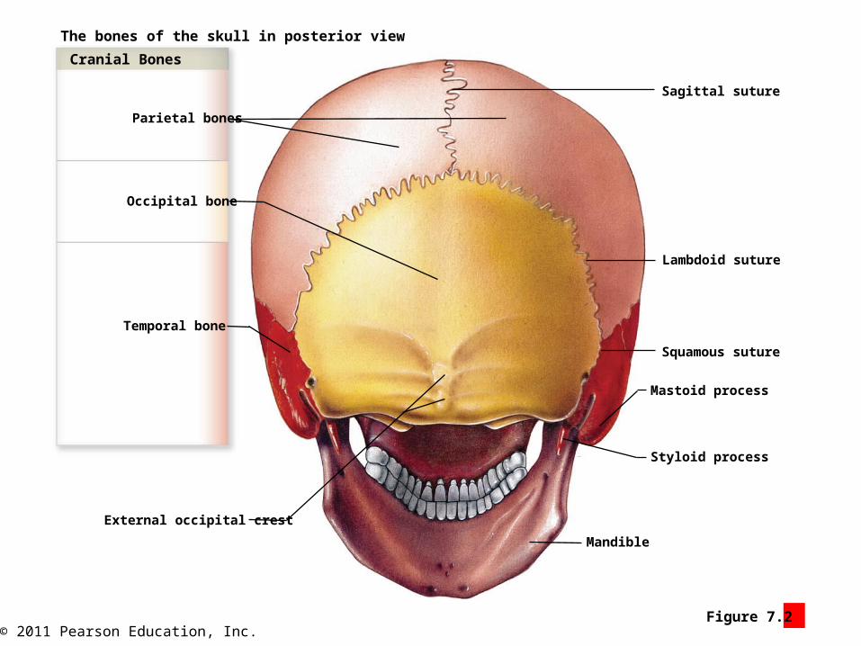
The bones of the skull in posterior view
Parietal bones
Occipital bone
Temporal bone
External occipital crest
Mandible
Styloid process
Mastoid process
Squamous suture
Lambdoid suture
Sagittal suture
Cranial Bones

© 2011 Pearson Education, Inc.Figure 7.3 1
The skull in lateral view
Frontal squama (forehead)Superior and inferior
temporal lines
Coronalsuture
Parietalbone
Frontalbone
Temporalbone
Occipitalbone
Zygomaticbone
Alveolarprocesses
Sphenoid
Ethmoid
Lacrimal bone
Nasal bone
Mandible
Mental protuberance Mandibular angle
Zygomatic arch (cheekbone)
Styloid process
Mastoid process
Lambdoid suture
Externalacousticmeatus
Squamous suture
Squamous part(of temporal bone)
Maxilla

© 2011 Pearson Education, Inc.Figure 7.3 2
Squamous suture
Frontal sinuses
Coronalsuture
Parietalbone
Frontalbone
Temporalbone
Occipitalbone
Sphenoid
Ethmoid
Nasal bone
Maxilla
Lambdoid suture
Petrous part(of temporal bone)
Styloid process
Palatinebone
Internalacousticmeatus
Mandible
Sphenoidalsinus (right) Sella turcica
Hypoglossal canal
Vomer
The interior of the skull, as revealed by sagittalsection that passes just to the left of the midline

© 2011 Pearson Education, Inc.Figure 7.4 1
An inferior view of the skull
Zygomatic bone
Frontalbone
Palatinebone
Maxilla Vomer Foramina
Foramen lacerum
Foramen ovale
Carotid canal
Jugular foramen
Stylomastoid foramen
Foramen magnum
External occipital crest
Inferior and superiornuchal lines
Lambdoid suture
Occipital condyle
Temporal bone
Mandibular fossa
Styloid process
Zygomatic arch
Sphenoid
Occipital bone

© 2011 Pearson Education, Inc.Figure 7.4 2
The interior of the skull, as revealed by horizontal section
Temporal bone
Sphenoid
Frontal bone
Carotid canal
Occipital bone
Parietal bone
Mastoid foramen
Ethmoid
Foramen lacerum
Foramen ovale
Foramen rotundum
Crista galli
Cribriform plate
Sella turcica
Foramen spinosum
Jugular foramen
Internal occipital crest
Hypoglossal canal
Internal acoustic meatus
Nasal bones

© 2011 Pearson Education, Inc.Figure 7.5 1
Two views of the sphenoid
Optic canal
Superiorsurfaceof the
sphenoid
Foramenspinosum
Foramenovale
Foramenrotundum
Superiororbital fissure
Lesser wing
Sphenoidal spine
Sella turcica
Hypophyseal fossa
Greater wing

© 2011 Pearson Education, Inc.Figure 7.5 2
The ethmoid
Cribriform plate
Lateral masses
Superior and middlenasal conchae
Perpendicular plate
Superior surface Posterior surface
Crista galli

© 2011 Pearson Education, Inc.Figure 7.5 3
The palatine bones
Perpendicular plateof the palatine bone
Horizontal plate
Nasalcrest
Orbital process

© 2011 Pearson Education, Inc.Figure 7.6 1
The bones of the orbital complex
Lacrimal fossa
Supra-orbitalmargin
Sphenoid
Temporal bone
Zygomatic bone
Zygomaticofacialforamen
Intra-orbitalforamen
Maxilla
Middle nasal concha
Inferior nasal concha
Nasolacrimal canal
Lacrimal sulcus
Ethmoid
Frontal bone
Palatine bone
Supra-orbital notch

© 2011 Pearson Education, Inc.Figure 7.6 2
The bones of the nasal complex
Frontal bone
Maxilla
Frontal section
Mandible
Cranial cavity
Zygomatic bone
Maxillary sinus
Nasal cavities
Ethmoidal air cells
Orbit

© 2011 Pearson Education, Inc.Figure 7.6 3
Frontal bone
The bones that form and surround the nasal cavity, asrevealed by a sagittal section with nasal septum removed
Lacrimal bone
Inferior nasal concha
Maxilla
Frontal sinuses
Sphenoidal sinus
Sphenoid
EthmoidNasal bone
Perpendicular plateof palatine bone
Hard palate
Pterygoid plates
Middle nasal concha
Superior nasal concha

© 2011 Pearson Education, Inc.Figure 7.7 1
The mandible in lateral view
Teeth (molars)
Condylar process
Mandibular notch
Ramus of the mandible
Body of the mandible
Mental foramen
Alveolar process
Coronoid process
© 2011 Pearson Education, Inc.Figure 7.7 2
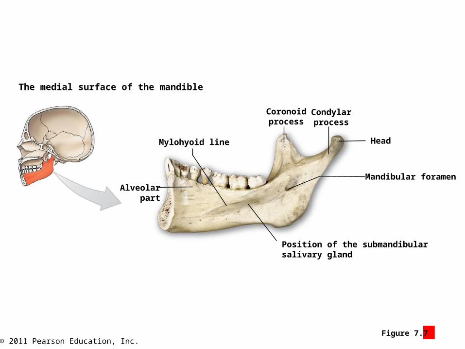
The medial surface of the mandible
Mylohyoid line
Coronoidprocess
Condylarprocess
Alveolarpart
Head
Mandibular foramen
Position of the submandibular salivary gland

© 2011 Pearson Education, Inc.Figure 7.7 3
The hyoid bone
Body of the hyoid
Greater horn
Lesser horn

© 2011 Pearson Education, Inc.Figure 7.7 4
Ethmoid
Maxillary bones
Lacrimal bones
Frontal bone
Mandible
Hyoid boneTemporal bones
Sphenoid
Palatine bones
Nasal bones
CRANIUMFACE
Zygomatic bones
Vomer
Inferior nasal conchae
Occipital bone
Parietal bones
SKULL
ASSOCIATED BONES
Auditory ossiclesenclosed in
temporal bones(see Chapter 15)
The auditory ossicles, bones associatedwith the skull
6
78
1
1 1
1
1
1
1
22
2
2 2
2
2
2
14

© 2011 Pearson Education, Inc.Figure 7.8 1
The anterior fontanelle (“soft spot”) and associated sutures in theskull of an infant
Rightparietal
bone
Leftparietal
bone
Sagittal suture
Anterior fontanelle
Coronal suture
Frontal suture
Frontal bone
Frontal suture
© 2011 Pearson Education, Inc.Figure 7.8 2
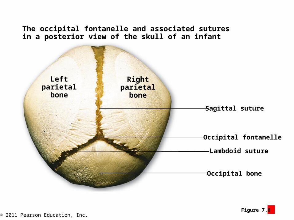
Sagittal suture
Rightparietal
bone
Leftparietal
bone
Lambdoid suture
Occipital fontanelle
Occipital bone
The occipital fontanelle and associated suturesin a posterior view of the skull of an infant
© 2011 Pearson Education, Inc.Figure 7.8 3
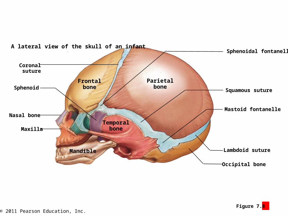
A lateral view of the skull of an infant
Parietalbone
Frontalbone
Temporalbone
Coronalsuture
Sphenoid
Nasal bone
Maxilla
Mandible
Occipital bone
Lambdoid suture
Sphenoidal fontanelle
Mastoid fontanelle
Squamous suture
© 2011 Pearson Education, Inc.Figure 7.8 4
Frontal suture
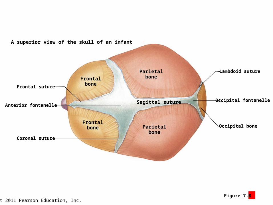
A superior view of the skull of an infant
Frontalbone
Parietalbone
Frontalbone Parietal
boneCoronal suture
Anterior fontanelleSagittal suture Occipital fontanelle
Occipital bone
Lambdoid suture

© 2011 Pearson Education, Inc.Figure 7.9 1
The spinal curves and vertebral regionsin the adult vertebral column
Spinal Curves
Primary curves develop beforebirth, and secondary curves afterbirth.
Cervical curve(a secondary curve)
Thoracic curve(a primary curve)
Lumbar curve(a secondary curve)
Sacral curve(a primary curve)
Sacral
Coccygeal
Lumbar(5 vertebrae)
Thoracic(12 vertebrae)
Cervical(7 vertebrae)
Vertebral Regions
Regions are definedby anatomicalcharacteristics ofindividual vertebrae.C1
C2C3C4C5C6C7T1
T2T3
T4
T5
T6
T7
T8
T9
T10
T11
T12
L1
L2
L3
L4
L5

© 2011 Pearson Education, Inc.Figure 7.9 1

© 2011 Pearson Education, Inc.Figure 7.9 2
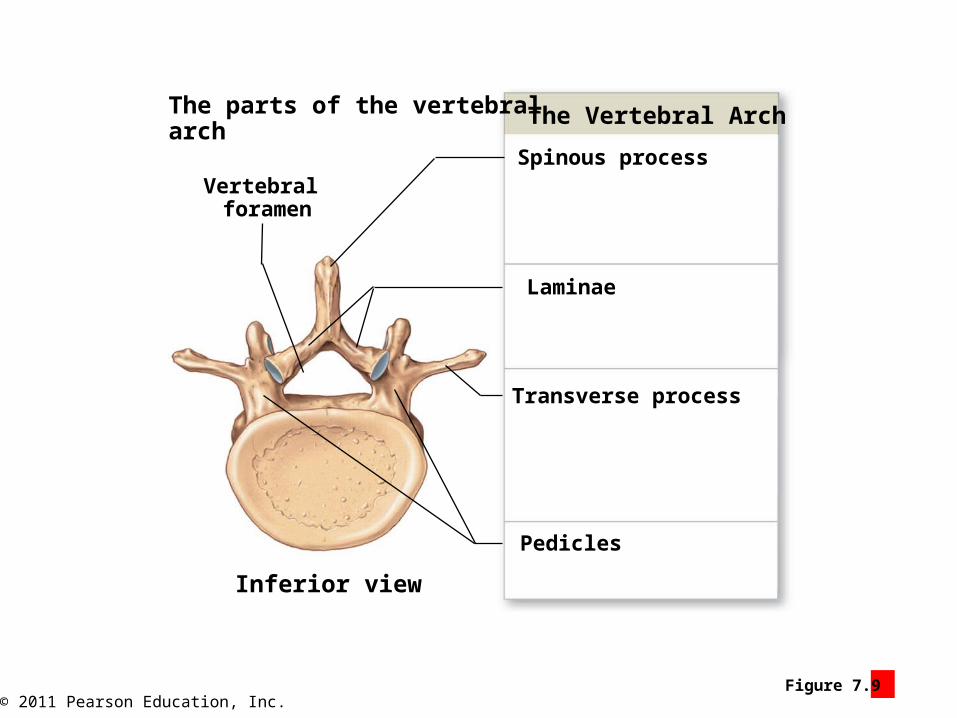
The parts of a typical vertebra Parts of a Vertebra
Articular processes
Vertebral arch
Vertebral body
Superior view
© 2011 Pearson Education, Inc.Figure 7.9 3
The parts of the vertebralarch
Vertebral foramen
Inferior view
Pedicles
Laminae
Transverse process
Spinous process
The Vertebral Arch

© 2011 Pearson Education, Inc.Figure 7.9 4
A lateral view of three vertebrae
Pedicle
Intervertebral disc
Intervertebral foramina
Vertebral canal
Vertebralbody

© 2011 Pearson Education, Inc.Figure 7.9 5
A posterior view of two vertebrae
Articular facet
Superior articular process
Inferior articular process
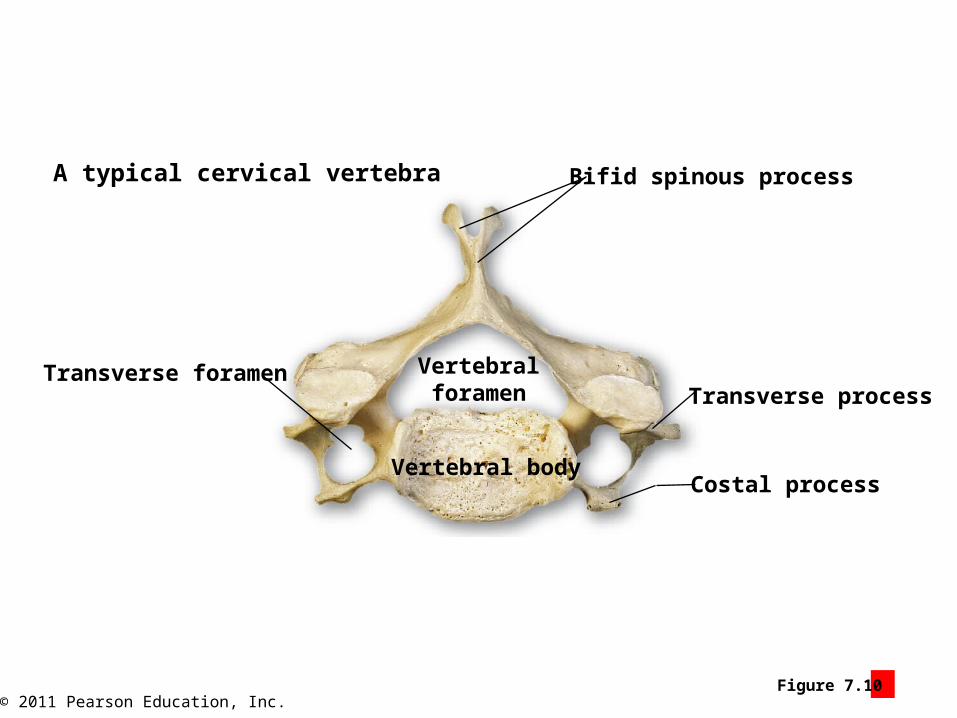
© 2011 Pearson Education, Inc.Figure 7.10 1
A typical cervical vertebra
Transverse foramen
Vertebral body
Vertebralforamen
Bifid spinous process
Transverse process
Costal process
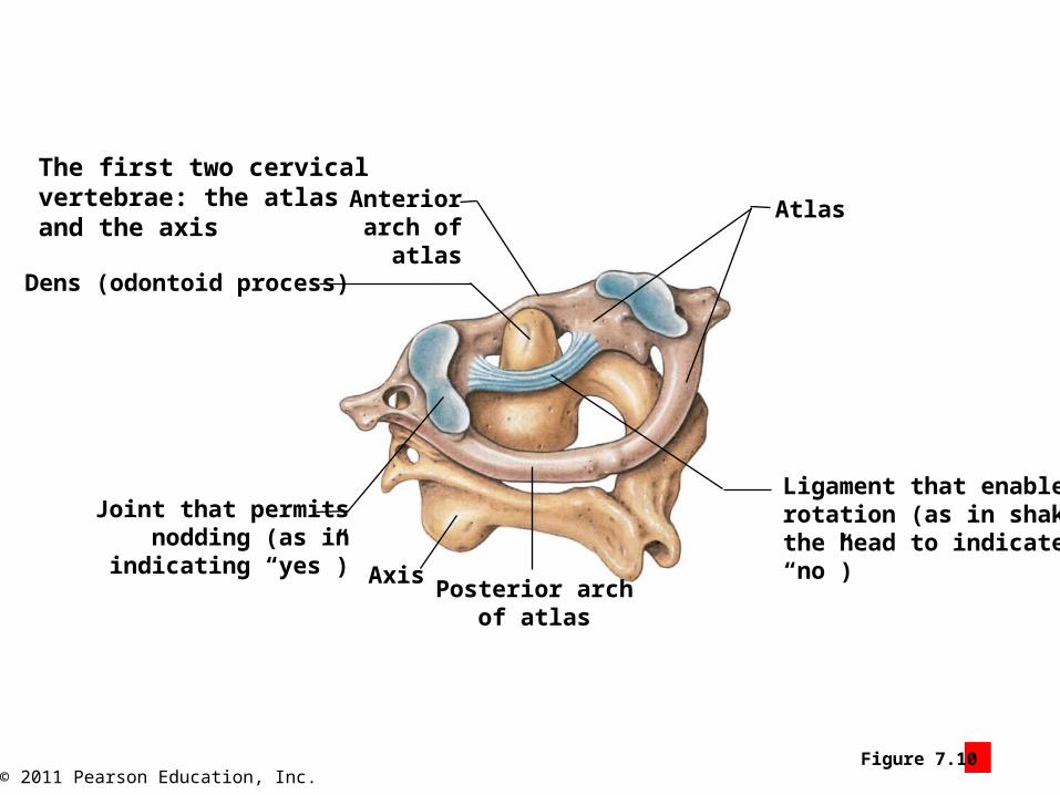
© 2011 Pearson Education, Inc.Figure 7.10 2
The first two cervicalvertebrae: the atlasand the axis
Anteriorarch of
atlasDens (odontoid process)
Joint that permitsnodding (as in
indicating “yes”)Posterior arch
of atlas
Axis
Atlas
Ligament that enablesrotation (as in shakingthe head to indicate“no”)

© 2011 Pearson Education, Inc.Figure 7.10 3
A lateral view of the seven cervical vertebrae
Vertebraprominens

© 2011 Pearson Education, Inc.Figure 7.10 4

© 2011 Pearson Education, Inc.Figure 7.10 5
Transverse process
Vertebralforamen
Spinous process
A typical thoracic vertebra in superior view
Vertebralbody
Superiorarticular facet
Superiorcostal facet
© 2011 Pearson Education, Inc.Figure 7.10 6
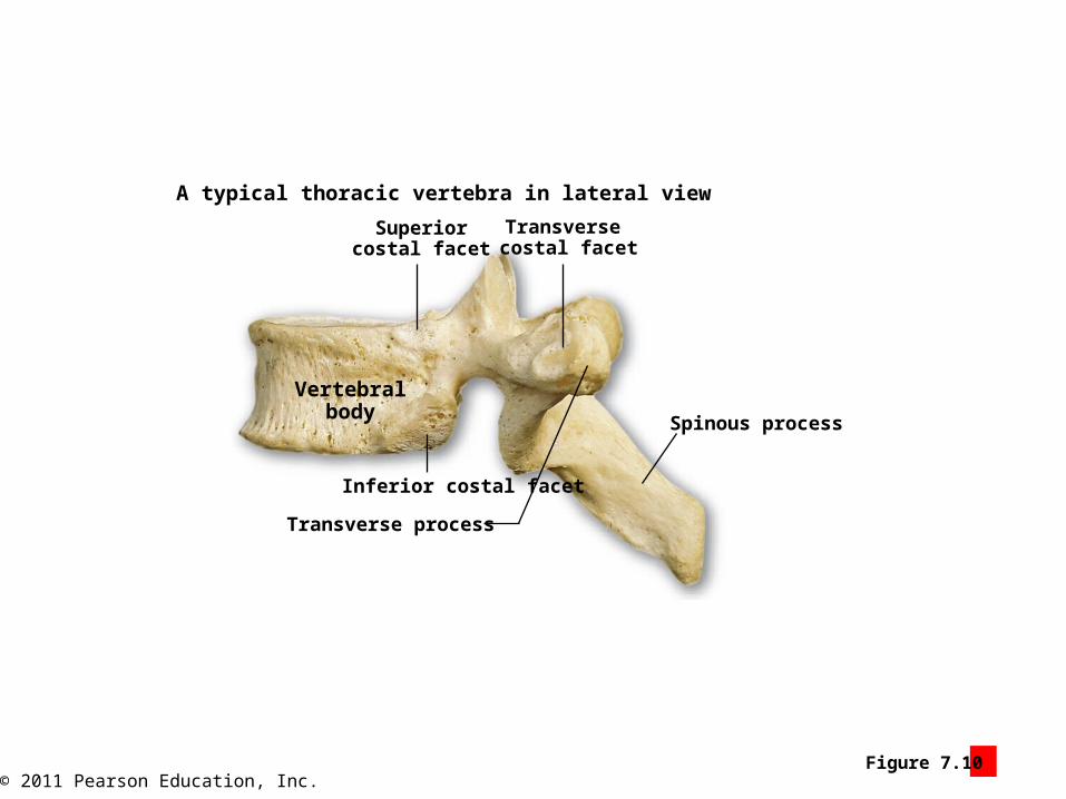
Vertebralbody
Spinous process
Transverse costal facet
Superiorcostal facet
A typical thoracic vertebra in lateral view
Transverse process
Inferior costal facet

© 2011 Pearson Education, Inc.Figure 7.12 1
An anterior view of the thoracic cage
Jugular notch
Vertebrosternal ribs(ribs 1–7)
Vertebrosternal ribs(ribs 8–10)
Floating ribs(ribs 11 and 12)
Ribs
Costal cartilages
Xiphoid process
Body
Manubrium
Sternum
T1
T11
T1212
11
1
2
3
4
5
6
7
8
9
10
© 2011 Pearson Education, Inc.Figure 7.12 2
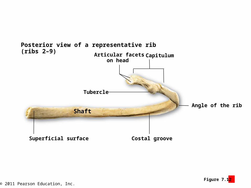
Posterior view of a representative rib(ribs 2–9)
Tubercle
Articular facetson head
Shaft
Superficial surface Costal groove
Capitulum
Angle of the rib

© 2011 Pearson Education, Inc.Figure 7.12 4
Sternum
Ribs
The action of a typical rib, whichcan be likened to the movementof a bucket’s handle
© 2011 Pearson Education, Inc.Figure 7.12 3
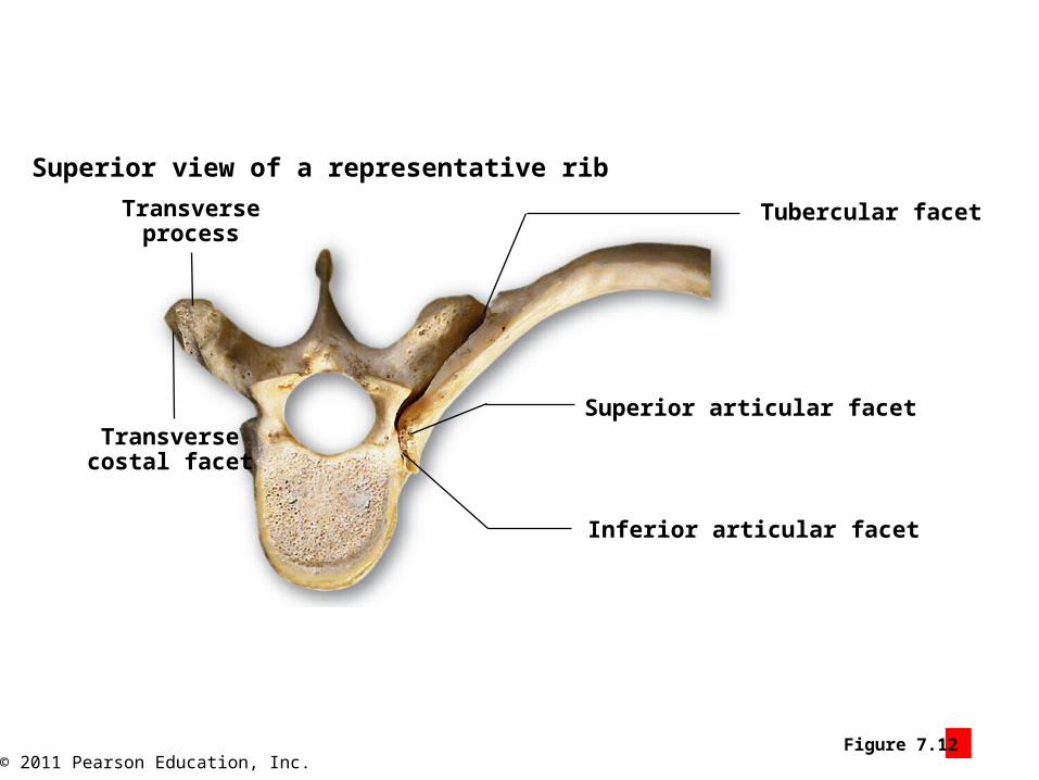
Tubercular facet
Superior articular facet
Inferior articular facet
Transversecostal facet
Transverseprocess
Superior view of a representative rib

© 2011 Pearson Education, Inc.Figure 7 Section 2
SKELETAL SYSTEM
AXIALSKELETON
The bones of the appendicular skeleton
80
2
2
2
2
2
16
Clavicle
Scapula
Humerus
Radius
Ulna
Carpal bones
Metacarpal bones
Phalanges(proximal,
middle, distal)
Hip bone(coxal bone)
Femur
Patella
Tibia
Fibula
Tarsal bones
Metatarsal bones
Phalanges
Lowerlimbs
Pelvicgirdle
Upper limbs
Pectoralgirdle
4
60
2
126
2
2
2
2
2
28
28
10
14
10
206
60
APPENDICULARSKELETON

© 2011 Pearson Education, Inc.Figure 7.13 1
The relationship of the clavicleto adjacent bones
Anterior view
Humerus
Scapula
Clavicle
Jugular notch
© 2011 Pearson Education, Inc.Figure 7.13 2
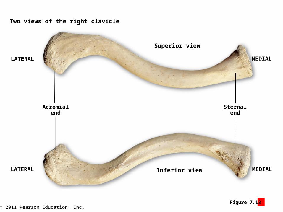
Two views of the right clavicle
Superior view
LATERAL MEDIAL
Acromialend
Sternalend
LATERAL Inferior view MEDIAL

© 2011 Pearson Education, Inc.Figure 7.13 3 – 4
Two views of the right scapula
Acromion Coracoidprocess
Superiorborder
Superiorangle
Medialborder
Inferiorangle
Lateralborder
Process thatsupports thecup-shaped
glenoid cavity
Anterior view
Acromion
Scapular spine
Supraspinous fossa
Infraspinous fossa
Posterior view
Subscapular fossa

© 2011 Pearson Education, Inc.Figure 7.13 5
A lateral view of the rightscapula
Acromion
Glenoid cavity
Coracoidprocess
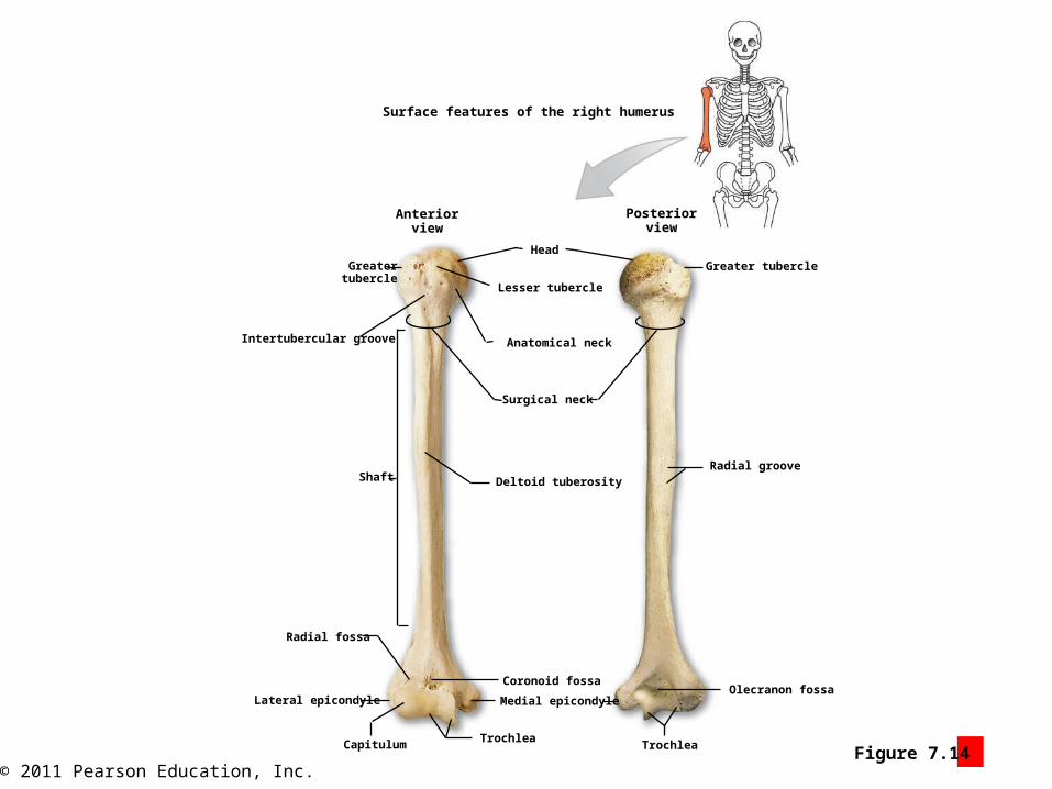
© 2011 Pearson Education, Inc.Figure 7.14 1
Surface features of the right humerus
Anteriorview
Greatertubercle
Head
Posteriorview
Greater tubercle
Lesser tubercle
Anatomical neck
Surgical neck
Intertubercular groove
Shaft Deltoid tuberosity
Radial fossa
Lateral epicondyle
Coronoid fossa
Medial epicondyle
CapitulumTrochlea
Trochlea
Olecranon fossa
Radial groove

© 2011 Pearson Education, Inc.Figure 7.14 2
Olecranon
Posteriorview
Surface features of the right ulna and radius
Proximal radio-ulnar joint
Ulna Radius
Ulnar head
Ulnar notch
Interosseous membrane
Styloid process of the ulna
Styloid process of the radius
Radius
Ulnar head
Ulna
Distal radio-ulnar joint
Radial notchat proximalradio-ulnar joint
Radial tuberosity
Neck of the radius Coronoid process
Trochlear notchRadial head
Anteriorview

© 2011 Pearson Education, Inc.Figure 7.15 1
The bones of the carpus (wrist)
Proximal Carpal Bones
Scaphoid
Right wrist and hand,anterior (palmar) view
Lunate Pisiform Triquetrum
Metacarpalbones
Trapezium Trapezoid Capitate Hamate
Distalphalanx
Middlephalanx
Proximalphalanx
Radius Ulna
VIVIIIIII
Distal Carpal Bones

© 2011 Pearson Education, Inc.Figure 7.15 2
I
IIIIIIV
V
Proximal phalanx
Middle phalanx
Distal phalanx
Pisiform
Triquetrum
Lunate
Scaphoid
Proximal Carpal Bones
The metacarpal bones(designated I–V) and thephalanges of the hand Ulna Radius
Proximal phalanx of pollex
Distal phalanx of pollex
Metacarpal bones
Right wrist and hand,posterior (dorsal) view
Hamate
Capitate
Trapezoid
Trapezium
Distal Carpal Bones
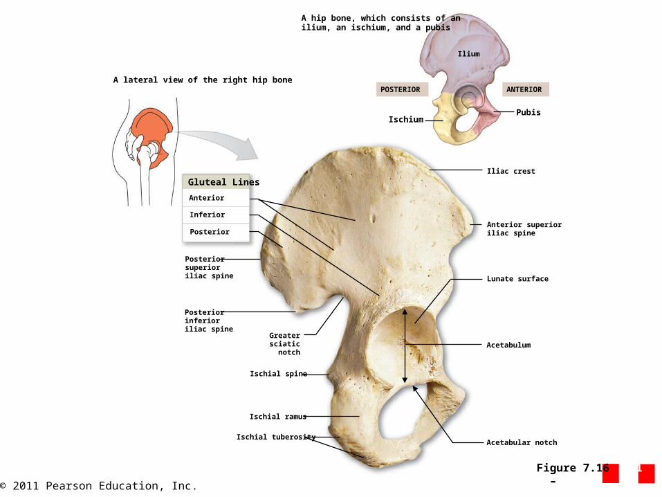
© 2011 Pearson Education, Inc.Figure 7.16 1 – 2
A lateral view of the right hip bone
A hip bone, which consists of anilium, an ischium, and a pubis
Gluteal Lines
Posterior
Posteriorsuperioriliac spine
Posteriorinferioriliac spine
Greatersciaticnotch
Ischial spine
Ischial ramus
Ischial tuberosityAcetabular notch
Acetabulum
Lunate surface
Anterior superioriliac spine
Iliac crest
IschiumPubis
ANTERIORPOSTERIOR
Ilium
Inferior
Anterior

© 2011 Pearson Education, Inc.Figure 7.16 3
ANTERIOR POSTERIOR
Ilium
Pubis Ischium
Iliac crest
Iliac fossa
Arcuate line of the ilium
Pectineal line
Superior pubic ramus
Pubic symphysis
Inferior pubic ramus
Ischial ramus
Obturator foramen
Greater sciatic notch
Auricular surfaceof the ilium
Iliac tuberosity
A medial view of the right hip bone
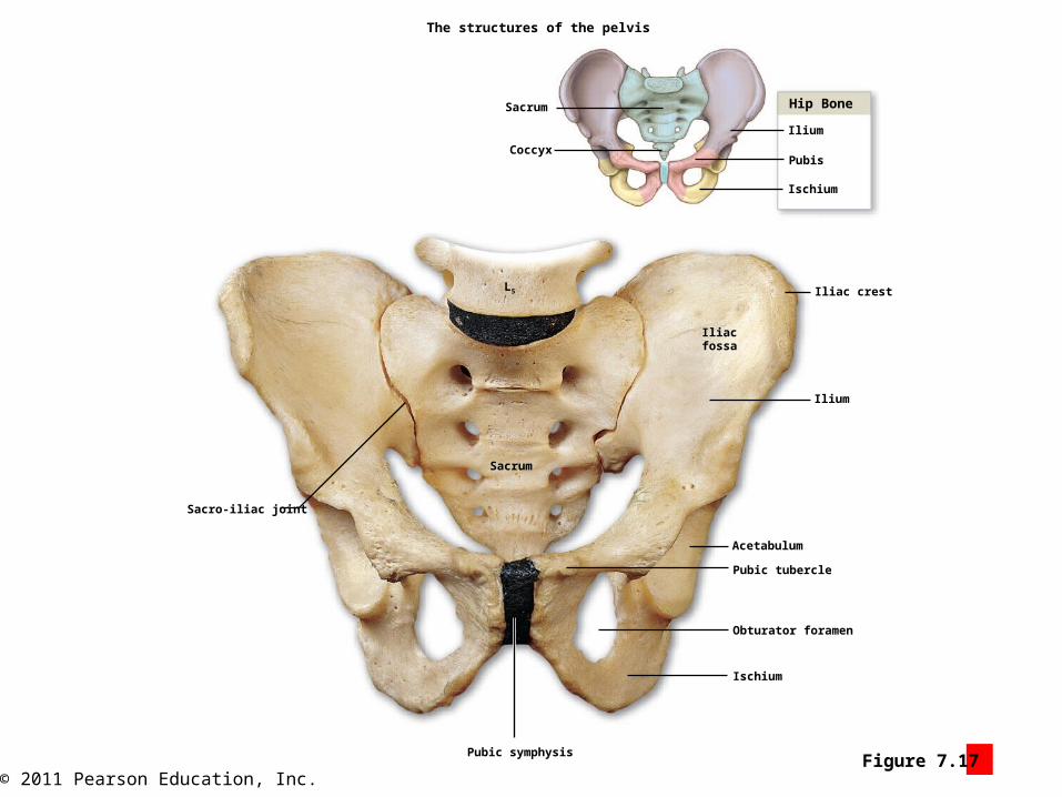
© 2011 Pearson Education, Inc.Figure 7.17 1
The structures of the pelvis
Sacrum
Coccyx
Hip Bone
Ilium
Pubis
Ischium
Iliac crest
Iliacfossa
Ilium
Acetabulum
Pubic tubercle
Obturator foramen
Ischium
Pubic symphysis
Sacrum
Sacro-iliac joint
L5

© 2011 Pearson Education, Inc.Figure 7.17 2
The locations and extents of the true (lesser) pelvis(in purple) and the false (greater) pelvis
Superior view Inferior view
Ischialspine
Pelvic outlet
Pelvicoutlet
Pelvic inletPelvic brim
False pelvis

© 2011 Pearson Education, Inc.Figure 7.17 3
The shapes of the pelvis in females and males
The pelvis of a female The pelvis of a male
Female Male
Ischialspine
Ischialspine
100°or more 90°
or less
© 2011 Pearson Education, Inc.Figure 7.18 1 - 2
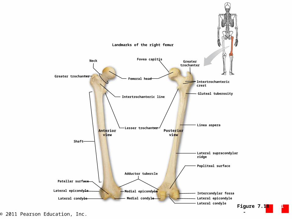
Landmarks of the right femur
Neck
Greater trochanter
Shaft
Patellar surface
Lateral epicondyle
Lateral condyle Medial condyle
Medial epicondyle
Adductor tubercle
Lesser trochanterAnterior
viewPosterior
view
Intertrochanteric line
Femoral head
Fovea capitisGreater
trochanter
Intertrochantericcrest
Gluteal tuberosity
Linea aspera
Lateral supracondylarridge
Popliteal surface
Intercondylar fossa
Lateral epicondyle
Lateral condyle

© 2011 Pearson Education, Inc.Figure 7.18 3
Anterior view Posterior view
Attachment area forthe patellar ligament,
which attaches thepatella to the tibia
Attachmentarea for
quadricepstendon
Baseof patella
The surface features of the patella
Apexof patella
Articular surfaceof patella
Medial facet, for medialcondyle of femur
Lateral facet, forlateral condyleof femur

© 2011 Pearson Education, Inc.Figure 7.18 4
Lateral tibial condyle
Head of the fibula
Fibula
Lateral malleolusof the fibula
Inferiorarticular surface
Medial malleolusof the tibia
Tibia
Anterior marginof the tibia
Interosseous membrane
Anteriorview
Posteriorview
Tibial tuberosity
Medial tibial condyle
Superiortibiofibular joint
Articular surface ofmedial tibial condyle
Intercondylar eminence
Articular surface oflateral tibial condyle
Lateral tibial condyle
Head of fibula
Fibula
Inferior tibiofibularjoint
Lateral malleolus(fibula)
The features of the right tibia and fibula

© 2011 Pearson Education, Inc.
The Ankle (Tarsus)
The ankle consists of seventarsal bones.
Calcaneus
Talus
Navicular
Cuboid
Cuneiform bones
The bones of the ankle and foot
Trochlea
Metatarsals
Articulations of the cuboid and thecuneiform bones with the metatarsalbones
Metatarsal bones (designated I–V)
Phalanges
Proximal, middle, and distal phalanges
Proximal phalanx
Distal phalanx
Hallux
V IV III II I
Figure 7.19 1
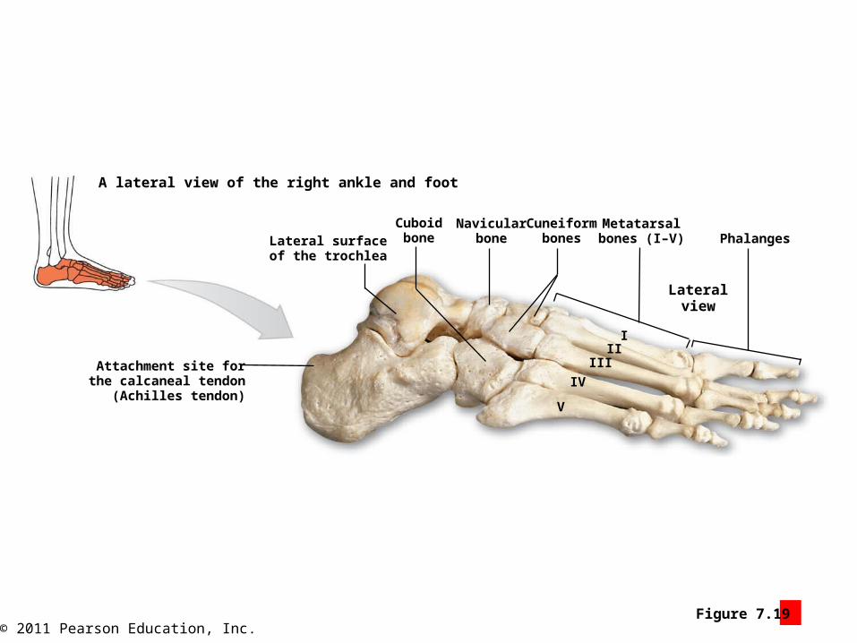
© 2011 Pearson Education, Inc.Figure 7.19 2
Lateralview
A lateral view of the right ankle and foot
PhalangesMetatarsalbones (I–V)
Cuneiformbones
Navicularbone
CuboidboneLateral surface
of the trochlea
Attachment site forthe calcaneal tendon
(Achilles tendon)V
IV
IIIII
I
© 2011 Pearson Education, Inc.Figure 7.19 2
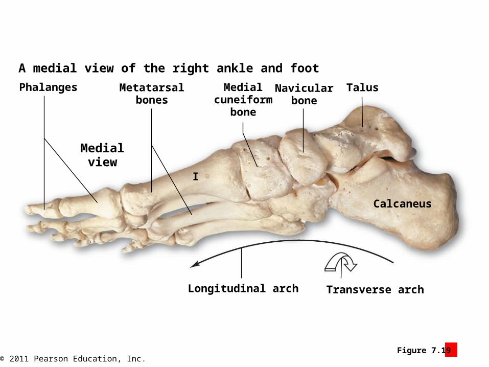
Phalanges
Medialview
Metatarsalbones
Medialcuneiform
bone
Navicularbone
Talus
Calcaneus
Longitudinal arch Transverse arch
A medial view of the right ankle and foot
I