11lomaoo state college: of a !~h
TRANSCRIPT

MEMOIR 254
EXAMINED AMD CHEC~USTti .
. '. I.lt::lkAkY NOVEMBER 1943
.' 11lOMAOO STATE COLLEGE: OF A ~ !~h~e~T €!~b.L'NS COLO.,. ...
CORNELL UNIVERSITY - ",,"00
AGRICULTURAL EXPERIMENT STATION
COMPARATIVE STUDY OF MOUTH PARTS
OF REPRESENTATIVE HEMIPTERA-HOMOPTERA
F. H. BUTT
ITHACA. NEW YORK
PUBLISHED BY THE UNIVERSITY
Received for publication April 29, 1943
Butt, F. H. 1943.pdf Memoir. Cornell University, Agricultural Experimental Station. 254:1-20.

CONTENTS
PAGEThe head................................................................ 3The syringe and the hypopharyngeal wings. . . . . . . . . . . . . . . . . . . . . . . . . . . . . . . . . . 5The lora.... . . . 6The food pump. . . . . . . . . . . . . . . . . . . . . . . . . . . . . . . . . . . . . . . . . . . . . . . . . . . . . . . . . . . 8The labium of Cicada and of Cephisus. . . . . . . . . . . . . . . . . . . . . . . . . . . . . . . . . . . . . . . 9The labium of Notonecta. . . . . . . . . . . . . . . . . . . . . . . . . ... . . . . .. . . . . . . . . . . . . . . . . 10The labium of Oncopeltus. . . . . . . . . . . . . .. . . . . . . . . . . . . . . . . . . . . . . . . . . . . . . . . .. 11The mandible and maxilla of Cicada. . . . . . . . . . . . . . . . . . . . . . . . . . . . . . . . . . . . . . .. 13The mandible and maxilla of Hemiptera. . . . . . . . . . . . . . . . . . . . . . . . . . . . . . . . . . . . . 15Summary................................................................ 18References. . . . . . . . . . . . . . . . . . . . . . . . . . . . . . . . . . . . . . . . . . . . . . . . . . . . . . . . . . . . . .. 19Abbreviations. . . . . . . . . . . . . . . . . . . . . . . . . . . . . . . . . . . . . . . . . . . . . . . . . . . . . . 20
Plates I to VIn
Butt, F. H. 1943.pdf Memoir. Cornell University, Agricultural Experimental Station. 254:1-20.

COMPARATIVE STUDY OF MOUTH PARTS
OF
REPRESENTATIVE HEMIPTERA-HOMOPTERA
F. H. BUTT
The members of the order Hemiptera, commonly known as the true bugs,and their close relatives, the members of the order Homoptera, among whichare aphids, leafhoppers, and others, constitute a group of insects of greateconomic importance to man. Some of them cause widespread damage tovegetation through the withdrawal of heavy amounts oj plant juices; othersspread causative organisms of plant diseases; and some attack man anddomestic animals, causing irritation and great economic loss. They all havepiercing and sucking mouth parts that act as efficient hypodermic syringes.The mechanism that operates these mouth parts, the methods of penetrationof plant or animal tissues, and the withdrawal of food from these tissueshave long been subjects of interest to entomologists and much work has beendone in those fields. This memoir is the result of comparative studies ofthe food pump and of the mouth parts and their muscle attachments inseveral representative species of the Homoptera and the Hemiptera, todetermine wherein the mouth parts of the two orders are similar and whereinthey differ in their methods of operation.
TibicinG septendecim '(L.), the seventeen-year Cicada (Plate II, A), andthe nymph of Cephisus siccifolius (\Valker), a South American memberof the family Cercopidae (Plate I, C) were used as representatives of theHomoptera. BenGclts griseus (Say) (Plate I, A), and N otonecta sp.(Plate I, B), were used to represent the aquatic Hemiptera; Oncopeltusfascia/us (Dallas), the milkweed bug, (Plate I, E), and Acrosternurn hilare(Say) a stinkbug (Plate CD) represent the terrestrial plant-feeding forms.
The author is indebted to R. E. Snodgrass of the United States Bureauof Entomology and Plant Quarantine for his suggestions, which have beenhelpful in the conduct of the investigations leading to this memoir, and forthe use of his illustration of the mandibles of the roach (Plate VIII, B).
THE HEAD
The head of Cicada is triangular in outline with eyes at the outer cornersand the mouth parts suspended from the lower point. The most prominentfeature of the head is a convex bulge between the compound eyes with horizontal corrugations on its surface marking the attachment of powerfulmuscles within (Plate II, A). This bulge is the postclypeus (Pclp). Beneaththe postclypeus is the anteclypeus (Aclp) and attached to its distal marginis the small labrum (Lm) which is applied closely to the base of the labium.The labium (Lb) itself is elongate, grooved on its anterior surface to containthe bristles constituting the feeding apparatus, and suspended freely fromthe membrane posterior to the maxillary plates.
Bounding the lower lateral edges of the postclypeus are two plates, thelora (Lo), or mandibular plates, and laterad of these the maxillary plates(Mx) continuous with the cranial wall. They merge into the genae beneath
Butt, F. H. 1943.pdf Memoir. Cornell University, Agricultural Experimental Station. 254:1-20.

4 CORNELL EXPERIMENT STATION MEMOIR 254
the eyes. .The antennae are close to the epistomal sutures and project frombeneath overhanging bulges mesad of the compound eyes.
The cephalic aspect of Cephisus shows the same characteristics as Cicada(Plate I, C). The clypeus is divided into the enormous postclypeus (Pclp)and distally the smaller anteclypeus (Aclp). Attached to the distal end ofthe anteclypeus is the small pointed labrum (Lm) covering the base of thetrough in the labium (Lb). Laterad of the post- and anteclypeus on eachside are the exposed loral plates (Lo) and beneath them the maxillary plates(Mx). Cephisus is included mainly to show the musculature of the labiummore clearly than Cicada does.
In Notonecta the eyes are enlarged and the area between them, the clypeus,extends across the top of the head where it merges into the front (Plate I,B). There are no frontal sutures or ocelli.
The maxillary plates are partially covered by the extension of the clypealwall of the head. Laterally two oval sclerites (Mx) of the maxillary segmentare visible but the median lobes to which the maxillary protractors areattached lie below the lora adjacent to the hypopharynx. The clypeus extendsto a line on the front of the head along the lower extremity of the maxilla.At this point is the clypeo-Iabral suture, and below it lies the labrum, atriangular flap that covers the trough and part of the basal segment of thelabium.
Specializations found in the head of Benacus are likewise due to theenlargement of the eyes in adaptation to aquatic life (Plate I, A). But theclypeus is divided into postclypeus fused with the front (Pclp + Fr) andanteclypeus or tylus (Aclp). The lateral areas of the postclypeus extenddownward between the eyes alongside the tylus.
The maxillary plates (Mx) are greatly altered by enlargement of theeyes and are pushed distally until they form flap-like covers for the sides ofthe basal segment of the labium. These flaps nearly meet in front along themedian line overlapping the anteclypeus or tylus (Aclp) for a short distance.The labrum (Lm) is a long striated lobe attached to the lower extremityof the anteclypeus. It forms a cover for the bristle trough in the labium (Lb).The antennae are pushed to the caudal surface of the head where they havebecome embedded in cavities immediately beneath each eye. If the surfaceof an eye is removed the inner surface of one of these cavities appears as adome-like protuberance (Plate IV, A, Y), just above the antennal fossa.
The heads of the milkweed bug, Oncopeltus fasciatus, (Plate I, E) andof Acrosternum (Plate I, D) are typical of the terrestrial forms of theHemiptera. From the dorsal side of the head, the anteclypeus (Aclp)appears as a narrow, well defined median sclerite marked off from the jugum (Ju) on each side by deep clefts (Plate I, E, D). The cranium is unbroken dorsally by other sutures than these. Caudad of the anteclypeus aretwo ocelli, mesad of the compound eyes. Between the ocelli are the fusedpostclypeus and frons (Pclp + Fr).
The beak arises at the anterior point of the head (Plate VI, B) and iscurved back beneath the thorax when not in use. The anteclypeus mergesinto the long pointed labrum, or upper lip, (Lm), which helps to hold thebristles within the trough of the beak. Below and in front of the antennalbase is the maxillary plate (Mx). The suture (s) between the jugum andmaxillary plate is a constant feature and is always well marked in the forms
Butt, F. H. 1943.pdf Memoir. Cornell University, Agricultural Experimental Station. 254:1-20.

MOUTH PARTS, HEMIPTERA-HOMOPTERA 5
studied. Below the maxillary plate is the narrow buccula (Buc). On thelower surface of the head extending back from the antenna is a suture(Ocs), not always distinct, which marks off the gula plate (Gu).
The hypopharynx is a median lobe protruding from the lower surfaceof the head between the mandibular plates, concealed by the anteclypeusand labrum in the Cicada, by the unbroken front surface of the head inNotonecta, and by the anteclypeus in other Hemiptera. The upper surfaceof the hypopharynx is grooved narrowly toward the tip to form the floorof the tube that carries food to the sucking pump. This groove runs into amedian basin-like enlargement forming the floor of the sucking pump, whichis well sclerotized and braced to withstand the suction resulting from theaction of the dilator muscles of the pump. The hypopharynx (Hphy) and thefloor of the food pump (Flpmp) are clearly seen when the anteclypeus iscut away in Oncopeltus (Plate VIII, I) or in Acrosternum (Plate VIII, H).A mid-sagittal section through the head of Cicada (Plate II, B) or ofCephisus (Plate III, A) shows that the food duct on the upper surface ofthe hypopharynx runs vertically into the pump in these insects.
THE SYRINGE AND THE HYPOPHARYNGEAL WINGS
In more generalized insects, at the point where the ventral or posteriorwall of the hypopharynx (figure 1) becomes the dorsal or anterior wall ofthe labium, a pocket forms, called the salivarium (Slv), into which thesalivary ducts empty. On its dorsal wall is inserted a pair of dorsal salivarymuscles (Is), which originate either on the suspensory sclerites (Hs) of thehypopharynx or on the lateral walls of the hypopharynx when these areabsent. On its ventral wall are inserted the salivary muscles of the labium'(2s and 3s). In the Orthoptera the dorsal wall of the salivarium is somewhat concave (Snodgrass, 1935). In both the Homoptera and Hemipterathis concave surface is modified into a closed tube which delivers the salivaryjuices to the tip of the hypopharynx ilnd from there into the salivary canalin the bristle bundle (Plate VI, C, Duct). The salivarium of the Orthopterahas become modified in the Homoptera and Hemiptera into a pump, thesalivary syringe, that is equipped wi::h a piston and valves to control thedirection of flow of the salivary liquid (Plate II, B; Plate III, A; Plate VI,A, D; Syr).
The lower lateral surfaces of the hypopharynx in Homoptera and Hemiptera are extended back into the head as two divergent trough-like platesor wings (Plate IV, A, D, Hphyw; Plate V, C, Hphyw). To the under sideof these the dorsal salivary muscles of the salivarium, now greatly enlargedinto the dilator muscles of the syringe (Plate IV, D, dlsyr), are fastened. Thesalivary muscles from the labium have disappeared. Plate IV, D shows therelationship of the syringe to the backward flaring wings of the hypopharynxin Notonecta. The hypopharynx has been cut away for this figure to showthe salivary pump (Syr). The posterior extremities of the wings are narrowly extended for the origin of the dilator muscles (dlsyr). In Cicada andin Cephisus the wings remain broad and flat and are united closely with theposterior tentorial arm (Plate II, F, Tnt). In Oncopeltus they showconsiderable modifications; they extend back into the head as in Cicada andNotonecta, but beyond the point where the maxillary lever -is located theybecome membranous, are deeply folded, and are attached to the ventral head
Butt, F. H. 1943.pdf Memoir. Cornell University, Agricultural Experimental Station. 254:1-20.

6 CORNI::LL EXPERIMENT STATION i\!E),IOIR 254
Fen .-------Tnl
-vdlphy
,Hphy) 'Lb
FJ<;URE 1. SAGITTAL SECTION OF GENERALIZED INSECT ilEAl) TO THE LH,. OF Tin: MEDI"NLINF.
wall (Plate VIT, B, D; Hphyw) by means of fibers resembling tonofibrillae.As a consequence of this modification the origins of the dilators of the syringe(dlsyr) have migrated to the ventral lateral surfaces of the head.
Other muscles originating on the hypopharYllgcal wings are the firstadductors (add 1) of the lahium in Notonecta (Plate V, C, D) and inOncopcltus (Plate VI. A). Short fihers (Ii.) extend from the upper innersurface of the wings (Hphyw) to the under surface of the food pump floor(Flpmp) in these insects (Plate V, C. I), and Plate VI. D, E). These fibershold the floor of the food pump firm against the pull of the pump muscles.
THE LORA
If the labrum and the alHec1ypetts of Cicada arc lifted up to expose thehypopharynx (Hphy) (figure 2), the loral plates (La) are seen to becontinuous with the LIpper surface of the hypopharynx and the pump (Snod-
Butt, F. H. 1943.pdf Memoir. Cornell University, Agricultural Experimental Station. 254:1-20.

MOUTH PARTS, HEMIPTERA-HOMQPTERA
Pclp--
Lo----
Mx----
""~'~~.' -,
--Lm
7
FIGCRE 2. CEPHALIC ASPECT or CICADA WITH LABRUM AND ANTECLYPEUS LlI'TED TOSHOW IIYPOPIIARYNX AND rLOOR or rOOD PUMP
grass, 1938). In Notonecta the loral surfaces are enclosed entirely withinthe head and can be seen only when the front surface of the head capsulehas been removed (Plate IV, B, Lo).
In Benacus (Plate I, A), Oncopcltus (Plate I, E), Acrosternum (Plate I,D), and other Heteroptera the lora are modified in a different way. In theseforms the postdypcus (Pdp) fuses with the front (Fr) of the head but theanteclypeus (Aclp), or tylus, remains distinct. The lora (Lo) are not flatplates, but form vertical walls continuous with the upper surface of thehypopharynx inside the head (Plate VI, D). The anteclypeus (Ac1p) fits
Butt, F. H. 1943.pdf Memoir. Cornell University, Agricultural Experimental Station. 254:1-20.

8 CORNELL EXPERIMENT STATION MEMOIR 254
, tightly in the groove between the loral plates (Lo) so as to form a lid forthe food 'pump.
THE FOOD PUMP
The cibarium (Cb) of the generalized insect is a space just in front ofthe opening into the stomodaeum enclosed anteriorly by the epipharyngealsurface of the clypeus (figure 1, and Plate VIII, B). Its floor is the slightlyconcave upper surface of the hypopharynx; it is flanked by the hypopharyngeal suspensorial sclerites (Hs). The cibarium is compressed by the contraction of the retractor muscles of the mouth ang~es (rao) which help toforce the food into the buccal region of the stomodaeum (Buc).
Behind the buccal region is the pharynx (Phy), which crosses over thetentorium. The pharynx usually appears as an enlarged portion of the stomodaeum, and may be divided into the anterior pharynx (Phy), that portionin front of the cerebral nerve connectives, and the posterior pharynx (Pphy),which lies above the tentorial bar (Tnt). The dorsal dilators (dlphy) of thepharynx arise on the head wall before and behind the brain and the lateraland ventral dilators (vdlphy) arise on the tentorium.
Just behind the dilators of the cibarium (dlcb) is a pair of muscles alsoarising on the clypeus and inserted on the upper surface of the buccal region(Buc). These are the dilators buccales (dlbc) of Snodgrass. Behind themand separating them from the muscles of the pharynx is the frontal ganglion(Fr gng). The frontal ganglion and the frontal connectives (Frn) alwaysform a loop enclosing the dilators of the pharynx and the retractors of themouth angles (rao).
The food pump of Cicada evidently represents' the cibarial region of themore generalized insects (Snodgrass, 1935). As has already been pointedout, its floor (figure 2, Flpmp) is the basin on the upper surface of the hypopharynx (Hphy); the cover, which is flexible and acts as a diaphragm,is the epipharyngeal surface of the anteclypeus. The dilator muscles (dlcb)of the pump originate on the corrugated walls of the postclypeus (Pdp)that form such a striking feature of the head (Plate II, A, B).
While the functional mouth of Cicada is represented by the tube leadingfrom the deep cleft (Duct) between the anteclypeus and the hypopharynx,the true mouth (Mth) is the posterior opening of the pump into the stomodaeum. The stomodaeum extends upward from the inner mouth and enlargesinto a small sac resting on the transverse bar of the tentorium (Plate II,B, C, Phy). This sac, according to Snodgrass ( 1935), is the true pharynxsince the frontal ganglion (not shown in figures of Cicada) lies on itsanterior end. The walls of the pharynx are muscular and the organ isprovided with dilator muscles (dlphy) arising on the postocular region ofthe head and on the tentorium (Plate II, C). The pharynx is also providedwith dilator muscles originating immediately behind the dilators of the foodpump (Plate II, B, dlphy). These dilator muscles are not large and theirorigin is separate from that of the more cephalic pump muscles, but in somemembers of the Hemiptera these two masses of muscles are difficult todistinguish and one must rely on the position of the frontal nerve andganglion to distinguish them. Probably the pharynx in Cicada takes littlepart in the pumping of the liquid food into the alimentary canal, but in otherforms the pharynx is more highly developed.
Butt, F. H. 1943.pdf Memoir. Cornell University, Agricultural Experimental Station. 254:1-20.

MOUTH PARTS, HEMIPTERA-HOMOPTERA 9
In Cephisits (Plate III, A) the food pump is similar to that of Cicada.The dilators of the pharynx (dlphy) however, are more highly developedand fan out along the forepart of the pharynx as they do in Notonecta(Plate V, B). The dilator muscles that arise on the back of the head andon the tentorium are similar to those of Cicada.
In Notonecta the food pump is similar to that already described (PlateV, B). The cibarial dilators (dIeb) arise on the front of the head and areinserted on the epipharyngeal cover of the pump (Pmp). The floor of thepump is sclerotized heavily at its anterior end. Underneath, the pump floor(Flpmp) is held firmly against the pull of the powerful cibarial muscles bymuscle fibers (fi) extending from the hypopharyngeal wings (Hphyw) tothe pump floor (Plate V, D). These fibers undoubtedly are present inCicada and Cephisus although they were not observed. The dilator musclesof the pharynx (dlphy) are compressed in a small opening between thefrontal nerve (Frn) and the brain, but they fan out along the upper surfaceof the pharynx (Plate V, B).
In Oncopeltus the muscles of the food pump are divided into two masses(Plate VI, A). The smaller anterior group (dIeb) is entirely within theanteclypeus and probably corresponds to the dilators of the cibarium in thegeneralized insect. The larger group (dlbc) originating on the front andinserted anteriorly on the pump, partly within the anteclypeus, would thencorrespond to the dilators of the buccal cavity (dlbc) of the generalizedinsect (figure 1).
The pharynx is elongated in Oncopeltus and the dilators (dlphy) arerelatively much larger than· in Notonecta and spread out over an extensivelength of the pharynx (Plate VI, A). Near their point of insertion thesemuscles are separated longitudinally into two parts by a broad thin bandof muscles (Im phy) , extending from a point just back of the dilator of thebuccal cavity (dlbc) to a point beneath the brain (Br). In cross sectionthis muscle (Im phy) appears larger than any of the vertical muscles near it(Plate VII, D). Its function appears to be to collapse the walls of thepharynx when tension is relaxed in the vertical dilators. While thepharyngeal pump is much more extensive in this insect than in Notonectaor Cicada, there are no fibers on the ventral walls of the pharynx to holdit firm against the pull of the dilators in this region. The pharynx undoubtedly aids in the pumping of the food, but probably, through the closingaction of the longitudinal muscle, it also acts as a valve to prevent theregurgitation of food when the anterior food pump is in action.
THE LABIUM OF CICADA AND CEPHISUS
Material suitable for preparing serial sections of the labium of Cicadawas lacking; therefore, histological sections of the labium of Cephisus wereprepared to show the arrangements of the muscles. Dissections made of theCephisus labium indicate that, with the exception of the transverse musclein the second segment (Plate III, D, trans 2), which is lacking in Cicada,the musculature of the two insects is the same. In the following descriptionof the muscles, both insects will be considered together.
The labium (Lb) of Cicada is a long slender organ consisting of threesegments, with its anterior surface deeply grooved to accommodate themandibular and maxillary bristles (Plate II, A, B, C). It is not firmly
Butt, F. H. 1943.pdf Memoir. Cornell University, Agricultural Experimental Station. 254:1-20.

10 CORNELL EXPERIMENT STATION MEMOIR 254
attached ~o the head capsule as in the Hemiptera but is suspended from theneck membrane back of the maxillary segment and is provided with twosets of cranial muscles to control its movements (Plate II, F). A process(P), for the attachment of these muscles, extends into the head from theupper surface.
The muscles of the labium are as follows:1. The protractors of the labium (plb) originating on the sclerotized edge
of the head capsule and inserted on the ends of the labial process (P)(Plate II, C, F). .
2. The retractors of the labium (rlb), originating on the head wall beneaththe end of the tentorium (Tnt) ; fanning out at the other end to attach tothe membrane at the base of the labium (Plate II, F).
3. The first adductor (add 1) ; origin on under side of labial process (P) ;insertion on ridge between first and second segment (Plate II, B; Plate III,A, D ; and in cross section of labium of Cephisus, Plate III, C).
4. Paired transverse muscles of bristle groove (trans 1) ; origin laterallyon walls of second segment; insertion on back of bristle groove (Plate II, B ;Plate III,. A, D, and E). ' .
5. Retractors of terminal segment (r ter s) ; origin caudally on walls ofthe second segment; insertion on apodemes laterally placed on the ridgebetween the second and third segments (Plate II, B; Plate III, A, D, E,and F).
6. Transverse muscles of the second segment (trans 2) ; origin on therear surface of the labial wall in Cephisus; insertion on the bristle trough,not present in Cicada (Plate III, A, D, and F).
7. Transverse fibers in third segment (trans 3) ; extending between rearwall of the labium to the bristle groove (Plate III, D, G),
8. Transverse fibers in all segments extending from front walls of thelabium to the'lateral walls of the groove (dig). These fibers are found inall insects dissected for this study, but are poorly developed in Notonecta(Plate III, C, E, F, G). Kemper (1932) describes them in Cimex, but callsthem the transverse muscle bundle (trans).
THE LABIUM OF NOTONECTA
In Notonecta the labium, consisting of four segments, articulates withthe head capsule instead of the membrane of the neck region. Caudally thehead capsule forms a collar-like, heavily sclerotized area that mergesventrally into a median plate between the two maxillary sclerites; this plateis known as the gula or hypostomal bridge (Gu; Plate IV, C; Plate V, A).At its ventro-Iateral corners this plate invaginates and is continuous withthe upper surface of the first labial segment (Plate IV, C, D, Lb seg 1),which is in turn continuous with the ventro-Iateral hypopharyngeal wingplates (Hphyw). The labium is separated posteriorly from the gula orhypostomal bridge by a deeply infolded, flexible joint that extends to thesides of the labium, but not in front (Plate V, C). The front surface iscontinuously sclerotized but elasticity of this area allows the labium toswing in a vertical longitudinal plane.
The whole structure in the middle of the head, consisting of the gulaor hypostomal bridge (Gu; Plate V, C) and the wings of the hypopharynx
Butt, F. H. 1943.pdf Memoir. Cornell University, Agricultural Experimental Station. 254:1-20.

MOUTH PARTS, HEMIPTERA-HOMOPTERA 11
(Hphyw), fonns a rigid support for the mouth parts, the beak, the foodand salivary pumps, and a point of attachment for the muscles that ·operatethese movable parts of the head.
The muscles of the labium are as follows:1. First adducto~sof the labium (add 1) ; a pair of muscle bundles having
their origins on the inner surfaces of the hypopharyngeal wings; insertionson the ridge between the first and second segments (Plate V, B, C, D,and F).
2. Second adductors of the labium. (add 2) ; a pair of muscle bundlesoriginating laterally on the upper front surface of the first segment; insertedon lateral conjunctiva between the first and second segments, the posteriorfibers on a dorsally extending apodeme (Plate V, C, D, and F).
3. The median transverse muscles of the bristle trough (m trans) ; a medianmuscle bundle originating on the upper anterior surface of an apodemeextending inward from the floor of the groove between first and secondsegments; insertion on floor of groove in first segment (Plate V, B).
4. Third adductors of labium (add 3) ; a median bundle arising on lowersurface of apodeme (Ap) inserted on conjunctiva between second and thirdsegments (Plate V, B, C, D, and F).
5. First transverse muscles of bristle trough (trans 1) ; a pair of musclesarising laterally on front surfaces of second segment; inserted on median·ridge of bristle trough in second and third segments (Plate V, C, D, and E).
6. Retractors of tenninal segment (r ter s) ; a pair of bundles arisinglaterally on walls of third segment and on dorsal extensions of ridge betweensecond and third segments (Plate V, C, D, and E); inserted on ridgebetween third and fourth segments.
7. Second transverse muscles of bristle trough (trans 2) : a single groupof muscles arising on median ventral surface of third segment convergingand inserted on bristle trough (Plate V, B, C, and D).
8. Fibers in terminal segment to support bristle trough (not shown inplates; trans 3).
9. Transverse fibers from front wall of labium to side walls of trough(dIg). Rudimentary in Notonecta.
A study of these muscles reveals that there are· no abductor muscles inNotonecta. Probably the labium is used mostly in a vertical position, whichrequires not much movement from its normal resting position. From thelarge size of the cavities within the labium it seems that blood pressuremight aid in extending the labium.
THE LABIUM OF ONCOPELTUS
The labium of Oncopeltus is a long slender structure attached to the frontof the head, and when not in use is carried close to the under side of thehead and thorax with its tip extending back between the legs. It is verymobile and can be swung from the resting position forward until its axislies almost in the same plane as that of the insect's body.
Of the four insects studied, Oncopeltus has the most highly developedmusculature for the labium. The muscles are as follows:
1. The first adductors of the labium (add 1) : a pair of slender musclesarising on inner walls of the hypopharyngeal wings (Hphyw) inserted on
Butt, F. H. 1943.pdf Memoir. Cornell University, Agricultural Experimental Station. 254:1-20.

12 CORNELL EXPERIMENT STATION MEMOIR 254
conjunctiva between first and second segments, (Plate VI, and Plate VII,A). These correspond to muscles that are similar in Notonecta, but notpresent in Cicada or in Cephisus.
2. The first abductors (abd 1) : a pair of muscles originating on the frontwall of the first segment toward its base; inserted laterally on the ridgebetween the first and second segments (Plate VI, A, and Plate VII, A).
3. The second adductors (add 2) : a single large muscle mass originatingon front surface of first segment on inner wall of trough; inserted in twoparts on same apodemes as add 1 (Plate VI, A, and Plate VII, A).
4. The third adductor muscle (add 3) : a single muscle bundle originatingon the trough for a considerable distance in the second segment; insertedon apodeme from the ridge on rear surface between the second and thirdsegments (Plate VI, A, and Plate VII, C).
5. First transverse muscles of the trough (trans 1) : a pair arising laterallyin the third segment and slanting downward to attach to the bristle trough.There are five fibers in ea~h set (Plate VI, A, and Plate VII, E).
6. Median transverse muscles of the third segment (trans 1 a) : a seriesof muscles originating medially on back wall; nearly all horizontal; insertedon trough (Plate VI, A).
7. Paired retractors ofthe terminal segment (1' tel's). They arise laterally,three fibers on extreme outer portion of the front surface; the others, a muchlarger group, on the rear wall adjacent to the median line (not distinctlyshown in figures) ; inserted on ridge between third and fourth segments oneach side (Plate VI, A, and Plate VII, E).
8. Second transverse muscles of third segment (trans 2) : a median groupof muscles appearing as a continuation of the first transverse muscle· groupbut heavier and two-layered, arising on back wall running horizontally tothe trough (Plate VI, A, and Plate VII, F).
9. Transverse fibers in fourth segment to support trough (trans 3; PlateVI, A, and Plate VII, G).
10. Transverse fibers from front walls of labium to side walls of trough(dIg) ; well developed in Oncopeltus throughout length of trough (PlateVII, A, C, E, F, and G).
The musculature of the terminal segment of all four insects is similar;it consists of the transverse fibers at the back of the bristle groove (trans 3)and the transverse fibers originating on the front walls of the labium, insertedon the lateral walls of the groove (dig). The next segment is also' similarin three of the insects, and but for one muscle, also in Cicada. In Cicadathe second transverse muscle of the second segment (trans 2) is missing butis present in the others. The retractor of the terminal segment in Cephisus(1' tel' s) varies slightly in the position of its origin and is more highlydeveloped in Oncopeltus but is similar in position and function in all theforms studied and in Cimex as described by Kemper. The first adductor(add 1), situated in the first segment in Cicada and Cephisus of theHomoptera, is similar in function to the same muscle in the second segmentof the Hemiptera but in origin it differs in the two orders.
The greatest difference between the two groups of insects is in the attachment of the labium to the head, and the different muscular arrangement inthe base of the labium. The labium of Cicada and Cephisus arises from theneck membrane behind the maxillary segment and its flexibility or mobility
Butt, F. H. 1943.pdf Memoir. Cornell University, Agricultural Experimental Station. 254:1-20.

MOUTH PARTS, HEMIPTERA-HOMOPTERA 13
is due to this position. It is controlled by protractor (plb) muscles andretractor (rib) muscles within the head. The labium of N otonecta orOncopeltus, consisting of four segments, is attached to the front of the headcapsule, its mobility depending entirely on the flexibility of the membranebetween segments.
Kemper (1932) states that the abductors and adductors of the Cimexlabium arise on the clypeus. This is not true in more primitive insects, andis not like the arrangement of the same muscles in Hemiptera, as found bythe writer. The suture (8) between the jugum (Ju) and the maxillary lobes(Mx) in these insects extends backward on the side of the head to the baseof the antennae (Plate VI, B). In this same region the anteclypeus (Aclp) ,or tylus, lies in the trough between the loral surfaces (Lo) of the head, closing tightly the food canal on the upper surface of the hypopharynx (PlateVI, D). Plate VI, C, represents a cross section posterior to the point wherethe first adductor muscles of the labium enter the head; these musclesoriginate upon the lower lateral walls of the mouth cavity ahead of point y.For any muscles of the labium to originate on the clypeus or any other dorsalarea of the head near this region is obviously impossible.
To bring out more clearly the relationships and dissimilarities that existbetween the muscles of the labium, they are classified in the table on page14, together with the muscles of Cimex, as given by Kemper (1932).
THE MANDIBLE AND MAXILLA OF CICADA
Both the mandibular and maxillary bristles of Cicada arise from the wallsof the bristle pouches. They are enlarged at their proximal ends and bothare forked; the branches of the mandible are longer than those of the maxilla.
The enlarged base of the mandibular bristle lies inside the bristle pouchjust under the overlying edge of the loral plate (Lo, Plate II, D). It isforked, one arm (ra) proceeding dorsally along the inner walls of the pouch,giving attachment to the first retractor muscles (1 rmd) that arise on thedorsal wall of the head; the other arm (pa) extends dorsally at the bottomof the groove between the lorum and the maxillary plate, its upper endarticulating with the posterior lateral margin of the lorum (Lo) (Snodgrass,1938). The protractor muscles (pmd) of the mandible originate on theinner face of the lorum (Lo) and are inserted on a wide, thin apodemalinflection along this protractor arm (not shown in Plate II, D).
The maxillary lever in Cicada is joined to the maxillary bristle by aflexible joint allowing considerable freedom of action in the lever, whilethe protractor arm of the mandible is a rigid part of the base of the bristleto which the protractor muscles are attached. The flexible joint lies at pointX (Plate II, E). It is evident, therefore, that the maxilla can be protrudedfarther than the mandible.
The inner end of the maxillary bristle serves as a point of attachmentfor retractor muscles (Plate II, E, rmx), which originate on the dorsalwalls of the head. A large protractor (2 pmx) originating ventrally on theinner face of the maxillary plate (Mx) is inserted on the end of the bristle.The lever (Lvr) of the maxilla is a modification of the wall of the bristlepouch and extends from the bristle to the posterior edge of the maxillaryplate (Plate II, E, Mx). Apparently this lever serves as a contrivance tohold the maxilla in line as the bristle moves in and out in the Hemiptera,
Butt, F. H. 1943.pdf Memoir. Cornell University, Agricultural Experimental Station. 254:1-20.

LABIAL MUSCLES .....j:o.
HOMOPTERA HEMIPTERA
Cicada Cephisus Notonecta Oncopeltus Cimex
Protractcr (plb) Protractor
Retractcr (rIb) Retractcr Firstsegment
add 2 (origin on c!ypeus similarto that of add 1 of Oncopeltus)
add 1 (similar to add 2 ofOncopeltus)
add 1
add 2 (not the same as inNotonecta)
add 1
add 2--------1----------1---------
add 1 (origin and insertion differ inl add 1Hemiptera but function same)
---_.
trans 1 Itrans 1 Itrans I
No trans 1 a trans 1 a No trans 1 a
Second Third j11_segment !tgmenl I r tersirter s Iadd 6
---~------ ------'---------trans 2 trans 2 trans 2
No trans 1 a I No trans 1 a
\l~Zt"l
~~X"tl
l:l~t"lZ..,(fl
~I-<oZ
~
~oI-<~
NU1.j:o.
add 3 (not the same as inNotonecta)
trans 3
trans
abd 1
No add 3 as in Cimex
trans 3
abd 1
digdig
trans 3
No add 3 as in CimexFirstsegment 1 No abd 1
ll' No abd 2 I No abd 2 I abd 2-------
Ii m trans I No m trans I No m trans
Fourth isegment I
l
(
"I No add 4 No add 4 add 4Second 1 _
segment I add 3 (origin differs from add 3 (origi;differ0rom add 5that of Oncopeltus) that of Notonect~)
trans 2No trans 2
trans 1 trans 1,--------
tl-ans 3
rters I rters
----------, II
trans 3 IIIdlg---------I dIg ;'~::;~nt
Butt, F. H. 1943.pdf Memoir. Cornell University, Agricultural Experimental Station. 254:1-20.

MOUTH PARTS, HEMIPTERA-HOMOPTERA 15
but in Cicada it also serves as a point of insertion for a heavy protractormuscle (1 pmx) , which originates on the lower maxillary plate with the firstprotractor, and for a fine retractor muscle (tm) which originates on thetentorium.
THE MANDIBLE AND MAXILLA OF HEMIPTERA
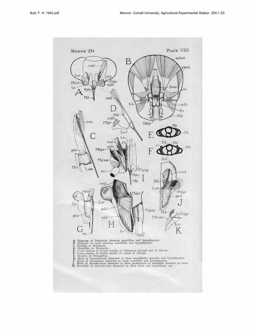
The mandibular bristle of Notonecta consists of a modification of the mesalwall of the mandibular sac. On its inner end is inserted the retractor muscle,(Plate VIII, D, rmd), the origin of which is on the back of the head at theedge of the foramen magnum beneath the eye. There is only one protractor(mdh), its origin being on the inner face of the lorum, its insertion on themandibular process (Mdpr).
The mandibular process (Mdpr) is one of the most important landmarksof the inner head skeleton. It is found in all members of the Hemipterathus far studied, and is constant in position. The outward invaginationmarking the inward projecting process i~ always just mesad and in front ofthe antennal fossa and in Notonecta and Benacus slightly below this point(Plate-IV, A, B; Mdpr).
In Oncopeltus removal of the upper surface of the jugum will reveal themandible and its muscles in place inside the head (Plate VIII, I, Md). Thelarge antennal base crowds the point of invagination of the mandibularprocess (Mdpr) forward. The process is enlarged to form the point ofinsertion for the fan-like hypopharyngeal transverse protractors of themandible (mdh). These muscles originate on the vertical walls of the foodtrough (Plate VIII, I, Lo; and Plate VI, D, Lo), which correspond to thelora of Cicada and the lateral extension of the upper surface of the hypopharynx in Notonecta (Plate VIII, A, Lo).
In most of the Hemiptera the inner edge of the mandibular sac (PlateVIII, I) is sclerotized to form a strong bar (Lvr) connecting the process(Mdpr) with the bristle (Md). This is the lever of the mandible. In Notonecta the lever (Lvr) is not heavily sclerotized and thus not so highlydeveloped as in Oncopeltus and Acrosternum, (Plate VIII, D). In thelatter insects the lever is highly developed and is bent at a sharp anglenear its middle (Plate VIII, I), so that its inner half lies parallel to and
. on top of the mandibular bristle (Md). Plate VIII, K, shows the process(Mdpr) and lever (Lvr) of Acrosternum after muscles have been removed.The lever has been lifted by a needle to show the fold in the sac (Md sac)that begins at this bend of the lever (Lvr) .
In Oncopeltus as in Notonecta the hypopharyngeal-mandibular muscleis the only mandibular protractor present. In Acrosternum there is a secondprotractor of the mandible (Plate VIII, H, J; pmd) inserted on the outerhalf of the lever and arising in the anterior extremity of the lobe, the topsurface of which is the jugum (Plate I, D, Ju; and Plate VIII, H, Ju).The point of insertion of this group of muscles (pmd) on the lever(Lvr) is revealed if the hypopharyngeal-mandibular protractor muscles areremoved (Plate VIII, J).
Snodgrass (1938) says, "In the Hemiptera both the primary articulationsof the mandible on the subgenal margin of the cranium have been suppressedto give freedom of movement to the mandibular base; the articulation withthe lorum represents the mesal point of contact between the mandibular base
Butt, F. H. 1943.pdf Memoir. Cornell University, Agricultural Experimental Station. 254:1-20.

16 CORNELL EXPERIMENT STATION MEMOIR 254
and the b'lse of the hypopharynx. The retractor muscle of the hemipterousmandible (Plate VIII, A, rmd) arising on the vertex is evidently theadductor muscle of the biting jaw (Plate VIII, B, admd) ; in Cicada theprimitive cranial abductor appears to be represented by a second retractormuscle (Plate II, D, rmd) arising on the gena and inserted laterally onthe mandibular base. The reduction in size of the mandibular bases inHemiptera has exposed the lateral parts of the hypopharynx on the sidesof the head, and the retraction of the mandibles has converted the primitively hypopharyngeal adductor muscles of the mandibles (Plate VIII,B, mdh) into protractor muscles (Plate II, D, pmd)." (Snodgrass heredoes not use "Hemiptera" in the restricted sense, but includes as well themembers of the Homoptera.)
Snodgrass's diagram of the mandible and hypopharynx of the roach hasbeen included h~re for comparison with the same structures in Notonecta.The same relations described for Cicada exist in Notonecta. In Notonectathe hypopharyngeal muscle of the mandibles (Plate VIII, A, mdh) retainsits transverse position in the head. Its origin is on the inner surface of theloral plate (Lo), an expansion of the upper surface of the hypopharynx(Hphy). Its insertion is on the mandibular process (Mdpr). Retraction ofthe mandible in the head has caused the hypopharyngeal muscle to act as aprotractor of the bristle. It works against the retractor muscle in Notonecta,instead of with it as in the roach.
A difference between otonecta and Cicada should be pointed out. InCicada the hypopharyngeal muscle is inserted directly on the protractor armof the mandible. In Notonecta this muscle acts upon the mandibular process(Plate VIII, A, D; Mdpr), which is connected with the mandible itself bythe lever (Lvr). The lever is a sclerotized bar lying in the inner wall ofthe mandibular bristle sac and is connected to the mandible by a flexiblejoint. The structure of the mandible, lever, and mandibular process is similarin all members of the Hemiptera studied and constitutes a fundamentaldifference between the mandible of this group and the mandible of Cicada.
Beneath and mesad of the mandible of Notonecta lies the maxilla formedas a heavily sderotized rod in the membrane of the bristle pouch. The maxillais less complex than the mandible. It has a single retractor bundle ofmuscles (rmx) originating on the inner wall of the head capsule at the edgeof the foramen adjacent to the mandibular retractor (Plate VIII, C) andinserted on the mesal edge of the maxillary bristle.
The maxillary segment of the head consists of two parts: the maxillaryplate (Plate IV, B, Mx), the external sclerotized plate beneath the eye,and a fleshy lobe, probably the galea, not visible because it is covered bythe lower part of the clypeal region of the head and by the labrum. Theprotractor muscles (pmx) have their origins on the lower surface of thefleshy lobe (Plate VIII, C) ; the edge of the lobe is apparently reinforcedby a sclerotized margin extending from the external plate to take the pullof the muscles. No maxillary lever is present in Notonecta.
Dissections of Oncopeitus made to expose the maxilla reveal it as asclerotized bristle which broadens at its inner end (Plate VIII, G, Mx).The maxillary sac is expanded inwardly to accommodate a curved transverse bar; this is the lever (Lvr) in the membrat:le of the upper surfaceof the sac, articulating with the inner side of the maxillary bristle and
Butt, F. H. 1943.pdf Memoir. Cornell University, Agricultural Experimental Station. 254:1-20.

MOUTH PARTS, HEMIPTERA-HOMOPTERA 17
extending to the head wall at a point beneath and slightly caudad of thecompound eye. The lever is bowed and can best be seen from the underside as shown in Plate VIII, G. The protractor muscle (pmx) originates onthe end of the maxillary segment and is inserted on the broad base of thebristle. It is encircled by the lever on the under side. The retractors (rmx)are inserted on the inner end of the bristle and extend to the edge of theforamen magnum laterad of the mandibular muscles. The arrangement of themaxilla is similar in Acrosternum; and, except for the more elaboratemusculature, it is similar in Cicada (Plate II, E). The lever (Lvr) ofOncopeltus at its outer end is joined to the head wall by a series of fibers(fi) extending from the epidermal layer of the maxillary sac to the epidermallayer of the head capsule (Plate VII, B). These fibers probably correspondto tonofibrillae. The inner vertical wall of the sac is formed from the wingof the hypopharynx (Hphyw). At the line along which the wing approachesthe lower surface of the head and where the sac (Mx sac) bends outwardunder the lever, another series of fibers (fi) connects the head wall withthe sac.
Back of the lever, the walls of the sac (Plate VII, D, Hphyw) becomemembranous and highly convoluted and the muscles of the salivary syringe(dlsyr), which in Cicada, Cephisus, and Notonecta are attached to the underside of the hypopharyngeal wings (Plate II, F, dlsyr; Plate IV, C, and D,dlsyr), have migrated upon the head capsule.
At the posterior end of the food pump in Oncopeltus (Plate VI, E) across section shows that from here forward the hypopharyngea~ wings(Hphyw) are joined to the floor of the food pump (Pmp) by fibers (fi)that support the pump by opposing the pull of the pump muscles (dlbe).A section taken through the antennal base (Plate VI, D) indicates that themandibular and maxillary sacs have merged into the bristle sac (B sac) infront of the compound eyes and behind the base of the antennae.
The maxillary protractors (pmx) each divide into three bundles nearthe region where they attach to the wan of the head (Plate VI, D). Theinner maxillary protractor bundle (a) originates on the wall of the bristlepouch. The lower bundle (b) (Plate VI, C) fastens to the wall of themaxillary lobe toward its tip on the lower side, and the upper lateral bundle(c) has its origin dorsally in the maxillary lobe toward its anteriorextremity (Plate VI, C).
The mandibular and maxillary bristles are hollow, seta-like organs. Outside the bristle sac and within the trough of the labium the mandibles (Md)are on the outside of the bristle bundle (Plate VIII, E, F,), acting as guidesfor the maxillae (Mx) within. The mandibles are flanged on their uppersurface in Notonecta, the flange overlying the maxilla on the side facing theopening in the labial trough. Near the proximal end of the mandible, whereit lies under the labrum, the mesal margin of the flange is ridged (Plate VIII,F, r). The ridge on each mandible runs in a slot on the inner surface of thelabrum (Plate V, F, r). Evidently these grooves are effective in forcing themandibles and maxillae together beyond the point of the hypopharynx. InOncopeltus the flange and ridge are not present and the bristles are forcedinto a compact bundle in a different way. The 1abrum (Lm) of Oncopeltusis grooved on its lower surface and fits tightly around the bristle bundle
Butt, F. H. 1943.pdf Memoir. Cornell University, Agricultural Experimental Station. 254:1-20.

18 CORNELL EXPERIMENT STATION MEMOIR 254
(Plate VII, A). Evidently the labral groove forces the mandibles (Md)and maxillae (Mx) together beyond the end of the hypopharynx.
In Notonecta the bristle bundle (Bb) is flattened and apparently unableto turn in the labial groove (Plate V, E). In Oncopeltus the bristle bundleis nearly cylindrical and can turn in the labial groove. Serial sections of thelabium with the bristle bundle in place show the axis of the bristle bundleto be different in each section. Possibly this indicates that the bristles havea twisting motion, like that of a drill, as they penetrate the plant tissue(Plate VII, A, C, E, F, and G; Md, Mx).
SUMMARY
In Cicada and Cephisus, the clypeus is divided into postclypeus andanteclypeus; the loral plates are exposed at the sides of the clypeus. InNotonecta the entire clypeus is fused with the front; the loral plates lieinside the head and are entirely covered. In Benacus, Oncopeltus, andAcrosternum, the postclypeus is fused with the front; the anteclypeus ortylus is narrow and lies in a trough between the vertical loral plates.
The lower~lateral surfaces of the hypopharynx are extended into the headas two divergent trough-like plates. In Cicada and Cephisus' a tentoriumis present and these plates rest on the under surface of the tentorium andappear to be united closely with it. The dilator muscles of the salivary syringe:ire attached to their under surfaces.
In other forms studied no tentorium is present. The wings of the hypopharynx extend backward in Notonecta and give support to the inner endsof the salivary retractor muscles. In Oncopeltus the hypopharyngeal wingsare greatly modified. They become membranous caudally and are attachedto the head wall along their lower edges. The origins of the salivary-pumpretractors are on the head capsule instead of the wings.
The food pump of Cicada represents the cibarial region of generalizedinsects; The functional mouth is a tube leading from the cleft between theanteclypeus and the hypopharynx. The true mouth is the posterior openingof the food pump into the pharynx which lies above the tentorial bar. .
The muscles of the food pump are the dilators of the cibarium, a largemass arising in the large clypeal bulge on the front of the head. Behind theseare the pharyngeal dilators. Cephisus and Notonecta are similar. In Notonecta and Oncopeltus the frontal nerve and ganglion lie between these twomasses. The pharyngeal dilators reach their greatest development in Oncopeltus, where they fan out on the upper surface of the pharynx in two parts,with a longitudinal muscle lying between.
The three-segmented labium of Cicada and of Cephisus is attached to theneck membrane. The four-segmented labium of the Hemiptera studied arisesfrom the head capsule at its anterior end. The protractor and retractormuscles of the labium of Cicada and of Cephisus originate on the posterioredge of the head capsule under the end of the tentorium. These musclesdo not exist in the Hemiptera; the adductor muscles and abductors, whenpresent, originate on the hypopharyngeal wings. The muscles of the terminallabial segment and of the next-to-the-Iast segment in Cicada and in Cephisusare similar to the muscles. in the same segments in the Hemiptera studied.Other muscles in the labium differ in the two groups of insects except that
Butt, F. H. 1943.pdf Memoir. Cornell University, Agricultural Experimental Station. 254:1-20.

MOUTH PARTS, HEMIPTERA-HOMOPTERA 19
the first adductor in Cicada and Cephisus is similar in function to the firstadductor in Notonecta and Oncopeltus.
The mandibles and maxillae of the Homoptera and Hemiptera arise fromthe walls of the bristle pouches. The protractor muscles of the mandibles inCicada are attached directly to the protractor arm and arise on the loralplates. In Notonecta and other Hemiptera studied they arise on the loralplates, but are inserted on the mandibular process, a constant feature in theHemiptera. The process is joined by a lever to the mandible. In Acrosternuman additional set of protractor muscles is attached directly to this lever.
The maxillary bristle of Cicada is equipped with a lever attaching to thehead capsule behind the eye, as are the maxillary bristles of Oncopeltus andAcrosternum. No maxillary lever is present in Notonecta.
REFERENCESCRAMPTON, G. C.
1921. The sc1erites of the head, and the mouthparts of certain immatureand adult insects. Ent. Soc. Amer. Ann. 14: 65-110. ,
KEMPER, HEINRICH1932. Beitrage zur Biologie der Bettwanze (Cimex lectularius L.) III.
tJber den Mechanismus des Stechsaugaktes. Ztschr. Morph.u. Okol. Tiere 24: 491-518.
MUIR, F., and KERSHAW, J. C.1912. The development of the mouthparts in the Homoptera, with obser
vations on the embryo of Siphanta. Psyche 19: 77-89.
SNODGRASS, R. E.1921. The mouth parts of the cicada. Ent. Soc. Wash. Proc. 23: 1-15.1927. The head and mouth parts of the cicada. Ent. Soc. Wash. Proc.
29: 1-16.1935. Principles of insect morphology. 666 pages.1938. The loral plates and the hypopharynx of Hemiptera. Ent. Soc.
Wash. Proc. 40: 228-236.
WEBER, HERMANN1929. Kopf und Thorax von Psylla mali Schmidb (Hemiptera-Homop
tera). Ztschr. Morph. u. ako!. Tiere 14: 59-165.1930. Biologie der Hemipteren. 543 pages.1933. Lehrbuch der Entomologie. 726 pages.
Butt, F. H. 1943.pdf Memoir. Cornell University, Agricultural Experimental Station. 254:1-20.

ABBREVIATIONS
mdh = hypopharyngeal-mandibularmuscle
Mdpr = mandibular processMd sac = mandibular sacMth = mouthMx = maxillaMx sac = maxillary sacOcs = occipital sutureOes = oesophagusOp n = optic nerveP = process of labiumpa = protractor armPclp = postclypeusPhy = pharynxplb = protractor of labiumpmd = protractor of mandiblePmp = food pumppmx = protractor of maxillaPphy = posterior pharynxr = ridge on upper surface of mandiblera = retractor armrao = retractors of mouth anglerhphy = retractor of hypopharynxrib = retractor of labiumrmd = retractor of mandiblermx = retractor of maxillaRnv = recurrent nerver ter s = retractor of terminal segments = sutureIs = dorsal salivary muscle2s, 3s = salivary muscles of the labiumSid = salivary ductSiv = salivariumSm = salivary meatusStom = stomodaeumSyr = syringe (salivary)tm = tentorial retractor muscle of maxillaTnt = tentoriumTr = tracheatrans = transverse musclevdlphy = ventral dilators of pharynxY = dome-like protuberance in eye of
Benacus
abd = abductor muscleabmd = abductor of mandibleAclp = anteclypeusadd = adductoradmd = adductor of mandibleAnt = antennaantm = antennal muscleAp = apodemeAt = anterior tentorial armBb = bristle bundleB sac = bristle sacBr = brainBuc = buccula, or buccal cavityCb = cibariumClp = clypeuscplm = compressor of labrumdlbc = dilator of buccalesdleb = dilator of cibariumdig = transverse dilators of the groovedlphy = dilator of pharynxdlsyr = dilator of syringeFd = food ductfi = muscle fibersFlpmp = floor of pumpFr = fronsFr gng = frontal ganglionFrn = frontal nerveg = groove in labium of NotonectaGu = gulaHphy = hypopharynxHphyw = hypopharyngeal wingsHs = hypopharyngeal suspensoriumJu = jugumLb = labiumLb seg 1 = first labial segmentLm = labrumlm phy = longitudinal muscle of pharynxLa = loraLvr = leverm trans = median transverse muscleMd = mandibleMd b = mandibular bristle
20
Butt, F. H. 1943.pdf Memoir. Cornell University, Agricultural Experimental Station. 254:1-20.

MEMOIR 254 PLATE I
A
D
L'
'Q, Pelp' Fr
\Ju
c~Lm
-Cb
[
--Lb
~ Pdp .. fr
A. o,pha!;c as~t of Be"onu griulOS.B. C<:ph;o!;c aspect of N<nl>ttulo .p.C. Cephalic aspect of Ctl.i...s ncrifl)/i"s nr.mph.D. Dor l aspect of bud of Ac."....U .... ,,'" lillo_e. a .tinkl.>ug.E. Do l aspect of hud of O"cotdr..J flUriallOS, the milkweed hUi.
Butt, F. H. 1943.pdf Memoir. Cornell University, Agricultural Experimental Station. 254:1-20.

MEMOIR 254 PLATE II
c-- Br
_~id--Phy
-dhyr
-Syr--Hphy
B
Lb-
M._Pelp_
dlch
L'-
o
AIrm~
Lo__
pmd---
A. C~phali~ as{""'t of Ti/>i";",, urt~..dui..., the s~v~nteen'y""r Cicada.B. M~dian sarmal seetion of he.ad of Cicada.C. Caudal aspect of head of Cicada.D. Mandibl~ and iu musclu. 0' Cicada.E. Maxilla and its muscles. of Cicada.F. B...~ 0' labium of Cicada ",ith in mUKI~s.
Butt, F. H. 1943.pdf Memoir. Cornell University, Agricultural Experimental Station. 254:1-20.

ME),IOIR 254
Pmp--Duct- •
Hphy--'-: is",J
A
c
___Tn!
Aclp- -
Lm-
B
PLATE III
-Lo
"x_Lb
Butt, F. H. 1943.pdf Memoir. Cornell University, Agricultural Experimental Station. 254:1-20.

MEMOIR 254
!i:~'\ 1~
y--.-~._---
ALb----
dlphyOe~~~""
M)C·-~
Gu-- ~,,/~~,
Lbsegl \lI,~,VI
c
-g
PLATE IV
B
A. Head ofB~ gnuu with parI of head alld ~r<' cuI away 10 show hypopbar:rna:~al wings.B. Hud of NOIOnecta with part of bead and ~rc cut away to show food pump. moutb parts.
and their IDlUCI~s.
C. Caudal upeet of head of Notoll«!a.D. Hypopharynrn.\ willi" alld oalinl1' aydnr~ of Notone<:ta.
Butt, F. H. 1943.pdf Memoir. Cornell University, Agricultural Experimental Station. 254:1-20.

MEMOIR 254
Lm·
A
c
E FA. 1.,.I~ral Ul'~<:t <), bud <)f NMO"rrla 51'.B. MN-ian ""lJiual oe<:t;on of bud.C. Me:lian UlJittal oe<:ti<)n <)f labium.D. ~'rontal .outi"" 'hrough labium.E. Cro" ....,.ion Ihrough Ihi«l lahial ""1lmen'.F. Cr<)" """.ion thr<)ulJh bual labial selJn,~nt.
PLATE V
-lr/llns2
Butt, F. H. 1943.pdf Memoir. Cornell University, Agricultural Experimental Station. 254:1-20.

MEMOIR 254
D
c,md -1;\lf~IAL.;&Mel s..c-··..~",· ,
MXS4C·J.L."'-
<>{ hud ''''1wtt" 3n1~""a
PLATE VI
Butt, F. H. 1943.pdf Memoir. Cornell University, Agricultural Experimental Station. 254:1-20.

:\1EMOIR 254 PLATE VIf
,bd I
E
o
, d'.g F GA. Crou M<1;... of Ialli..... 'II~II but.1 ..........11 (J'b.lt , .•• A).B. C.... M<1'''' of """d .1I " t~ (Plait , .•• A. 8).C. Crou O«t.... of Ialli ..... 11I 1I~ wc-"" (J'b.lt \"1, .").D. Crou t«tiool 11I.-1I head bdlind ~ (Plait \"1. A. BJ.E. C ...."ion of Ialli"", 11I...-h .Ioim (J'b.lt VI. A).F. C KCI.... of blli 110.--," d....] port of "um~I (Plait V., .\).G. Crou l'tdion of Ialli IhrooP f<>artll ~I (Plait "1. A).
Butt, F. H. 1943.pdf Memoir. Cornell University, Agricultural Experimental Station. 254:1-20.
-flpmp Mdt0--MdprMdsac-_.
,
"'K
MEMOIR 254
Mdpr--l6:::"\/J,1.0 ~---A H,by-
Md-
GMx-/
pmd-----
H
Hphy-
E
F
I
PLATE VIII
% 4bmd
A'
~\l\J:LIV--Md
,~~.-tl:l.
SId)
A. D,arram of NOIoneeta aho.. inr mandibles and hypopharynx.B. Oiagram of r""ch Ibowing mandibles and hn>opharynx.C. Maxilla of NOIOlleela.D. )Iandible of NOlonecla.E. Cr.,.. section of bristle bundle of Noton""ta beyond end of labrum.F. Cr",s seclion of bri.t!e bundle in Te'gion of labrum.G. )laxil1a of Onco~1tu•.H. Hea,! of Acrosternum di •..ecled to show mandibular muocle. and hypopharynx.I. Head of OncopeU"s di.uecled to .bow mandible and bYl>Opbarynx.
J_, Head of Acro..ernum dls.o«led f<> show protrsew,s of mandible in....rted on le,-er.l!: "bndible of ACTo.temurn di• ..ecled 10 show lever and mandibular sac.
Butt, F. H. 1943.pdf Memoir. Cornell University, Agricultural Experimental Station. 254:1-20.