you can't teach a middle-aged ganglion new tricks
TRANSCRIPT
these histological methods, however wellthey resolve fine details, work only on fixedtissue, thus completely missing informationon neural activity and, even more importantin the present context, being unable to detectthe dynamics of individual morphologicalfeatures. To assay such dynamics requiresrepeated imaging of the very same neuron inthe very same animal, which to date onlyoptical microscopy can deliver. But imagingin intact tissue or intact animals still facessubstantial obstacles.
In a study reported in this issue, Ganand colleagues have overcome these obsta-
many orders of magnitude larger than whatcould be possibly genetically determined. Itis all the more important, therefore, tounderstand the rules governing neural(self-) organization, particularly duringdevelopment, repair and adult plasticity.
We have known how neurons look, downto the finest axonal and dendritic branches,since Golgi discovered the eponymous stain-ing method1 and highly corrected micro-scope optics were perfected. More recently,electron microscopy refined this picture andallowed, in particular, the unambiguousidentification of synaptic connections. All
N E W S A N D V I E W S
A typical neuron has 10,000 neighbors, ascompared to only tens or at most hundredsfor other cells of the body. It matters, ofcourse, not only how many neighbors youhave but also which of your neighbors youtalk to and who talks back to you.Furthermore, the complexity of the fullydeveloped mammalian nervous system is
908 VOLUME 6 | NUMBER 9 | SEPTEMBER 2003 NATURE NEUROSCIENCE
You can’t teach a middle-aged ganglion new tricksWinfried Denk
Repeated imaging of the same individual neuron for over a year in mice allows the authors of a new study in this issue to showthat presynaptic axon terminals become progressively more stable as the animals age, changing little after 6 to 12 months.
Winfried Denk is in the Department of Biomedical
Optics, Max-Planck Institute for Medical Research,
Jahn Strasse 29, Heidelberg D-69120, Germany.
e-mail: [email protected]
very well suited to be a trigger mechanism forupward transitions.
This leaves as a candidate mechanism trueCICR via ryanodine receptors. But ryan-odine receptors (and for that matter IP3receptors) usually show at least some cal-cium-dependent inactivation (and adapta-tion). These phenomena would lead toproblems such as unwanted drifts in thewave-front between inputs, integrationerrors, and even termination of the signal.Indeed, to our knowledge, CICR-basedwaves are experimentally observed to propa-gate10 and are not ‘balanced’ to give a non-moving front. However, ryanodine receptorinactivation is heterogeneous; the (rare)RyR3 subtype is steeply calcium-activatedand is only weakly inactivating11, providinga possible substrate for the model.
Some of the problems outlined above canbe circumvented by returning to voltage-based mechanisms. At the mathematicallevel there is no difference between thecable equation (which describes the spreadof charge) and the reaction-diffusion equa-tion (which describes the spread of ions andmolecules). Furthermore, there is no realmathematical difference between voltage6
and calcium bistability (Fig. 1a,b). The ideathat spatial activation patterns in bistabledendrites could contribute a single-cellmechanism to multistable persistent neuralactivity is also being explored in other inte-grator models12. A natural extension wouldbe to implement the wave-front model by
combining local voltage bistability and thecable equation.
Mechanisms based on voltage multistabilityhave several advantages. The voltage-depend-ent conductances underlying such a schemecould include persistent sodium, long-lastingcalcium and NMDA conductances, all ofwhich have an established track record ofinvolvement in bistability and indeed multista-bility in many different neuron types13. In par-ticular, NMDA conductance is abundant incandidate integrator neurons (Wang, S.S.-H. etal. Soc. Neurosci. Abstr. 24, 602.5, 1998) and isnecessary for normal integration in vivo14.Because these conductances are voltage-gated,inputs could drive both upward and down-ward transitions. A voltage-based mechanismalso removes the need for a calcium-to-firingconversion step. One weakness is that the den-dritic cable would be much more compactelectrically than for calcium diffusion, but ifmembrane conductance is sufficiently high, asmay occur in vivo, dendrites may be suffi-ciently ‘long’ in electrical terms to supportplateau potential wave-fronts. Cable nonuni-formities or heterogeneity12 may be requiredfor correct readout. Another weakness of thevoltage wave-front idea is that it does notexplain why transient intracellular currentinjections in integrator neurons do not evokepersistent changes in firing1,15. However, thisdiscrepancy can also be explained if currentinjections affect additional conductances thatmask changes in dendritic state15. In short, avoltage-based proposal would be mathemati-
cally similar to the existing calcium-basedmodel, and may be more plausible biologically.
Models that have aimed to explain howneural circuits generate persistent activityhave suggested various underlying mecha-nisms, including tuned recurrent feedback,cellular or dendritic bistability, and now cal-cium release wave-fronts. Neural circuits mayeven use some combination of these toimprove robustness12. The definitive tests,however, will be experimental. To paraphrasethe embryologist Viktor Hamburger,“Our realteacher is the nervous system, who is, inciden-tally, the only teacher who is always right.”
1. Loewenstein, Y. & Sompolinsky, H. Nat. Neurosci. 6,961–967 (2003).
2. Robinson, D.A. Annu. Rev. Neurosci. 12, 33–45(1989).
3. Aksay, E., Baker, R., Seung, H.S. & Tank, D.W. J. Neurophysiol. 84, 1035–1049 (2000).
4. Goldman-Rakic, P.S. Neuron 14, 477–485 (1995).5. Arnold, D.B. & Robinson, D.A. Exp. Brain Res. 113,
57–74 (1997).6. Koulakov, A.A., Raghavachari, S., Kepecs, A. &
Lisman, J.E. Nat. Neurosci. 5, 775–782 (2002).7. Berridge, M.J., Lipp, P. & Bootman, M.D. Nat. Rev.
Mol. Cell Biol. 1, 11–21 (2000).8. Seung, H.S., Lee, D.D., Reis, B.Y. & Tank, D.W.
J. Comp. Neurosci. 9, 171–185 (2000).9. Khodakhah, K. & Ogden, D. J. Physiol. 487, 343–358
(1995).10. Jaffe, L.F. BioEssays 21, 657–667 (1999).11. Fill, M. & Copello, J.A. Physiol. Rev. 82, 893–922
(2002).12. Goldman, M.S., Levine, J.H., Major, G., Tank, D.W. &
Seung, H.S. Cereb. Cortex (in press).13. Simon, M., Perrier, J.F. & Hounsgaard, J. Eur. J.
Neurosci. 18, 258–266 (2003).14. Mettens, P., Cheron, G. & Godaux, E. Neuroreport 5,
1333–1336 (1994).15. Aksay, E., Gamkrelidze, G., Seung, H.S., Baker, R. &
Tank, D.W. Nat. Neurosci. 4, 184–193 (2001).
©20
03 N
atu
re P
ub
lish
ing
Gro
up
h
ttp
://w
ww
.nat
ure
.co
m/n
atu
ren
euro
scie
nce
clusters on the salivary ducts, makingrelocation and imaging easier.
This work follows on the heels of twostudies5,6 that examined neuronal morphol-ogy over time in the cerebral cortex and,incidentally, used very similar transgenicanimals7. Interestingly, these earlier studiescame to contradictory conclusions concern-ing the stability of dendritic spines. Onefound that in the mouse barrel cortex,although dendritic structure was stable overweeks, spines appeared and disappeared5.But according to the other study6, whichused similar GFP-expressing mice, spines onpyramidal neurons in layer 5 of primaryvisual cortex show remarkable plasticityduring the critical period early in develop-ment, but become much more stable in theadult. The current study by Gan et al.2 nowadds complementary results on presynapticmorphological stability and thus providesfurther information about how stable theadult nervous system really is.
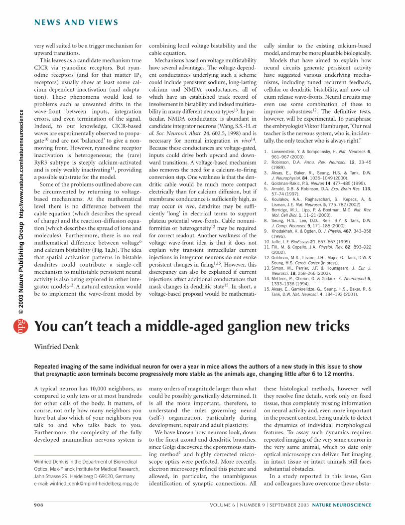
cles and pushed long-term in vivo imagingto a new level2. Using the rodent parasym-pathetic submandibular ganglion prepa-ration that the Lichtman and Purves labshave been perfecting over the pastdecades3,4, Gan et al.2 now show that atleast some synaptic connections becomeincreasingly morphologically stable as thenervous system matures, changing hardlyat all after middle age (in a mouse ∼ 6–12months). The reported observations wereonly possible by combining a number ofcutting-edge technologies: microsurgicalpreparation, allowing the same area to beimaged several times during a period ofmore than a year, confocal microscopyand transgenic expression of green fluo-rescence protein (GFP), allowing stablefluorescent labeling of cells. Also, the sub-mandibular ganglion preparation used byGan et al.2 may be particularly conduciveto such studies as the neurons are eithercompletely isolated or present in small
60 xwater
Saline
Submandibular ganglion
N E W S A N D V I E W S
NATURE NEUROSCIENCE VOLUME 6 | NUMBER 9 | SEPTEMBER 2003 909
Figure 1 Diagram of experimental methods.
There are, however, a number of issuesthat still need to be resolved, including thephysiological relevance of morphologicalstability or plasticity. Could a ganglionicsynapse, which seems to serve as a relay, withreliable action potential initiation in thepostsynaptic cell following every presynapticspike, ever be the site of and hence model for‘memory’? Are morphological changes cor-related to memory formation at all, as hasbeen suggested, for example, by observationsof spine genesis during synaptic stimula-tion8,9? Then there is, of course, the questionof what the biochemical mechanisms arethat provide stability and malleability,respectively, to synaptic morphology. Thesystem used by Gan et al.2 may be well suitedto study this very question, as the effects oftransgenic and pharmacological manipula-tions on long-term morphological stabilitycan probability be assessed more clearly andquantitatively in the submandibular gan-glion than, say, in cortex.
1. Golgi, C. Gazzetta Medica Italiana, Lombardia 33,244–246 (1873).
2. Gan, W.B., Kwon, E., Feng, G., Sanes, J.R. &Lichtman, J.W. Nat. Neurosci. 6, 956–960(2003).
3. Lichtman, J.W. J. Physiol. (Lond.) 273, 155–177(1977).
4. Purves, D. & Lichtman, J.W. J. Neurosci. 7,1492–1497 (1987).
5. Trachtenberg, J.T. et al. Nature 420, 788–794(2002).
6. Grutzendler, J., Kasthuri, N. & Gan, W.B. Nature420, 812–816 (2002).
7. Feng, G. et al. Neuron 28, 41–51 (2000).8. Engert, F. & Bonhoeffer, T. Nature 399, 66–70
(1999).9. Maletic-Savatic, M., Malinow, R. & Svoboda, K.
Science 283, 1923–1927 (1999).
Seeing after blindnessRichard L Gregory
The unusual case of a man who regained his sight after 40 years of blindness allows researchers to examine the neural andbehavioral effects of losing visual experience on the establishment and maintenance of visual system function in humans.
Sight given to an adult following blindnessfrom infancy is interesting for what thisunusual occurrence can tell us about the nor-mal development of vision. But of course theadult is not a baby suspended in time, as hehas lost the normal ‘critical development’periods and is armed with knowledge of the
The author is Emeritus Professor of
Neuropsychology, Department of Experimental
Psychology, University of Bristol, 8 Woodland Road,
Clifton, Bristol BS8 1TN, UK.
e-mail: [email protected]
world from years of exploratory touch andfrom sighted people through language. Inthis issue, a thorough characterization ofsuch a subject shows that he had instant sightfor identification of simple shapes like a cir-cle or triangle, with interesting abnormalitiesand a need for learning about more compli-cated visual objects, and almost completelyspared motion perception.
The case reported here by Fine et al.1 is ofa man (MM) who regained sight in his earlyforties, having lost one eye and the sight inhis other eye in a chemical accident at threeand a half years of age. The study was car-
ried out by an expert team of visual scien-tists who used psychophysics, functionalmagnetic resonance imaging (fMRI) andelectroretinograms to characterize MM’svisual abilities, brain function and eye func-tion. The case is also unusual in that thesubject is an intelligent and remarkably suc-cessful person who was a champion blindskier. The results broadly confirm severalreported cases over the last half-century, aswell as add important new findings.
The empiricist philosopher John Locke2
first addressed the issue of whether experi-ence is important in the development of
©20
03 N
atu
re P
ub
lish
ing
Gro
up
h
ttp
://w
ww
.nat
ure
.co
m/n
atu
ren
euro
scie
nce