x-ray free electron lasers: principles, properties and applications
TRANSCRIPT
X-Ray Free Electron Lasers: Principles, Properties and Applications
Claudio Pellegrini, UCLA, and Joachim Stöhr , SSRL
1. INTRODUCTION
Creating matter from the vacuum, taking an atomic scale motion picture of a chemical process in a time of a few femtoseconds (1 fs = 10-15 sec) or unraveling the complex molecular structure of a single protein or virus. These are some of the new exciting experiments that a novel radiation source, the X-ray free-electron laser, will make possible. The free-electron laser has been developed over many years, starting from the first one built at Stanford by John Madey and his collaborators in the 1970s. It is a powerful and challenging combination of particle-accelerator and laser physics and technology. Until recently free-electron lasers have only been operating at infrared or near ultraviolet wavelengths. Because of a combination of theoretical, experimental, and technological advances, it is now possible to have X-ray free-electron lasers. They can revolutionize the way we study matter at the atomic and molecular level, allowing atomic resolution snapshots on the ultrafast timescale associated with the intrinsic atomic motions of atoms in matter.
Through the centuries photons have been the primary tool for the investigation of matter, starting with the use of sunlight and culminating with the laser. During the last century x-rays have allowed us to see the invisible and their impact on materials and life sciences is well documented and cannot be overstated. In particular, the advent of synchrotron radiation sources has led to a genuine scientific revolution during the last three decades. The importance of synchrotron radiation research has led to the construction of a large number of dedicated synchrotron radiation facilities. Today, the most advanced, so called 3rd generation facilities, are based on undulators and thousands of scientists are using such sources around the world.
Useful as they are these facilities have limitations. The minimum pulse length of the X-
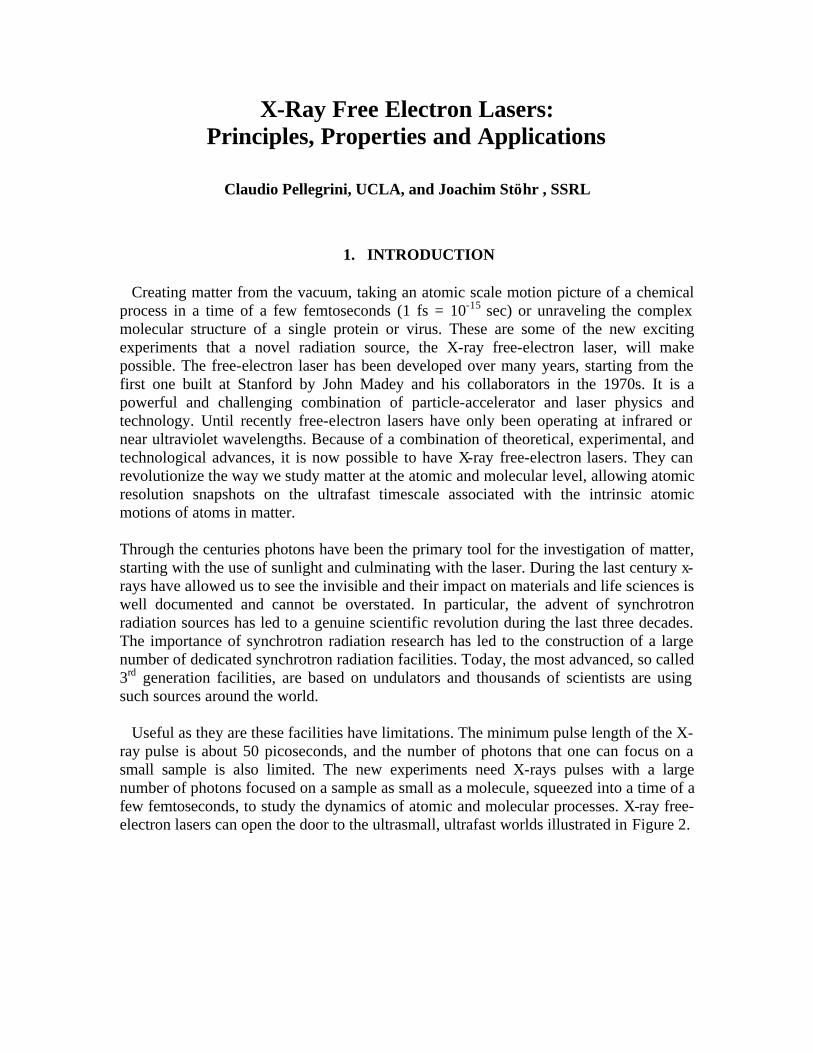
ray pulse is about 50 picoseconds, and the number of photons that one can focus on a small sample is also limited. The new experiments need X-rays pulses with a large number of photons focused on a sample as small as a molecule, squeezed into a time of a few femtoseconds, to study the dynamics of atomic and molecular processes. X-ray free-electron lasers can open the door to the ultrasmall, ultrafast worlds illustrated in Figure 2.
2
Fig. 1 Length and times scales in the ultrasmall, ultrafast worlds opened up by XFELs. A comparison between the main properties of an advanced synchrotron radiation source
and an X-ray free-electron laser is shown in Table 1.
3rd Gen. SASE-FREE-ELECTRON
LASER
Short pulse SASE-FREE-ELECTRON
LASER Wavelength range, nm 1-0.1 Emittance, nm rad 2 0.03 0.03 Pulse length, ps 15-30 0.06 0.01 Average brightness 1020 1022 1021
Peak brightness 1023 1033 1033
Peak power, W 103 1010 1010
Table 1 Some typical characteristics of the undulator radiation from 3rd generation ring based light sources, and FREE-ELECTRON LASER light sources. The emittance is in nm rad; the pulse length in ps; the average and peak brigthness are in photons/sec/mm2/mrad2/0.1%bandwidth; the peak power in W.
2. X-RAY FREE-ELECTRON LASERS AND THE FREE-ELECTRON LASER COLLECTIVE INSTABILITY
A free-electron laser that emits X-rays with a wavelength of the size of an atom (about 1 Å) can be built because of a favorable and interesting phenomenon of self-organization
3
of the electrons in a relativistic beam, known as the free-electron laser collective instability. This instability takes an electron beam with a random electron position distribution, and changes it into a distribution with electrons regularly spaced at about the x-ray wavelength, producing what could be called a 1-dimensional electron crystal. The radiation from this crystal has the new and exciting properties just discussed.
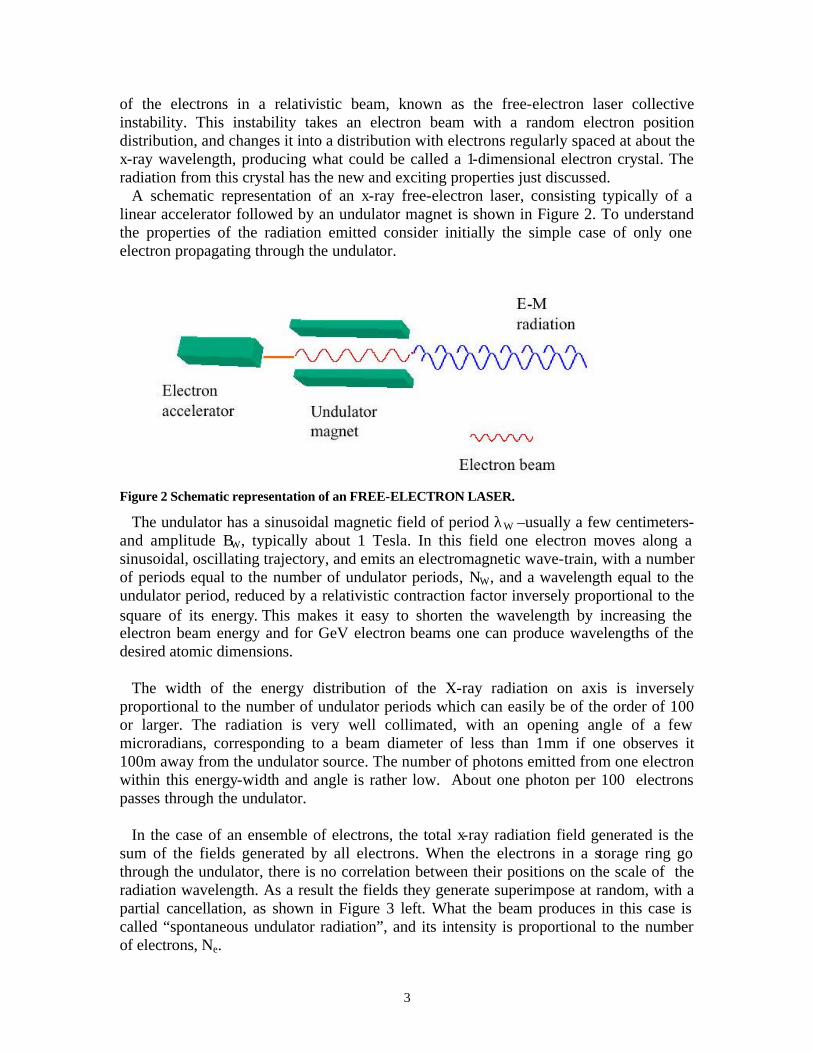
A schematic representation of an x-ray free-electron laser, consisting typically of a linear accelerator followed by an undulator magnet is shown in Figure 2. To understand the properties of the radiation emitted consider initially the simple case of only one electron propagating through the undulator.
Figure 2 Schematic representation of an FREE-ELECTRON LASER.
The undulator has a sinusoidal magnetic field of period λW –usually a few centimeters- and amplitude BW, typically about 1 Tesla. In this field one electron moves along a sinusoidal, oscillating trajectory, and emits an electromagnetic wave-train, with a number of periods equal to the number of undulator periods, NW, and a wavelength equal to the undulator period, reduced by a relativistic contraction factor inversely proportional to the square of its energy. This makes it easy to shorten the wavelength by increasing the electron beam energy and for GeV electron beams one can produce wavelengths of the desired atomic dimensions.
The width of the energy distribution of the X-ray radiation on axis is inversely
proportional to the number of undulator periods which can easily be of the order of 100 or larger. The radiation is very well collimated, with an opening angle of a few microradians, corresponding to a beam diameter of less than 1mm if one observes it 100m away from the undulator source. The number of photons emitted from one electron within this energy-width and angle is rather low. About one photon per 100 electrons passes through the undulator.
In the case of an ensemble of electrons, the total x-ray radiation field generated is the
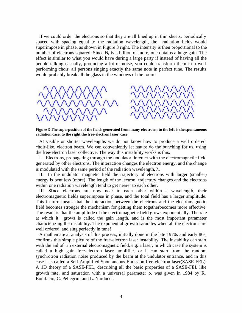
sum of the fields generated by all electrons. When the electrons in a storage ring go through the undulator, there is no correlation between their positions on the scale of the radiation wavelength. As a result the fields they generate superimpose at random, with a partial cancellation, as shown in Figure 3 left. What the beam produces in this case is called “spontaneous undulator radiation”, and its intensity is proportional to the number of electrons, Ne.
4
If we could order the electrons so that they are all lined up in thin sheets, periodically
spaced with spacing equal to the radiation wavelength, the radiation fields would superimpose in phase, as shown in Figure 3 right. The intensity is then proportional to the number of electrons squared. Since Ne is a billion or more, one obtains a huge gain. The effect is similar to what you would have during a large party if instead of having all the people talking casually, producing a lot of noise, you could transform them in a well performing choir, all persons singing exactly the same note in perfect tune. The results would probably break all the glass in the windows of the room!
Figure 3 The superposition of the fields generated from many electrons; to the left is the spontaneous radiation case, to the right the free-electron laser case.
At visible or shorter wavelengths we do not know how to produce a well ordered, choir-like, electron beam. We can conveniently let nature do the bunching for us, using the free-electron laser collective. The way this instability works is this.
I. Electrons, propagating through the undulator, interact with the electromagnetic field generated by other electrons. The interaction changes the electron energy, and the change is modulated with the same period of the radiation wavelength, λ.
II. In the undulator magnetic field the trajectory of electrons with larger (smaller) energy is bent less (more). The length of the lectron trajectory changes and the electrons within one radiation wavelength tend to get nearer to each other.
III. Since electrons are now near to each other within a wavelength, their electromagnetic fields superimpose in phase, and the total field has a larger amplitude. This in turn means that the interaction between the electrons and the electromagnetic field becomes stronger the mechanism for getting them togetherbecomes more effective. The result is that the amplitude of the electromagnetic field grows exponentially. The rate at which it grows is called the gain length, and is the most important parameter characterizing the instability. The exponential growth saturates when all the electrons are well ordered, and sing perfectly in tune!
A mathematical analysis of this process, initially done in the late 1970s and early 80s, confirms this simple picture of the free-electron laser instability. The instability can start with the aid of an external electromagnetic field, e.g. a laser, in which case the system is called a high gain free-electron laser amplifier, or it can start from the random synchrotron radiation noise produced by the beam at the undulator entrance, and in this case it is called a Self Amplified Spontaneous Emission free-electron laser(SASE-FEL). A 1D theory of a SASE-FEL, describing all the basic properties of a SASE-FEL like growth rate, and saturation with a universal parameter ρ, was given in 1984 by R. Bonifacio, C. Pellegrini and L. Narducci.

5
In the X-ray region ρ is of the order of 1/1000, or less, which means that to reach saturation we need about 1000 undulator periods, and that one thousandth of the electron beam kinetic energy is transformed into photons. Consider as an example a 1 Å X-ray SASE-FEL with 15 GeV electron energy, and ρ ~ 1/1000. In this case The number of photons per electron - within the same frequency band and angle - is about 1000, a gain of 100,000 over the spontaneous radiation case
In a SASE-FEL the x-ray radiation propagates slightly faster than the electrons - the electrons slip back by one radiation wavelength per undulator period. Electrons can only interact with the radiation generated by the electrons preceding them in the beam, and that it can catch up with them because of their lower velocity. These are the electrons within the “cooperation length”, equal to the distance the electrons slip back in one gain length. This property of the interaction has the effect of smoothing out the temporal distribution of the radiation intensity from an initial variation on the scale of λ, typical of spontaneous radiation, to a final variation at saturation on the scale of the cooperation length, as shown in Figure 4. The photons are said to come in “spikes”, and the intensity in each of them changes at random. For a long electron bunch, with many spikes, the relative intensity fluctuation is 1/(number of spikes )1/2
Figure 4 The temporal structure of the LCLS SASE-FREE-ELECTRON LASER near the beginning of the undulator, during the exponential growth and at saturation, for the LCLS case. At saturation the intensity is in the form of “spikes”, with a length of the order of the cooperation length.
The instability can only develop if some conditions are satisfied: a) the electrons must
have nearly the same energy; b) the electron beam radius and angular spread must be similar, “matched”, to that of the X-ray beam; c) the gain must be large enough to overcome the losses due to diffraction of the radiation from the beam.
These conditions can be used to show that as the free-electron laser wavelength is reduced, the electron accelerator driving it must satisfy more stringent requirements. In particular collective effects in the accelerator, due to the electromagnetic interaction of the electrons between themselves, and with the accelerator itself, must be strictly controlled. Operating an X-ray SASE-FEL is a balancing act between controlling the unwanted collective effects in the accelerator, and letting the free-electron laser collective instability develop in the undulator.
3. THE DEVELOPMENT OF SASE-FREE-ELECTRON LASERS In 1992 a workshop was held at SLAC, to discuss the possibility of developing novel
X-ray sources, described as 4th generation sources, with performance exceeding the best
6
of the third generation storage rings. At the workshop Claudio Pellegrini showed that the introduction of a novel electron source, the photoinjector, initially developed by Fraser, Sheffield, and Gray at Los Alamos, and the progress in accelerating high quality beams in linacs due to the linear collider development at SLAC, opened the possibility to have a 1Å X-ray SASE-FEL, based on the SLAC linac. A study group, coordinated by Herman Winick developed this concept, and gave it a name, the Linac Coherent Light Source (LCLS). A design group, directed by Max Cornacchia, continued the work and in 1998 produced a report demonstrating the feasibility of LCLS. A similar group was also formed at DESY and developed the idea of a joint linear collider, X-ray SASE-FEL system, called TESLA. Recently other efforts have begun in Japan, Italy and other countries.
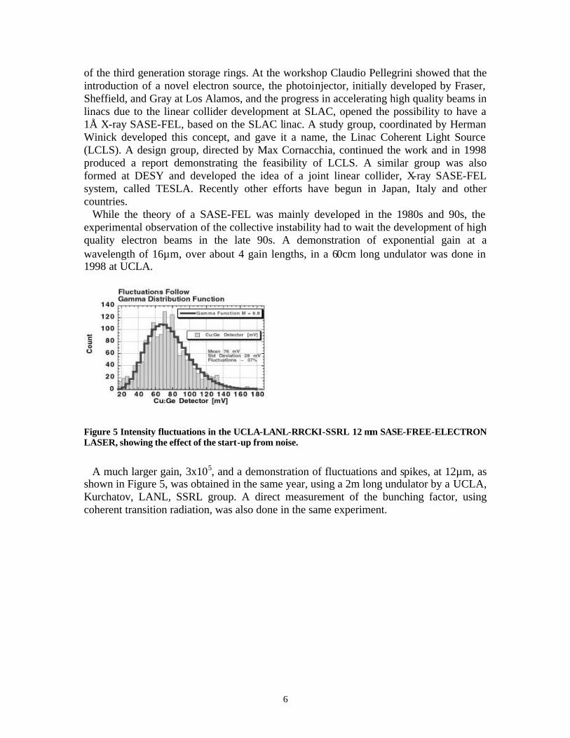
While the theory of a SASE-FEL was mainly developed in the 1980s and 90s, the experimental observation of the collective instability had to wait the development of high quality electron beams in the late 90s. A demonstration of exponential gain at a wavelength of 16µm, over about 4 gain lengths, in a 60cm long undulator was done in 1998 at UCLA.
Figure 5 Intensity fluctuations in the UCLA-LANL-RRCKI-SSRL 12 µm SASE-FREE-ELECTRON LASER, showing the effect of the start-up from noise.
A much larger gain, 3x10⁵, and a demonstration of fluctuations and spikes, at 12µm, as
shown in Figure 5, was obtained in the same year, using a 2m long undulator by a UCLA, Kurchatov, LANL, SSRL group. A direct measurement of the bunching factor, using coherent transition radiation, was also done in the same experiment.
7
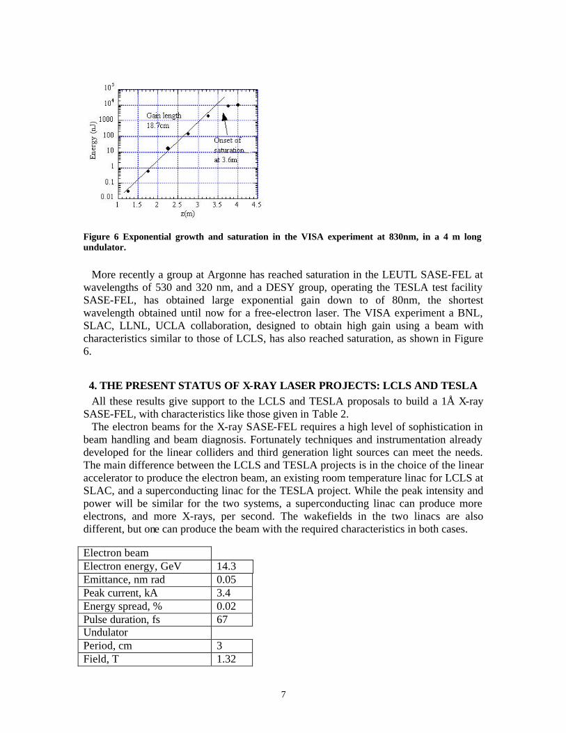
Figure 6 Exponential growth and saturation in the VISA experiment at 830nm, in a 4 m long undulator.
More recently a group at Argonne has reached saturation in the LEUTL SASE-FEL at
wavelengths of 530 and 320 nm, and a DESY group, operating the TESLA test facility SASE-FEL, has obtained large exponential gain down to of 80nm, the shortest wavelength obtained until now for a free-electron laser. The VISA experiment a BNL, SLAC, LLNL, UCLA collaboration, designed to obtain high gain using a beam with characteristics similar to those of LCLS, has also reached saturation, as shown in Figure 6.
4. THE PRESENT STATUS OF X-RAY LASER PROJECTS: LCLS AND TESLA All these results give support to the LCLS and TESLA proposals to build a 1Å X-ray
SASE-FEL, with characteristics like those given in Table 2. The electron beams for the X-ray SASE-FEL requires a high level of sophistication in
beam handling and beam diagnosis. Fortunately techniques and instrumentation already developed for the linear colliders and third generation light sources can meet the needs. The main difference between the LCLS and TESLA projects is in the choice of the linear accelerator to produce the electron beam, an existing room temperature linac for LCLS at SLAC, and a superconducting linac for the TESLA project. While the peak intensity and power will be similar for the two systems, a superconducting linac can produce more electrons, and more X-rays, per second. The wakefields in the two linacs are also different, but one can produce the beam with the required characteristics in both cases.
Electron beam Electron energy, GeV 14.3 Emittance, nm rad 0.05 Peak current, kA 3.4 Energy spread, % 0.02 Pulse duration, fs 67 Undulator Period, cm 3 Field, T 1.32
8
K 3.7 Gap, mm 6 Total length, m 100 Radiation Wavelength, nm 0.15 FREE-ELECTRON LASER parameter, ρ
5x10⁻⁴
Field gain length, m 11.7 Bunches/sec 120 Average brightness 4x10²² Peak brightness 1033 Peak power, GW 109 Intensity fluctuations, % 8
Table 2 LCLS Parameters. Energy spread, pulse length, emittance are rms values. Brightness is in the same units of Table 1. The energy spread is the local energy spread within 2π cooperation lengths. A correlated energy chirp of 0.1% is also present along the bunch.
4.1 Status and time lines
4.2 Future developments: shorter pulses. The LCLS in its initial configuration will be able to provide X-ray pulses with a
duration of about 230 femtoseconds. Even though this is about hundred times shorter than in storage rings, there is already a strong scientific interest in shorter pulses. A further reduction of ten times of the pulse length will open new possibilities to explore the structure of complex molecule, and to study non-linear physics.
Several schemes to achieve such ultra short pulses have been proposed. They all start by introducing an energy-position correlation (chirping) in the electron bunch. A similar correlation then exists between the photon wavelength and position along the pulse. A short part of the X-ray pulse is then obtained by selecting a certain wavelength range with a monochromator. A very promising scheme using this approach and two undulators has been studied recently, and could be easily implemented in the future in LCLS.
5. SCIENTIFIC RESEARCH WITH X-RAY FREE-ELECTRON LASERS At a time when we have only started to utilize the most advanced properties of 3rd generation light sources one may ask whether we are ready to effectively utilize the unusual radiation properties of an XFEL for scientific research. It might be argued that in the development of x-ray sources an XFEL represents a revolutionary leap into unchartered territory. Indeed, when the characteristics of x-ray radiation emitted by an XFEL were first discussed, an overriding concern was related to the destructive power of
9
such a “death ray” with a peak saturation power of about 10 GW. Wouldn’t such a beam simply destroy everything put in its path, from x-ray optics to the sample itself? Traditionally we have used x-rays as a gentle probe of matter, studying its natural equilibrium state and avoiding changes in the probed material, in particular radiation damage, caused by the x-ray beam itself. A prominent example is structural molecular biology where great efforts are made not to alter the delicate state of organic matter by the x-ray beam during the measurement. While some of the excitement surrounding XFELs clearly relates to exploring the interaction of the powerful x-ray beam with a sample and creating new forms of matter, there are fortunately also ways to use XFEL radiation as a gentle probe. The flexible use of XFEL radiation is one of the key factors of its usefulness for research and it will be discussed first. 5.1 Flexible use of XFEL radiation: manipulation and probing of matter The flexible use of XFEL radiation is based on variable focusing and tuning the x-ray energy. These methods allow one to change the power density of the incident x-rays by many orders of magnitude. In addition, the interaction of the beam with the sample strongly depends on the atomic number of the elements in the ma terial. The unfocussed XFEL beam size at the exit of the undulator is about 100x100µm2, the diameter of a fine needle (see Fig. 1). At a typical experimental station located about 200m from the undulator exit, the beam has expanded to a size of only 200x200µm2. This beam can be focused by state-of-the-art techniques to about 0.1 x 0.1 µm2 with maybe a factor of 10 loss in power. This is about the size of a virus or the smallest structures that can be lithographically fabricated today on an electronic chip (see Fig. 1). Through variable focusing the flux density of the x-ray beam can therefore be tuned by a staggering factor of about one million.
Figure 7 Left: Energy dependent cross sections of the interaction of x-rays with copper. The range covered by LCLS is shaded yellow. Right: Relative photoabsorption (photoelectric) cross section of 8 keV x-rays as a function of atomic number .
10
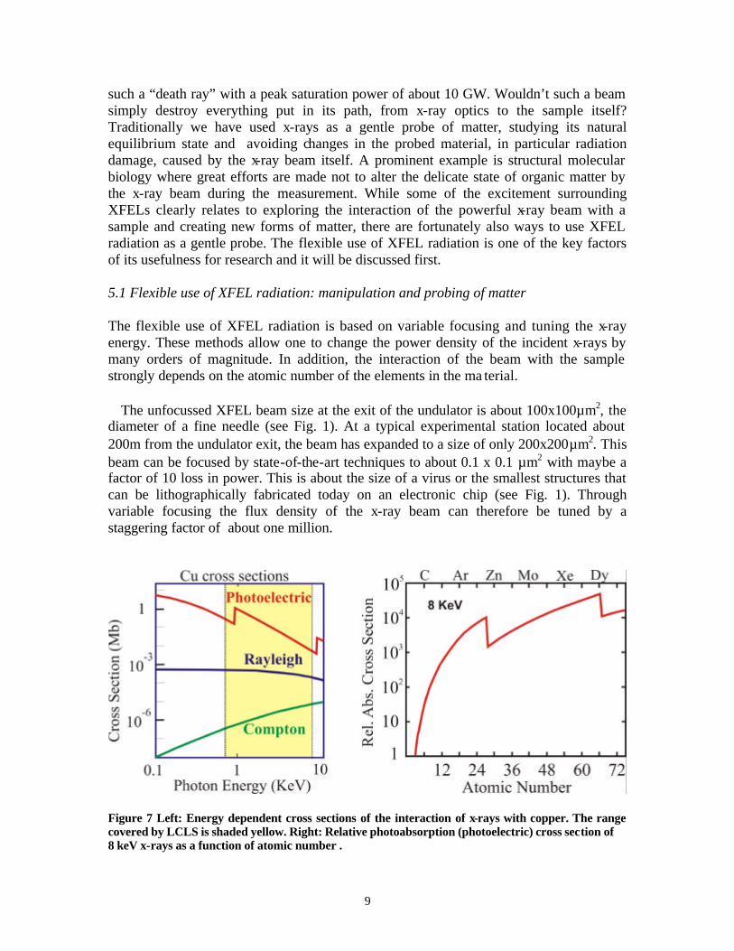
Fig. 7 shows, for a copper sample, that the interaction of the x-ray beam with matter in the 800eV – 8 keV range covered by LCLS is dominated by the photoelectric effect. Its cross section (red line) is larger than the Rayleigh scattering (blue) and Compton (green) scattering cross sections. In the photoelectric effect the absorbed x-ray energy is transferred to valence and core photoelectrons and the aftereffects of the electronic excitations result in adiabatic heating and possibly bond breaking. In the simplest approximation one may assume that the total energy of the absorbed photons is converted into heat and the increase in sample temperature is simply proportional to Nσ E, where N is the number of photons, σ is the photoelectric cross section and E the photon energy. The red curve in Fig. 7 shows that σ decreases with photon energy by about a factor of 100 and exhibits changes of a factor of 10 or more at the characteristic absorption edges of the elements. In contrast, the elastic scattering cross section (blue) which determines the signal in conventional diffraction experiments, is nearly constant with energy, except for anomalous effects close to absorption edges. For diffraction experiments one can therefore utilize the tunability of the photon energy to significantly reduce sample damage effects caused by the photoelectric effect. Finally, the photoelectric effect is strongly dependent on atomic number, Z, as shown in Fig. 6b, reflecting the intuitive picture that it should increase with the number of electrons per atom. Thus materials with low-Z atoms are expected to experience significantly less heating and radiation damage than high-Z materials. For this very reason low-Z materials such as beryllium or diamond appear to be good choices for beam line optical elements. The absorption cross section is seen to change by almost a factor of one hundred thousand for 3 < Z < 70 . Three general classes of experiments have been proposed for LCLS. The first class consists of experiments where the x-ray beam is used to probe the sample without modifying it, as is done in most experiments at current synchrotron sources. In the second class, the LCLS beam is used to induce non-linear photo-processes or transform matter into extreme conditions. In the third class the ultra-fast nature of the LCLS pulse is used to obtain a snapshot of the structure of the sample before the aftereffects of radiation damage set in. The same source can be used for all three types of experiments by utilizing the huge changes in photon flux density available by focusing the LCLS beam, by exploiting the strong dependence of the photoabsorption cross section on photon energy and atomic number or by use of the ultrafast nature of the x-ray pulse – getting the information before the sample realizes what hit it. 5.2 Unique aspects of XFEL radiation: short intense pulses, fields of high amplitude and frequency, and coherence In today’s advanced synchrotron radiation experiments we typically utilize an x-ray flux of about 1012 - 1013 photons/s. This number has experimental significance since, today, we can record a diffraction pattern of a sample in about one second. From the diffraction pattern we can then construct a real space image of the atomic positions in the sample. Because of the time duration of the measurement we determine the average position of
11
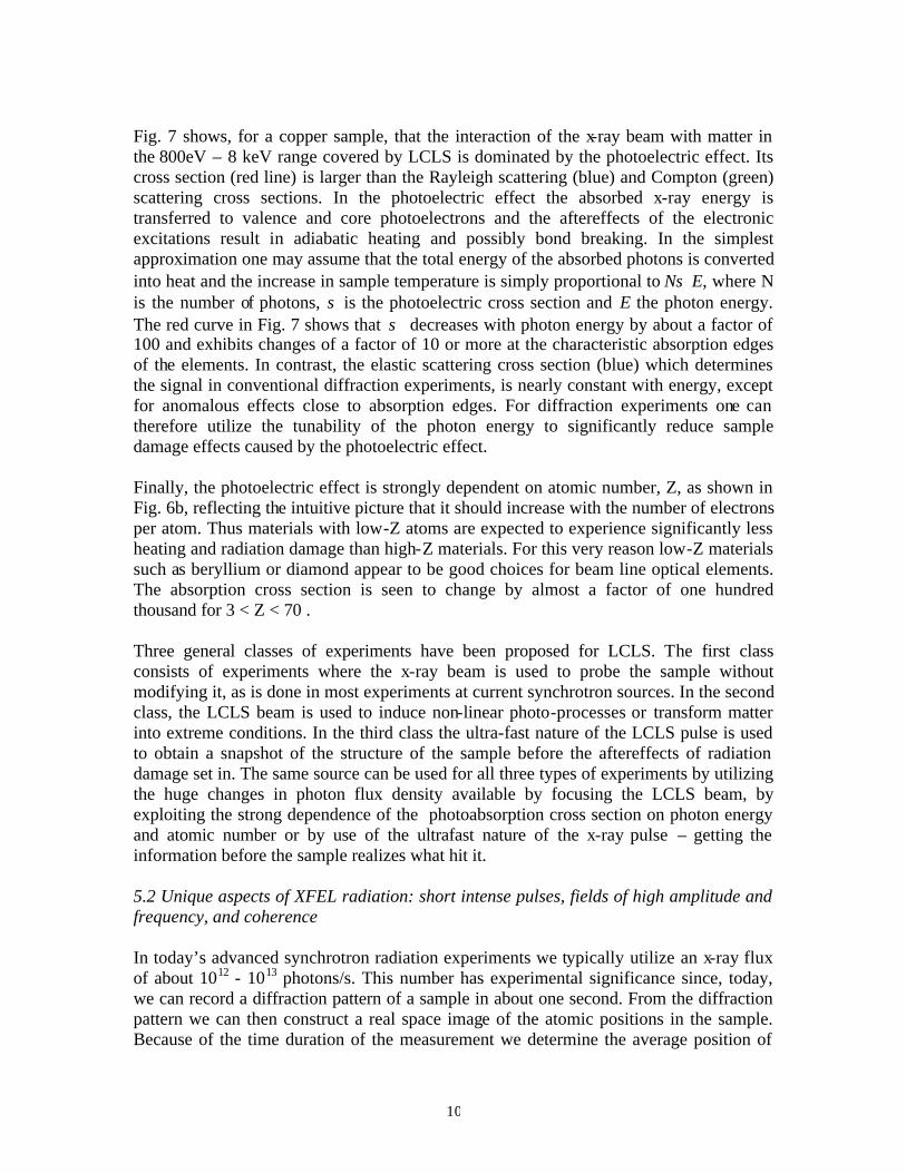
the vibrating atoms. One unique capability of XFELs, their peak flux or brightness (flux per source size and per emission angle), is best demonstrated by the fact that such a source emits the same number of X-rays that we can get today in 1 second in an ultra-fast single burst of about 100 femtoseconds. The shortness of this time is hard to imagine and some help is provided by Fig. 1. It is so short that sound would travel only the distance of one atom, and the fastest “thing” we know, light, would travel only about 50 µm or the width of human hair. It is also the time with which atoms, the building blocks of materials, vibrate. Hence XFELs allow us not only to “see” the invisible, down to the size of an atom, but to take snapshots of the motion of atoms and ultra-small, so called nanoscale, objects like molecules and clusters. This is illustrated in Fig. 8 where we schematically show how a small molecule separates from a bigger molecule, a chemical process called dissociation. Today we can only imagine such a movie but with an XFEL we will be able to record it and then play it back to look at the detailed process.
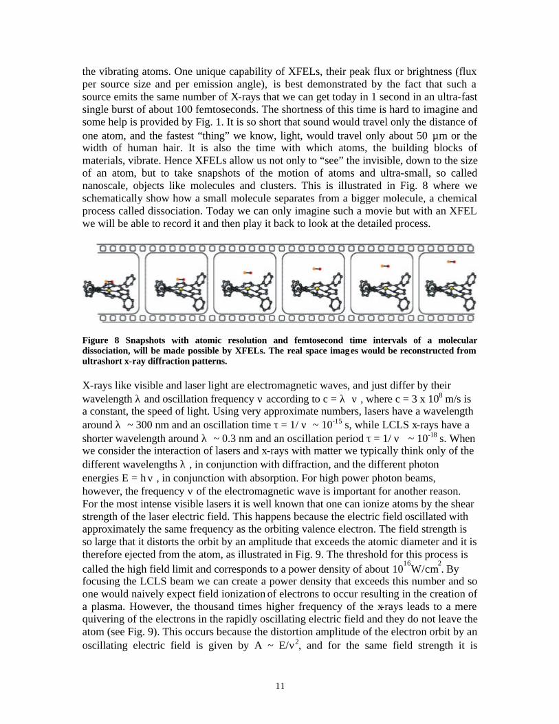
Figure 8 Snapshots with atomic resolution and femtosecond time intervals of a molecular dissociation, will be made possible by XFELs. The real space images would be reconstructed from ultrashort x-ray diffraction patterns. X-rays like visible and laser light are electromagnetic waves, and just differ by their wavelength λ and oscillation frequency ν according to c = λ ν , where c = 3 x 10⁸ m/s is a constant, the speed of light. Using very approximate numbers, lasers have a wavelength around λ ~ 300 nm and an oscillation time τ = 1/ ν ~ 10-15 s, while LCLS x-rays have a shorter wavelength around λ ~ 0.3 nm and an oscillation period τ = 1/ ν ~ 10-18 s. When we consider the interaction of lasers and x-rays with matter we typically think only of the different wavelengths λ , in conjunction with diffraction, and the different photon energies E = h ν , in conjunction with absorption. For high power photon beams, however, the frequency ν of the electromagnetic wave is important for another reason. For the most intense visible lasers it is well known that one can ionize atoms by the shear strength of the laser electric field. This happens because the electric field oscillated with approximately the same frequency as the orbiting valence electron. The field strength is so large that it distorts the orbit by an amplitude that exceeds the atomic diameter and it is therefore ejected from the atom, as illustrated in Fig. 9. The threshold for this process is called the high field limit and corresponds to a power density of about 10
16W/cm
2. By
focusing the LCLS beam we can create a power density that exceeds this number and so one would naively expect field ionization of electrons to occur resulting in the creation of a plasma. However, the thousand times higher frequency of the x-rays leads to a mere quivering of the electrons in the rapidly oscillating electric field and they do not leave the atom (see Fig. 9). This occurs because the distortion amplitude of the electron orbit by an oscillating electric field is given by A ~ E/ν2, and for the same field strength it is
12
therefore more than a million times smaller for x-rays than for visible laser light! The high frequency of the x-ray field is therefore expected to stabilize the atom against ionization - a concept that has not been checked experimentally – but certainly will be with LCLS.
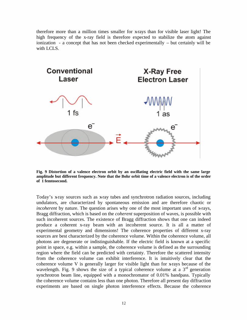
Fig. 9 Distortion of a valence electron orbit by an oscillating electric field with the same large amplitude but different frequency. Note that the Bohr orbit time of a valence electron is of the order of 1 femtosecond. Today’s x-ray sources such as x-ray tubes and synchrotron radiation sources, including undulators, are characterized by spontaneous emission and are therefore chaotic or incoherent by nature. The question arises why one of the most important uses of x-rays, Bragg diffraction, which is based on the coherent superposition of waves, is possible with such incoherent sources. The existence of Bragg diffraction shows that one can indeed produce a coherent x-ray beam with an incoherent source. It is all a matter of experimental geometry and dimensions! The coherence properties of different x-ray sources are best characterized by the coherence volume. Within the coherence volume, all photons are degenerate or indistinguishable. If the electric field is known at a specific point in space, e.g. within a sample, the coherence volume is defined as the surrounding region where the field can be predicted with certainty. Therefore the scattered intensity from the coherence volume can exhibit interference. It is intuitively clear that the coherence volume V is generally larger for visible light than for x-rays because of the wavelength. Fig. 9 shows the size of a typical coherence volume at a 3rd generation synchrotron beam line, equipped with a monochromator of 0.01% bandpass. Typically the coherence volume contains less than one photon. Therefore all present day diffraction experiments are based on single photon interference effects. Because the coherence
13
volume is large relative to atomic distances we observe interference between waves scattered off atoms and atomic planes – that is, Bragg diffraction.
Figure 9 Comparison of the coherence properties of a state-of-the-art 3rd generation beam line with that of an XFEL Even waves scattered off larger “objects” within the coherence volume show interference. An example are the peaks in the small angle scattering intensity which allow one to detect, for example, the presence of characteristic nanometer to micrometer structures in macromolecular systems like polymers. In many systems no long range order exists and conventional small angle x-ray scattering can only determine statistical or average structural parameters. This is due to the fact that the illuminated sample area is much larger than the coherence volume and the measured intensities are therefore an incoherent superposition of the intensities from many coherence volumes in the sample. The situation changes if the illuminated sample area matches the coherence volume. Now the scattered intensity consists of an interference or “speckle” pattern that contains the detailed information on the true structure of the sample in the illuminated area. An example is given in Fig. 10 for a sample that contains magnetic worm domains with an average size of 300 nm. Inversion of the speckle pattern, using techniques such as oversampling, promise to give real space images of nanostructures.
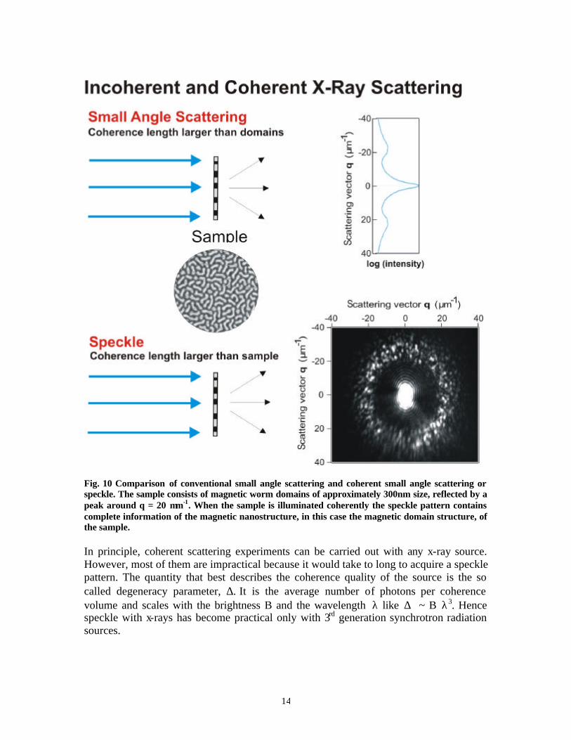
14
Fig. 10 Comparison of conventional small angle scattering and coherent small angle scattering or speckle. The sample consists of magnetic worm domains of approximately 300nm size, reflected by a peak around q = 20 µm-1. When the sample is illuminated coherently the speckle pattern contains complete information of the magnetic nanostructure, in this case the magnetic domain structure, of the sample. In principle, coherent scattering experiments can be carried out with any x-ray source. However, most of them are impractical because it would take to long to acquire a speckle pattern. The quantity that best describes the coherence quality of the source is the so called degeneracy parameter, ∆. It is the average number of photons per coherence volume and scales with the brightness B and the wavelength λ like ∆ ~ B λ3. Hence speckle with x-rays has become practical only with 3rd generation synchrotron radiation sources.
15
The radiation from XFELs exhibits unprecedented coherence. Now all x-rays in a given intensity spike (see Fig. 4) are completely coherent while photons in different spikes are incoherent. Since the total intensity per pulse is about 1012 photons and there are about 1000 spikes per pulse there are now about 109 photons in the coherence volume. This is illustrated in Fig. 9 where we have assumed the natural 0.2% bandwidth of the XFEL beam. We can now begin to do multiphoton experiments. Since coherent photons are equivalent we can pool their energy and create multiphoton excitations and carry out various non-linear x-ray experiments. This is a largely unexplored area of science. We can also use the large number of coherent x-rays per pulse to explore the ultra-small, ultra-fast worlds illustrated in Fig. 1. Here the interest lies in systems which exhibit a great deal of dynamical disorder. Examples are materials undergoing phase transitions, simple and complex fluids or glasses. Because of the large transverse coherence area of 100x100µm2 and the large number of coherent photons per pulse one can record a speckle pattern in a single shot. Inversion of this pattern will yield a femtosecond snapshot of the nanoscale structure of the sample, allowing the direct observation of the dynamics of complex charge and spin ordering phenomena.
6.3 Discussion of selected experiments The proposed experiments for the LCLS and Tesla facilities cover a wide range of fields such as high energy physics, atomic physics, plasma physics, chemistry, materials science, condensed matter physics, and biology. The interested reader is referred to the detailed descriptions available on the web. Here we can only mention some of the ideas and explain why XFELs are expected to truly break new ground. The very first experiment with XFELs will be aimed at understanding the fundamental interaction process of the high power x-ray beam with atoms, molecules and clusters. It will explore the formation of hollow atoms, which is possible because in the x-ray range the photoelectric cross section for core electron excitation exceeds that for valence electron excitation and atoms can therefore be stripped of electrons from the inside out. This process requires that an excitation of a core electron by a photon is followed by another core electron excitation by a second photon before the intermediate excited state decays. The experiment will also study various multiphoton processes enabled by the large degeneracy parameter. Finally, it will investigate the disintegration and explosion of clusters, yielding information on the time scale of the damage caused by the x-ray beam, a fundamental question important for all other experiments. A second proposed experiment uses the LCLS beam to create and investigate an interesting state of matter, called warm dense matter (WDM). WDM exists in the cores of large planets and it is of interest in the study of inertial fusion. Conventional solids are “dense” and the binding energy between atoms is much larger than their thermal energy. On the other hand, plasmas consist of weakly interacting particles whose thermal energy is large. Hence both cases can be treated by theories where one interaction is taken as a weak perturbation on the other. WDM is both hot and dense, that is its density is that of
16
condensed matter and its thermal energy is nearly that of a plasma. For this state of matter few theories are even capable of making predictions. Conventional lasers have provided limited information because they cannot penetrate WDM and other experimental difficulties have been associated with the controlled production of WDM, e.g. its ultrashort life time and the existence of spatial gradients. LCLS has the capability to both create and probe WDM, for example with a second delayed x-ray pulse. The study of molecular reactions lies at the very heart of chemistry and ultrafast laser spectroscopy has allowed us a glimpse at molecular motions in response to an optical excitation pulse. Despite their great success, conventional lasers can only study electronic excitations but they cannot “see” the positions of the atoms during the various transformation stages. This, of course, is the domain of x-rays, and with LCLS one will be able to take diffraction snapshots as shown in Fig. 8. The importance of actually knowing the atomic structure is underscored by the fact that our view of chemistry is largely based structure and many important discoveries in biology and chemistry can be traced back to the determination of a structure. Another experiment pushes the envelope in probing ordering phenomena in hard and soft condensed matter on the important nanometer length scale, which cannot be seen by optical photons, over a broad range of time scales. Nanoscale dynamics, illustrated by Fig. 1, is not only scientifically interesting but it constitutes the competitive arena of advanced technological devices. Finally, LCLS also holds great promise for biology. Today, radiation damage is one of the main obstacles in determining the structure of proteins that cannot be crystallized. The most important ones being cell membrane proteins which constitute nearly half of all proteins. The proposed experiment is based on the use of LCLS pulses that are fast and intense enough to determine the structure by x-ray scattering in a single shot, before radiation damage sets in. Like the application of conventional lasers has been accompanied by R&D on lasers themselves, the above experiments will certainly be accompanied by an R&D program to explore new accelerator and optics concepts with LCLS.
7. CONCLUSIONS
The history of and experience with three generations of synchrotron radiation sources has taught us that the above experiments are at best the tip of the iceberg of scientific opportunities. It is safe to predict that we have not yet thought of the most important experiments that eventually will be done with this new class of radiation sources – x-ray free electron lasers!