x chromosome inactivation: a silence that needs to be broken
TRANSCRIPT

REVIEW
X Chromosome Inactivation: A Silence That Needs to beBroken
Reelina Basu and Li-Feng Zhang*
School of Biological Sciences, Nanyang Technological University, 60 Nanyang Drive, Singapore
Received 28 June 2011; Revised 4 August 2011; Accepted 6 August 2011
Summary: Each mammalian female cell transcription-ally inactivates one X chromosome to balance X-linkedgene dosage between males and females. This phe-nomenon, called X chromosome inactivation, is a per-fect epigenetic event, in which two chromosomes withidentical DNA sequences are solely distinguished byepigenetic modifications. In this case, epigeneticmarks, such as histone modifications, histone variants,DNA methylation, and ncRNAs, are all enriched on onechromosome, the inactive X chromosome (Xi), to estab-lish its chromosome-wide gene silencing. At face value,it seems that the gene silencing mechanism of Xi is wellunderstood. However, the ‘‘silence’’ of Xi in somaticcells is so tightly maintained that it remains largelyintact even after almost all known epigenetic modifica-tions are artificially depleted. To understand how thegene silence of Xi is maintained in soma is a major chal-lenge in current research. We summarize the currentknowledge related with this issue and discuss futureresearch directions. genesis 49:821–834, 2011. VVC 2011
Wiley Periodicals, Inc.
Key words: epigenetics; noncoding RNA; chromatin;nuclear organization
INTRODUCTION
In mammals, each female cell independently silencesthe transcription of one X chromosome to balance theX-linked gene dosage between males (XY) and females(XX). This phenomenon, called X chromosome inactiva-tion (XCI), has been studied for half a century sinceMary Lyon first put forth the hypothesis of XCI in 1961(Lyon, 1961). XCI remains an intriguing topic in mod-ern biology. Four aspects of XCI research attract atten-tion of scientists from different research fields. (1) XCIis regarded as a perfect epigenetic event. The two Xs in
each female cell are identical in DNA sequences. How-ever, one is active in transcription, the other is silenced.The regulation of gene expression in this case solelydepends on epigenetic mechanisms. (2) In each undif-ferentiated female embryonic stem (ES) cell, the two Xsare both transcriptionally active. Based on currentknowledge, they are genetically and epigenetically iden-tical at this stage. Upon the onset of XCI, one of the twoXs is randomly silenced in each female cell. How is thisallele-specificity achieved during XCI? This question,also regarded as ‘‘symmetry break’’ or ‘‘counting/choice,’’ is being intensively investigated in the XCI field(Barakat et al., 2010). (3) It is known that, at least in themouse, the regulations of XCI and pluripotency aremechanistically coupled. When ES cells are differenti-ated, pluripotency is lost and XCI occurs. When somaticcells are dedifferentiated to regenerate pluripotency, forexample in iPS cells (induced pluripotent stem cell) andPGCs (primordial germ cell), the inactive X chromosome(Xi) is reactivated. Therefore, XCI serves as a goodresearch subject or experimental readout in studies onmolecular mechanism of pluripotency (Orkin andHochedlinger, 2011). (4) XCI is an interesting topic froman evolutionary point of view (Chaumeil et al., 2011).Dosage compensation of genes located on sex chromo-somes is a common task faced by all heterogameticorganisms. Different strategies of dosage compensationwere established by different organisms. XCI, as oneway of dosage compensation, is specific to mammals.
* Correspondence to: Li-Feng Zhang, School of Biological Sciences,
Nanyang Technological University, 60 Nanyang Drive, Singapore.
E-mail: [email protected]
Contract grant sponsor: Nanyang Technological University; Contract
grant sponsor: Singapore Stem Cell Consortium.
Published online 26 August 2011 in
Wiley Online Library (wileyonlinelibrary.com).
DOI: 10.1002/dvg.20792
' 2011 Wiley Periodicals, Inc. genesis 49:821–834 (2011)

In fact, XCI mechanism evolves rapidly even within mam-mals. Different mammals utilize different strategies toachieve XCI (Okamoto et al., 2011).
In this review, we focus on the first aspect. For morein-depth information on other XCI related topics, werefer readers to the more specialized literatures cited.
XCI, the Perfect Epigenetic Event
Epigenetics is defined as a heritable regulation ofgene expression unrelated to DNA sequence. Epigeneticmodifications include DNA methylation, histone modifi-cations, histone variants, and higher order of chromatinstructures. Other than DNA and histone modifications,long noncoding RNAs (ncRNA) are also frequentlyobserved in many epigenetic events. These epigeneticmodifications are regarded as important informationwritten on top of the genetic code (the DNA sequence)to annotate the genome and to regulate gene expres-sion. Epigenetic studies help to explain selective, adapt-ive, and inheritable gene expression in different celltypes of a multicellular organism. Therefore, epigeneticsis related to many fundamental questions in biology.
The inactive X chromosome (Xi) has been known asa peculiar structure in cytological stains for a long time.Xi was first observed by Barr and colleagues in 1949(Barr and Bertram, 1949). The structure was identifiedin female cat neurons stained by basic dyes such as cre-syl violet. Nowadays, it is clear that the peculiar charac-ters of Xi in cytological stains are due to its heavy epige-netic coat. Epigenetic marks, such as histone modifica-tions, histone variants, DNA methylation, and ncRNAs,are all enriched on one chromosome, the Xi, to estab-lish its chromosome-wide and allele-specific gene silenc-ing. Many epigenetic modifications on Xi have beenidentified. Most of them can be used as good markers tohighlight the Xi in cytological stains.
At face value, it seems the gene silencing mechanismof Xi is well understood. However, the silence of Xi insomatic cells is so tightly maintained that it remainedlargely intact even after almost all known epigeneticmodifications were artificially depleted (Csankovszkiet al., 1999, 2001; Zhang et al., 2007). Currently, westill do not have any artificial method to efficientlybreak down the silencing of Xi in somatic cells. Theunderstanding of the epigenetic mechanisms involvedin maintaining Xi silencing in soma has important bio-logical significance. It is also directly relevant to the bio-medical aspect of X-linked genetic disorders in female,for example the Rett syndrome (Guy et al., 2007). Here,we will first summarize all the factors (proteins, RNAs,or other molecular identities), which are known to beinvolved in the maintenance of Xi silencing in soma. Af-ter that, we will discuss new perspectives and futureresearch directions.
Xist, the Noncoding RNA
Xist is a critical factor involved in XCI. Xist stands forXi specific transcript, which is a long (18 kb) ncRNA al-lele specifically transcribed from the Xi (Borsani et al.,1991; Brockdorff et al., 1991; Brown et al., 1991). TheXist gene is located on the X chromosome. The onset ofXCI occurs in early embryonic development. In eachfemale cell of the inner cell mass in E4.5 blastocyst,both X chromosomes are transcriptionally active. Atthis time point, both Xs have Xist gene expressed at alow level. The Xist RNA transcripts associate in cis withthe DNA region from which they are transcribed. InRNA FISH (fluorescence in situ hybridization), Xist
RNAs can be visualized as two pinpoint RNA signals(Lee et al., 1999). At early postimplantation stage(�E5.5–6.5), each cell independently and randomlyselects one X as the future Xi. On the chosen Xi, Xistexpression is allele-specifically upregulated. Upregu-lated Xist RNA transcripts spread and coat the X chro-mosome territory (Clemson et al., 1996). At this timepoint, Xist RNAs can be visualized as a cloud signal coat-ing the X chromosome territory in RNA FISH. Coatingof Xist RNA on the Xi further recruits other gene silenc-ing factors to establish the allele-specific and chromo-some-wide gene silencing. On the chosen Xa (active X),Xist expression is allele-specifically repressed. Theonset of XCI can be recapitulated in vitro by differentia-tion of ES cells.
The critical role of Xist in establishing gene silencingof Xi during the initiation stages of XCI was demon-strated by gene knockout studies (Marahrens et al.,1997; Penny et al., 1996). Loss of Xist caused XCI fail-ure and cell death. However, the role of Xist in main-taining gene silencing of Xi in somatic cell was shownto be minimal if not dispensable. When the Xist genewas conditionally removed from the Xi in somatic cells,gene silencing of Xi remained largely intact (Costanziet al., 2000; Csankovszki et al., 2001). Using an Xi-linked GFP reporter, it was shown that only 0.05% ofthe cells reactivated the GFP gene after Xist deletion insomatic cells. The reactivation rate was merely raised to0.8% when the mutant cells were further treated with5-azadC to induce limited DNA demethylation and TSA(Trichostatin A) to inhibit histone deacetylase. The func-tional role of Xist in establishing and maintaining genesilencing of Xi was further studied by inducible expres-sion of Xist. It was found that gene silencing dependedon Xist expression during an early time window,roughly the first 72 h of in vitro differentiation of EScells (Wutz and Jaenisch, 2000). After this time window,the gene silencing status becomes irreversible and inde-pendent of Xist expression.
How does Xist RNA establish gene silencing of Xi dur-ing the early developmental time window? This criticalquestion is yet to be answered. One observation closely
822 BASU AND ZHANG

related to this issue is a so-called ‘‘chromosomal mem-ory’’ established by Xist RNA during the early develop-mental time window. Chromosomal memory can beexemplified by Xist-induced H3K27me3 enrichment onthe Xi. H3K27me3 (trimethylation of histone H3 lysine27) is one layer of epigenetic coating of the Xi. Enrich-ment of H3K27me3 on Xi depends on Xist RNA. WhenXist RNA is deleted from the Xi, the enrichment ofH3K27me3 is also depleted (Plath et al., 2004; Zhanget al., 2007). Using the inducible expression system,Xist expression was turned on, off, then back on againduring ES cell differentiation. As expected, H3K27me3enrichment on Xi was depleted after Xist expressionwas turned off. Interestingly, it was found that earlyexpression of Xist enabled the RNA to reestablishH3K27me3 enrichment on the Xi at a later time point(Kohlmaier et al., 2004). This property was termed as‘‘chromosomal memory.’’ Chromosomal memory isestablished by Xist during the early developmental timewindow. The molecular nature of chromosomal mem-ory is unknown. Unfortunately, chromosomal memorywas found to be independent of gene silencing. Using atruncated Xist allele, which is unable to induce genesilencing, to repeat the experiment, it was found thatthe mutant Xist RNA successfully established chromo-somal memory (Kohlmaier et al., 2004). Using a differ-ent experimental system, a recent study reported thatH3K27me3 enrichment could be established by autoso-mal transgenic Xist expression in somatic cells (Jeonand Lee, 2011).
Besides Xist, there are other ncRNAs involved in XCI.Tsix, as its name suggests, is the antisense transcript ofXist (Lee, 2000; Lee et al., 1999). The expression ofTsix from one X allele counteracts Xist expression fromthe same allele. Therefore, Tsix helps to establish allele-specific expression of Xist (choice), and Xist helps toestablish chromosome-wide gene inactivation(silencing). Other than Xist and Tsix, there are otherncRNAs identified from the nearby DNA region, forexample Jpx (Tian et al., 2010) and Xite (Ogawa andLee, 2003). Jpx and Xite involve in XCI by affecting Xist
and Tsix expression. Tsix and Xite RNAs are notexpressed in somatic cells. Therefore they are notinvolved in maintaining Xi gene silencing in soma. Jpxis expressed in both male and female somatic cells. It isunclear whether Jpx is involved in maintaining Xi genesilencing in soma.
DNA Methylation
The cytosine residues of the CpG island of a gene’spromoter region are often methylated when the gene issilenced. DNA methylation is a common mechanismwidely involved in regulation of many genes: for exam-ple, the silencing of retroviral genome during mouseembryogenesis (Jahner et al., 1982) and the allele-spe-
cific expression of genetically imprinted genes. Geneticimprinting occurs on a small group of genes (about 100genes), which are allele-specifically expressed from ei-ther the paternal or the maternal allele. Obviously,genetic imprinting is an epigenetic event. Parentallyinherited epigenetic marks (genetic imprints) areresponsible for the allele-specific expression pattern ofimprinted genes. The critical role of DNA methylationin genetic imprinting has been clearly demonstrated (Liet al., 1993).
Interestingly, an imprinted form of X inactivationexists, in which the paternal X is non-randomly inacti-vated (Sharman, 1971). Compared to random XCI,imprinted XCI is born with an obvious genetic disad-vantage. Because the paternal X is always silenced inimprinted XCI, a maternally inherited defective X alleleinevitably causes trouble. Imprinted XCI is believed tobe the evolutionarily older form of XCI. In corrobora-tion with this notion, imprinted XCI occurs exclusivelyin all tissues of marsupials (ancient mammals, such askangaroos). In the mouse, imprinted XCI can bedetected in early embryogenesis starting from the 4- to8-cell stage embryo (Huynh and Lee, 2003; Mak et al.,2004; Okamoto et al., 2004). In blastocyst (E3.5–4.5),imprinted XCI is only maintained in the trophectodermand primitive endoderm, which further carry imprintedXCI into the future extraembryonic tissues (Takagi andSasaki, 1975). In the inner cell mass, imprinted XCI iserased and random XCI is set to occur around E5.5–6.5(Mak et al., 2004) in all cells of the embryonic tissues.Unlike the situation in mouse, recent data suggest thatthe Xist gene expression is not subject to imprinting inrabbit and human embryos (Okamoto et al., 2011).
Interestingly, the DNA methylation pattern of Xist
gene promoter correlates with its gene activity (Arielet al., 1995; Norris et al., 1994; Zuccotti and Monk,1995), which suggests that DNA methylation is involvedin XCI. To test this idea, a series of functional analysiswere done using gene knockout mutants of Dnmt1
(maintenance DNA methylase) and Dnmt 3a/3b (denovo DNA methylases). In these mutants, DNA hypome-thylation status was confirmed both at the genome leveland locally on the Xist gene. Mutant embryos or mutantES cell lines were examined in various aspects for XCIdefects, such as imprinted XCI status, random XCI sta-tus, allele-specific expression pattern of Xist gene, andallele-specific silencing of other X-linked genes.
Mutants of Dnmt gene knockouts did show mis-regu-lated Xist expression (up-regulation of Xist from the sin-gle X in male and from both Xs in female). However,this only occurred in a low percentage of cells in mu-tant embryos (Beard et al., 1995; Panning and Jaenisch,1996; Sado et al., 2004). Consistent results wereobtained from mutant ES cell lines. Undifferentiated mu-tant ES cell lines did not upregulate Xist expression(Beard et al., 1995; Panning and Jaenisch, 1996; Sado
823X CHROMOSOME INACTIVATION IN SOMATIC CELLS

et al., 2004). Xist misregulation only occurred in mu-tant ES cells after prolonged in vitro differentiation (Pan-ning and Jaenisch, 1996; Sado et al., 2004). Theseresults suggest that DNA methylation may only berequired in repressing Xist expression in vitro in a latedifferentiation ES population. In addition, Xist upregula-tion in late-differentiation ES cells did not induce genesilencing (Sado et al., 2004), as Xist-mediated genesilencing requires a critical time window during earlydifferentiation. Thus, current data revealed a dispensa-ble role of DNA methylation in regulating the allele-spe-cific expression pattern of Xist.
Dnmt gene knockouts also showed limited effect onthe gene silencing status of other X-linked genes. Mu-tant embryos were able to survive to E9.5 stage, whichsuggests that both imprinted XCI and random XCI werelargely intact in the mutant embryos. In addition, ifDNA demethylation activated the inactive X chromo-some in mutant embryos, mutant females with imbal-anced X-linked gene dosage are expected to die earlierthan mutant males. However, this was not observed(Beard et al., 1995). In Dnmt1 (maintenance DNAmethylase) knockout mutants, an X-linked lacZ trans-gene was tightly silenced in extraembryonic tissuesupon paternal transmission (Sado et al., 2000), whichshowed that imprinted XCI was intact in Dnmt1mutantembryos. The same transgene only showed leakyexpression in embryonic tissues. This result showedthat Dnmt1 was dispensable for both forms of XCI. Onthe other hand, the dispensable role of de novo DNAmethylases in XCI was also confirmed by double geneknockout of the two de novo DNA methylases (Dnmt3a/3b), (Sado et al., 2004). Taken together, a series ofgene knockout studies on Dnmt enzymes revealed thedispensable role of DNA methylation in XCI.
Besides DNA methylation, methylated DNA bindingprotein Mbd2 also showed limited effect in repressingXist expression in somatic cells (Barr et al., 2007).Mbd2 was shown to physically interact with Xist gene.In male mutant fibroblasts of Mbd2 gene knockout, alow level of Xist expression was detected. Other meth-ylated DNA binding proteins, Mbd1, MeCP2, and Kaiso,had no effect on Xist expression.
Heterochromatic Histone Modifications(H3K27me3 and H2AK119ub)
Polycomb group (PcG) proteins were identified ingenetic screening of Drosophila melanogaster as fac-tors involved in heritable silencing of homeotic genes(Schuettengruber et al., 2007). These proteins are con-served in evolution and play extensive biological roles.Interestingly, PcG proteins are also involved in X chro-mosome inactivation and genetic imprinting.
Mammalian PcG proteins form two multiprotein com-plexes, polycomb repressive Complexes 1 and 2 (PRC1
and PRC2). PRC1 ubiquitylates histone H2A at lysine119 (Wang et al., 2004). PRC1 proteins andH2AK119ub are enriched on the Xi (de Napoles et al.,2004; Fang et al., 2004; Plath et al., 2004). PRC2 cata-lyzes tri-methylation of histone H3 lysine 27 (Cao et al.,2002; Czermin et al., 2002; Kuzmichev et al., 2002; Mul-ler et al., 2002). PRC2 proteins (namely Ezh2, Eed andSuz12) and H3K27me3 also showed enriched localiza-tion on the inactive X chromosome (de la Cruz et al.,2005; Erhardt et al., 2003; Mak et al., 2002; Plath et al.,2003; Silva et al., 2003). Despite a well-established func-tional role in heritable gene silencing in many biologicalevents, the functional roles of PcG proteins and theirassociated histone modifications in XCI remain elusive.
It is clear that PcG proteins and their associated his-tone modifications are dispensable for maintaining Xisilencing in soma. In Drosophila, a DNA regulatory ele-ment called PcG response element (PRE) is responsiblefor recruiting PcG proteins. In XCI, recruiting PcG pro-teins and the Xi enrichment of PcG-associated histonemodifications depend on Xist RNA (Plath et al., 2004;Schoeftner et al., 2006; Zhang et al., 2007). BecauseXist is dispensable in somatic cells, so are the PcG pro-teins and their associated histone modifications.
Current data also ruled out a critical role of PcG pro-teins in establishing random XCI. First, in Ring1B (aPRC 1 protein) and Eed (a PRC2 protein) deficient EScells, enrichment of H2AK119ub and H3K27me3 onthe Xi were depleted, but random XCI was undisrupted(Kalantry and Magnuson, 2006; Leeb and Wutz, 2007).Second, a truncated Xist RNA, which lost its ability tointroduce gene silencing, was able to recruitH3K27me3 (Kohlmaier et al., 2004).
The role of PcG proteins in imprinted XCI remainslargely unknown. Attempts to address this question areprecluded by maternally expressed proteins in preim-plantation embryos. Currently, evidence is available tosupport a functional role of Eed in maintaining thesilencing status of the paternal X in differentiated TScells, which represents trophectoderm cell lineage(Kalantry et al., 2006; Wang et al., 2001). Meanwhile, itwas found that PRC2 proteins were not enriched on theimprinted paternal Xi in XEN cells, which representsextraembryonic endoderm cell lineage (Kunath et al.,2005).
PcG proteins and trithorax group proteins (trxG)were originally identified in Drosophila melanogaster
as repressors and activators of Hox genes. While manyPcG proteins showed Xi-enrichment, a trxG proteinAsh2I (absent, small or homeotic discs 2) showed sur-prising Xi-enrichment (Pullirsch et al., 2010). However,it is believed that, as a trxG protein, Ash2I only plays aregulatory role unrelated to gene activation. Xi-enrich-ment of Ash2I is Xist-dependent and independent ofgene silencing. The enrichment is also regulated bychromosomal memory, such that Xist expression in
824 BASU AND ZHANG

early ES cell differentiation enables Xist to efficientlyrecruit Ash2I at a later time point of differentiation.
Polycomblike protein 2 (Pcl2), one of the three Pclproteins in the mouse, was recently identified as a pro-tein enriched on Xi (Casanova et al., 2011). Pcl2 inter-acts with PRC2. After shRNA knockdown of Pcl2, theXist RNA foci were unaffected, however the Ezh2enrichment on Xi was diminished. Therefore, Pcl2 facili-tates the loading of PRC2 complex onto the Xi.
Euchromatic Histone Modifications (H3K4me3and H4Ac)
The PcG protein associated histone modifications(H3K27me3 and H2AK119ub) are usually associatedwith gene silencing, therefore, they are considered asheterochromatin marks. In contrast, histone modifica-tions such as H3K4me3 and H4Ac (histone 4 acetyla-tion) are generally associated with active transcription,therefore, they are considered as euchromatin marks.The chromatin of Xi is defined by an enrichment of het-erochromatin marks (H3K27me3 and H2AK119ub) anda dearth of euchromatin marks (H3K4me3 and H4Ac).Enrichment of H3K27me3 and H2AK119ub on the Xi isinduced by Xist RNA. It is currently unclear what mech-anism is involved in generating the dearth of euchroma-tin marks on the Xi. Upon Xist deletion from Xi in so-matic cells, enrichment of H3K27me3 and H2AK119ubwere depleted, however the dearth of euchromatinmarks on Xi remained unchanged (Zhang et al., 2007).When Xist expression was induced from the Xa in so-matic cells by DNA demethylation, the Xist cloud colo-calized with the X chromosome territory. However, itdid not result in gene silencing and dearth of euchroma-tin marks on the chromosome (Thorogood and Brown,2010). When a mutant form of Xist, which lacked genesilencing capacity, was induced in ES cells, it reas-sembled heterochromatic modifications on the chromo-some territory (Pullirsch et al., 2010). Such a chromo-some territory, with all the heterochromatic modifica-tions resembling Xi but without gene silencing, wastermed as Xiag (Xi with active genes). Hypoacetylationof histone 4 was also established to a certain degreealong Xiag. However, on comparing the immunostainpattern of H4Ac on the metaphase chromosomespreads of Xiag and Xi, numerous H4Ac ‘‘bright bands’’could be recognized along the overall hypoacetylatedXiag. It was believed that such acetylated chromatinregions corresponded to active gene activity. In sum-mary, the current results show that the dearth ofeuchromatin marks along the Xi is independent of Xistfunction. It will be interesting to test if the dearth ofeuchromatin marks on Xi is responsible for the tightgene silencing maintained on Xi after Xist deletion.
Another observation related to euchromatic modifica-tion along the Xi is that the gene silencing along the Xi
is not complete. Profiling of the human inactive X(Carrel and Willard, 2005) showed that 75% of geneswere inactivated, 15% escape inactivation (escapegenes) and 10% escape inactivation in some females(heterogeneous genes). Compared to the human, thegene silencing along the mouse Xi is more complete.Among the murine X-linked genes, which escape inacti-vation, are Smcx, Sts, Utx (Agulnik et al., 1994; Green-field et al., 1998; Salido et al., 1996). The fact thathumans have a much higher number of active genes onthe inactive X may explain the phenotype differencebetween the Turners syndrome patients and the XOmice. The DNA loci of escapee genes carry euchromaticmodifications. One possible mechanism to escape het-erochromatization on the inactive X is the presence ofboundary elements as DNA insulators for shieldinggenes from the position effect of their neighboringregions (Filippova et al., 2005).
macroH2A, the Histone Variant
macroH2A (mH2A) is a histone2A variant with 370amino acid residues, three times the size of H2A (130a.a.), (Costanzi and Pehrson, 1998). mH2A has threevariants (mH2A1.1, mH2A1.2 and mH2A2) encoded bytwo genes (Chadwick and Willard, 2001; Costanzi andPehrson, 1998). The N-terminal 120 a.a. of mH2A showshomology with H2A. The rest of the peptide forms a do-main called the ‘‘macro’’ domain, which showed ADP-ribose binding ability (Karras et al., 2005) and is homol-ogous with other ADP-ribose binding proteins such aspoly-ADP-ribose polymerases (PARPs).
mH2A showed enriched localization on Xi in somaticcells (Chadwick and Willard, 2001; Costanzi and Pehr-son, 1998), preimplantation embryos (Costanzi et al.,2000) and primordial germ cells (Nesterova et al.,2002). Using mH2A-GFP fusion proteins, it was shownthat the H2A-homologous domain of mH2A alone wasable to cause enrichment on the Xi (Chadwick et al.,2001). The mH2A-dense focus in cell nucleus wasnamed as MCB (macrochromatin bodies). Interestingly,before the onset of XCI, MCB was observed in bothmale and female ES cells as a structure not associatedwith the X chromosome (Mermoud et al., 1999), butassociated with the centrosome (Rasmussen et al.,2000). Time course study further showed that enrich-ment of mH2A on the Xi was a late event (Mermoudet al., 1999). This result argued against a functional roleof mH2A in the initiation of random XCI. When the Xist
gene is conditionally deleted in somatic cells, mH2Aenrichment on the Xi was also depleted (Csankovszkiet al., 1999). Therefore, mH2A is dispensable in main-taining XCI in somatic cells. In corroboration with thisnotion, the gene knockout of mH2A1 showed nodefects of X chromosome inactivation (Changolkaret al., 2007).
825X CHROMOSOME INACTIVATION IN SOMATIC CELLS

Other Factors
PARP-1. Poly(ADP-ribose)polymerase 1 (Parp-1) is anuclear enzyme, which transfers and polymerizes ADP-ribose units from donor nicotinamide adenine dinucleo-tide (NAD1) molecules to target proteins. PARP pro-teins are involved in modulating chromatin structuresand regulating gene expression. For example, H1 andH2B are poly(ADP-ribosyl)ated in response to DNAdamage.
Female-specific embryonic lethality at E9.5 wasobserved in Parp-1
1/2Parp-2
2/2 mutant mice due to aspecific instability of the X chromosome (Menissier de
Murcia et al., 2003). Parp-1 showed enriched localiza-tion within the nucleolus in wild-type cells (Mederet al., 2005) and was identified as one of the proteinscoimmunoprecipitated with macroH2A1.2 (mH2A1.2)(Nusinow et al., 2007). Interestingly, when mH2A1.2was tagged with GFP, Parp-1 showed a stronger interac-
tion with the tagged mH2A1.2 in the coimmunoprecipi-tation assay and an enriched localization on the Xi to-gether with the tagged mH2A1 (Nusinow et al., 2007).The same study also showed that mH2A inhibited theenzyme activity of Parp-1. When somatic cells weretreated with shRNA against Parp-1, together with 5-
azadC and TSA, about 7% of cells showed reactivationof a Xi-linked GFP reporter (Nusinow et al., 2007).
Cullin3/Spop E3 ubiquitin ligase. Identified asproteins interacting with Bmi1 (a PRC1 protein), theCullin3/Spop Ubiquitin E3 ligase was shown to have alink with XCI (Hernandez-Munoz et al., 2005), Cullin3/Spop ubiquitinate Bmi1, and macroH2A1. When cellswere treated with 5-azadC, TSA, and shRNA againstCullin3 or Spop, about 3–6% of cells showed reactiva-tion of a Xi-linked GFP reporter (Hernandez-Munozet al., 2005).
SmcHD1. Multiple copy transgenes often displaymosaic or variegated expression patterns in individualcells (Martin and Whitelaw, 1996). Clearly, epigeneticmodifications play a role in this phenomenon. A mecha-nistic link between XCI and variegated transgeneexpression was speculated (Blewitt et al., 2005).SmcHD1 (structural maintenance of chromosomeshinge domain containing 1) was identified through arandom mutagenesis genetic screening of mice insearch for defects of a variegated expression pattern ofa GFP transgene (Blewitt et al., 2005). In femaleSmcHD1 mutant embryos, leakiness of both imprintedand random XCI was observed using an X-linked GFP re-porter (Blewitt et al., 2008). It was also reported thatreactivation of a small sampling of X-linked genes wasdetected using quantitative RT-PCR. The DNA hyper-methylation status of the promoter region of genes sub-jected to X chromosome inactivation was disrupted inmutants. Immunofluorescence showed that theSmcHD1 protein was enriched on the Xi. However, in
SmcHD1 knockout cells, other epigenetic characters ofXi, such as the coating of Xist cloud and enrichment ofH3K27me3, were not affected. The biochemical func-tion of SmcHD1 is currently unclear.
ATRX. The alpha thalassemia/mental retardation X-linked (ATRX) protein belongs to the SWI/SNF2 ATP-de-pendent helicase family, which is involved in a varietyof cellular activities through its chromatin remodelingfunction (Gibbons, 2006). Male embryos carrying anATRX-null allele failed to form a normal placenta (Gar-rick et al., 2006). Surprisingly, individual femaleembryos carrying a maternally inherited ATRX-null al-lele showed ATRX expression in extraembryonic tis-sues. This result showed that the paternally inheritedwild type ATRX allele escaped the imprinted XCI in cer-tain animals. Leakiness of the imprinted form of XCIwas observed in previous studies, for example in thecase of maternally inherited Tsix (antisense transcript ofXist) mutant allele (Lee, 2000). Therefore, the resultdoes not directly support a role of ATRX protein in XCI.However, ATRX protein showed enriched localizationon the Xi in immunostain (Baumann and De La Fuente,2009). The Xi enrichment was more easily recognizablein metaphase chromosome spreads than in interphasenuclei, as the protein also showed enrichment on cen-tromeric heterochromatin.
RNF12. Ring finger protein 12 (RNF12) was identi-fied initially as an XCI activator (Jonkers et al., 2009).RNF12 gene is X-linked. The expression level of RNF12helps cells to sense X chromosome copy numbers percell. Over-expression of RNF12 in male cells resulted inectopic XCI in male cells. RNF12 is known as an E3ubiquitin ligase. It was shown that RNF12 directlyaffected Xist expression (Barakat et al., 2011). Interest-ingly, heterozygous RNF12 gene knockout ES cellsshowed a biased XCI toward the mutant X allele.Because RNF12 gene knockout showed no deleteriouseffect in males (Shin et al., 2010), this result suggestedRNF12 was required in maintaining Xist expression. Ifan RNF121/2 cell chooses to inactivate the wild typeX allele, the single functional RNF12 allele is silenced.As a consequence, Xist expression cannot be main-tained properly on the wild type X allele. In corrobora-tion with this notion, maternally inherited RNF12 wasrequired for imprinted Xist expression from the pater-nal X allele (Shin et al., 2010). However, RNF12 expres-sion decreases and eventually disappears during ES celldifferentiation (Jonkers et al., 2009). Therefore, the roleof RNF12 in maintaining Xist expression is only essen-tial during initiation of XCI. RNF12 is not required forXist expression in somatic cells and is unrelated tomaintenance of Xi gene silencing.
Terra. Terra (telomeric repeat-containing RNA) isRNA transcribed from the telomeric DNA region. Detec-tion of Terra has been reported in various organisms,such as birds (Solovei et al., 1994), trypanosomes
826 BASU AND ZHANG
(Rudenko and Van der Ploeg, 1989), and mammals(Azzalin et al., 2007; Schoeftner and Blasco, 2008). InRNA FISH, Terra RNA could be visualized as pinpointfoci overlapping with the telomere DNA. Terra RNAfoci number per cell varies per cell. Not every telomereis associated with Terra RNA. Among all the Terra RNAfoci detected in each cell, bright and major Terra RNAsignals could be clearly distinguished. Interestingly,such bright Terra signals consistently showed up incells, and these signals were associated with sex chro-mosomes (Zhang et al., 2009). The expression patternof Terra RNA was developmentally regulated. In undif-ferentiated ES cells, bright Terra foci were associatedwith all sex chromosomes (the two Xs in female, and X/Y in males). In somatic cells, bright Terra RNA signalswere allele-specifically associated with the Xi in femaleand the Y chromosome in male. The allele-specificexpression pattern of Terra suggests the RNA may beinvolved in maintaining Xi silencing in somatic cells. Todate, Terra’s functional role has been studied in differ-ent aspects of telomere biology (Luke and Lingner,2009). Its role in XCI remains unclear.
Higher Order of Chromatin Structures
Besides histone and DNA modifications, higher orderof chromatin structure is also regarded as epigeneticmodification. Currently, a growing body of evidencesuggests that nuclear organization or nuclear matrixmay play an important role in XCI. Noticeably, tworecently identified proteins Saf-A and SATB1 are bothnuclear matrix proteins. In addition, studies on LINE-1elements (long interspersed nuclear element) suggestthat they participate in chromosome territory organiza-tion of Xi.
SATB1. SATB1 (special AT-rich binding protein) wasidentified as a factor involved in Xist-mediated genesilencing through a sophisticated genetic screen. Xist-mediated gene silencing requires strict developmentalcontext (Wutz and Jaenisch, 2000). Using an inducibleXist cDNA transgene, it was found that, other than EScells, hematopoietic precursor cells also provided a per-missive environment for Xist-mediated gene silencing(Savarese et al., 2006). The inducible Xist system wasthen combined with a thymic lymphoma model (Agreloet al., 2009). The inducible Xist cDNA transgene on thesingle male X chromosome was able to repress tumordevelopment upon doxycycline induction. Dox-resist-ant lymphoma cells evolved after the tumor cells werecultured in vitro for 1 month. Global gene expressionprofiling identified that SATB1 was down-regulated indox-resistant tumor cells. The role of SATB1 in Xist-mediated gene silencing was confirmed by induction ofSATB1 expression in fibroblast cells. Induced expres-sion of Xist in fibroblasts cannot initiate gene silencing.However, SATB1 expression alone is sufficient to create
a permissive environment for Xist-mediated genesilencing.
Interestingly, it was observed that SATB1 and Xist
influenced each other’s pattern of localization. In nor-mal thymocyte nuclei, SATB1 forms a cage-like structurecircumscribing heterochromatin (Cai et al., 2003). Inmore than 50% of the female thymocytes, Xist did notcoat the Xi, but localized along the SATB1 cage. InSATB1-deficient mice, more female thymocytes showeda Xist cloud coating Xi. Upon Xist induction on bothfemale Xs in thymocytes, SATB1 delocalized from a cagestructure and showed a two-focus pattern overlappingwith two Xist clouds (Agrelo et al., 2009).
These results demonstrated a functional role ofSATB1 in Xist-mediated gene silencing in lymphomacells and fibroblast. However, the role of SATB1 in em-bryonic stem cells is less clear. SATB1 does not colocal-ize with Xist RNA in ES cells. Single gene disruption ofeither SATB1 or SATB2 was compatible with female de-velopment (Alvarez et al., 2000; Britanova et al., 2006;Dobreva et al., 2006). Data of double gene disruption ofSATB1 and SATB2 are currently not available. In addi-tion, SATB1 is not involved in maintenance of Xi genesilencing in somatic cells, as SATB1 is not expressed inall somatic cell types (Agrelo et al., 2009).
Saf-A. Saf-A, also known as hnRNP U or SP120, is anuclear matrix protein, which was found to be concen-trated on the inactive X chromosome (Helbig and Fack-elmayer, 2003). FRAP (fluorescence recovery after pho-tobleaching) experiments showed that the enrichmentof Saf-A on Xi was stable with low protein mobility(Fackelmayer, 2005). Saf-A enrichment on Xi was Xist-dependent and independent of gene silencing (Pullirschet al., 2010). Interestingly, Saf-A affected Xist RNA coat-ing on Xi. It was found Xist cloud formation was dis-rupted after RNAi knockdown of Saf-A in somatic cells(Hasegawa et al., 2010). Using RNAi to knock down Saf-A in embryonic stem cells, it was also found that Saf-Awas required in establishing XCI (Hasegawa et al.,2010). Homologous Saf-A gene disruption in mouseembryos was post-implantation lethal at around E6.5(Roshon and Ruley, 2005). This result argued against arole of Saf-A in imprinted XCI. It is currently unknownwhether mutant female embryos suffered a more severedefect than males due to failure of random XCI.
It is interesting to note that Saf-A and SATB1 both arenuclear matrix proteins (Dickinson et al., 1992; Romiget al., 1992) and both affect Xist cloud formation. Nu-clear matrix is defined as the insoluble filamentary rem-nant of cell nuclei after cells are treated with DNase Iand high-salt extraction of proteins (Pederson, 2000). Itis long-known that Xist RNA remained with the nuclearmatrix fraction after DNA extraction (Clemson et al.,1996). These results suggest the involvement of nuclearmatrix in Xist cloud formation and XCI. The detailedmechanism awaits future research.
827X CHROMOSOME INACTIVATION IN SOMATIC CELLS
LINE-1. LINE-1 is a major type of repetitive DNA ele-ments in mammalian genome. Full-length LINE-1 ele-ments, when being transcribed, can function as autono-mous retrotransposon. Most of the LINE-1 elements inthe genome, however, are inactive LINE-1 carrying atruncated 50 end. It has been proposed that LINE-1 ele-ments work as ‘‘way stations’’ facilitating the spreading ofXistmediated gene silencing along the host chromosome(Lyon, 1998). Consistent with this hypothesis, studies onthe localization of genes within the Xi chromosome terri-tory showed that a gene silencing compartment wasformed by intergenic DNA sequences, and X-linked geneswere moved into this compartment when being silenced(Chaumeil et al., 2006). The distribution profiles of LINE-1 were shown to be consistent with the distribution ofXist-mediated gene silencing (Bailey et al., 2000; Carrelet al., 2006; Chow et al., 2010; Tang et al., 2010). InRNA FISH, large LINE-1 RNA foci (LINE-1 RNA cloud)were detected in differentiating ES cells (Chow et al.,2010). In Day 4 in vitro differentiation cell population,some cells showed two LINE-1 clouds adjacent to Xa andXi. On Day 8, some cells showed one LINE-1 cloud spe-cifically overlapping with Xi chromosome territory. LINE-1 clouds were not detected in the fully differentiatedMEF cells (mouse embryonic fibroblast). It is important
to extend this finding to human cells, as LINE-1 elementsshow particularly interesting behavior in humans(Ostertag and Kazazian, 2001). First, LINE-1 distributionis significantly more enriched on human X chromosomethan human autosomes. Second, 11 of 14 recent (79%)L1 insertions in the human genome are insertions intothe X chromosome. It is also important to extend thefindings in mouse ES cells to early mouse embryos, as invitro differentiation of ES cell is a highly artificial process.Observations made in the late in vitro differentiationpopulation might be artificial. Furthermore, it is neces-sary to confirm whether LINE-1 transposon activities areinvolved in XCI, as the LINE-1 activities detected in differ-entiating ES cells were confirmed as full-length LINE-1RNAs, which could be functionally active. Nonetheless,mounting evidence suggests LINE-1 elements areinvolved in the chromosome territory organization of Xi.
YY1. YY1 (yin-yang 1) was first reported as a DNA-binding protein, which represses the adeno-associatedvirus P5 promoter (Shi et al., 1991). YY1 is alsoinvolved in transcription regulation of other genes,including the LINE-1 elements (Becker et al., 1993; Kur-ose et al., 1995; Minakami et al., 1992). Immunostain ofYY1 did not show an enrichment of YY1 on the Xi(unpublished data). Interestingly, a recent study showedthat YY1 was involved in Xist RNA coating of the Xichromosome territory (Jeon and Lee, 2011). shRNAknockdown of YY-1 in female somatic cells disruptedXist cloud formation. Therefore, emerging evidencesuggests the involvement of LINE-1 elements and nu-clear architecture in XCI.
Perspectives and Future Research Directions
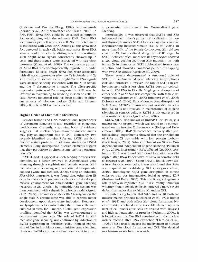
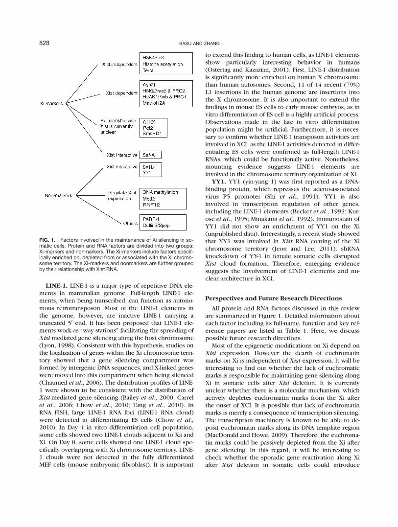
All protein and RNA factors discussed in this revieware summarized in Figure 1. Detailed information abouteach factor including its full-name, function and key ref-erence papers are listed in Table 1. Here, we discusspossible future research directions.
Most of the epigenetic modifications on Xi depend onXist expression. However the dearth of euchromatinmarks on Xi is independent of Xist expression. It will beinteresting to find out whether the lack of euchromaticmarks is responsible for maintaining gene silencing alongXi in somatic cells after Xist deletion. It is currentlyunclear whether there is a molecular mechanism, whichactively depletes euchromatin marks from the Xi afterthe onset of XCI. It is possible that lack of euchromatinmarks is merely a consequence of transcription silencing.The transcription machinery is known to be able to de-posit euchromatin marks along its DNA template region(MacDonald and Howe, 2009). Therefore, the euchroma-tin marks could be passively depleted from the Xi aftergene silencing. In this regard, it will be interesting tocheck whether the sporadic gene reactivation along Xiafter Xist deletion in somatic cells could introduce
FIG. 1. Factors involved in the maintenance of Xi silencing in so-matic cells. Protein and RNA factors are divided into two groups:Xi-markers and nonmarkers. The Xi-markers include factors specif-ically enriched on, depleted from or associated with the Xi chromo-some territory. The Xi-markers and nonmarkers are further groupedby their relationship with Xist RNA.
828 BASU AND ZHANG

euchromatic marks into corresponding DNA regions,and whether such euchromatic ‘‘lesions’’ could graduallyspread into neighboring regions after a number of celldivisions.
A growing list of recent discoveries shows that nu-clear matrix, nuclear organization or higher order ofchromatin structures may play important roles in XCI.Clearly, future research is required in this direction. It
should be noted that nuclear organization can only bestable for one cell cycle. Upon cell division, correct nu-clear organization has to be re-established in eachdaughter cell. If Xi is specifically organized into a cer-tain nuclear compartment but not Xa, there must besome kind of epigenetic modifications on the Xi, whichcan be recognized by cells in rebuilding the Xi nuclearcompartment after each cell cycle. Such an epigenetic
Table 1Factors Involved in Xi Silencing in Somatic Cell
Name Full name Function/identity Involvement in XCI Reference
Ash2l absent, small or homeoticdiscs 2
belongs to trithoraxgroup protein
enrichment on Xi Pullirsch et al., 2010
ATRX alpha thalassemia/mentalretardation X-linked
belongs to SWI/SNF2ATP-dependenthelicase family
enrichment on Xi Baumann et al., 2009
Cullin3/Spop N/A E3 ubiquitin ligase ubiquitinate macroH2A1 Hernandez-Munozet al., 2005
Dnmt1 DNA methylatranferase 1 maintenance DNAmethylase
regulate Xist and other X-linked gene expression
Beard et al., 1995;Panning andJaenisch, 1996;Sado et al., 2000
Dnmt3a/3b DNA methylatranferase3a/3b
de novo DNAmethylases
regulate Xist and other X-linked gene expression
Sado et al., 2004
H2AK119ub histone2A lysine119ubiquitination
histone modification enrichment on Xi Plath et al., 2004; Fanget al., 2004; deNapoles et al., 2004
H3K4Me3 histone3 lysine4trimethylation
histone modification depletion from Xi Zhang et al., 2007
H3K27Me3 histone3 lysine27trimethylation
histone modification enrichment on Xi Silva et al., 2003; Plathet al., 2003; Erhardtet al., 2003; Maket al., 2002; de laCruz et al., 2005
H4Ac histone4 acetylation histone modification depletion from Xi Pullirsch et al., 2010Line-1 long interspersed nuclear
elementretrotransposon facilitate spreading of Xist
along X chromosome;involved in Xi chromosometerritory organization
Chow et al., 2010
macroH2A macro histone2A histone variant enrichment on Xi Costanzi and Pehrson,1998
Mbd2 methylated DNA bindingprotein 2
methylated DNAbinding protein
regulate Xist expression Barr et al., 2007
PARP-1 Poly(ADP-ribose)polymerase 1
Poly(ADP-ribose)polymerase
interact with macroH2A1.2 Nusinow et al., 2007
Pcl2 Polycomblike Protein 2 Facilitate loading ofPRC2 proteins on Xi
Enrichment on Xi Casanova et al., 2011
Saf-A scaffold attachmentfactor A
nuclear matrix protein enrichment on Xi; influenceXist localization
Helbig andFackelmayer, 2003;Pullirsch et al., 2010;Hasegawa et al.,2010
SATB1 special AT-rich bindingprotein
nuclear matrix protein influence Xist localization Agrelo et al., 2009
SmcHD1 structural maintenance ofchromosomes hingedomain containing 1
structural maintenanceof chromosomeshinge domaincontaining protein
enrichment on Xi Blewitt et al., 2008
Terra telomeric repeat-containing RNA
ncRNA large and bright Terra RNAfoci attach to Xichromosome territory
Zhang et al., 2009
Xist Xi specific transcript ncRNA play critical role duringinitiation of XCI
Costanzi et al., 2000;Csankovszki et al.,2001
YY1 Yin Yang-1 transcription factor Required for loading Xist RNAon Xi
Yesu Jeon and Lee,2011
829X CHROMOSOME INACTIVATION IN SOMATIC CELLS

modification may still be missing in our current list. Itshould also be noted that there might not be a nuclearcompartment specially built just for Xi. Xi may belongto a nuclear environment that is generally shared by fac-ultative heterochromatin. The perinucleolar region maybe such a nuclear domain. Xi is frequently localizedwithin the perinucleolar region and evidence suggeststhat Xi is targeted to perinucleolar region during the Sphase (Zhang et al., 2007).
Based on our current knowledge, marsupials undertakeimprinted XCI in all tissues, but they do not have Xist
gene. Eutherians (placental mammals) have Xist gene andundertake random XCI in all somatic tissues. It is reasona-ble to argue that Xist was evolved to achieve the ‘‘symme-try break’’ required in random XCI. If so, the Xist-depend-ent XCI may be built up upon a core gene silencing mech-anism established in the ancient imprinted XCI ofmarsupials. Therefore, studies on imprinted XCI in marsu-pials may eventually provide an answer for the genesilencing mechanism in random XCI.
Because of the lack of knowledge on gene silencingmechanism of XCI, we still do not have an artificialmethod to efficiently breakdown gene silencing on Xi.Xi can only be reactivated in vivo in three known bio-logical events. First, the imprinted XCI in early mouseembryos is reactivated during epiblast lineage specifi-cation in the inner cell mass. Second, Xi is reactivatedin PGCs, right after PGC specification. Third, Xi reacti-vation occurs in generation of iPS cells. Studies on Xireactivation will no doubt help us understand thegene silencing mechanism of XCI. A recent study onepiblast specification has shown that gene reactiva-tion occurred on Xi when its chromosome territorywas still covered by the Xist cloud (Williams et al.,2011). This result is consistent with the existence of aXist-independent gene silencing mechanism. Such agene silencing mechanism is established by Xist dur-ing the initiation stage of XCI. However, in somaticcells, the gene silencing mechanism can efficientlymaintain gene silencing without depending on Xist
expression. During epiblast specification, the silencedgenes on Xi can also be reactivated regardless of Xistexpression.
CONCLUSION
The inactive X chromosome carries multiple layers ofepigenetic modifications to achieve its chromosome-wide gene silencing. Many of the Xi-associated epigeneticmodifications and their associated proteins have beenidentified. However, the Xi silencing remained largelyintact even after almost all known factors were artificiallydepleted from the Xi. Current data showed that Xi silenc-ing in soma was tightly controlled by a multilayer mecha-nism. The difficulty in breaking down Xi silencing couldbe a result of mechanistic redundancy. On the other
hand, there might be unknown epigenetic factors/mech-anisms responsible for maintaining Xi silencing. Tounderstand how Xi silencing is epigenetically maintainedin soma is a challenging question bearing important bio-logical and biomedical significance.
LITERATURE CITED
Agrelo R, Souabni A, Novatchkova M, Haslinger C, LeebM, Komnenovic V, Kishimoto H, Gresh L, Kohwi-Shi-gematsu T, Kenner L, Wutz A. 2009. SATB1 definesthe developmental context for gene silencing byXist in lymphoma and embryonic cells. Dev Cell16:507–516.
Agulnik AI, Mitchell MJ, Mattei MG, Borsani G, AvnerPA, Lerner JL, Bishop CE. 1994. A novel X gene witha widely transcribed Y-linked homologue escapesX-inactivation in mouse and human. Hum Mol Genet3:879–884.
Alvarez JD, Yasui DH, Niida H, Joh T, Loh DY, Kohwi-Shi-gematsu T. 2000. The MAR-binding protein SATB1orchestrates temporal and spatial expression of mul-tiple genes during T-cell development. Genes Dev14:521–535.
Ariel M, Robinson E, McCarrey JR, Cedar H. 1995. Ga-mete-specific methylation correlates with imprint-ing of the murine Xist gene. Nat Genet 9:312–315.
Azzalin CM, Reichenbach P, Khoriauli L, Giulotto E,Lingner J. 2007. Telomeric repeat containing RNAand RNA surveillance factors at mammalian chromo-some ends. Science 318:798–801.
Bailey JA, Carrel L, Chakravarti A, Eichler EE. 2000. Mo-lecular evidence for a relationship between LINE-1elements and X chromosome inactivation: The Lyonrepeat hypothesis. Proc Natl Acad Sci USA 97:6634–6639.
Barakat TS, Gunhanlar N, Pardo CG, Achame EM, Ghaz-vini M, Boers R, Kenter A, Rentmeester E, Groote-goed JA, Gribnau J. 2011. RNF12 activates Xist andis essential for X chromosome inactivation. PLoSGenet 7:e1002001.
Barakat TS, Jonkers I, Monkhorst K, Gribnau J. 2010. X-changing information on X inactivation. Exp CellRes 316:679–687.
Barr H, Hermann A, Berger J, Tsai HH, Adie K, Pro-khortchouk A, Hendrich B, Bird A. 2007. Mbd2 con-tributes to DNA methylation-directed repression ofthe Xist gene. Mol Cell Biol 27:3750–3757.
Barr ML, Bertram EG. 1949. A morphological distinctionbetween neurones of the male and female, and thebehaviour of the nucleolar satellite during acceler-ated nucleoprotein synthesis. Nature 163:676.
Baumann C, De La Fuente R. 2009. ATRX marks theinactive X chromosome (Xi) in somatic cells andduring imprinted X chromosome inactivation introphoblast stem cells. Chromosoma 118:209–222.
830 BASU AND ZHANG
Beard C, Li E, Jaenisch R. 1995. Loss of methylation acti-vates Xist in somatic but not in embryonic cells.Genes Dev 9:2325–2334.
Becker KG, Swergold GD, Ozato K, Thayer RE. 1993.Binding of the ubiquitous nuclear transcription fac-tor YY1 to a cis regulatory sequence in the humanLINE-1 transposable element. Hum Mol Genet2:1697–1702.
Blewitt ME, Gendrel AV, Pang Z, Sparrow DB, WhitelawN, Craig JM, Apedaile A, Hilton DJ, Dunwoodie SL,Brockdorff N, Kay GF, Whitelaw E. 2008. SmcHD1,containing a structural-maintenance-of-chromo-somes hinge domain, has a critical role in X inactiva-tion. Nat Genet 40:663–669.
Blewitt ME, Vickaryous NK, Hemley SJ, Ashe A, BruxnerTJ, Preis JI, Arkell R, Whitelaw E. 2005. An N-ethyl-N-nitrosourea screen for genes involved in variega-tion in the mouse. Proc Natl Acad Sci USA102:7629–7634.
Borsani G, Tonlorenzi R, Simmler MC, Dandolo L,Arnaud D, Capra V, Grompe M, Pizzuti A, Muzny D,Lawrence C. 1991. Characterization of a murinegene expressed from the inactive X chromosome.Nature 351:325.
Britanova O, Depew MJ, Schwark M, Thomas BL, Mile-tich I, Sharpe P, Tarabykin V. 2006. Satb2 haploinsuf-ficiency phenocopies 2q32-q33 deletions, whereasloss suggests a fundamental role in the coordinationof jaw development. Am J Hum Genet 79:668–678.
Brockdorff N, Ashworth A, Kay GF, Cooper P, Smith S,McCabe VM, Norris DP, Penny GD, Patel D, Rastan S.1991. Conservation of position and exclusiveexpression of mouse Xist from the inactive X chro-mosome. Nature 351:329.
Brown CJ, Ballabio A, Rupert JL, Lafreniere RG, GrompeM, Tonlorenzi R, Willard HF. 1991. A gene from theregion of the human X inactivation centre isexpressed exclusively from the inactive X chromo-some. Nature 349:38.
Cai S, Han HJ, Kohwi-Shigematsu T. 2003. Tissue-spe-cific nuclear architecture and gene expression regu-lated by SATB1. Nat Genet 34:42–51.
Cao R, Wang L, Wang H, Xia L, Erdjument-Bromage H,Tempst P, Jones RS, Zhang Y. 2002. Role of histoneH3 lysine 27 methylation in Polycomb-group silenc-ing. Science 298:1039–1043.
Carrel L, Park C, Tyekucheva S, Dunn J, Chiaromonte F,Makova KD. 2006. Genomic environment predictsexpression patterns on the human inactive X chro-mosome. PLoS Genet 2:e151.
Carrel L, Willard HF. 2005. X-inactivation profile revealsextensive variability in X-linked gene expression infemales. Nature 434:400–404.
Casanova M, Preissner T, Cerase A, Poot R, Yamada D, LiX, Appanah R, Bezstarosti K, Demmers J, Koseki H,Brockdorff N. 2011. Polycomblike 2 facilitates the
recruitment of PRC2 Polycomb group complexes tothe inactive X chromosome and to target loci in em-bryonic stem cells. Development 138:1471–1482.
Chadwick BP, Valley CM, Willard HF. 2001. Histone vari-ant macroH2A contains two distinct macrochroma-tin domains capable of directing macroH2A to theinactive X chromosome. Nucleic Acids Res29:2699–2705.
Chadwick BP, Willard HF. 2001. Histone H2A variantsand the inactive X chromosome: Identification of asecond macroH2A variant. Hum Mol Genet10:1101–1113.
Changolkar LN, Costanzi C, Leu NA, Chen D, McLaugh-lin KJ, Pehrson JR. 2007. Developmental changes inhistone macroH2A1-mediated gene regulation. MolCell Biol 27:2758–2764.
Chaumeil J, Le Baccon P, Wutz A, Heard E. 2006. A novelrole for Xist RNA in the formation of a repressive nu-clear compartment into which genes are recruitedwhen silenced. Genes Dev 20:2223–2237.
Chaumeil J, Waters PD, Koina E, Gilbert C, Robinson TJ,Graves JA. 2011. Evolution from XIST-independentto XIST-controlled X-chromosome inactivation: Epi-genetic modifications in distantly related mammals.PLoS One 6:e19040.
Chow JC, Ciaudo C, Fazzari MJ, Mise N, Servant N, GlassJL, Attreed M, Avner P, Wutz A, Barillot E, GreallyJM, Voinnet O, Heard E. 2010. LINE-1 activity in fac-ultative heterochromatin formation during X chro-mosome inactivation. Cell 141:956–969.
Clemson CM, McNeil JA, Willard HF, Lawrence JB. 1996.XIST RNA paints the inactive X chromosome atinterphase: Evidence for a novel RNA involved innuclear/chromosome structure. J.Cell Biol 132:259.
Costanzi C, Pehrson JR. 1998. Histone macroH2A1 isconcentrated in the inactive X chromosome offemale mammals. Nature 393:599–601.
Costanzi C, Stein P, Worrad DM, Schultz RM, PehrsonJR. 2000. Histone macroH2A1 is concentrated in theinactive X chromosome of female preimplantationmouse embryos. Development 127:2283–2289.
Csankovszki G, Nagy A, Jaenisch R. 2001. Synergism ofXist RNA, DNA methylation, and histone hypoacety-lation in maintaining X chromosome inactivation. JCell Biol 153:773–784.
Csankovszki G, Panning B, Bates B, Pehrson JR, JaenischR. 1999. Conditional deletion of Xist disrupts his-tone macroH2A localization but not maintenance ofX inactivation. Nat Genet 22:323–324.
Czermin B, Melfi R, McCabe D, Seitz V, Imhof A, PirrottaV. 2002. Drosophila enhancer of Zeste/ESC com-plexes have a histone H3 methyltransferase activitythat marks chromosomal Polycomb sites. Cell111:185–196.
de la Cruz CC, Fang J, Plath K, Worringer KA, NusinowDA, Zhang Y, Panning B. 2005. Developmental regu-
831X CHROMOSOME INACTIVATION IN SOMATIC CELLS
lation of Suz 12 localization. Chromosoma 114:183–192.
de Napoles M, Mermoud JE, Wakao R, Tang YA, EndohM, Appanah R, Nesterova TB, Silva J, Otte AP, VidalM, Koseki H, Brockdorff N. 2004. Polycomb groupproteins Ring1A/B link ubiquitylation of histoneH2A to heritable gene silencing and X inactivation.Dev Cell 7:663–676.
Dickinson LA, Joh T, Kohwi Y, Kohwi-Shigematsu T.1992. A tissue-specific MAR/SAR DNA-binding pro-tein with unusual binding site recognition. Cell70:631–645.
Dobreva G, Chahrour M, Dautzenberg M, Chirivella L,Kanzler B, Farinas I, Karsenty G, Grosschedl R.2006. SATB2 is a multifunctional determinant of cra-niofacial patterning and osteoblast differentiation.Cell 125:971–986.
Erhardt S, Su IH, Schneider R, Barton S, Bannister AJ,Perez-Burgos L, Jenuwein T, Kouzarides T, Tarakhov-sky A, Surani MA. 2003. Consequences of the deple-tion of zygotic and embryonic enhancer of zeste 2during preimplantation mouse development. Devel-opment 130:4235–4248.
Fackelmayer FO. 2005. A stable proteinaceous structurein the territory of inactive X chromosomes. J BiolChem 280:1720–1723.
Fang J, Chen T, Chadwick B, Li E, Zhang Y. 2004.Ring1b-mediated H2A ubiquitination associates withinactive X chromosomes and is involved in initiationof X inactivation. J Biol Chem 279:52812–52815.
Filippova GN, Cheng MK, Moore JM, Truong JP, Hu YJ,Nguyen DK, Tsuchiya KD, Disteche CM. 2005.Boundaries between chromosomal domains of Xinactivation and escape bind CTCF and lack CpGmethylation during early development. Dev Cell8:31–42.
Garrick D, Sharpe JA, Arkell R, Dobbie L, Smith AJ,Wood WG, Higgs DR, Gibbons RJ. 2006. Loss of Atrxaffects trophoblast development and the pattern ofX-inactivation in extraembryonic tissues. PLoSGenet 2:e58.
Gibbons R. 2006. Alpha thalassaemia-mental retarda-tion, X linked. Orphanet J Rare Dis 1:15.
Greenfield A, Carrel L, Pennisi D, Philippe C, Quaderi N,Siggers P, Steiner K, Tam PP, Monaco AP, Willard HF,Koopman P. 1998. The UTX gene escapes X inactiva-tion in mice and humans. Hum Mol Genet 7:737–742.
Guy J, Gan J, Selfridge J, Cobb S, Bird A. 2007. Reversalof neurological defects in a mouse model of Rettsyndrome. Science 315:1143–1147.
Hasegawa Y, Brockdorff N, Kawano S, Tsutui K, Naka-gawa S. 2010. The matrix protein hnRNP U isrequired for chromosomal localization of Xist RNA.Dev Cell 19:469–476.
Helbig R, Fackelmayer FO. 2003. Scaffold attachmentfactor A (SAF-A) is concentrated in inactive X
chromosome territories through its RGG domain.Chromosoma 112:173–182.
Hernandez-Munoz I, Lund AH, van der Stoop P, BoutsmaE, Muijrers I, Verhoeven E, Nusinow DA, Panning B,Marahrens Y, van Lohuizen M. 2005. Stable X chro-mosome inactivation involves the PRC1 Polycombcomplex and requires histone MACROH2A1 and theCULLIN3/SPOP ubiquitin E3 ligase. Proc Natl AcadSci USA 102:7635–7640.
Huynh KD, Lee JT. 2003. Inheritance of a pre-inacti-vated paternal X chromosome in early mouseembryos. Nature 426:857–862.
Jahner D, Stuhlmann H, Stewart CL, Harbers K, Lohler J,Simon I, Jaenisch R. 1982. De novo methylation andexpression of retroviral genomes during mouseembryogenesis. Nature 298:623–628.
Jeon Y, Lee JT. 2011. YY1 Tethers Xist RNA to the inac-tive X nucleation center. Cell 146:119–133.
Jonkers I, Barakat TS, Achame EM, Monkhorst K, KenterA, Rentmeester E, Grosveld F, Grootegoed JA, Grib-nau J. 2009. RNF12 is an X-Encoded dose-dependentactivator of X chromosome inactivation. Cell139:999–1011.
Kalantry S, Magnuson T. 2006. The Polycomb groupprotein EED is dispensable for the initiation ofrandom X-chromosome inactivation. PLoS Genet2:e66.
Kalantry S, Mills KC, Yee D, Otte AP, Panning B, Magnu-son T. 2006. The Polycomb group protein Eed pro-tects the inactive X-chromosome from differentia-tion-induced reactivation. Nat Cell Biol 8:195–202.
Karras GI, Kustatscher G, Buhecha HR, Allen MD,Pugieux C, Sait F, Bycroft M, Ladurner AG. 2005.The macro domain is an ADP-ribose binding mod-ule. EMBO J 24:1911–1920.
Kohlmaier A, Savarese F, Lachner M, Martens J, Jenu-wein T, Wutz A. 2004. A chromosomal memory trig-gered by Xist regulates histone methylation in Xinactivation. PLoS Biol 2:E171.
Kunath T, Arnaud D, Uy GD, Okamoto I, Chureau C,Yamanaka Y, Heard E, Gardner RL, Avner P, RossantJ. 2005. Imprinted X-inactivation in extra-embryonicendoderm cell lines from mouse blastocysts. Devel-opment 132:1649–1661.
Kurose K, Hata K, Hattori M, Sakaki Y. 1995. RNA poly-merase III dependence of the human L1 promoterand possible participation of the RNA polymerase IIfactor YY1 in the RNA polymerase III transcriptionsystem. Nucleic Acids Res 23:3704–3709.
Kuzmichev A, Nishioka K, Erdjument-Bromage H,Tempst P, Reinberg D. 2002. Histone methyltransfer-ase activity associated with a human multiproteincomplex containing the enhancer of Zeste protein.Genes Dev 16:2893–2905.
Lee JT. 2000. Disruption of imprinted X inactivation byparent-of-origin effects at Tsix. Cell 103:17–27.
832 BASU AND ZHANG

Lee JT, Davidow LS, Warshawsky D. 1999. Tsix, a geneantisense to Xist at the X-inactivation centre. NatGenet 21:400.
Leeb M, Wutz A. 2007. Ring1B is crucial for the regula-tion of developmental control genes and PRC1 pro-teins but not X inactivation in embryonic cells. JCell Biol 178:219–229.
Li E, Beard C, Jaenisch R. 1993. Role for DNA methyla-tion in genomic imprinting. Nature 366:362–365.
Luke B, Lingner J. 2009. TERRA: Telomeric repeat-con-taining RNA. EMBO J 28:2503–2510.
Lyon MF. 1961. Gene action in the X-chromosome ofthe mouse (Mus musculus L.). Nature 190:372.
Lyon MF. 1998. X-chromosome inactivation: A repeat hy-pothesis. Cytogenet Cell Genet 80:133–137.
MacDonald VE, Howe LJ. 2009. Histone acetylation:Where to go and how to get there. Epigenetics4:139–143.
Mak W, Baxter J, Silva J, Newall AE, Otte AP, BrockdorffN. 2002. Mitotically stable association of polycombgroup proteins eed and enx1 with the inactive 3chromosome in trophoblast stem cells. Curr Biol12:1016–1020.
Mak W, Nesterova TB, de Napoles M, Appanah R, Yama-naka S, Otte AP, Brockdorff N. 2004. Reactivation ofthe paternal X chromosome in early mouseembryos. Science 303:666–669.
Marahrens Y, Panning B, Dausman J, Strauss W, JaenischR. 1997. Xist-deficient mice are defective in dosagecompensation but not spermatogenesis. Genes Dev11:156–166.
Martin DI, Whitelaw E. 1996. The vagaries of variegat-ing transgenes. Bioessays 18:919–923.
Meder VS, Boeglin M, de Murcia G, Schreiber V. 2005.PARP-1 and PARP-2 interact with nucleophosmin/B23 and accumulate in transcriptionally activenucleoli. J Cell Sci 118:211–222.
Menissier de Murcia J, Ricoul M, Tartier L, Nieder-gang C, Huber A, Dantzer F, Schreiber V, Ame JC,Dierich A, LeMeur M, Sabatier L, Chambon P, deMurcia G. 2003. Functional interaction betweenPARP-1 and PARP-2 in chromosome stability andembryonic development in mouse. EMBO J 22:2255–2263.
Mermoud JE, Costanzi C, Pehrson JR, Brockdorff N.1999. Histone macroH2A1.2 relocates to the inac-tive X chromosome after initiation and propagationof X-inactivation. J Cell Biol 147:1399–1408.
Minakami R, Kurose K, Etoh K, Furuhata Y, Hattori M,Sakaki Y. 1992. Identification of an internal cis-ele-ment essential for the human L1 transcription and anuclear factor(s) binding to the element. NucleicAcids Res 20:3139–3145.
Muller J, Hart CM, Francis NJ, Vargas ML, Sengupta A,Wild B, Miller EL, O’Connor MB, Kingston RE,Simon JA. 2002. Histone methyltransferase activity
of a Drosophila Polycomb group repressor complex.Cell 111:197–208.
Nesterova TB, Mermoud JE, Hilton K, Pehrson J, SuraniMA, McLaren A, Brockdorff N. 2002. Xist expressionand macroH2A1.2 localisation in mouse primordialand pluripotent embryonic germ cells. Differentia-tion 69:216–225.
Norris DP, Patel D, Kay GF, Penny GD, Brockdorff N,Sheardown SA, Rastan S. 1994. Evidence that ran-dom and imprinted Xist expression is controlled bypreemptive methylation. Cell 77:41–51.
Nusinow DA, Hernandez-Munoz I, Fazzio TG, Shah GM,Kraus WL, Panning B. 2007. Poly(ADP-ribose)polymerase 1 is inhibited by a histone H2A variant,MacroH2A, and contributes to silencing of theinactive X chromosome. J Biol Chem 282:12851–12859.
Ogawa Y, Lee JT. 2003. Xite, X-inactivation intergenictranscription elements that regulate the probabilityof choice. Mol Cell 11:731–743.
Okamoto I, Otte AP, Allis CD, Reinberg D, Heard E.2004. Epigenetic dynamics of imprinted X inactiva-tion during early mouse development. Science303:644–649.
Okamoto I, Patrat C, Thepot D, Peynot N, Fauque P,Daniel N, Diabangouaya P, Wolf JP, Renard JP, Duran-thon V, Heard E. 2011. Eutherian mammals usediverse strategies to initiate X-chromosome inactiva-tion during development. Nature 472:370–374.
Orkin SH, Hochedlinger K. 2011. Chromatin connec-tions to pluripotency and cellular reprogramming.Cell 145:835–850.
Ostertag EM, Kazazian HH Jr. 2001. Biology of mammalianL1 retrotransposons. Annu Rev Genet 35:501–538.
Panning B, Jaenisch R. 1996. DNA hypomethylation canactivate Xist expression and silence X-linked genes.Genes Dev 10:1991–2002.
Pederson T. 2000. Half a century of ‘‘the nuclear ma-trix’’. Mol Biol Cell 11:799–805.
Penny GD, Kay GF, Sheardown SA, Rastan S, BrockdorffN. 1996. Requirement for Xist in X chromosomeinactivation. Nature 379:131–137.
Plath K, Fang J, Mlynarczyk-Evans SK, Cao R, WorringerKA, Wang H, de la Cruz CC, Otte AP, Panning B,Zhang Y. 2003. Role of histone H3 lysine 27 methyl-ation in X inactivation. Science 300:131–135.
Plath K, Talbot D, Hamer KM, Otte AP, Yang TP, JaenischR, Panning B. 2004. Developmentally regulatedalterations in Polycomb repressive complex 1 pro-teins on the inactive X chromosome. J Cell Biol167:1025–1035.
Pullirsch D, Hartel R, Kishimoto H, Leeb M, Steiner G,Wutz A. 2010. The Trithorax group protein Ash2land Saf-A are recruited to the inactive X chromo-some at the onset of stable X inactivation. Develop-ment 137:935–943.
833X CHROMOSOME INACTIVATION IN SOMATIC CELLS
Rasmussen TP, Mastrangelo MA, Eden A, Pehrson JR, Jae-nisch R. 2000. Dynamic relocalization of histoneMacroH2A1 from centrosomes to inactive X chro-mosomes during X inactivation. J Cell Biol150:1189–1198.
Romig H, Fackelmayer FO, Renz A, Ramsperger U,Richter A. 1992. Characterization of SAF-A, a novelnuclear DNA binding protein from HeLa cells withhigh affinity for nuclear matrix/scaffold attachmentDNA elements. EMBO J 11:3431–3440.
Roshon MJ, Ruley HE. 2005. Hypomorphic mutation inhnRNP U results in post-implantation lethality.Transgenic Res 14:179–192.
Rudenko G, Van der Ploeg LH. 1989. Transcription oftelomere repeats in protozoa. EMBO J 8:2633–2638.
Sado T, Fenner MH, Tan SS, Tam P, Shioda T, Li E. 2000.X inactivation in the mouse embryo deficient forDnmt1: Distinct effect of hypomethylation onimprinted and random X inactivation. Dev Biol225:294–303.
Sado T, Okano M, Li E, Sasaki H. 2004. De novo DNAmethylation is dispensable for the initiation andpropagation of X chromosome inactivation. Devel-opment 131:975–982.
Salido EC, Li XM, Yen PH, Martin N, Mohandas TK, Sha-piro LJ. 1996. Cloning and expression of the mousepseudoautosomal steroid sulphatase gene (Sts). NatGenet 13:83–86.
Savarese F, Flahndorfer K, Jaenisch R, Busslinger M,Wutz A. 2006. Hematopoietic precursor cells transi-ently reestablish permissiveness for X inactivation.Mol Cell Biol 26:7167–7177.
Schoeftner S, Blasco MA. 2008. Developmentally regu-lated transcription of mammalian telomeres byDNA-dependent RNA polymerase II. Nat Cell Biol10:228–236.
Schoeftner S, Sengupta AK, Kubicek S, Mechtler K,Spahn L, Koseki H, Jenuwein T, Wutz A. 2006.Recruitment of PRC1 function at the initiation of Xinactivation independent of PRC2 and silencing.EMBO J 25:3110–3122.
Schuettengruber B, Chourrout D, Vervoort M, LeblancB, Cavalli G. 2007. Genome regulation by polycomband trithorax proteins. Cell 128:735–745.
Sharman GB. 1971. Late DNA replication in the pater-nally derived X chromosome of female kangaroos.Nature 230:231–232.
Shi Y, Seto E, Chang LS, Shenk T. 1991. Transcriptionalrepression by YY1, a human GLI-Kruppel-relatedprotein, and relief of repression by adenovirus E1Aprotein. Cell 67:377–388.
Shin J, Bossenz M, Chung Y, Ma H, Byron M, Tanigu-chi-Ishigaki N, Zhu X, Jiao B, Hall LL, Green MR,Jones SN, Hermans-Borgmeyer I, Lawrence JB,Bach I. 2010. Maternal Rnf12/RLIM is required for
imprinted X-chromosome inactivation in mice. Na-ture 467:977–981.
Silva J, Mak W, Zvetkova I, Appanah R, Nesterova TB,Webster Z, Peters AH, Jenuwein T, Otte AP, Brock-dorff N. 2003. Establishment of histone h3 methyla-tion on the inactive X chromosome requires tran-sient recruitment of Eed-Enx1 polycomb groupcomplexes. Dev Cell 4:481–495.
Solovei I, Gaginskaya ER, Macgregor HC. 1994. Thearrangement and transcription of telomere DNAsequences at the ends of lampbrush chromosomesof birds. Chromosome Res 2:460–470.
Takagi N, Sasaki M. 1975. Preferential inactivation of thepaternally derived X chromosome in the extraem-bryonic membranes of the mouse. Nature 256:640–642.
Tang YA, Huntley D, Montana G, Cerase A, NesterovaTB, Brockdorff N. 2010. Efficiency of Xist-mediatedsilencing on autosomes is linked to chromosomaldomain organisation. Epigenetics Chromatin 3:10.
Thorogood NP, Brown CJ. 2010. Active chromatin marksare retained on X chromosomes lacking gene orrepeat silencing despite XIST/Xist expression in so-matic cell hybrids. PLoS One 5:e10787.
Tian D, Sun S, Lee JT. 2010. The long noncoding RNA,Jpx, is a molecular switch for X chromosome inacti-vation. Cell 143:390–403.
Wang H, Wang L, Erdjument-Bromage H, Vidal M,Tempst P, Jones RS, Zhang Y. 2004. Role of histoneH2A ubiquitination in Polycomb silencing. Nature431:873–878.
Wang J, Mager J, Chen Y, Schneider E, Cross JC, Nagy A,Magnuson T. 2001. Imprinted X inactivation main-tained by a mouse Polycomb group gene. Nat Genet28:371–375.
Williams LH, Kalantry S, Starmer J, Magnuson T. 2011.Transcription precedes loss of Xist coating anddepletion of H3K27me3 during X-chromosomereprogramming in the mouse inner cell mass. Devel-opment 138:2049–2057.
Wutz A, Jaenisch R. 2000. A shift from reversible to irre-versible X inactivation is triggered during ES cell dif-ferentiation. Mol Cell 5:695–705.
Zhang LF, Huynh KD, Lee JT. 2007. Perinucleolar target-ing of the inactive X during S phase: Evidence for arole in the maintenance of silencing. Cell 129:693–706.
Zhang LF, Ogawa Y, Ahn JY, Namekawa SH, Silva SS, LeeJT. 2009. Telomeric RNAs mark sex chromosomes instem cells. Genetics 182:685–698.
Zuccotti M, Monk M. 1995. Methylation of the mouseXist gene in sperm and eggs correlates withimprinted Xist expression and paternal X-inactiva-tion. Nat Genet 9:316–320.
834 BASU AND ZHANG