world live neurovascular conference … op post op 1 month mra pre op 6 month cont pre op 6 month...
TRANSCRIPT
WORLD LIVE NEUROVASCULAR CONFERENCE Los Angeles, 15-17 MAY 2017
LIVE CASEs ANGIOGRAPHIC FOLLOW-UPs
WLNC 2017 ANKARA CASES F-U/BY PROF SAATCI
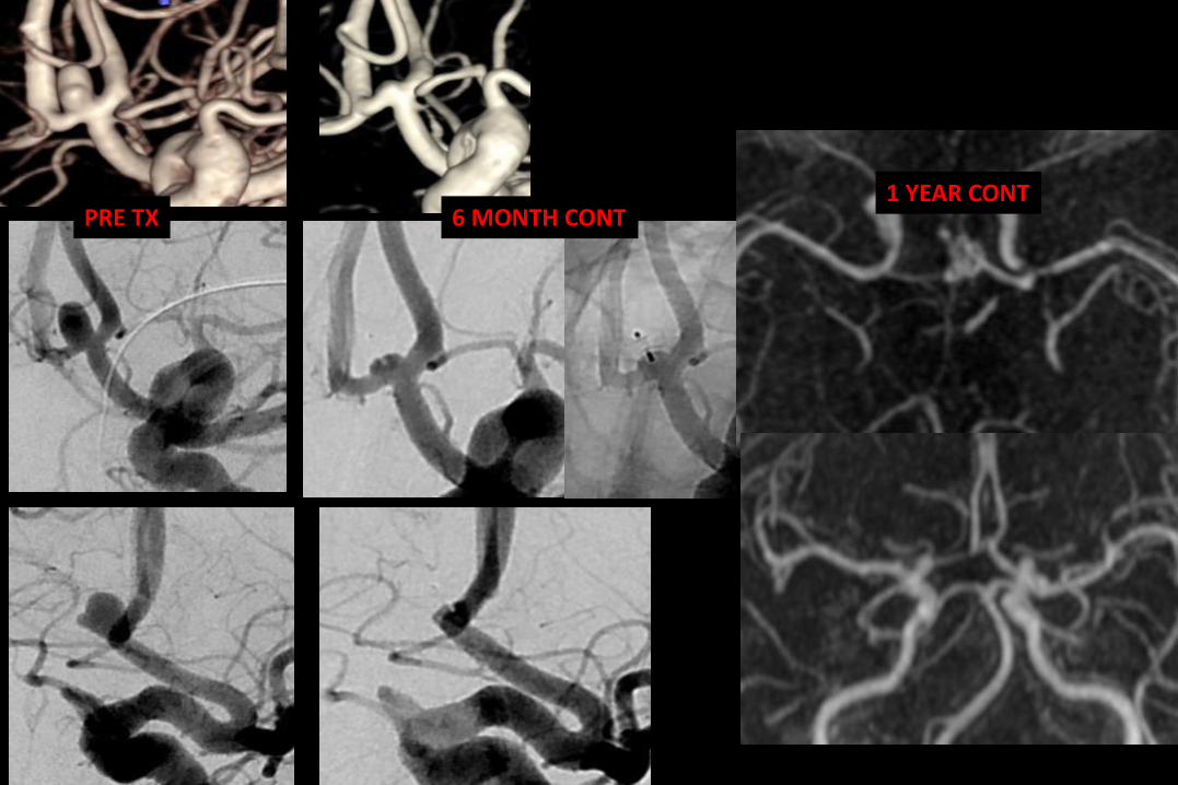
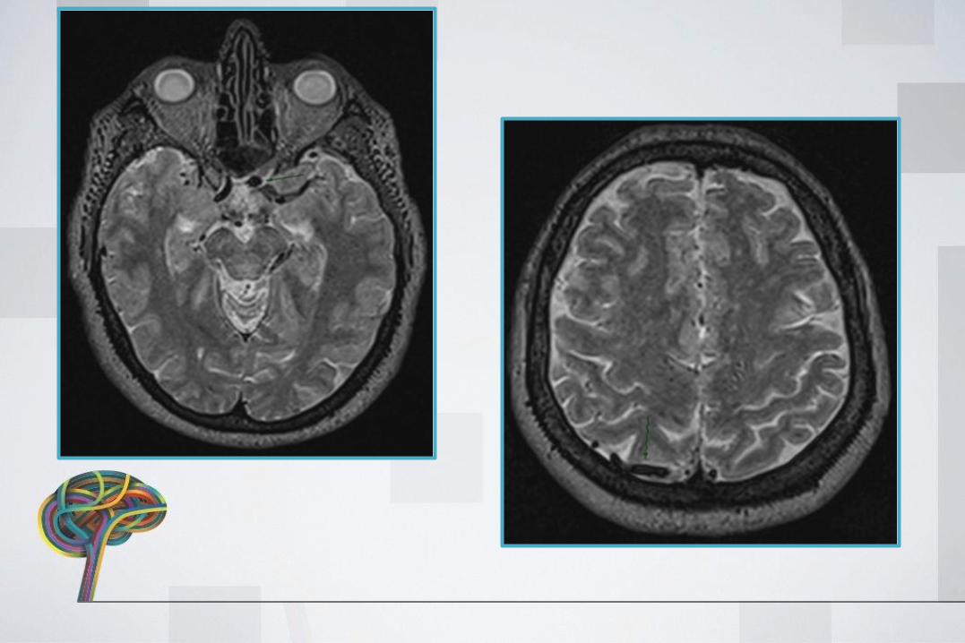
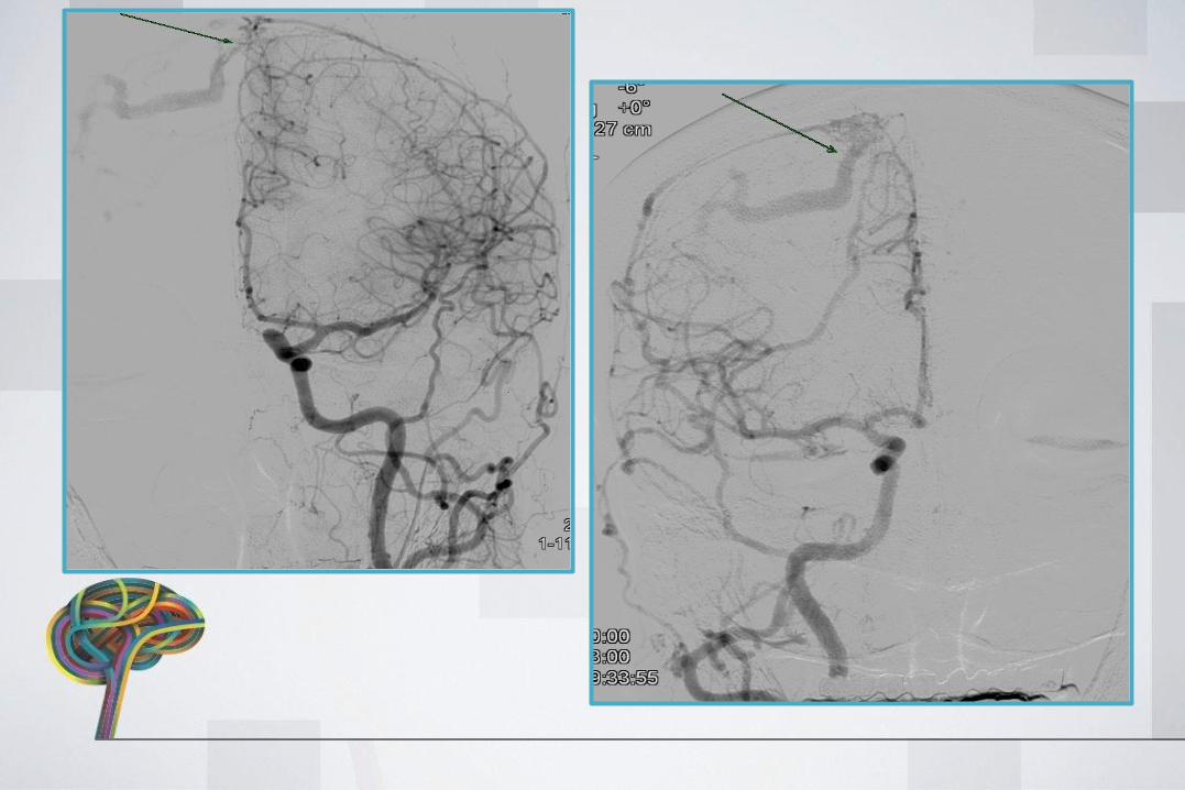
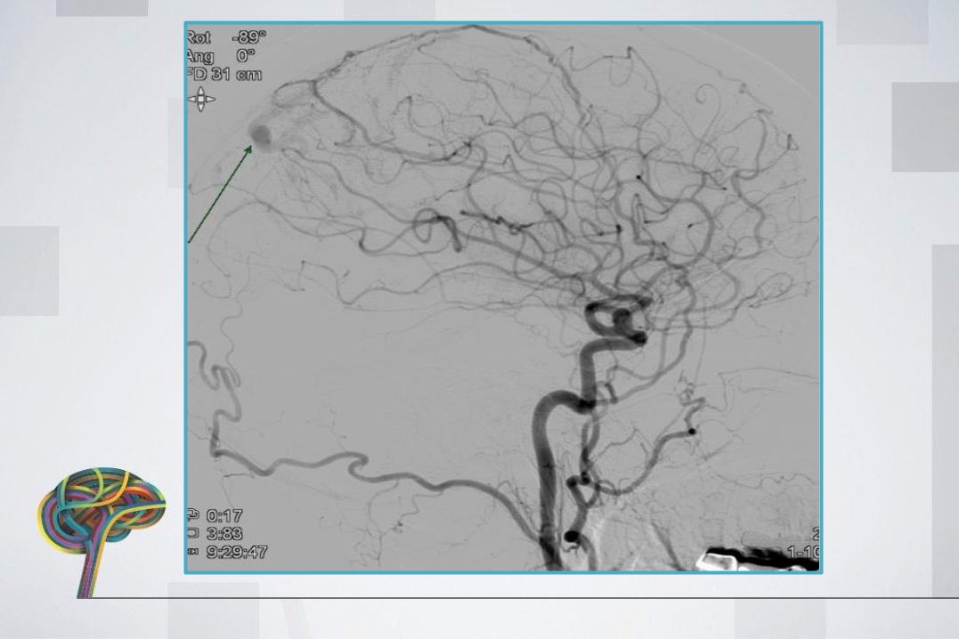
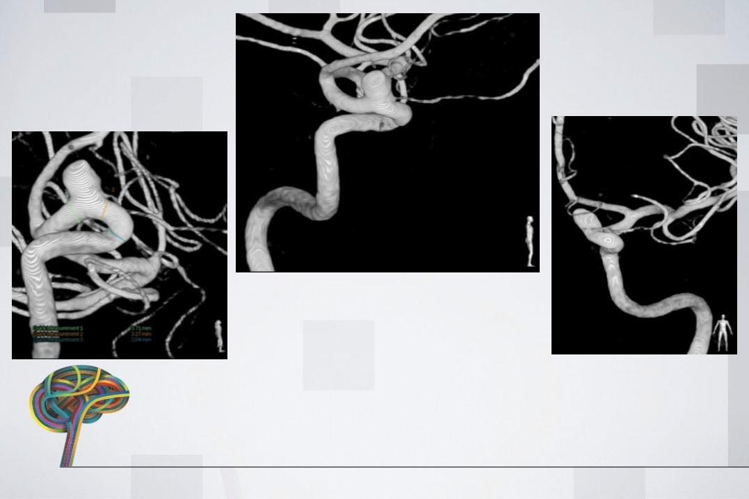
35y, M
• Very intense headache (SAH ?) 2 weeks ago.
• Multipl aneurysms (R MCA, rM1, ICA term)
• Has ankilosing spondylitis as his two siblings and his father : not on medication at the moment
• Had previous surgery for umblical hernia and nephrolithiasis
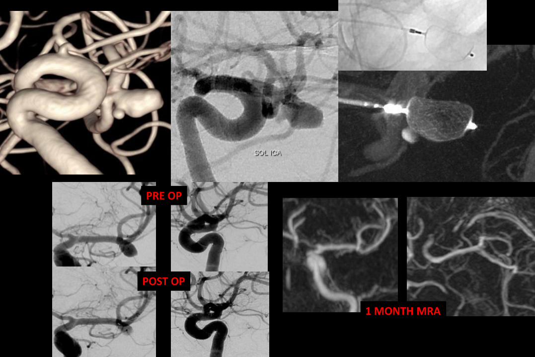
PRE OP
POST OP
1 MONTH MRA
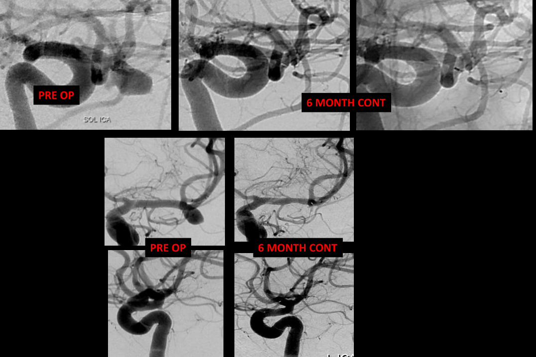
PRE OP6 MONTH CONT
PRE OP 6 MONTH CONT
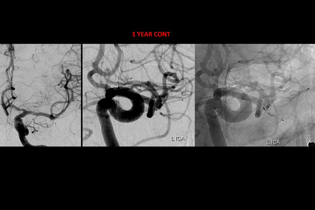
1 YEAR CONT
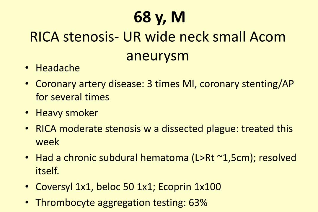
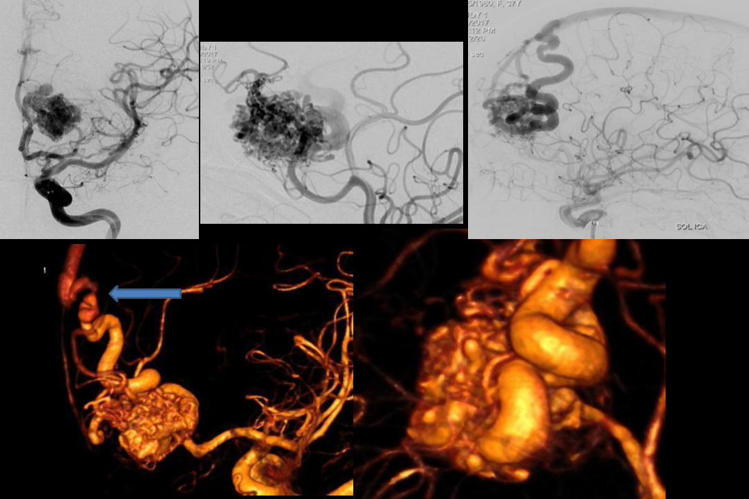
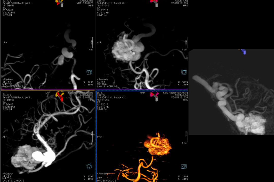
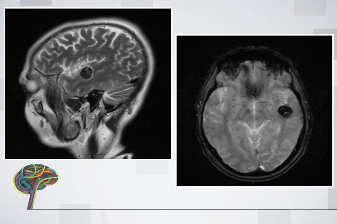
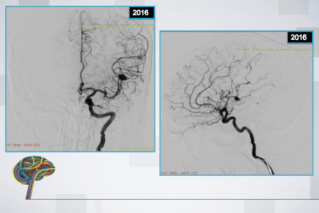
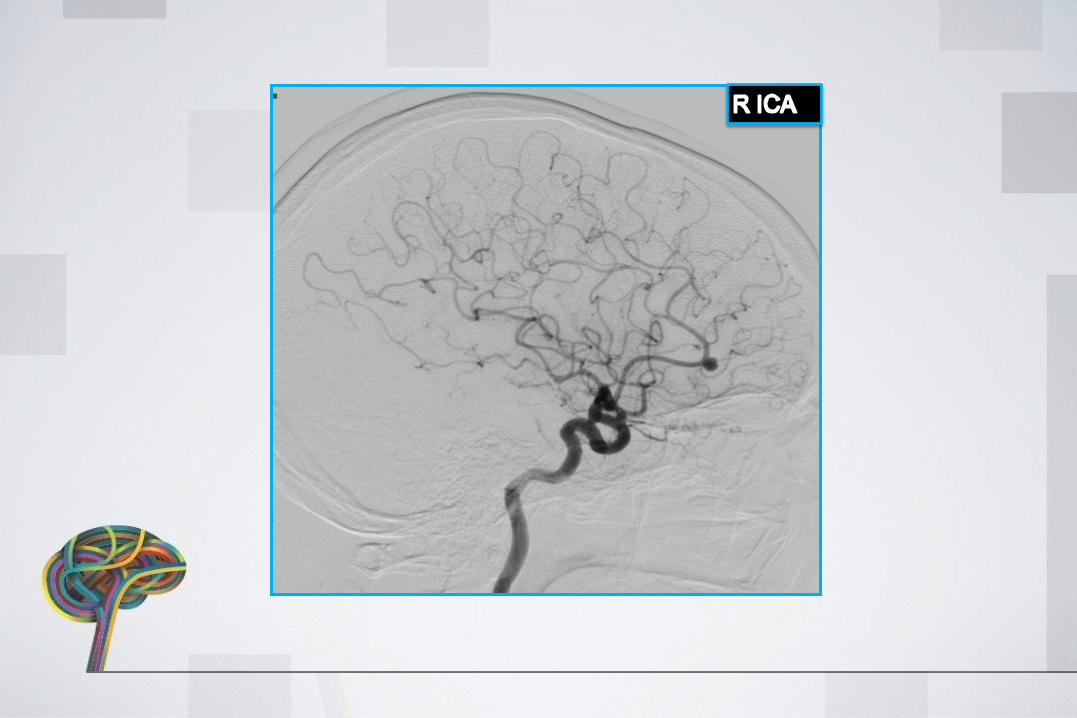
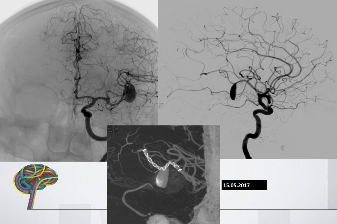
68 y, MRICA stenosis- UR wide neck small Acom
aneurysm • Headache
• Coronary artery disease: 3 times MI, coronary stenting/AP for several times
• Heavy smoker
• RICA moderate stenosis w a dissected plague: treated this week
• Had a chronic subdural hematoma (L>Rt ~1,5cm); resolved itself.
• Coversyl 1x1, beloc 50 1x1; Ecoprin 1x100
• Thrombocyte aggregation testing: 63%
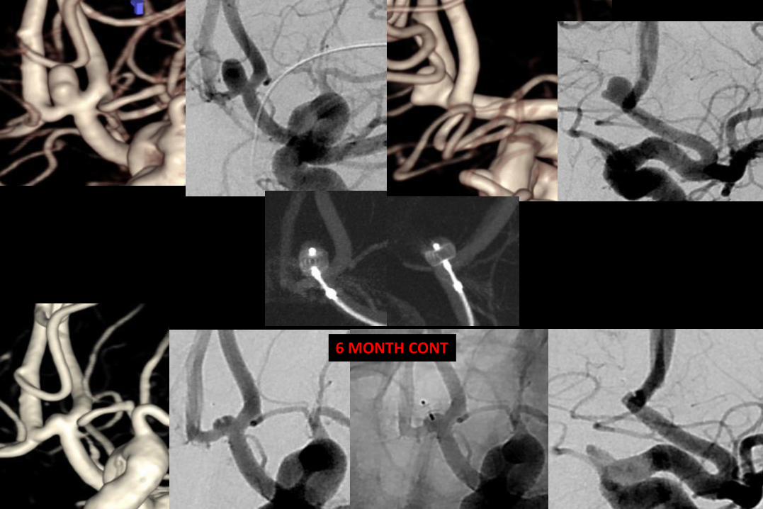
6 MONTH CONT
6 MONTH CONTPRE TX1 YEAR CONT
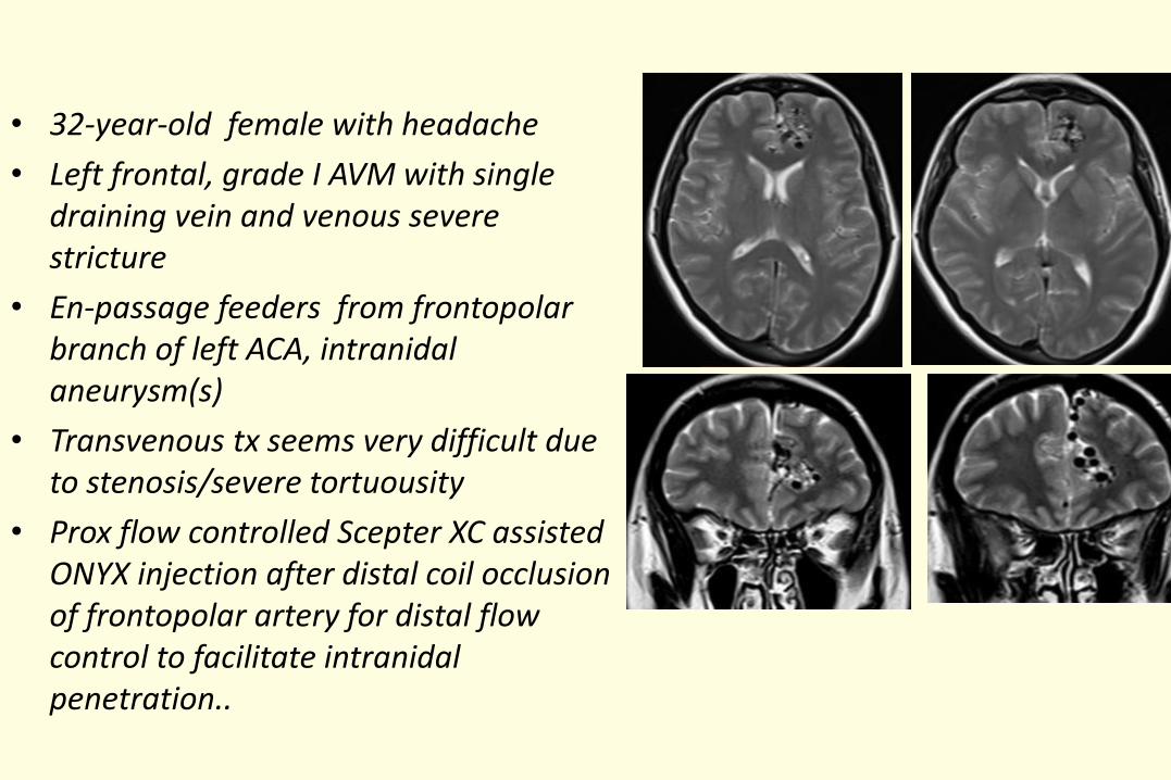
• 32-year-old female with headache
• Left frontal, grade I AVM with single draining vein and venous severe stricture
• En-passage feeders from frontopolar branch of left ACA, intranidal aneurysm(s)
• Transvenous tx seems very difficult due to stenosis/severe tortuousity
• Prox flow controlled Scepter XC assisted ONYX injection after distal coil occlusion of frontopolar artery for distal flow control to facilitate intranidal penetration..
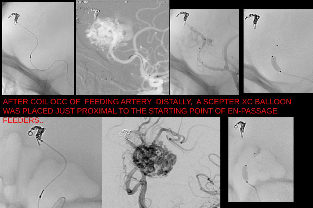
AFTER COIL OCC OF FEEDING ARTERY DISTALLY, A SCEPTER XC BALLOON
WAS PLACED JUST PROXIMAL TO THE STARTING POINT OF EN-PASSAGE
FEEDERS..
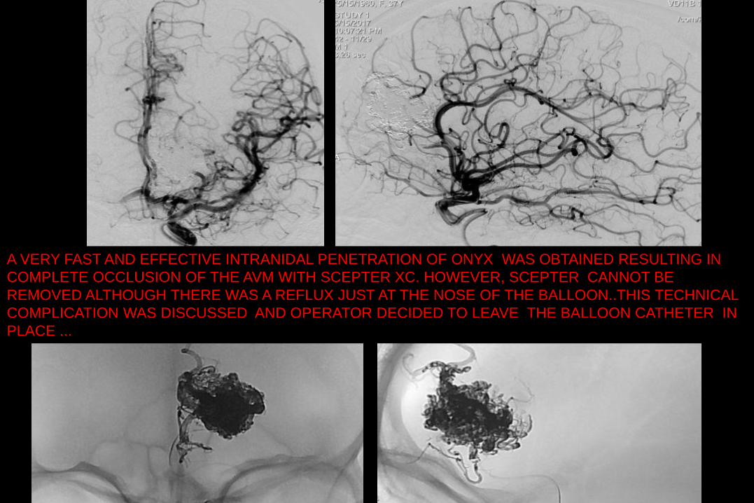
A VERY FAST AND EFFECTIVE INTRANIDAL PENETRATION OF ONYX WAS OBTAINED RESULTING IN
COMPLETE OCCLUSION OF THE AVM WITH SCEPTER XC. HOWEVER, SCEPTER CANNOT BE
REMOVED ALTHOUGH THERE WAS A REFLUX JUST AT THE NOSE OF THE BALLOON..THIS TECHNICAL
COMPLICATION WAS DISCUSSED AND OPERATOR DECIDED TO LEAVE THE BALLOON CATHETER IN
PLACE ...
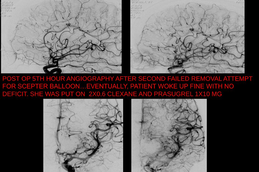
POST OP 5TH HOUR ANGIOGRAPHY AFTER SECOND FAILED REMOVAL ATTEMPT
FOR SCEPTER BALLOON…EVENTUALLY, PATIENT WOKE UP FINE WITH NO
DEFICIT. SHE WAS PUT ON 2X0.6 CLEXANE AND PRASUGREL 1X10 MG
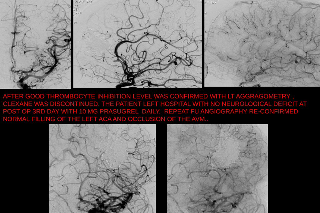
AFTER GOOD THROMBOCYTE INHIBITION LEVEL WAS CONFIRMED WITH LT AGGRAGOMETRY ,
CLEXANE WAS DISCONTINUED. THE PATIENT LEFT HOSPITAL WITH NO NEUROLOGICAL DEFICIT AT
POST OP 3RD DAY WITH 10 MG PRASUGREL DAILY. REPEAT FU ANGIOGRAPHY RE-CONFIRMED
NORMAL FILLING OF THE LEFT ACA AND OCCLUSION OF THE AVM..
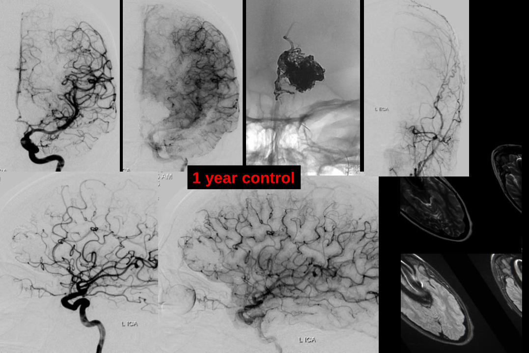
1 year control
WLNC 2017 LIMOGE CASE FU /BY PROF MOUNAYER
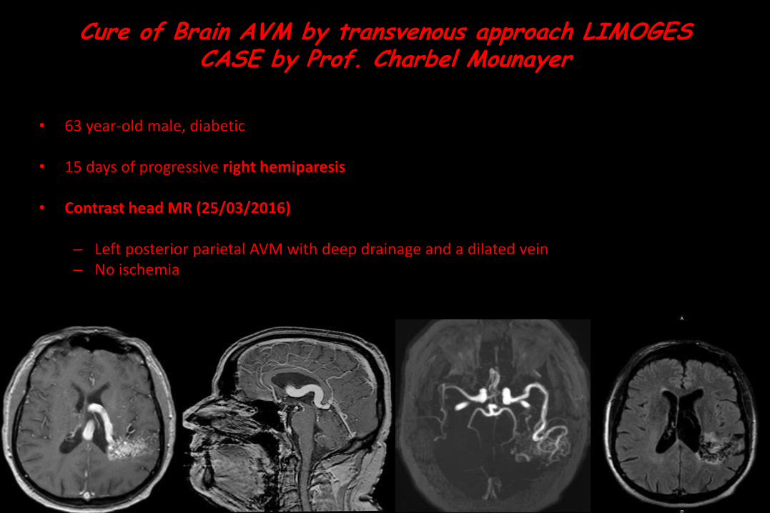
Cure of Brain AVM by transvenous approach LIMOGES CASE by Prof. Charbel Mounayer
• 63 year-old male, diabetic
• 15 days of progressive right hemiparesis
• Contrast head MR (25/03/2016)
– Left posterior parietal AVM with deep drainage and a dilated vein– No ischemia
First Session embolization – arterial approach (28/03/16)
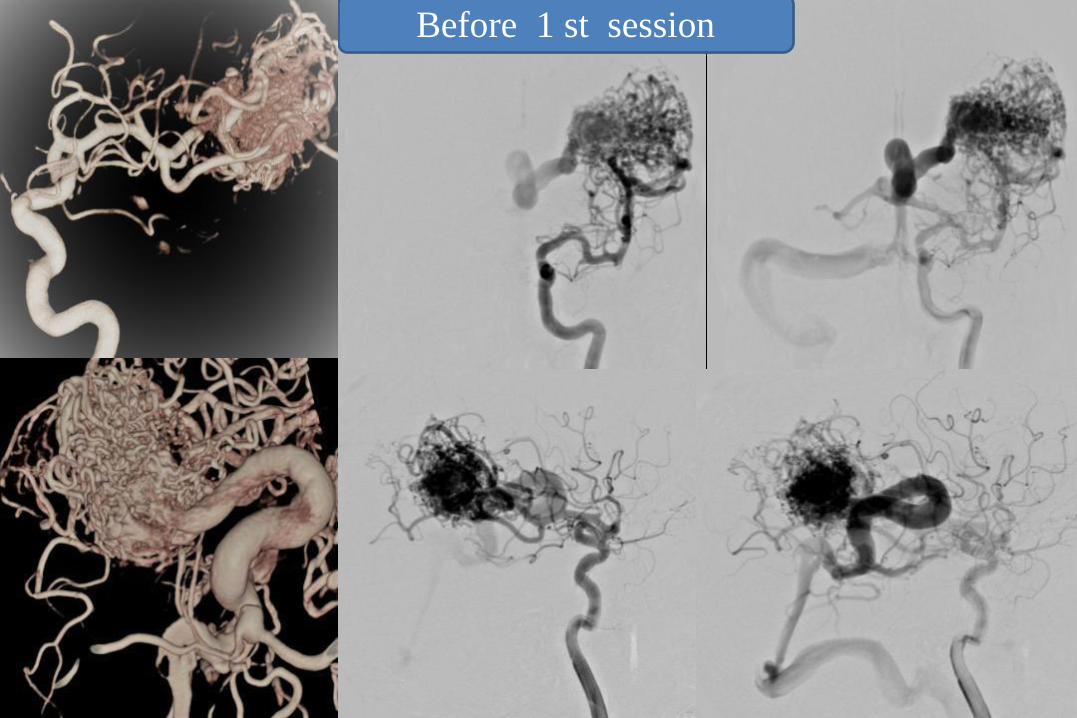
Before 1 st session
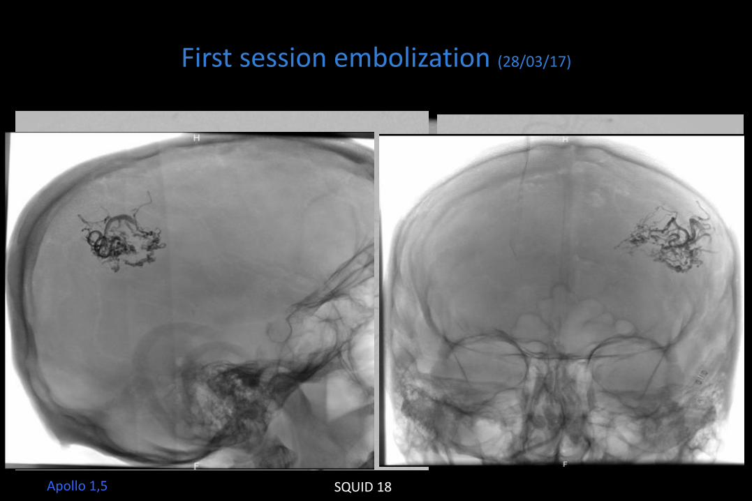
First session embolization (28/03/17)
Arterial approach
Apollo 1,5 SQUID 18
Apollo
SQUID 18
Apollo
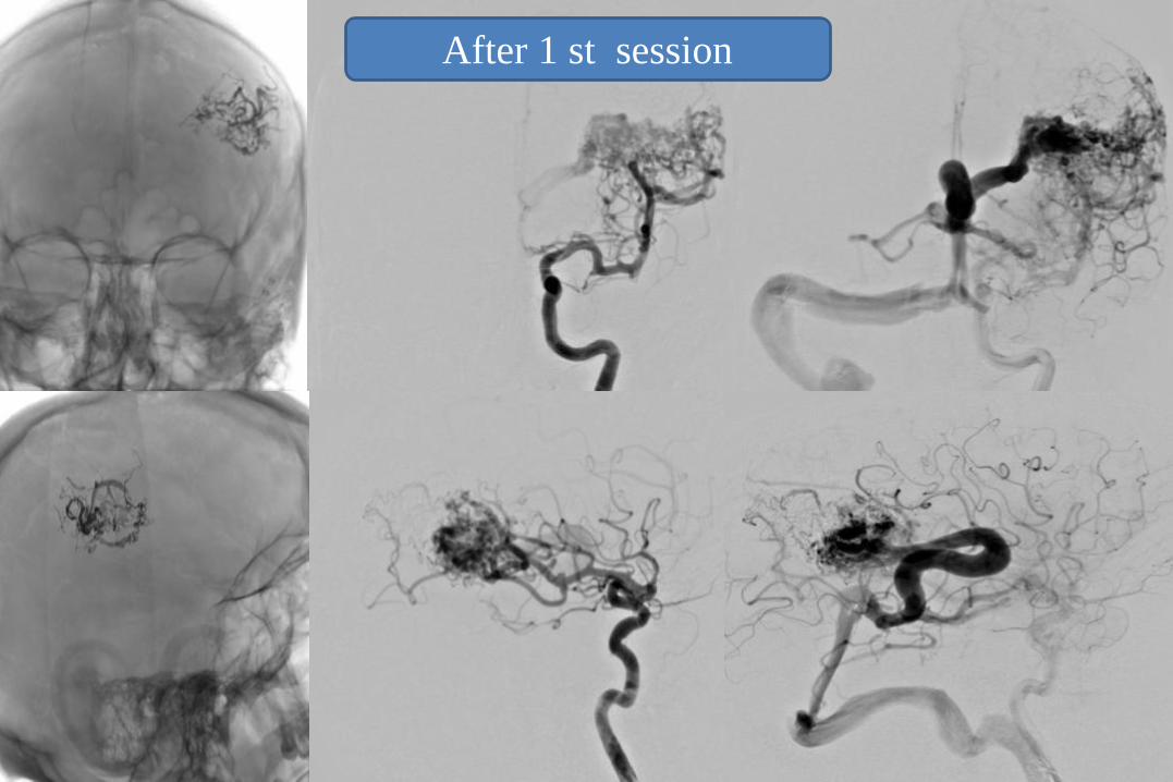
After 1 st session
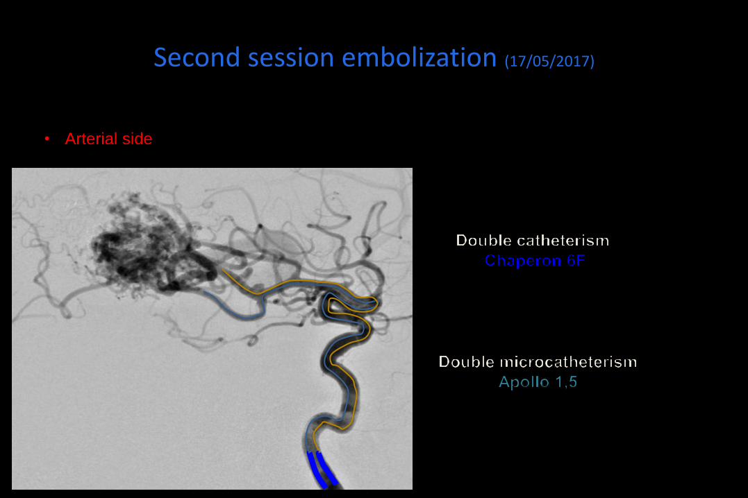
Second session embolization (17/05/2017)
• Arterial side
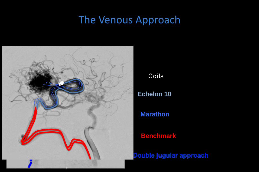
The Venous Approach
• Venous side
Benchmark
Marathon
Echelon 10
Onyx - 18
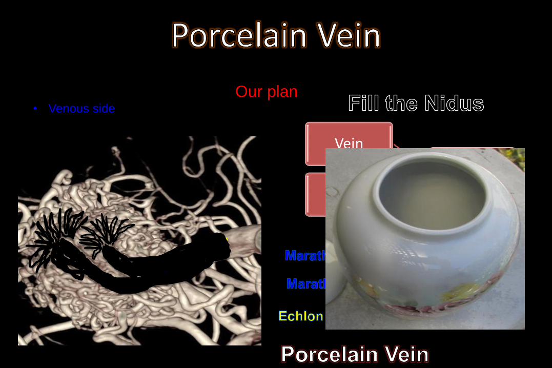
Our plan• Venous side
Posterior Parietal Vein
Vein
Vein
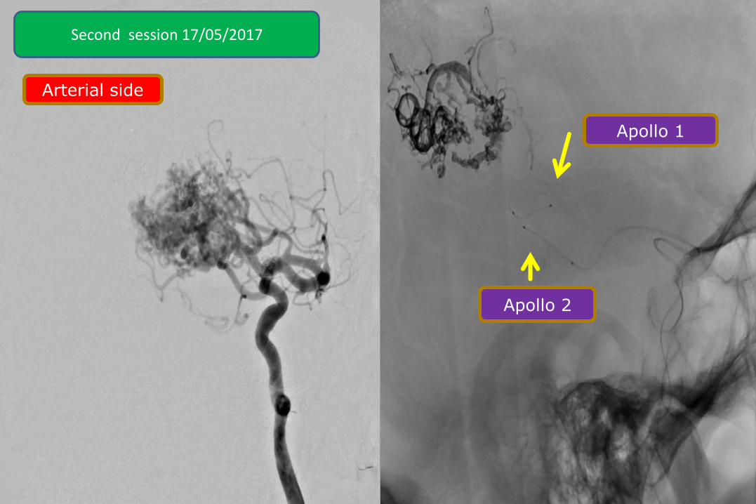
Arterial side
Apollo 1
Apollo 2
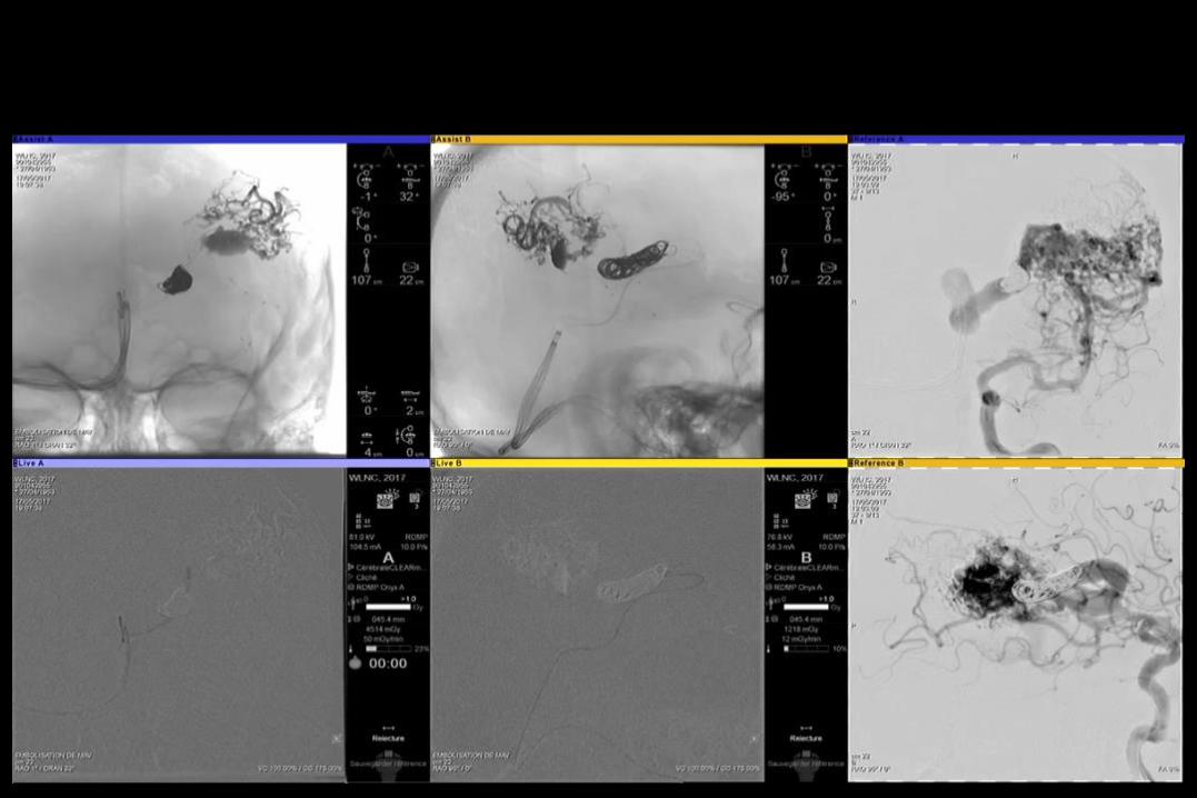
Second session 17/05/2017
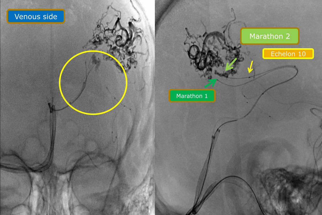
Venous side
Marathon 1
Marathon 2
Echelon 10
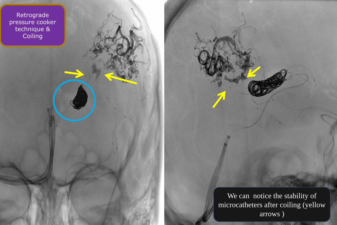
Retrograde pressure cooker
technique & Coiling
We can notice the stability of
microcatheters after coiling (yellow
arrows )
Onyx Injection &Coils Migration
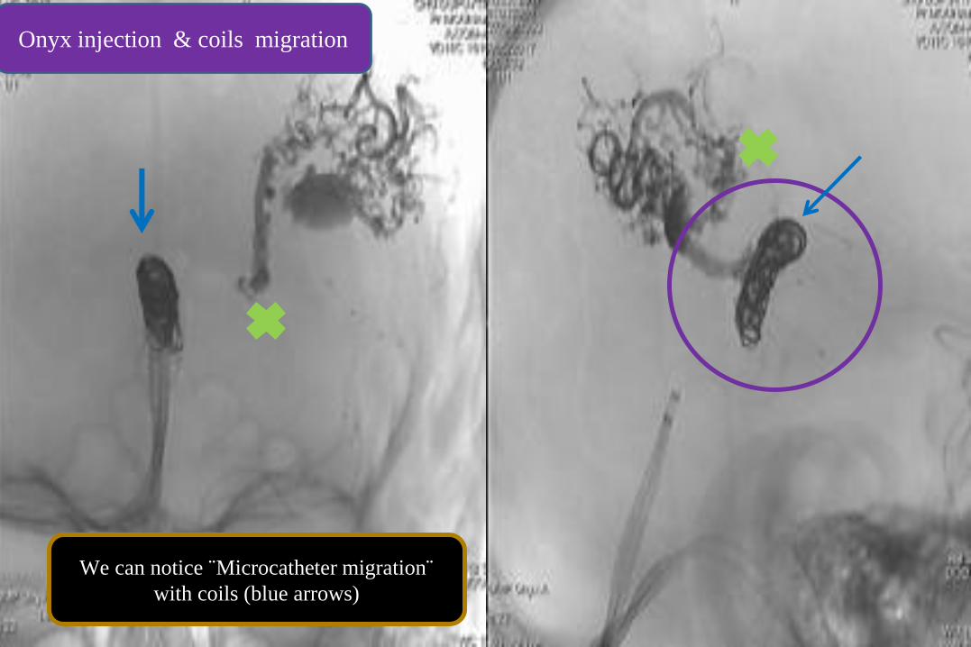
We can notice ¨Microcatheter migration¨
with coils (blue arrows)
Onyx injection & coils migration
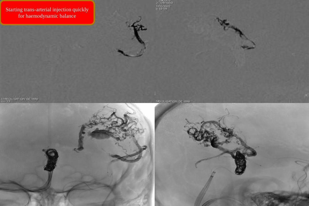
Starting trans-arterial injection quickly
for haemodynamic balance
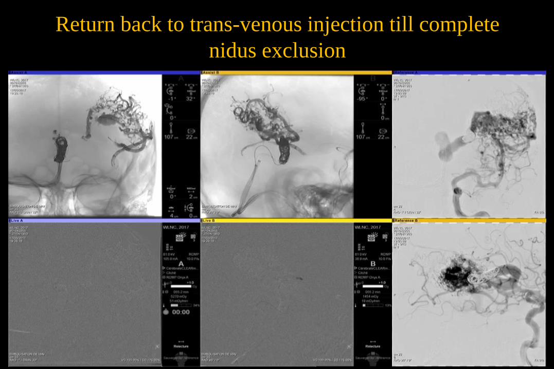
Return back to trans-venous injection till complete
nidus exclusion
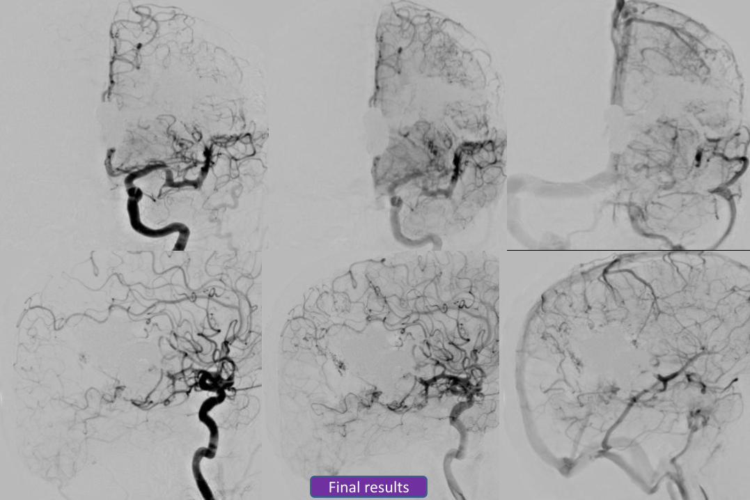
Final results
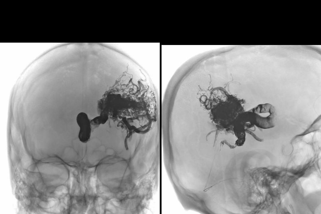
DYNA CT after treatment
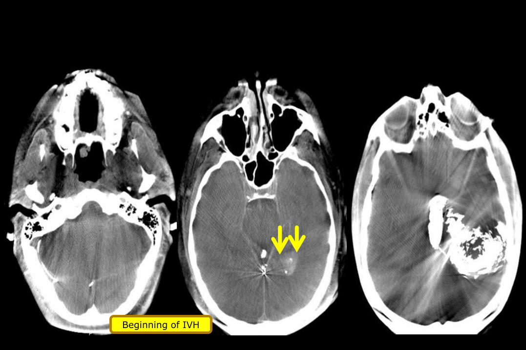
Beginning of IVH
CT BRAIN after 24 h
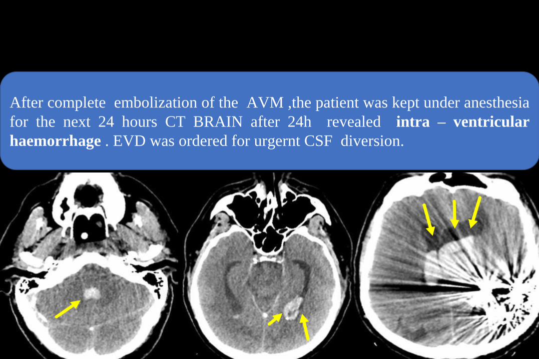
After complete embolization of the AVM ,the patient was kept under anesthesia
for the next 24 hours CT BRAIN after 24h revealed intra – ventricular
haemorrhage . EVD was ordered for urgernt CSF diversion.
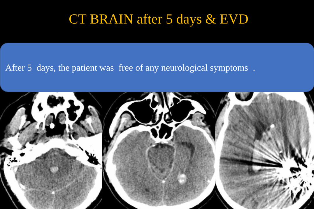
CT BRAIN after 5 days & EVD
After 5 days, the patient was free of any neurological symptoms .
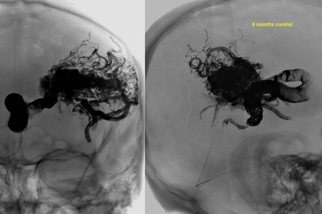
6 months control
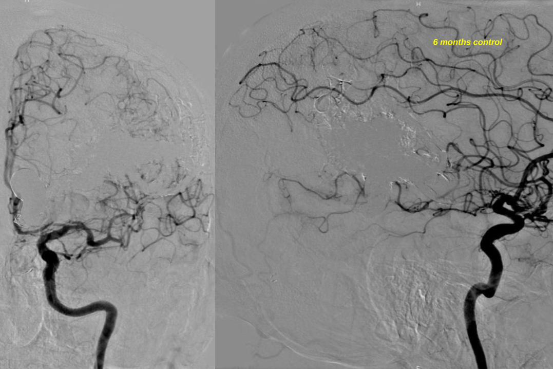
6 months control
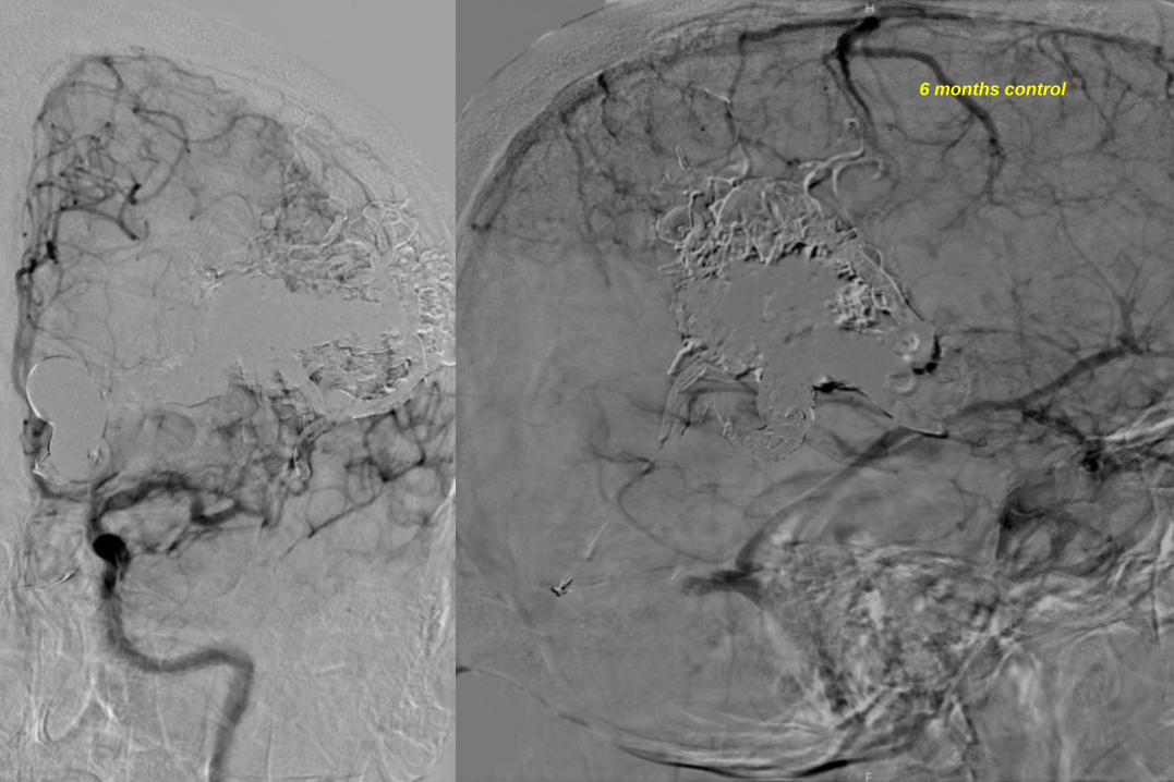
6 months control
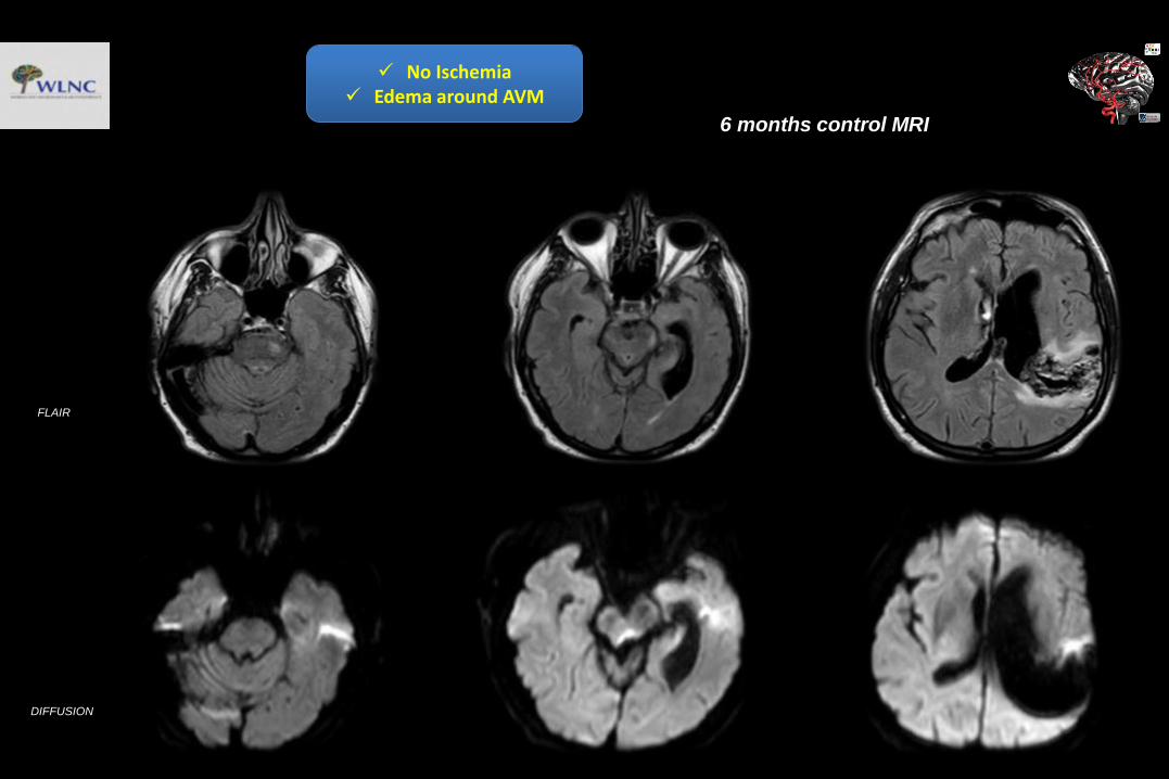
6 months control MRI
FLAIR
DIFFUSION
No Ischemia Edema around AVM
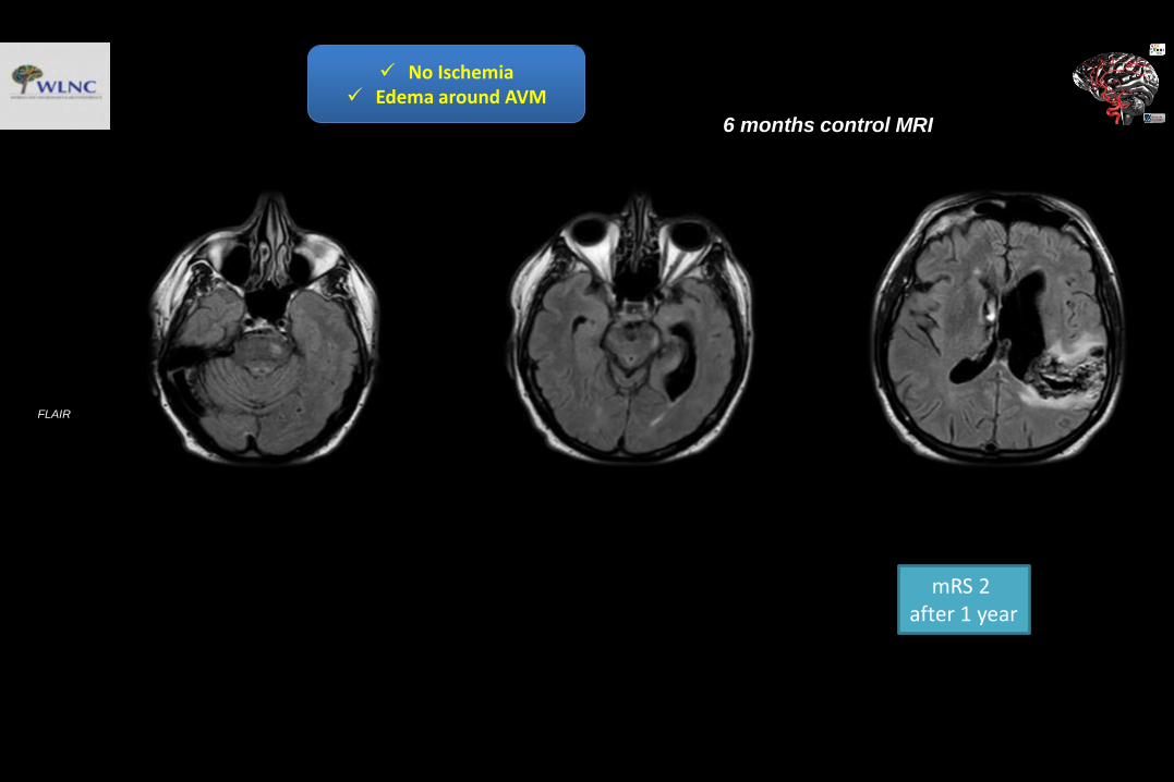
6 months control MRI
FLAIR
No Ischemia Edema around AVM
mRS 2 after 1 year
WLNC 2017 ISTANBUL CASES F-UBY PROF KIZILKILIC AND ISLAK
PT 1 DY
• 73 Y F• Presented with SAH 6 years ago• R ACA A1 segment aneurysm treated
with coils as parent artery occlusion. 1st year cont N
• Routine 5th year control MRA R ACA A1 Recurrent aneurysm
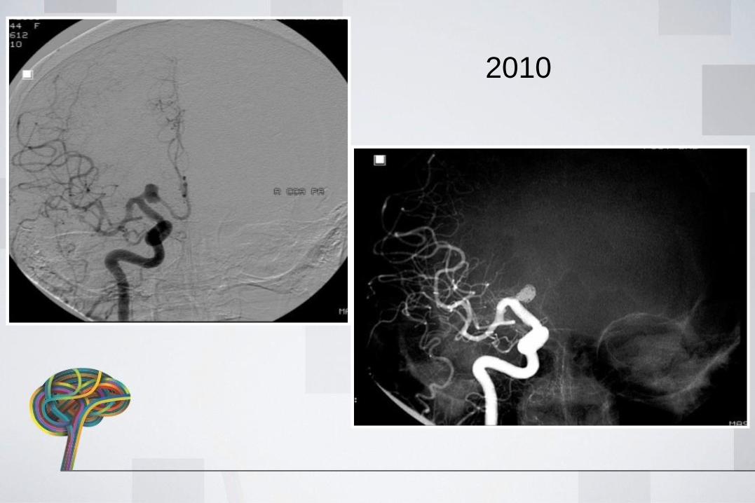
2010
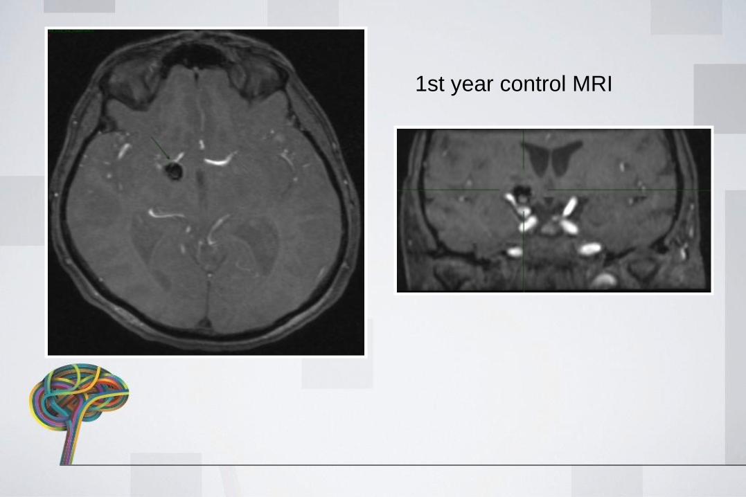
1st year control MRI
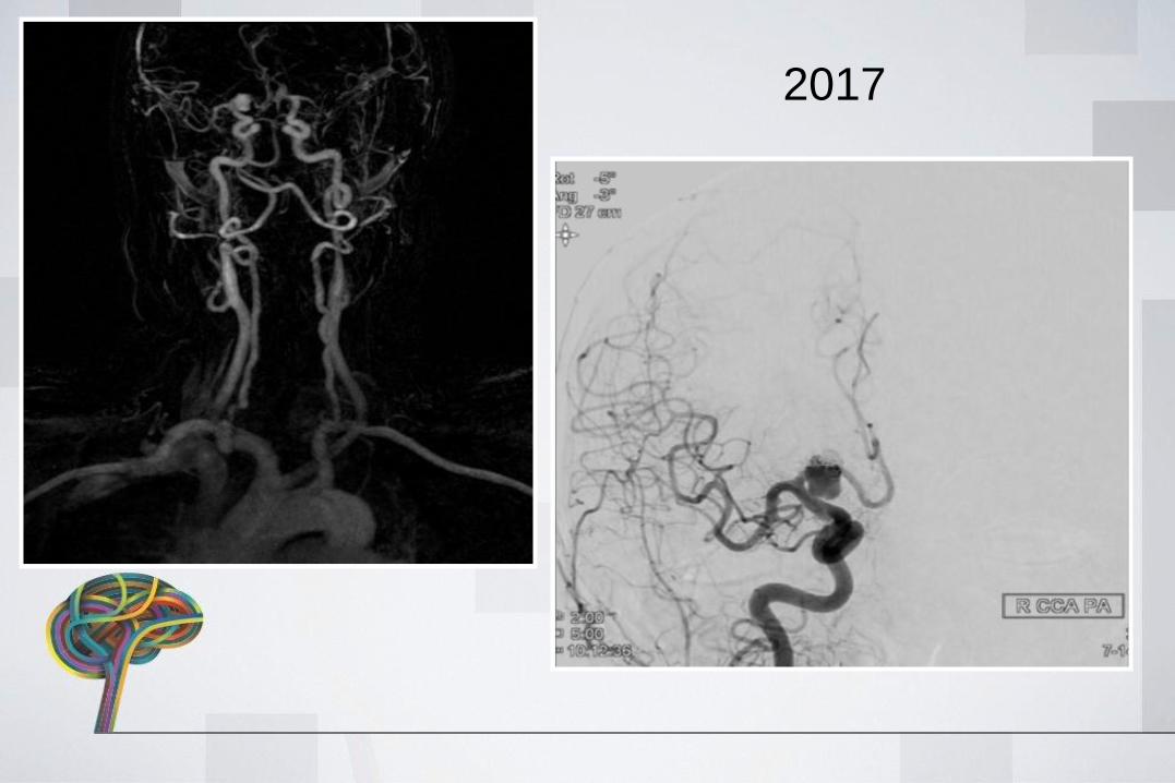
2017
TREATMENT
• Parent artery occlusion w coils versus FD
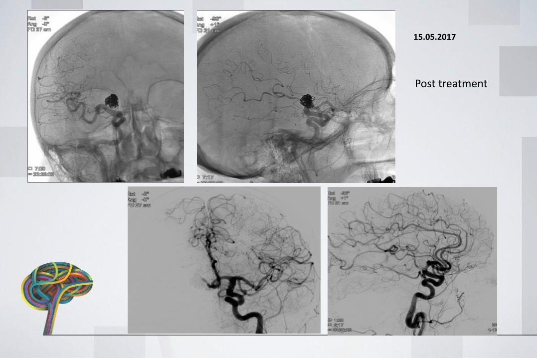
15.05.2017
Post treatment
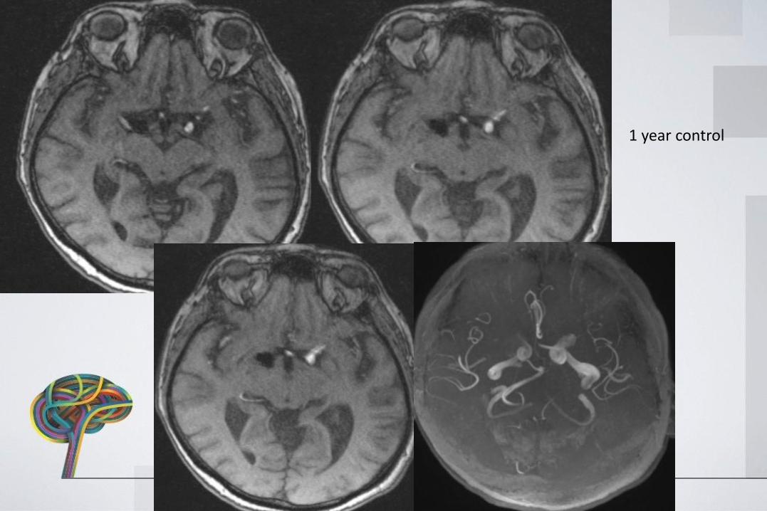
1 year control
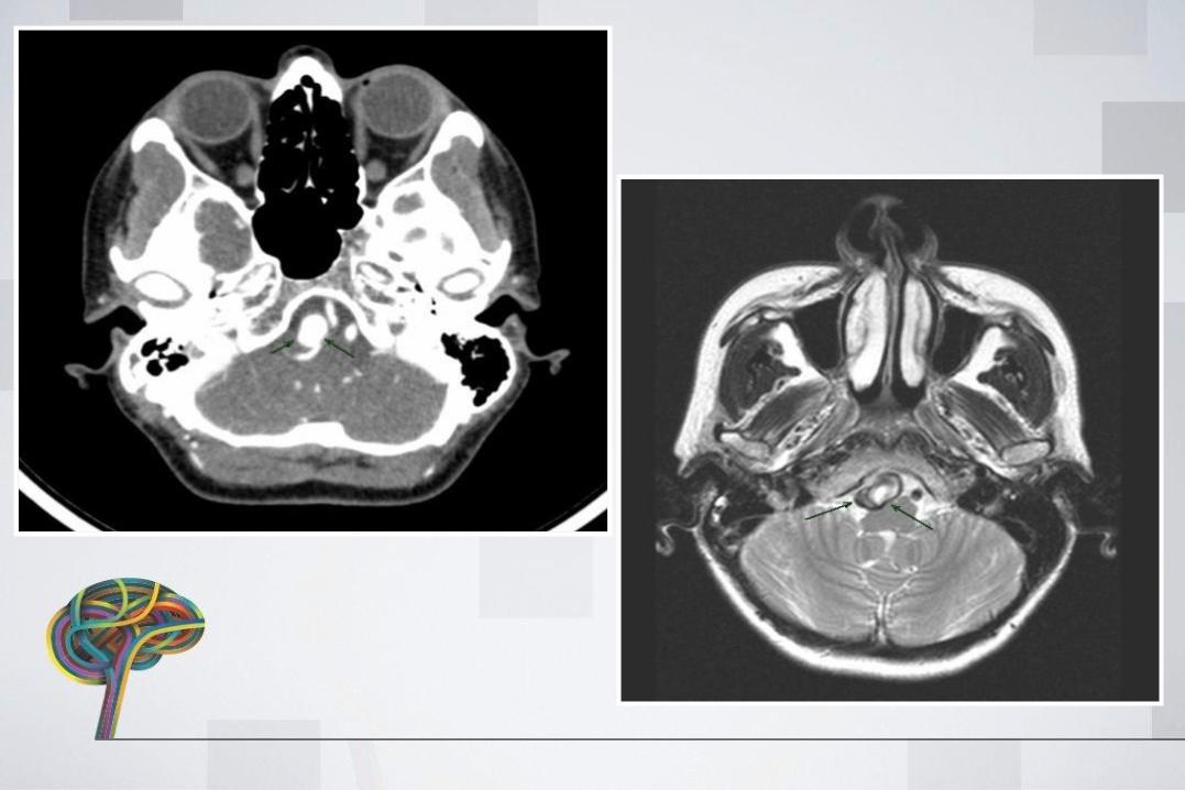
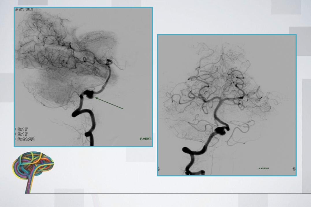
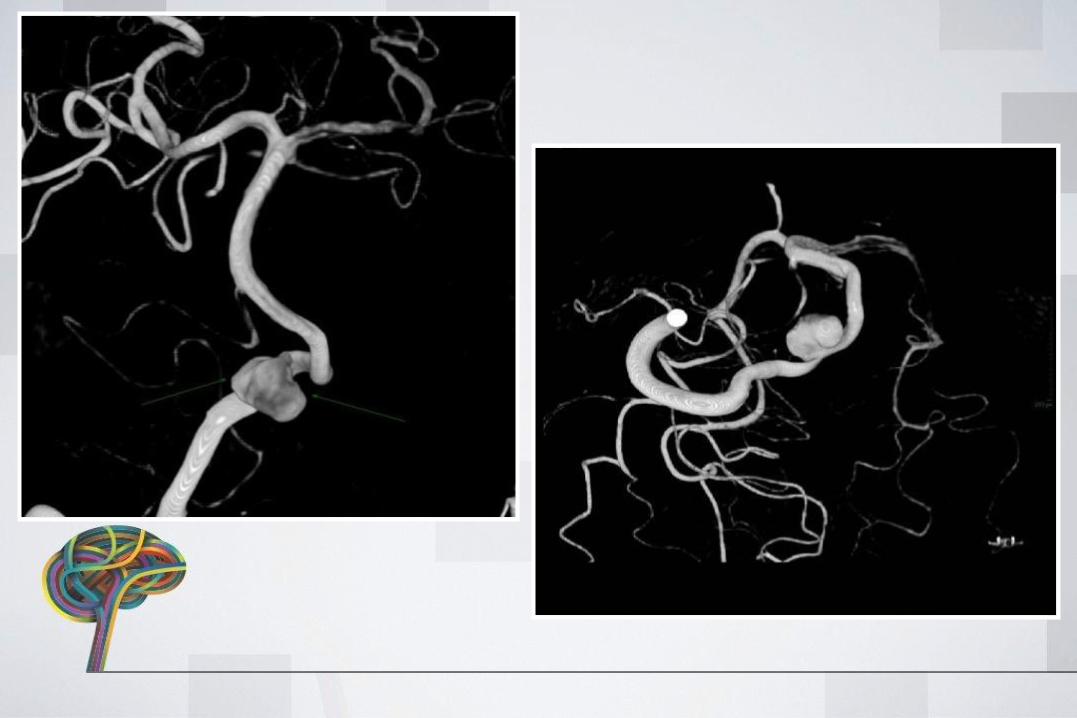
PT 2 BT• 43 Y F• Presented with headache• CTA-MRA-DSA: R V4 ANEURYSM• TREATMENT: FD
TREATMENT
• Flow diverter stent placement
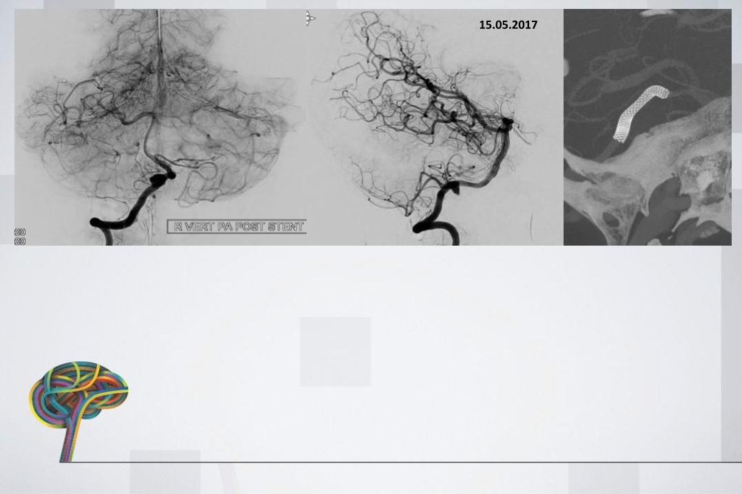
15.05.2017
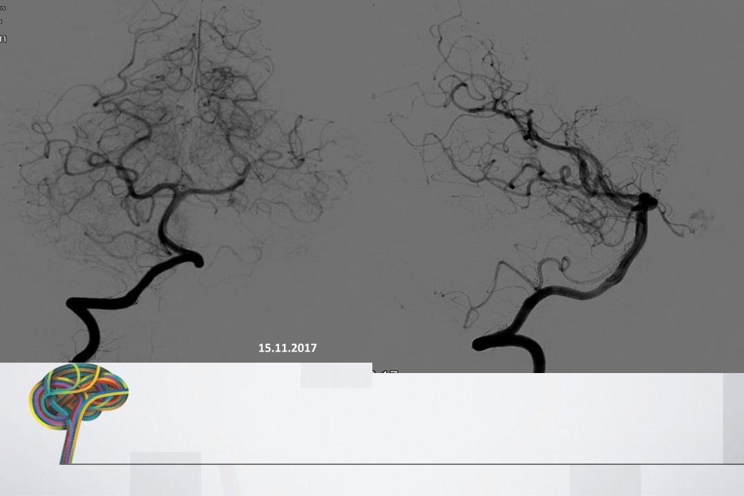
15.11.2017
PT3 HZ
• 64 Y F• 1994 basilar stroke ( L thalamic &
Mesencephalic)• normal basilar bif• 2016 Presented with headaches• MRA-DSA: Basilar apex aneurysm• Treatment: Y stent + coil embolization
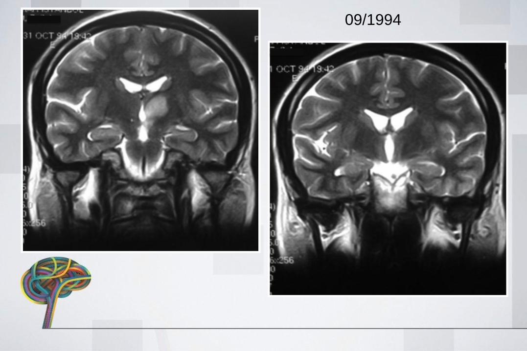
09/1994
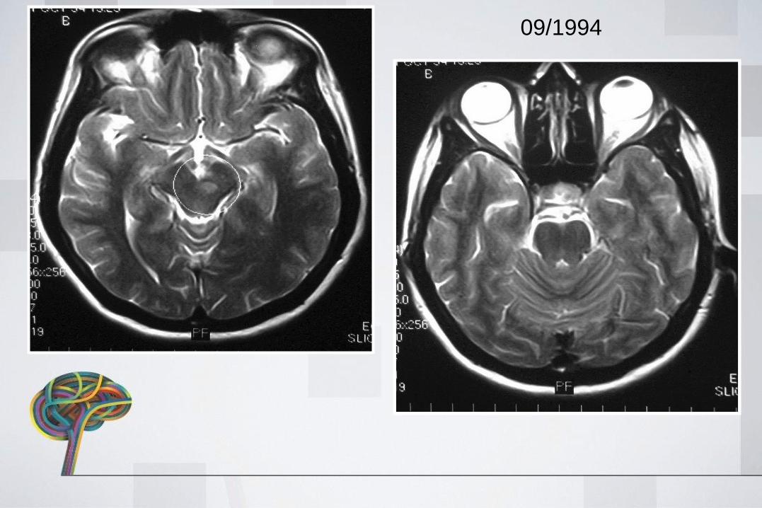
09/1994
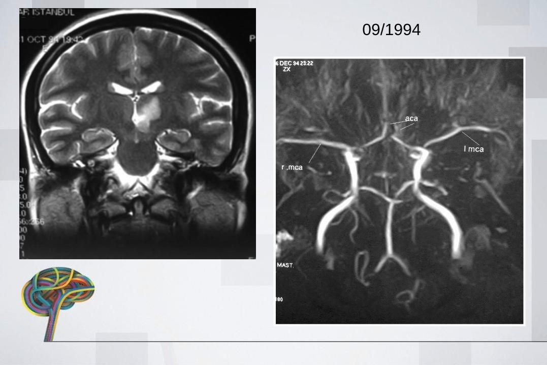
09/1994
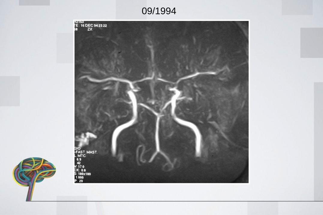
09/1994
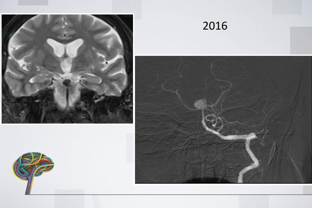
2016
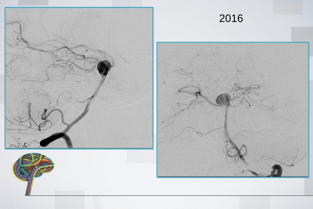
2016
TREATMENT
• Y stent + coil embolization
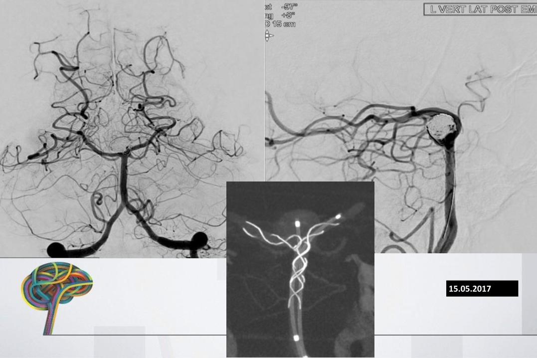
15.05.2017
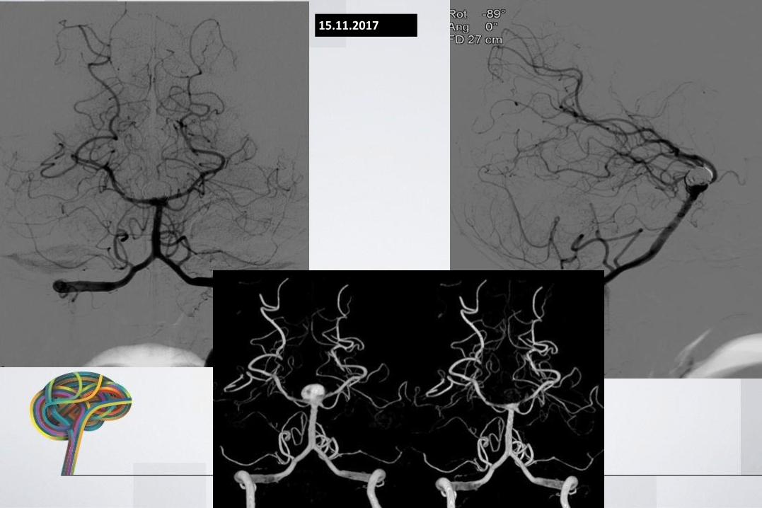
15.11.2017
PT 4 AS
• 61 Y F• Presented with headaches• MRA-DSA: L M2 dissecting Aneurysm• Treatment: Flow diverter stent placement

TREATMENT
• Flow diverter stent placement
15.05.2017

11.12.2017
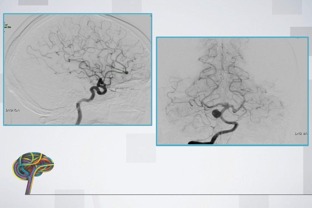
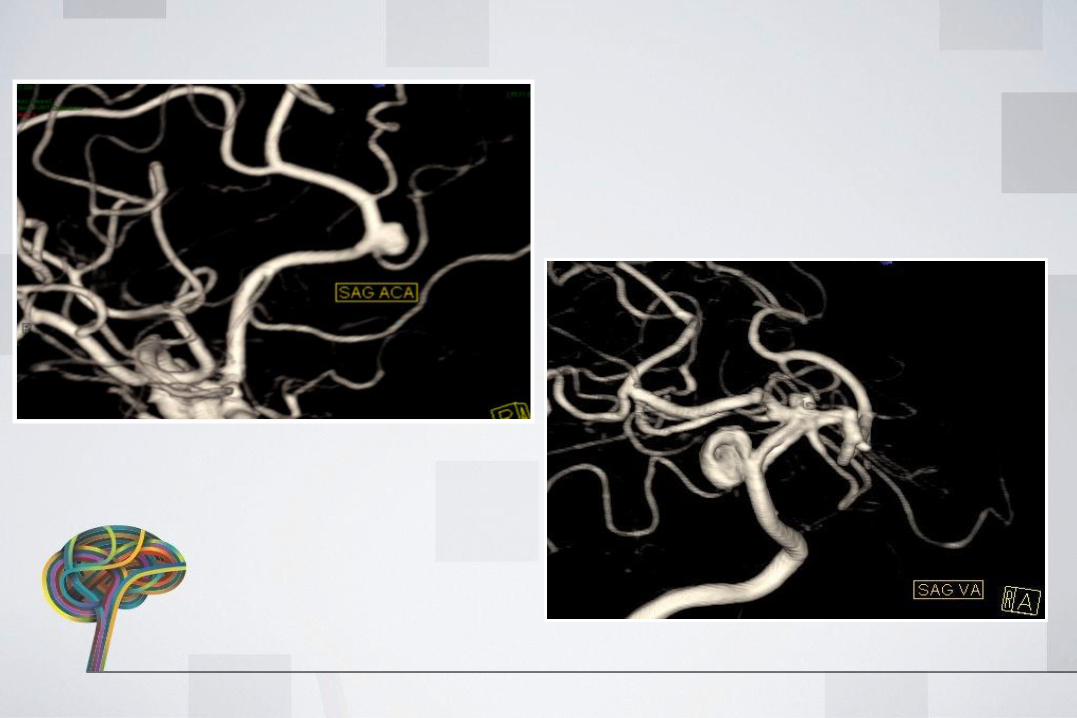
PT 5 NO
• 60 Y F• Presented with headache• DSA: Pericallosal aneurysm• Treatment: Flow diverter stent placement
TREATMENT
• Flow diverter stent placement
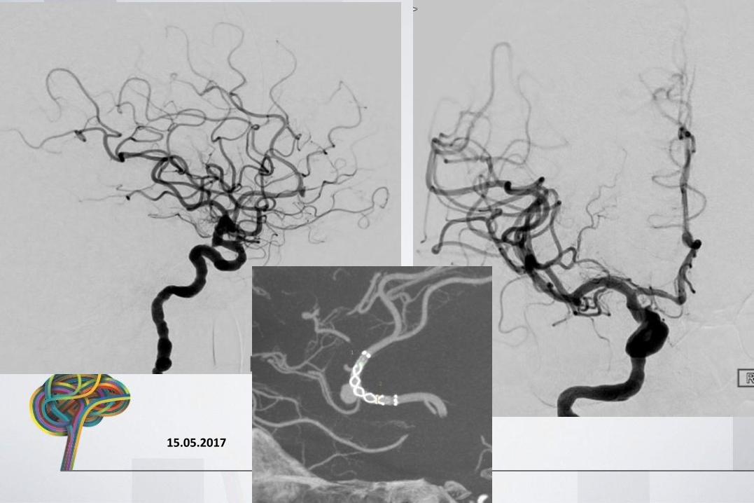
15.05.2017
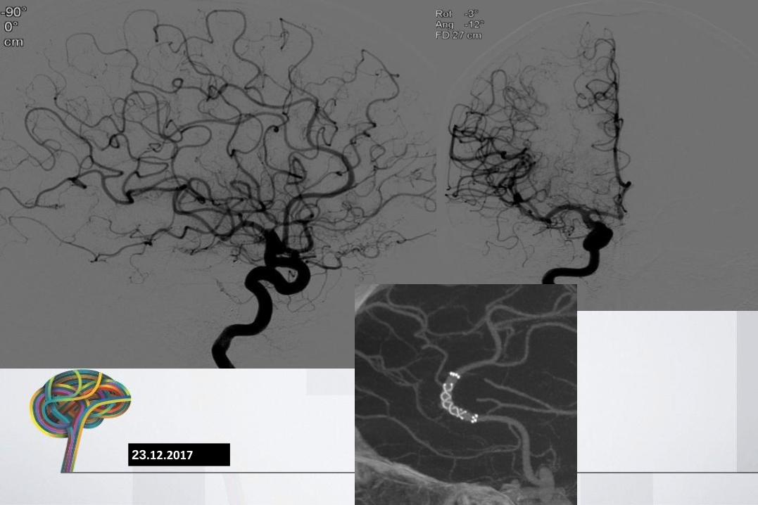
23.12.2017
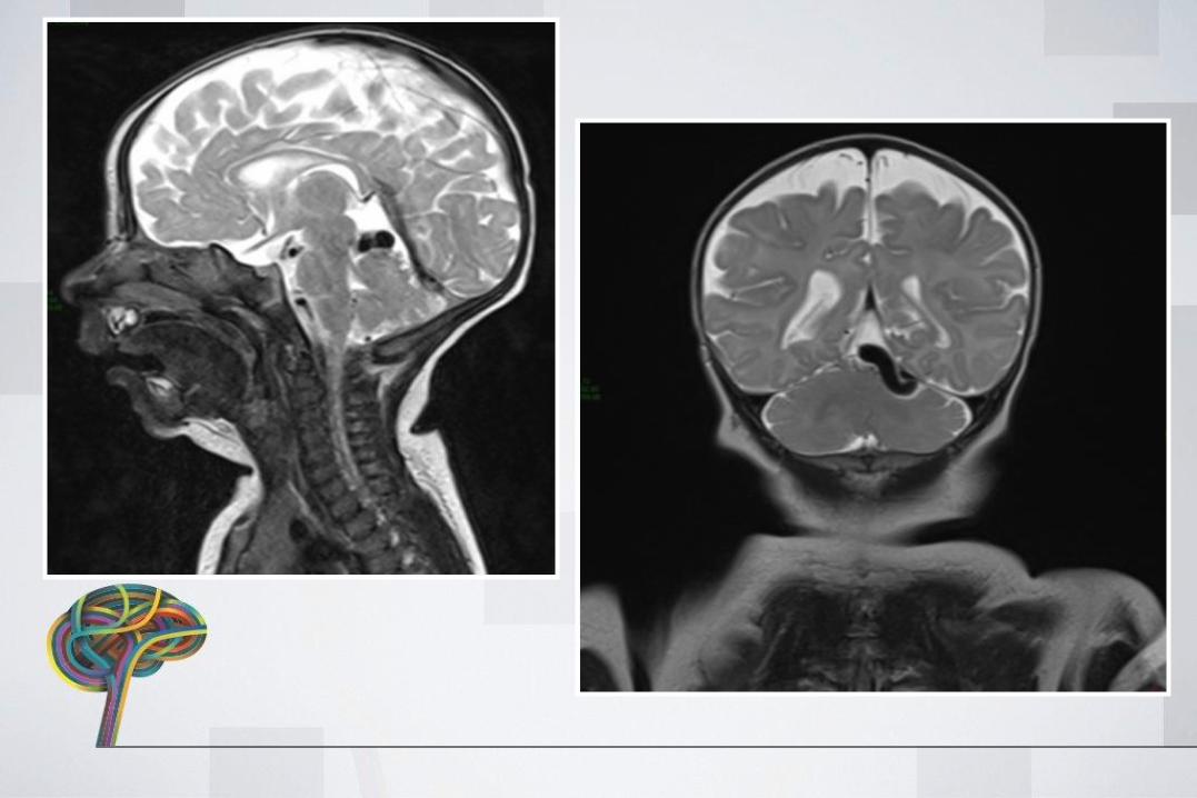
PT 6 RG
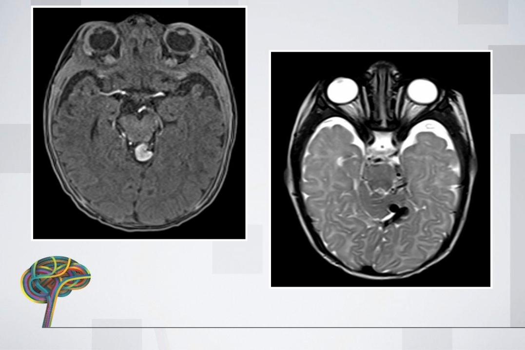
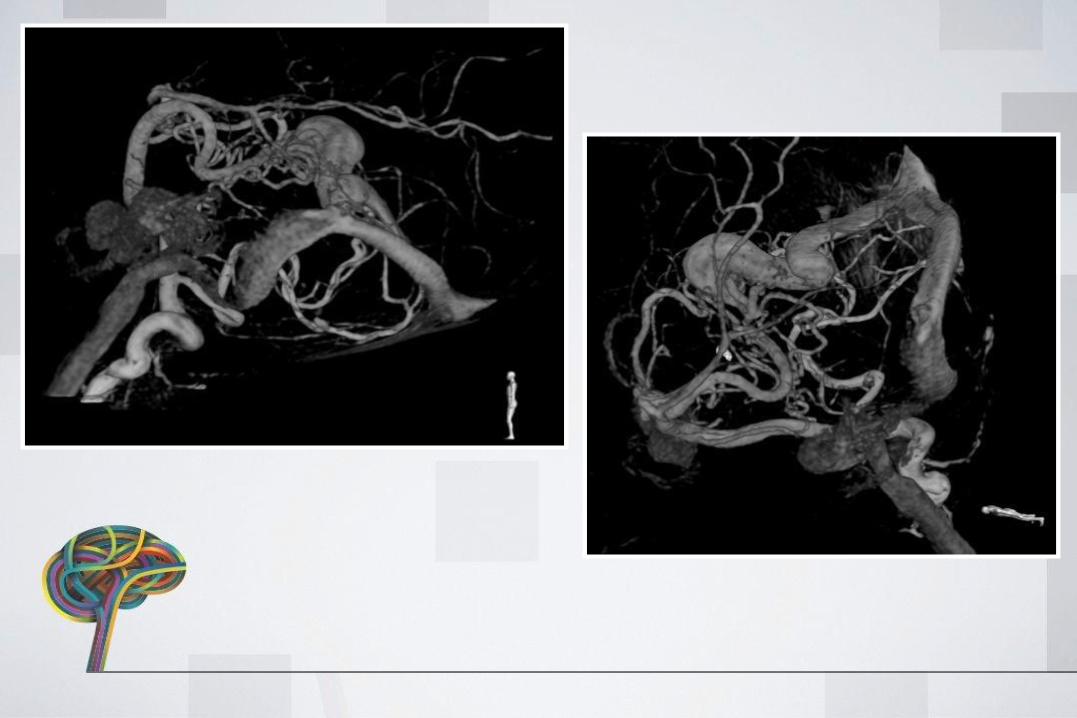
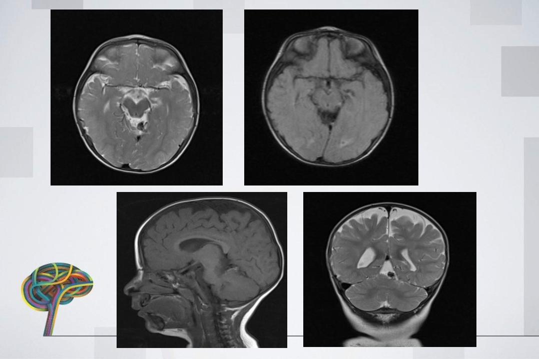
• 1 Y M• Presented with head enlargement• PE:Mild macrocephalia• US: w/o ventricular enlargement• MRA-DSA: Pial AVF in the posterior fossa• Treatment: Transarterial-transvenous
approach embolisation with liquid embolicagent
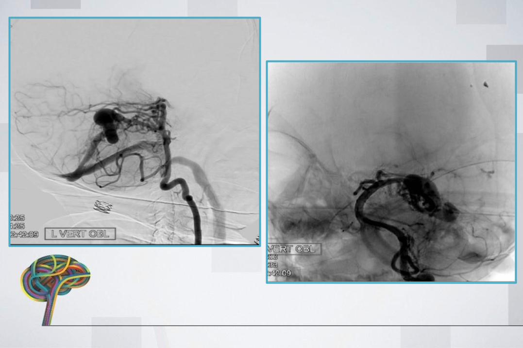
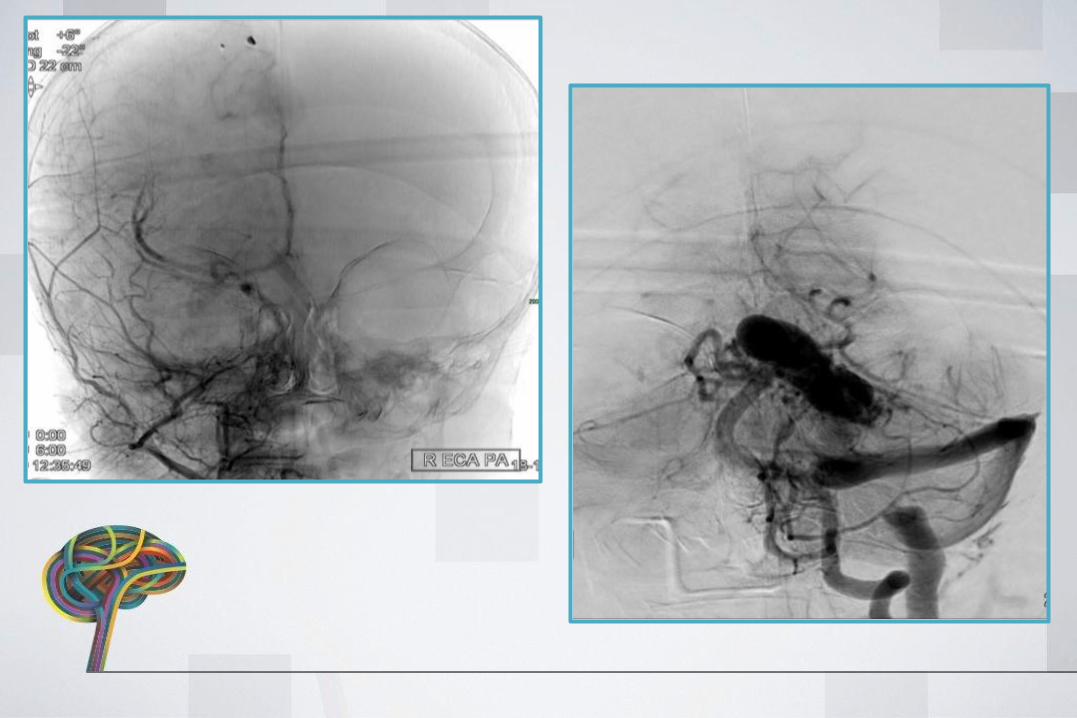
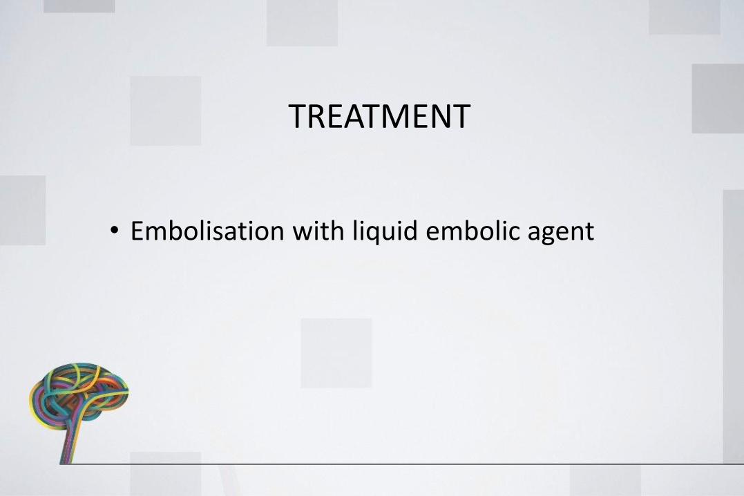
TREATMENT
• Embolisation with liquid embolic agent
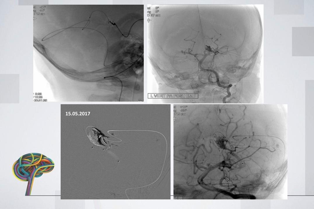
15.05.2017
PT 7 BI
• 33 Y F • Presented with: TIA in 2015• MRA-DSA: R ICA subpetrozal segment
obstruction due to dissection• Treatment with anticoagulation• 3rd M control opening of R ICA but 99%
stenosis• 18 months under Coumadin. Stenosis
persists• Treatment: Stent placement
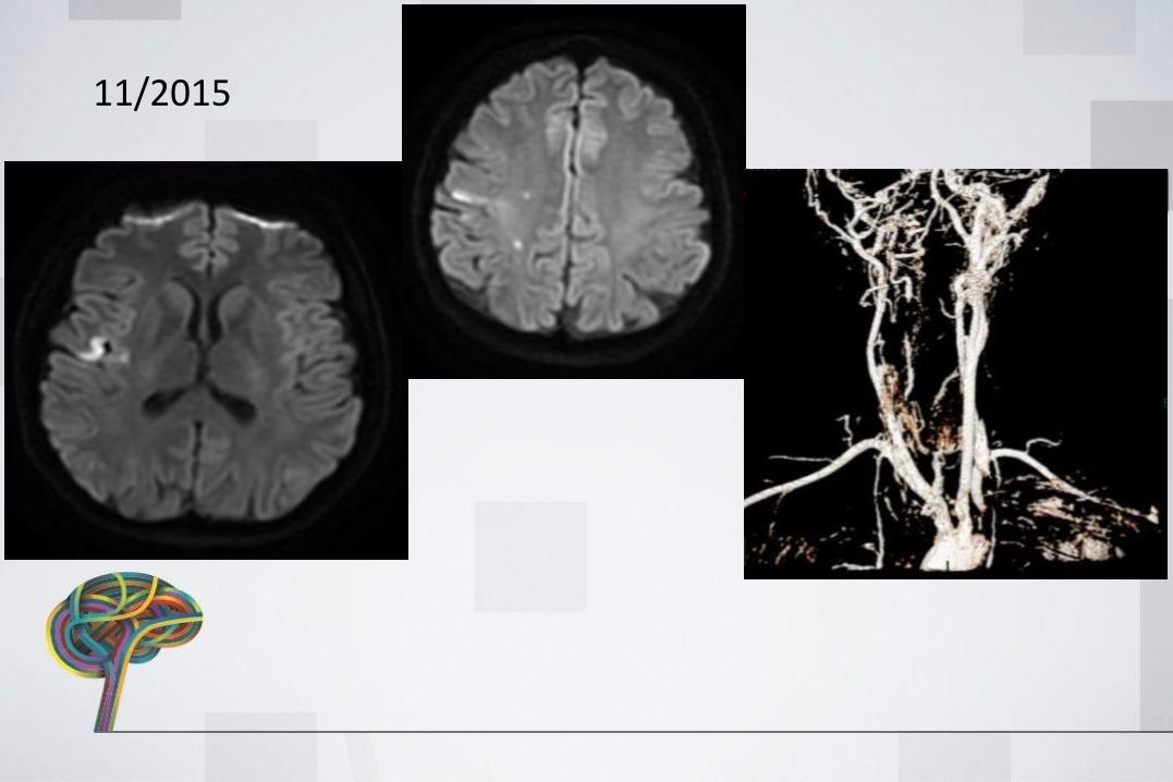
11/2015
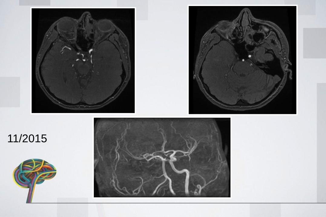
11/2015
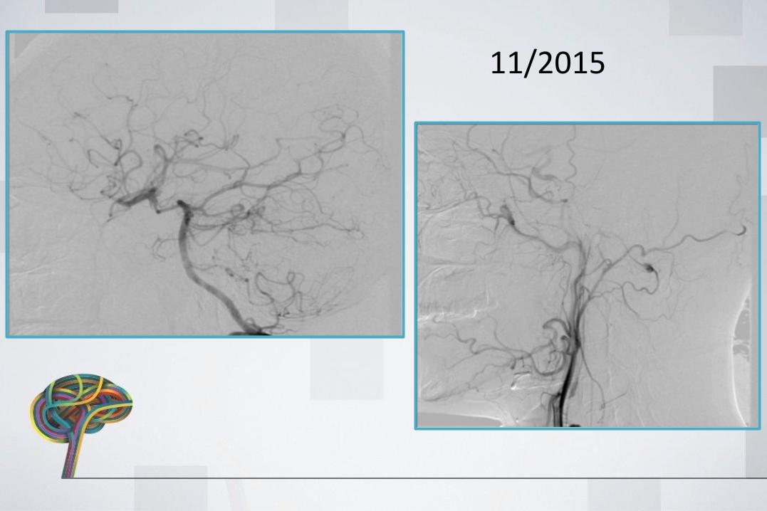
11/2015
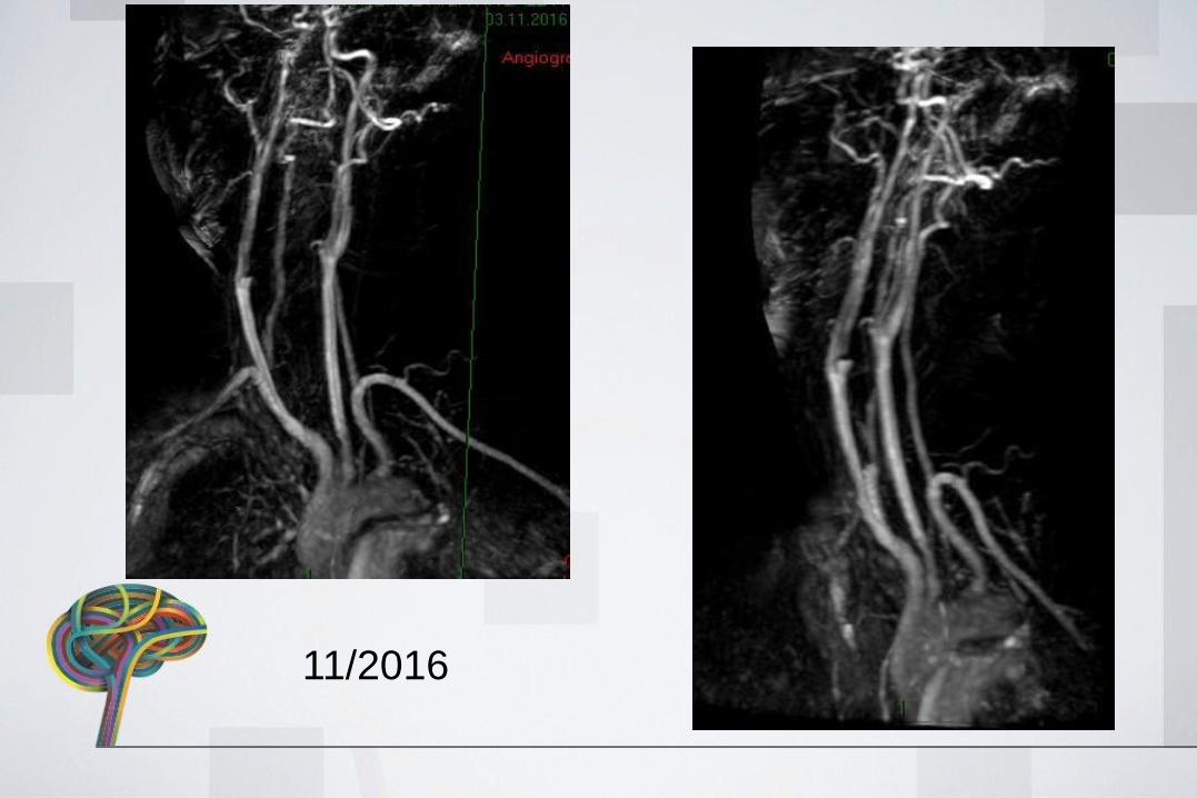
11/2016
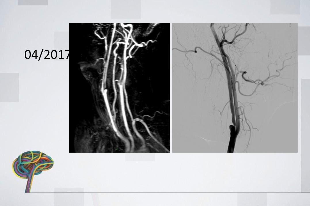
04/2017
TREATMENT
• Stent placement
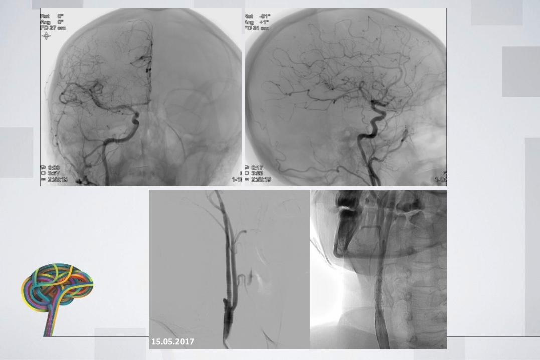
15.05.2017
PT 8 NE
• 60 Y F• Presented with headaches• DSA: Superior sagital sinus Dural AVF• Treatment plan: Embolisation with liquid embolic
agent
TREATMENT
• Embolisation with liquid embolic agent
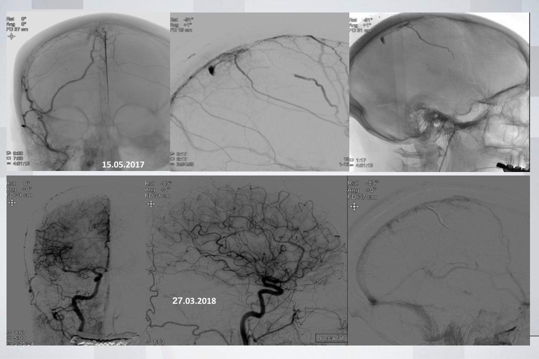
15.05.2017
27.03.2018
WLNC 2017 CHICAGO CASES F-UBY PROF DEMETRIUS LOPES
Pre Pre
Post - 6 mth Post - 6 mth
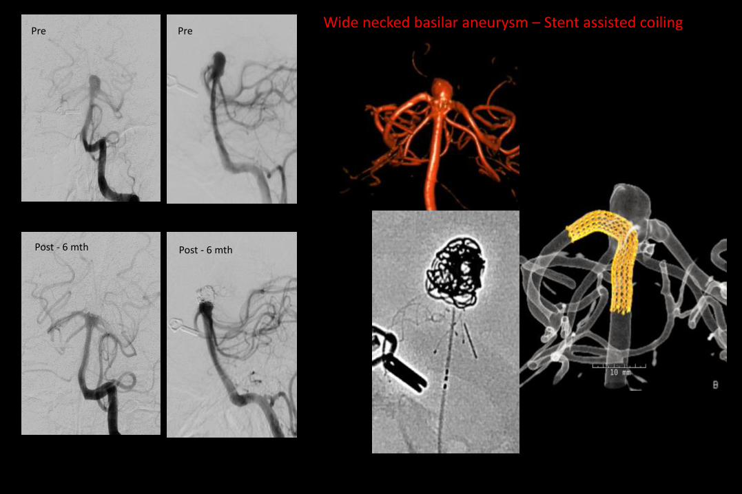
Wide necked basilar aneurysm – Stent assisted coiling
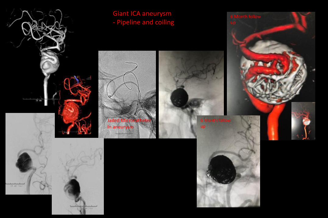
6 Month follow up
6 Month follow up
Giant ICA aneurysm- Pipeline and coiling
Jailed MicrocatheterIn aneurysm
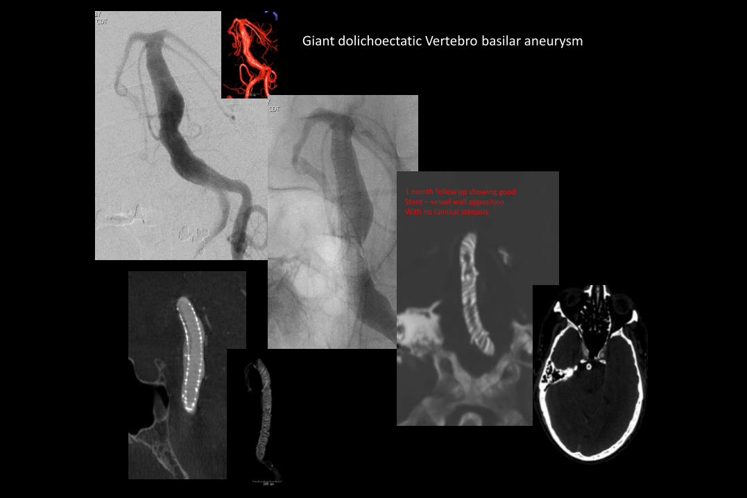
Giant dolichoectatic Vertebro basilar aneurysm
1 month follow up showing good Stent – vessel wall apposition With no luminal stenosis