what is urinary diversion? what is the urinary tract and · what is urinary diversion? urinary...
TRANSCRIPT
Urinary Diversion�National Kidney and Urologic Diseases Information Clearinghouse
What is urinary diversion? Urinary diversion is a surgical procedure that reroutes the normal flow of urine out of the body when urine flow is blocked. Urine flow may be blocked because of
• an enlarged prostate
• injury to the urethra
• birth defects of the urinary tract
• kidney, ureter, or bladder stones
• tumors of the genitourinary tract––which includes the urinary tract andreproductive organs––or adjacenttissues and organs
• conditions causing external pressure tothe urethra or one or both ureters
Bladder removal or a malfunctioning bladder may also cause blocked urine flow. When urine cannot flow out of the body, it can accumulate in the bladder, ureters, and kidneys. As a result, body wastes and extra water do not empty from the body, potentially resulting in pain, urinary tract infections, kidney failure, or, if left untreated, death. Urinary diversion can be temporary or permanent, depending on the reason for the procedure.
What is the urinary tract and how does it work? The urinary tract is the body’s drainage system for removing urine, which is composed of wastes and extra fluid. In order for normal urination to occur, all body parts in the urinary tract need to work together in the correct order.
Kidneys. The kidneys are two bean-shaped organs, each about the size of a fist. They are located just below the rib cage, one on each side of the spine. Every day, the kidneys filter about 120 to 150 quarts of blood to produce about 1 to 2 quarts of urine. The kidneys work around the clock; a person does not control what they do.
Ureters. Ureters are the thin tubes of muscle—one on each side of the bladder— that carry urine from each of the kidneys to the bladder.
Bladder. The bladder, located in the pelvis between the pelvic bones, is a hollow, muscular, balloon-shaped organ that expands as it fills with urine. Although a person does not control kidney function, a person does control when the bladder empties. Bladder emptying is known as
urination. The bladder stores urine until the person finds an appropriate time and place to urinate. A normal bladder acts like a reservoir and can hold 1.5 to 2 cups of urine. How often a person needs to urinate depends on how quickly the kidneys produce the urine that fills the bladder. The muscles of the bladder wall remain relaxed while the bladder fills with urine. As the bladder fills to capacity, signals sent to the brain tell a person to find a toilet soon.
Three sets of muscles work together like a dam, keeping urine in the bladder:
During urination, the bladder empties through the urethra, located at the bottom of the bladder. The urethra is the tube that carries urine outside of the body. The urethra is made up of muscles that stay closed while the bladder fills with urine. The area where the urethra joins the bladder is the bladder neck. The bladder neck is composed of muscles known as the internal sphincter. The urethra is surrounded by muscles called the pelvic floor muscles, also referred to as the external sphincter.
To urinate, the brain signals the muscular bladder wall to tighten, squeezing urine out of the bladder. At the same time, the brain signals the sphincters to relax. As the sphincters relax, urine exits the bladder through the urethra.
What is temporary urinary diversion? Temporary urinary diversion reroutes the flow of urine for several days or weeks. Temporary urinary diversions drain urine until the cause of blockage is treated or after urinary tract surgery. This type of urinary diversion includes a nephrostomy and urinary catheterization.
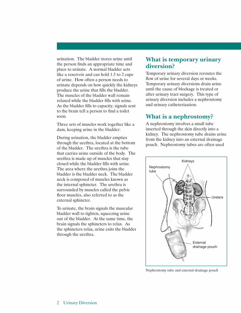
What is a nephrostomy? A nephrostomy involves a small tube inserted through the skin directly into a kidney. The nephrostomy tube drains urine from the kidney into an external drainage pouch. Nephrostomy tubes are often used
Nephrostomy tube
Ureters
External drainage pouch
Kidneys
Nephrostomy tube and external drainage pouch
2 Urinary Diversion
for less than a week after a percutaneous nephrolithotomy––a surgical procedure to break up and remove a kidney stone. This treatment is often used when a kidney stone is quite large or in a location that does not permit effective use of other treatments. For this procedure, a surgeon makes a tiny incision in the back and creates a tunnel into one of the kidneys. As the kidney heals after surgery, the nephrostomy provides an alternative route for urine drainage until normal urinary flow resumes. A person may also need a nephrostomy if narrowing, blockage, or inflammation of the ureters keeps urine from draining properly. Under these circumstances, the nephrostomy may stay in place for several weeks until the problem is resolved.
What is urinary catheterization? Urinary catheterization involves placing a thin, flexible tube––called a catheter––into the bladder to drain urine. Two methods of urinary catheterization include insertion of a catheter through the urethra or through an incision in the skin. For the first method, a special type of catheter, called a Foley catheter, is inserted through the urethra. A Foley catheter has a water-filled balloon on the end that a health care provider inserts into the bladder to keep the catheter in place. For the second method, called a suprapubic catheterization, a catheter is inserted through an incision in the skin beneath the belly button directly into the bladder. Urinary catheters may remain in place for several days or weeks while tissues heal after urinary tract surgery or treatment of urinary blockage.
What is permanent urinary diversion? Permanent urinary diversion requires surgery to reroute urine flow to an external pouch through an opening in the wall of the abdomen, called a stoma, or to a surgically created internal reservoir. Stomas range from three-fourths of an inch to 3 inches wide. Surgeons perform permanent urinary diversion when a patient has a damaged bladder or no longer has a bladder. Advanced bladder cancer ranks as the most common reason for bladder removals. Bladder damage may result from nerve damage, birth defects, or chronic––or long lasting––inflammation. Nerve damage severe enough to require permanent urinary diversion generally occurs from multiple sclerosis, among other diseases; spinal cord injuries; and damage caused by pelvic trauma or radiation injury. The most common birth defect requiring bladder surgery is spina bifida. Chronic bladder inflammation can result from severe cases of interstitial cystitis or chronic urinary retention. Interstitial cystitis is a condition that causes the bladder to become swollen and irritated, leading to decreased bladder capacity. Urinary retention is the inability to empty the bladder completely.
Read more in these publications at www.urologic.niddk.nih.gov:
• Interstitial Cystitis
• Urinary Retention
• What I need to know about InterstitialCystitis
3 Urinary Diversion
4 Urinary Diversion
The two permanent types of urinary diversion include urostomy and continent urinary diversion. A urostomy, also called a noncontinent urinary diversion, requires an external pouch––a disposable plastic bag that sticks to the skin of the abdomen. A continent urinary diversion involves the creation of an internal reservoir with a segment of bowel—also called the small and large intestines—that stores urine until it can be drained.
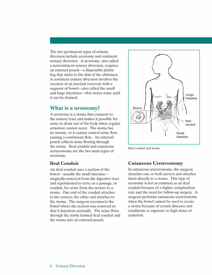
What is a urostomy? A urostomy is a stoma that connects to the urinary tract and makes it possible for urine to drain out of the body when regular urination cannot occur. The stoma has no muscle, so it cannot control urine flow, causing a continuous flow. An external pouch collects urine flowing through the stoma. Ileal conduit and cutaneous ureterostomy are the two main types of urostomy.
Ileal Conduit An ileal conduit uses a section of the bowel—usually the small intestine— surgically removed from the digestive tract and repositioned to serve as a passage, or conduit, for urine from the ureters to a stoma. One end of the conduit attaches to the ureters; the other end attaches to the stoma. The surgeon reconnects the bowel where the section was removed so that it functions normally. The urine flows through the newly formed ileal conduit and the stoma into an external pouch.
Stoma
Small intestine
Large intestine
Ileal conduit
Ileal conduit and stoma
Cutaneous Ureterostomy In cutaneous ureterostomy, the surgeon detaches one or both ureters and attaches them directly to a stoma. This type of urostomy is not as common as an ileal conduit because of a higher complication rate and the need for follow-up surgery. A surgeon performs cutaneous ureterostomy when the bowel cannot be used to create a stoma because of certain diseases and conditions or exposure to high doses of radiation.
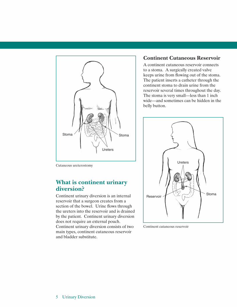
5 Urinary Diversion
Cutaneous ureterostomy
What is continent urinary diversion? Continent urinary diversion is an internal reservoir that a surgeon creates from a section of the bowel. Urine flows through the ureters into the reservoir and is drained by the patient. Continent urinary diversion does not require an external pouch. Continent urinary diversion consists of two main types, continent cutaneous reservoir and bladder substitute.
Continent Cutaneous Reservoir A continent cutaneous reservoir connects to a stoma. A surgically created valve keeps urine from flowing out of the stoma. The patient inserts a catheter through the continent stoma to drain urine from the reservoir several times throughout the day. The stoma is very small––less than 1 inch wide––and sometimes can be hidden in the belly button.
Stoma
Ureters
Stoma
Reservoir
Continent cutaneous reservoir
Ureters
Stoma
6 Urinary Diversion
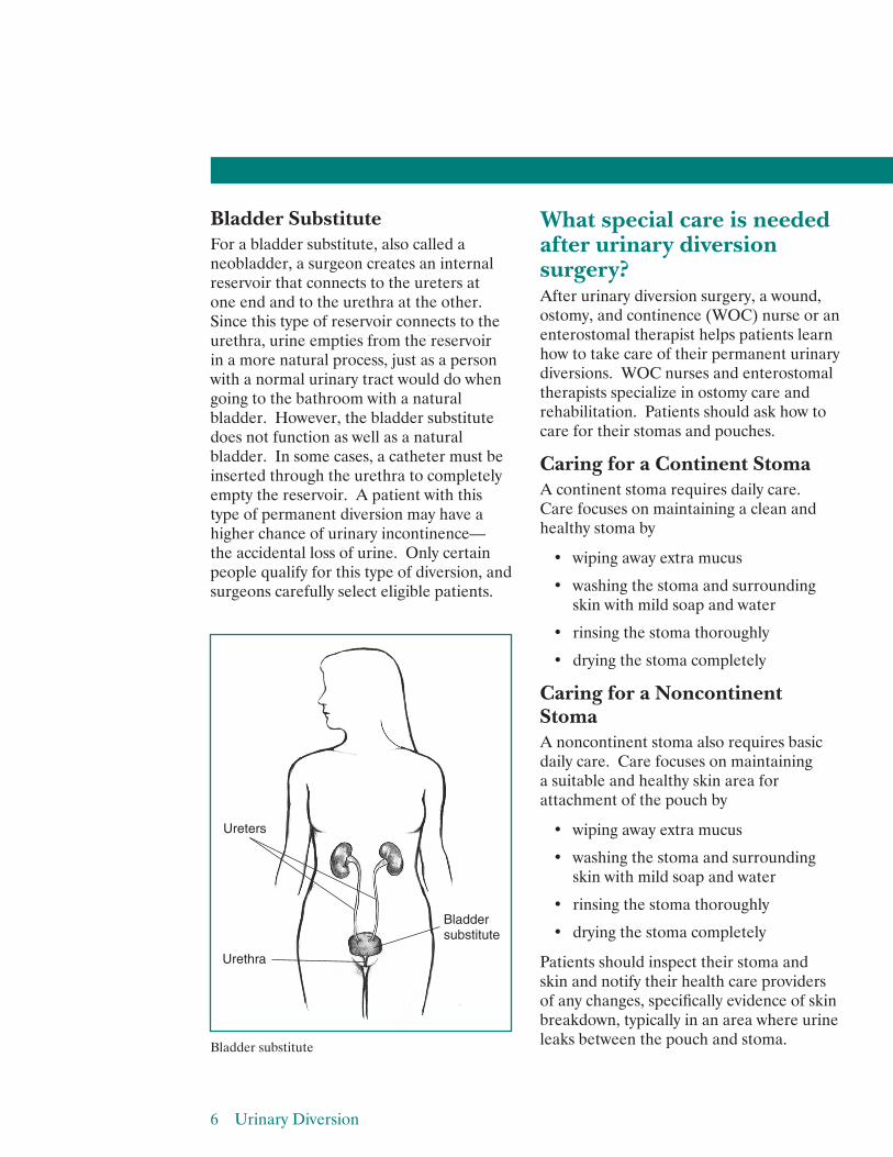
Bladder Substitute For a bladder substitute, also called a neobladder, a surgeon creates an internal reservoir that connects to the ureters at one end and to the urethra at the other. Since this type of reservoir connects to the urethra, urine empties from the reservoir in a more natural process, just as a person with a normal urinary tract would do when going to the bathroom with a natural bladder. However, the bladder substitute does not function as well as a natural bladder. In some cases, a catheter must be inserted through the urethra to completely empty the reservoir. A patient with this type of permanent diversion may have a higher chance of urinary incontinence–– the accidental loss of urine. Only certain people qualify for this type of diversion, and surgeons carefully select eligible patients.
Urethra
Ureters
Bladder substitute
Bladder substitute
What special care is needed after urinary diversion surgery? After urinary diversion surgery, a wound, ostomy, and continence (WOC) nurse or an enterostomal therapist helps patients learn how to take care of their permanent urinary diversions. WOC nurses and enterostomal therapists specialize in ostomy care and rehabilitation. Patients should ask how to care for their stomas and pouches.
Caring for a Continent Stoma A continent stoma requires daily care. Care focuses on maintaining a clean and healthy stoma by
• wiping away extra mucus
• washing the stoma and surroundingskin with mild soap and water
• rinsing the stoma thoroughly
• drying the stoma completely
Caring for a Noncontinent Stoma A noncontinent stoma also requires basic daily care. Care focuses on maintaining a suitable and healthy skin area for attachment of the pouch by
• wiping away extra mucus
• washing the stoma and surroundingskin with mild soap and water
• rinsing the stoma thoroughly
• drying the stoma completely
Patients should inspect their stoma and skin and notify their health care providers of any changes, specifically evidence of skin breakdown, typically in an area where urine leaks between the pouch and stoma.
Caring for a Pouch A person with an ileal conduit or with cutaneous ureterostomy also works with WOC nurses or enterostomal therapists to learn how to care for an external pouch. The pouch system usually consists of two pieces—a barrier that sticks to the skin, known as a wafer, and a disposable plastic bag or pouch that attaches to the barrier. Sometimes the barrier and pouch are one unit. The barrier protects the skin from urine and is designed to be as gentle as possible on the skin. The length of time the barrier stays sealed to the skin depends on many things, such as whether the barrier fits properly, the condition of the skin around the stoma, the patient’s physical activity level, and the shape of the body around the stoma.
The pouch has a drain valve at the bottom so the patient can empty it into a toilet without removing the pouch from the stoma. During the day, most patients need to empty the pouch about as often as they used the bathroom before having urinary diversion surgery. Patients should empty the pouch when it is about one-third to one-half full. At night, patients can attach a piece of flexible tubing to the drain valve on the pouch to let urine flow into a bigger pouch during sleep.
Patients should rinse and clean the pouch daily and change it every 5 to 7 days. When changing a pouch, patients need to clean the skin around the stoma with a wet towelette or washcloth. The skin should be completely dry before applying a new pouch. If the constant flow of urine from
the stoma irritates the skin, patients can use protective skin wipes or an ostomy powder designed to protect the skin around the stoma.
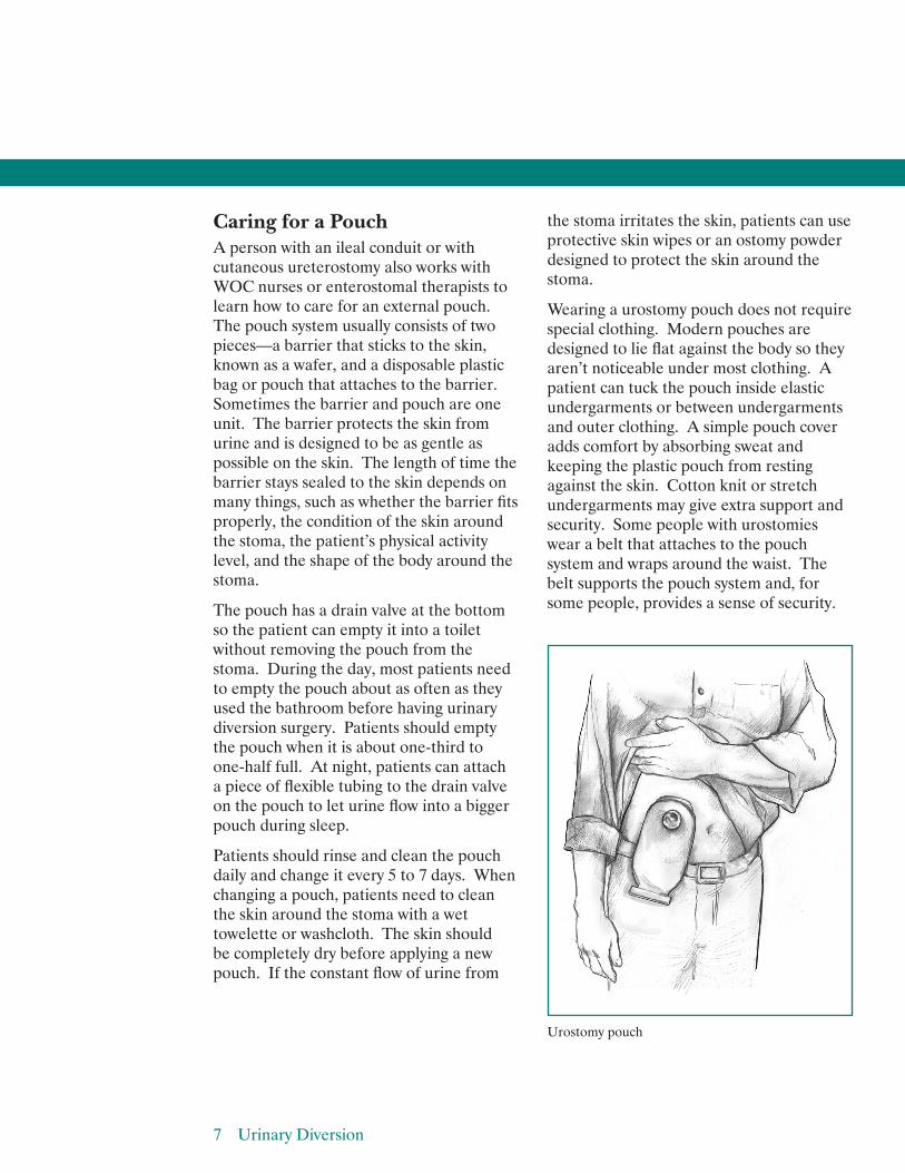
Wearing a urostomy pouch does not require special clothing. Modern pouches are designed to lie flat against the body so they aren’t noticeable under most clothing. A patient can tuck the pouch inside elastic undergarments or between undergarments and outer clothing. A simple pouch cover adds comfort by absorbing sweat and keeping the plastic pouch from resting against the skin. Cotton knit or stretch undergarments may give extra support and security. Some people with urostomies wear a belt that attaches to the pouch system and wraps around the waist. The belt supports the pouch system and, for some people, provides a sense of security.
Urostomy pouch
7 Urinary Diversion
Caring for a Continent Cutaneous Reservoir For a continent cutaneous reservoir, patients learn how to insert a catheter through the stoma or urethra to drain the internal reservoir. Patients can drain the reservoir by inserting the catheter while standing in front of the toilet or sitting on the toilet. During the first few weeks after urinary diversion surgery, patients need to drain the internal reservoir every couple of hours. Over time, the reservoir capacity will increase and patients will be able to go 4 to 6 hours between reservoir drainings. Patients should wash their hands with soap and water each time they use a catheter. Before and after catheterization, patients should clean the stoma and skin around it with a wet towelette or washcloth and completely dry the stoma and skin.
The reservoir is made from part of the bowel, so it may produce mucus that normally lines the digestive tract. To clear this mucus, patients may need to irrigate, or flush out, the reservoir using a syringe with sterile water or normal saline. Patients should talk with a WOC nurse, an enterostomal therapist, or a urologist––a doctor who specializes in the urinary tract–– about how often they should irrigate the reservoir.
Infection Bacteria often enter urostomies and continent urinary diversions and begin growing in number. At times, bacterial overgrowth causes a symptomatic urinary tract infection. Symptoms of infection may include
• fever
• milky urine or urine containing extra mucus
• strong-smelling urine
• back pain
• poor appetite
• nausea
• vomiting
Patients with symptoms of infection should call their health care providers at once. Drinking eight full glasses of water every day can help prevent infection by flushing out bacteria and keeping bacterial counts low. Patients should talk with their health care providers about appropriate times to have their urine tested and when to have treatment with antibiotics. Urine testing and infection treatment play a critical role in successful long-term care with minimal complications.
8 Urinary Diversion
Activities To help the stoma heal, patients need to restrict their activities, including driving and heavy lifting, during the first 2 to 3 weeks after urinary diversion surgery. Once the stoma has healed, patients should be able to do most of the activities they enjoyed before urinary diversion surgery, even swimming and other water sports. The only exceptions may be contact sports such as football or karate. Patients whose jobs include strenuous physical activities should talk with their health care providers and employers about making adjustments to their job responsibilities.
Relationships Patients may worry that people will have negative reactions to their urinary diversion. Most people will never know patients are wearing a pouch or have a continent urinary diversion. Friends and relatives are likely to be aware of the patient’s health problems. However, only a spouse, intimate partner, or primary caretaker needs to know the details of the urinary diversion. Patients can choose how much they share about their condition.
Urinary diversion surgery may reduce sexual function, especially when the bladder has been removed because of cancer. Patients who have good sexual function may resume sexual activities after urinary
diversion surgery as soon as their health care providers say it is safe. Patients should talk with their health care providers about any concerns they have about maintaining a satisfying sexual relationship. Health care providers can give information about ways to protect the stoma during sexual activity. Patients may want to ask about specially designed apparel to enhance intimacy for people with urostomies. Communicating with a sexual partner is essential. Patients should share their concerns and wishes and listen carefully to their partner’s concerns.
Eating, Diet, and Nutrition After urinary diversion surgery, patients will likely be able to resume their normal diet. Some foods, such as asparagus and seafood, may cause urine to have a stronger odor, which may be noticeable when emptying a pouch. If odor is a concern, patients should talk with their health care providers about changes in diet. Patients should also talk with their health care providers about their dietary needs. Some patients with continent urinary reservoirs have a chance of vitamin B deficiency and may require lifelong vitamin B injections. This requirement is only for a specific type of diversion and should be discussed with the health care provider in detail.
9 Urinary Diversion
Points to Remember • Urinary diversion is a surgical
procedure that reroutes the normal flow of urine out of the body when urine flow is blocked.
• Urinary diversion can be temporary or permanent, depending on the reason for the procedure.
• Temporary urinary diversion reroutes the flow of urine for several days or weeks. This type of urinary diversion includes a nephrostomy and urinary catheterization.
• A nephrostomy involves a small tube inserted through the skin directly into a kidney.
• Urinary catheterization involves placing a thin, flexible tube––called a catheter––into the bladder to drain urine.
• Permanent urinary diversion requires surgery to reroute urine flow to an external pouch through an opening in the wall of the abdomen, called a stoma, or to a surgically created internal reservoir.
• Surgeons perform permanent urinary diversion when a patient has a damaged bladder or no longer has a bladder.
• The two permanent types of urinary diversion include urostomy and continent urinary diversion.
• A urostomy is a stoma that connects to the urinary tract and makes it possible for urine to drain out of the body when regular urination cannot occur.
• Continent urinary diversion is aninternal reservoir that a surgeon creates from a section of the bowel.
• After urinary diversion surgery, a wound, ostomy, and continence (WOC) nurse or an enterostomal therapist helps patients learn how to take care of their permanent urinary diversions.
Hope through Research The National Institute of Diabetes and Digestive and Kidney Diseases (NIDDK) conducts and supports basic and clinical research into many kinds of kidney and urologic diseases. The knowledge gained from these studies advances scientific understanding of why kidney and urologic diseases develop and leads to improved methods of diagnosing, treating, and preventing them.
Researchers supported by the National Institutes of Health (NIH) are exploring ways to improve urinary diversions for patients who have had their bladders removed. One study compares two types of continent ileal neobladder construction. A Comparison of the Studer Pouch versus the T-Pouch Orthotopic Neobladder Urinary Diversion in Bladder Cancer Patients study is funded under NIH clinical trial number NCT01008865. Another study investigates whether a combination of a patient’s own cells and other materials can be used to form a conduit to let urine flow safely from the kidneys to outside the body. The Incontinent Urinary Diversion Using an Autologous Neo-Urinary Conduit study is funded under NIH clinical trial number NCT01087697.
10 Urinary Diversion
The National Cancer Institute also supports projects on the complications, emotional wellbeing, and quality of life of bladder cancer survivors with urinary diversion.
Clinical trials are research studies involving people. Clinical trials look at safe and effective new ways to prevent, detect, or treat disease. Researchers also use clinical trials to look at other aspects of care, such as improving the quality of life for people with chronic illnesses. To learn more about clinical trials, why they matter, and how to participate, visit the NIH Clinical Research Trials and You website at www.nih.gov/ health/clinicaltrials. For information about current studies, visit www.ClinicalTrials.gov.
For More Information Society of Urologic Nurses and Associates East Holly Avenue, Box 56 Pitman, NJ 08071–0056 Phone: 1–888–TAP–SUNA
(1–888–827–7862) Email: [email protected] Internet: www.suna.org
United Ostomy Associations of America, Inc. P.O. Box 512 Northfield, MN 55057–0512 Phone: 1–800–826–0826 Email: [email protected] Internet: www.ostomy.org
Urology Care Foundation 1000 Corporate Boulevard Linthicum, MD 21090 Phone: 1–800–828–7866 or 410–689–3700 Fax: 410–689–3998 Email: [email protected] Internet: www.UrologyHealth.org
Wound, Ostomy and Continence Nurses Society 15000 Commerce Parkway, Suite C Mount Laurel, NJ 08054 Phone: 1–888–224–9626 Fax: 856–439–0525 Email: [email protected] Internet: www.wocn.org
Acknowledgments Publications produced by the Clearinghouse are carefully reviewed by both NIDDK scientists and outside experts. This publication was reviewed by Joseph A. Costa, D.O., University of Florida College of Medicine, Jacksonville, FL.
You may also find additional information about this topic by visiting MedlinePlus at www.medlineplus.gov.
This publication may contain information about medications and, when taken as prescribed, the conditions they treat. When prepared, this publication included the most current information available. For updates or for questions about any medications, contact the U.S. Food and Drug Administration toll-free at 1–888–INFO–FDA (1–888–463–6332) or visit www.fda.gov. Consult your health care provider for more information.
11 Urinary Diversion
National Kidney and Urologic Diseases Information Clearinghouse
3 Information WayBethesda, MD 20892–3580Phone: 1–800–891–5390TTY: 1–866–569–1162Fax: 703–738–4929Email: [email protected]: www.urologic.niddk.nih.gov
The National Kidney and Urologic Diseases Information Clearinghouse (NKUDIC) is a service of the National Institute of Diabetes and Digestive and Kidney Diseases (NIDDK). The NIDDK is part of the National Institutes of Health of the U.S. Department of Health and Human Services. Established in 1987, the Clearinghouse provides information about diseases of the kidneys and urologic system to people with kidney and urologic disorders and to their families, health care professionals, and the public. The NKUDIC answers inquiries, develops and distributes publications, and works closely with professional and patient organizations and Government agencies to coordinate resources about kidney and urologic diseases.
This publication is not copyrighted. The Clearinghouse encourages users of this publication to duplicate and distribute as many copies as desired.
This publication is available at www.urologic.niddk.nih.gov.
NIH Publication No. 13–5629September 2013
The NIDDK prints on recycled paper with bio-based ink.