what does the following have in common? expulsion of newborn from the uterus wheeze of asthma spasm...
TRANSCRIPT
What does the following have in common?
Expulsion of newborn from the uterusWheeze of asthmaSpasm of coronary arteries
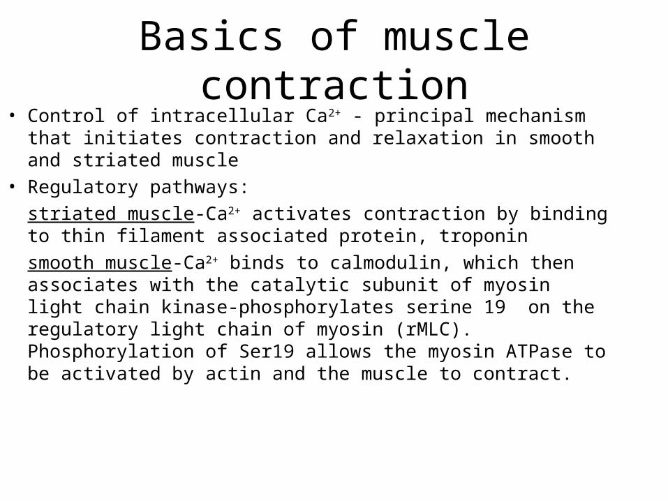
Basics of muscle contraction• Control of intracellular Ca2+ - principal mechanism that initiates
contraction and relaxation in smooth and striated muscle
• Regulatory pathways:
striated muscle-Ca2+ activates contraction by binding to thin filament associated protein, troponin
smooth muscle-Ca2+ binds to calmodulin, which then associates with the catalytic subunit of myosin light chain kinase-phosphorylates serine 19 on the regulatory light chain of myosin (rMLC). Phosphorylation of Ser19 allows the myosin ATPase to be activated by actin and the muscle to contract.
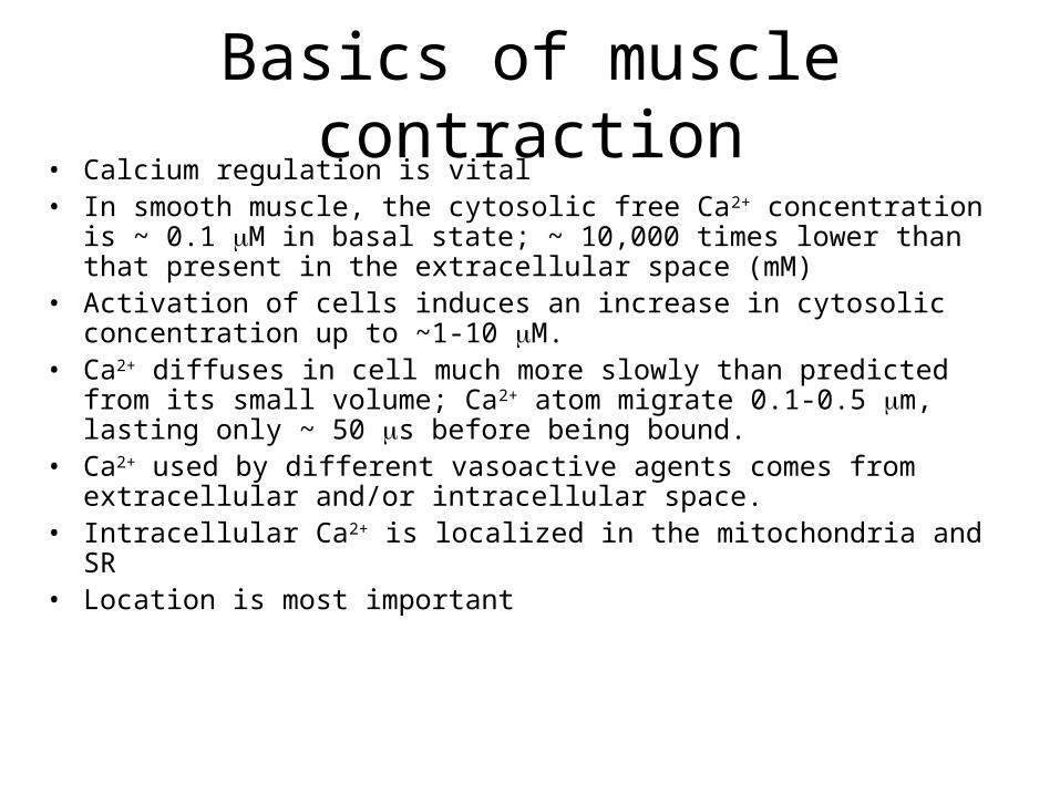
• Calcium regulation is vital• In smooth muscle, the cytosolic free Ca2+ concentration is ~ 0.1 M in basal
state; ~ 10,000 times lower than that present in the extracellular space (mM)• Activation of cells induces an increase in cytosolic concentration up to ~1-10
M. • Ca2+ diffuses in cell much more slowly than predicted from its small volume;
Ca2+ atom migrate 0.1-0.5 m, lasting only ~ 50 s before being bound.• Ca2+ used by different vasoactive agents comes from extracellular and/or
intracellular space.• Intracellular Ca2+ is localized in the mitochondria and SR• Location is most important
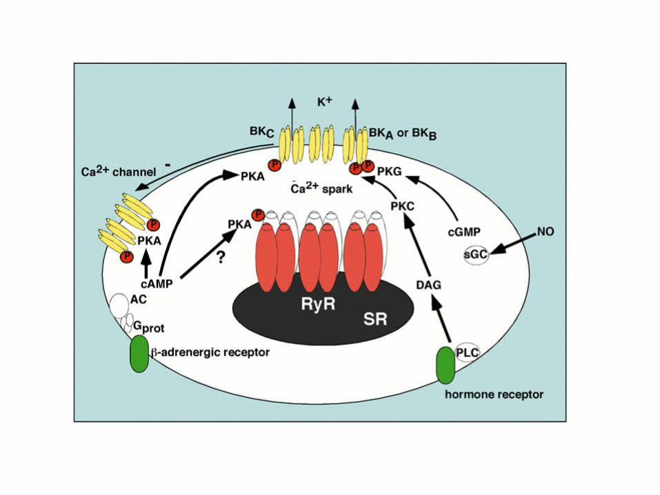
Basics of muscle contraction
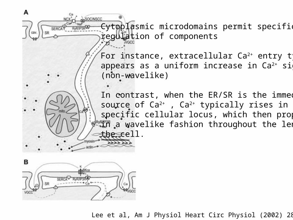
Lee et al, Am J Physiol Heart Circ Physiol (2002) 282:H1571
Cytoplasmic microdomains permit specific regulation of components
For instance, extracellular Ca2+ entry typicallyappears as a uniform increase in Ca2+ signal(non-wavelike)
In contrast, when the ER/SR is the immediatesource of Ca2+ , Ca2+ typically rises in aspecific cellular locus, which then propagatesin a wavelike fashion throughout the length of the cell.
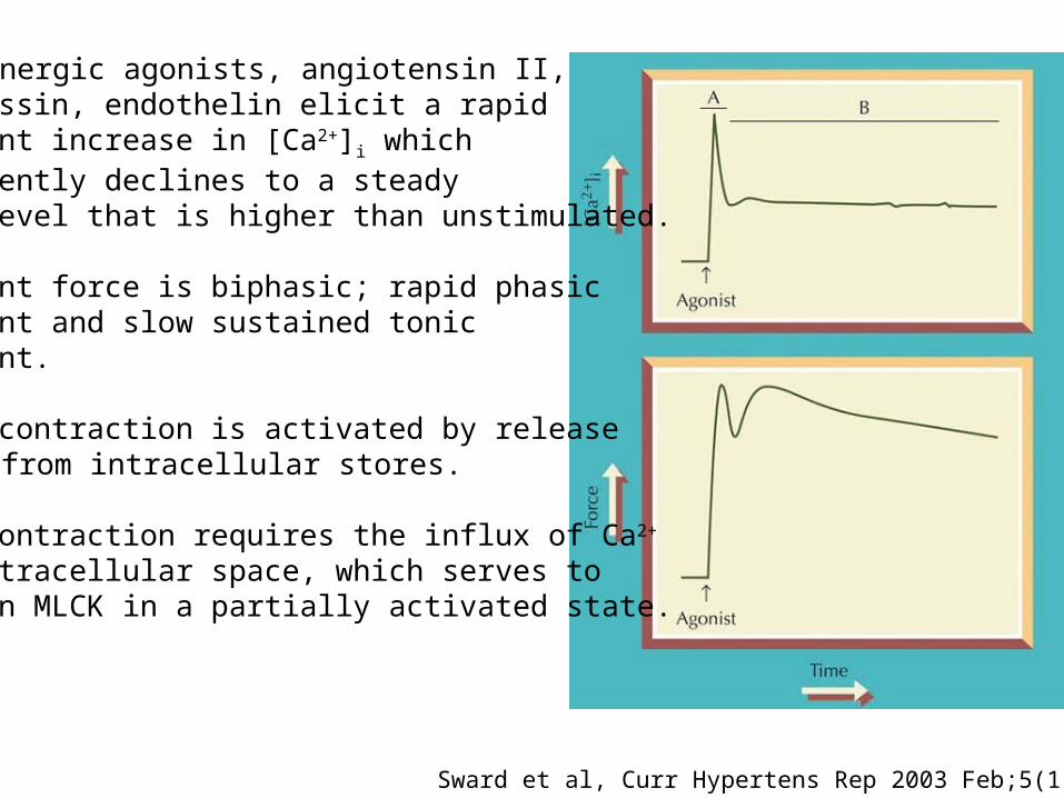
1-adrenergic agonists, angiotensin II, vasopressin, endothelin elicit a rapid transient increase in [Ca2+]i which subsequently declines to a steady state level that is higher than unstimulated.
Resultant force is biphasic; rapid phasic component and slow sustained tonic component.
Phasic contraction is activated by release of Ca2+ from intracellular stores.
Tonic contraction requires the influx of Ca2+
from extracellular space, which serves to maintain MLCK in a partially activated state.
Sward et al, Curr Hypertens Rep 2003 Feb;5(1):66-72
• The degree of interaction is determined by the net level of phosphorylation of the 20 kDa regulatory light chains of myosin II (rMLC).
• MLC is regulated by MLC kinase (MLCK) and MLC phosphatase (MLCP or PP1M).
• The extent of the rMLC phosphorylation and the amplitude of force production depends on the balance of the activities of MLCK and MLCP.
• Under certain conditions, force is also regulated independent of the changes in rMLC phosphorylation levels perhaps by thin filament associated proteins (caldesmon and calponin), which can be phosphorylated by MAP kinase and/or other kinases.
• Thin filament associated proteins might modulate the effect of rMLC phosphorylation, which is alone sufficient to initiate and maintain contraction.
• MLCP is a trimer comprising a 130 kD regulatory myosin binding subunit (MBS), a 37 kD catalytic subunit (PP1c), and a 20 kD protein of uncertain function (M20).
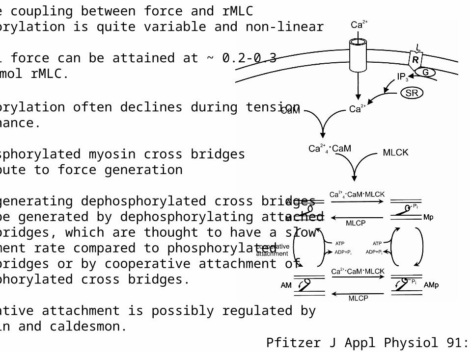
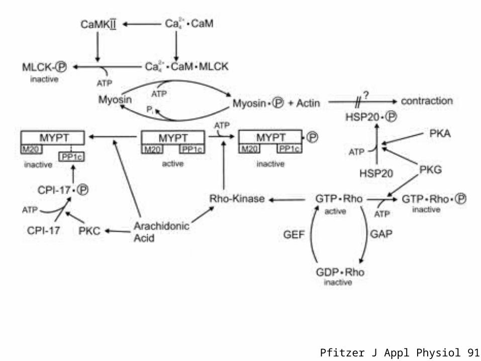
Pfitzer J Appl Physiol 91:497
Precise coupling between force and rMLCphosphorylation is quite variable and non-linear
Maximal force can be attained at ~ 0.2-0.3 mol Pi/mol rMLC.
Phosphorylation often declines during tensionmaintenance.
Nonphosphorylated myosin cross bridges contribute to force generation
Force generating dephosphorylated cross bridgescould be generated by dephosphorylating attachedcross bridges, which are thought to have a slowdetachment rate compared to phosphorylatedcross bridges or by cooperative attachment of dephosphorylated cross bridges.
Cooperative attachment is possibly regulated bycalponin and caldesmon.
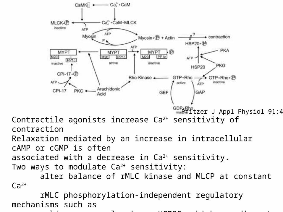
Pfitzer J Appl Physiol 91:497Contractile agonists increase Ca2+ sensitivity of contractionRelaxation mediated by an increase in intracellular cAMP or cGMP is often associated with a decrease in Ca2+ sensitivity.Two ways to modulate Ca2+ sensitivity:
alter balance of rMLC kinase and MLCP at constant Ca2+
rMLC phosphorylation-independent regulatory mechanisms such ascaldesmon or calponin or HSP20, which may disrupt phosphorylatedmyosin-actin cross bridges.
Pfitzer J Appl Physiol 91:497
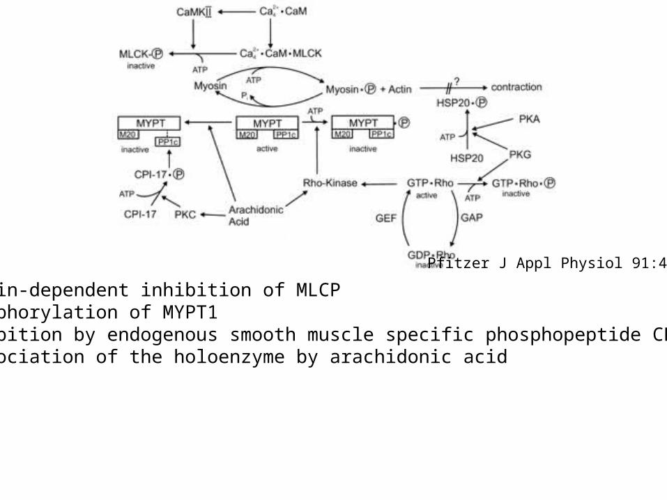
G protein-dependent inhibition of MLCP(1) Phosphorylation of MYPT1(2) Inhibition by endogenous smooth muscle specific phosphopeptide CPI-17(3) Dissociation of the holoenzyme by arachidonic acid
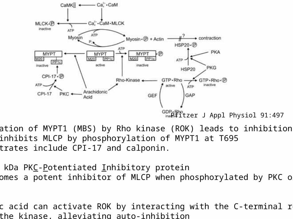
Phosphorylation of MYPT1 (MBS) by Rho kinase (ROK) leads to inhibition ofMLCP. ROK inhibits MLCP by phosphorylation of MYPT1 at T695 Other substrates include CPI-17 and calponin.
CPI-17= 17 kDa PKC-Potentiated Inhibitory proteinCPI-17 becomes a potent inhibitor of MLCP when phosphorylated by PKC or ROKat T38.
Arachidonic acid can activate ROK by interacting with the C-terminal regulatory domain of the kinase, alleviating auto-inhibition
Pfitzer J Appl Physiol 91:497
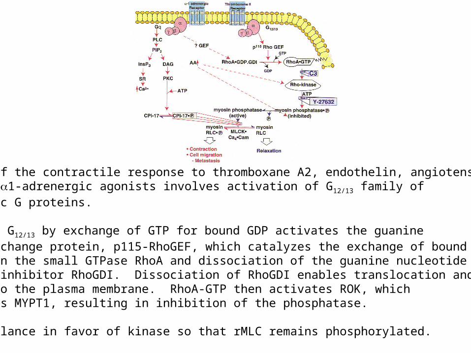
Tonic phase of the contractile response to thromboxane A2, endothelin, angiotensin II, vasopressin, 1-adrenergic agonists involves activation of G12/13 family ofheterotrimeric G proteins.
Activation of G12/13 by exchange of GTP for bound GDP activates the guaninenucleotide exchange protein, p115-RhoGEF, which catalyzes the exchange of bound GDP for GTP on the small GTPase RhoA and dissociation of the guanine nucleotidedissociation inhibitor RhoGDI. Dissociation of RhoGDI enables translocation andinsertion into the plasma membrane. RhoA-GTP then activates ROK, which phosphorylates MYPT1, resulting in inhibition of the phosphatase.
Shifts the balance in favor of kinase so that rMLC remains phosphorylated.
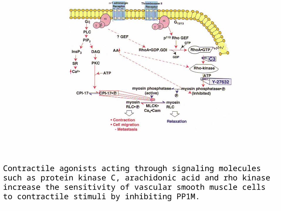
Contractile agonists acting through signaling moleculessuch as protein kinase C, arachidonic acid and rho kinaseincrease the sensitivity of vascular smooth muscle cells to contractile stimuli by inhibiting PP1M.
• Well-established that cAMP and cGMP decreases Ca2+ sensitivity of contraction in both intact and permeabilized smooth muscle.
• In vitro, PKA phosphorylates MLCK at two sites; site A decreases affinity of MLCK for Ca2+/calmodulin complex.
• However, agents that elevate PKA have negligible effects on phosphorylation of site A and Ca2+ activation of MLCK; suggests that cAMP/PKA desensitizes smooth muscle by an alternate mechanism.
• Phosphorylation of MLCK by PKG has no effect on activity.• Endogenous nitric oxide and related nitrovasodilators regulate blood pressure by
activation of soluble guanylate cyclase, elevation of cGMP, activation of cGMP dependent kinase (cGKIor PKG). cGMP-mediated vascular smooth muscle cell relaxation is characterized by a reduction in intracellular calcium concentration and activation of PP1M, which reduces the sensitivity of the contractile apparatus to intracellular calcium.
• The mechanism by which cGMP increases PP1M activity and myosin light chain dephosphorylation was elucidated in a series of experiments published by Surks et al.
Signals that decrease Ca2+ sensitivity
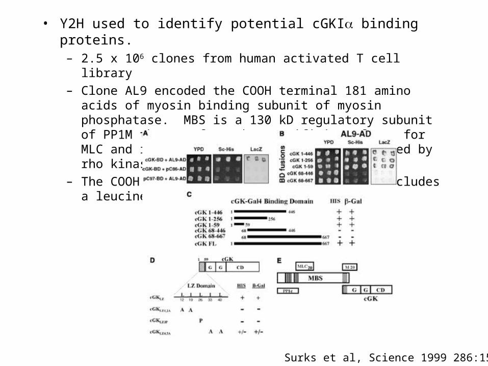
• Y2H used to identify potential cGKI binding proteins.– 2.5 x 10⁶ clones from human activated T cell library
– Clone AL9 encoded the COOH terminal 181 amino acids of myosin binding subunit of myosin phosphatase. MBS is a 130 kD regulatory subunit of PP1M that confers the specificity of PP1 for MLC and is the site on PP1M that is regulated by rho kinase.
– The COOH terminal 181 amino acids of MBS includes a leucine zipper domain.
Surks et al, Science 1999 286:1583
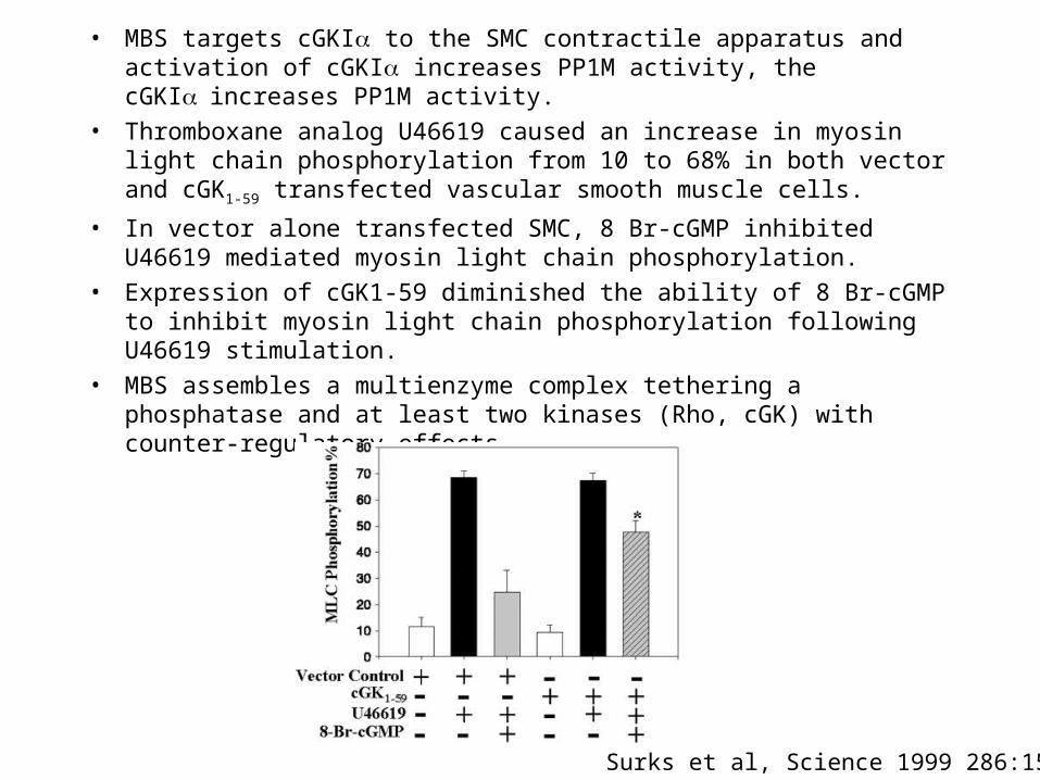
• MBS targets cGKI to the SMC contractile apparatus and activation of cGKI increases PP1M activity, the cGKIincreases PP1M activity.
• Thromboxane analog U46619 caused an increase in myosin light chain phosphorylation from 10 to 68% in both vector and cGK1-59 transfected vascular smooth muscle cells.
• In vector alone transfected SMC, 8 Br-cGMP inhibited U46619 mediated myosin light chain phosphorylation.
• Expression of cGK1-59 diminished the ability of 8 Br-cGMP to inhibit myosin light chain phosphorylation following U46619 stimulation.
• MBS assembles a multienzyme complex tethering a phosphatase and at least two kinases (Rho, cGK) with counter-regulatory effects.
Surks et al, Science 1999 286:1583
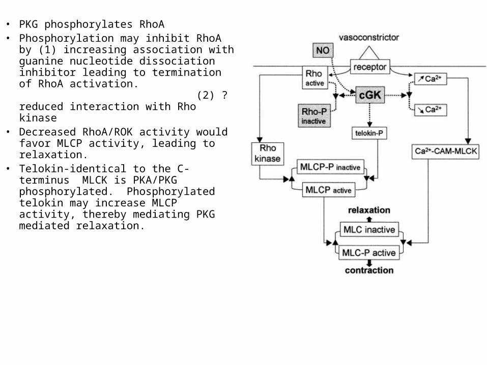
• PKG phosphorylates RhoA• Phosphorylation may inhibit RhoA by (1)
increasing association with guanine nucleotide dissociation inhibitor leading to termination of RhoA activation. (2) ? reduced interaction with Rho kinase
• Decreased RhoA/ROK activity would favor MLCP activity, leading to relaxation.
• Telokin-identical to the C-terminus MLCK is PKA/PKG phosphorylated. Phosphorylated telokin may increase MLCP activity, thereby mediating PKG mediated relaxation.
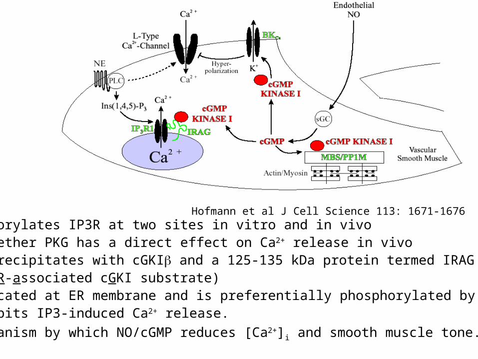
PKG phosphorylates IP3R at two sites in vitro and in vivoUnclear whether PKG has a direct effect on Ca2+ release in vivoIP3R1 co-precipitates with cGKI and a 125-135 kDa protein termed IRAG
(IP3R-associated cGKI substrate)IRAG is located at ER membrane and is preferentially phosphorylated by CGKIwhich inhibits IP3-induced Ca2+ release.
Major mechanism by which NO/cGMP reduces [Ca2+]i and smooth muscle tone.
Hofmann et al J Cell Science 113: 1671-1676
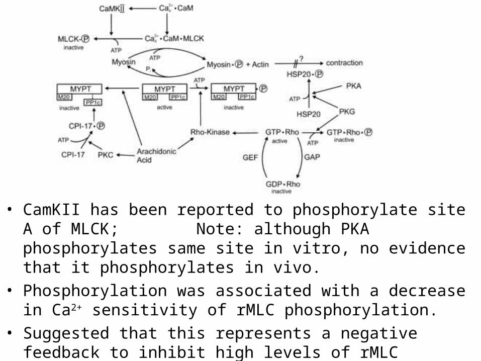
• CamKII has been reported to phosphorylate site A of MLCK; Note: although PKA phosphorylates same site in vitro, no evidence that it phosphorylates in vivo.
• Phosphorylation was associated with a decrease in Ca2+ sensitivity of rMLC phosphorylation.
• Suggested that this represents a negative feedback to inhibit high levels of rMLC phosphorylation.
Pfitzer J Appl Physiol 91:497

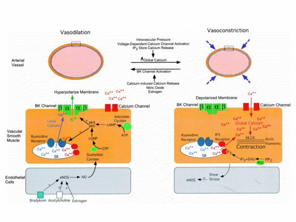
Ion channels in smooth muscle
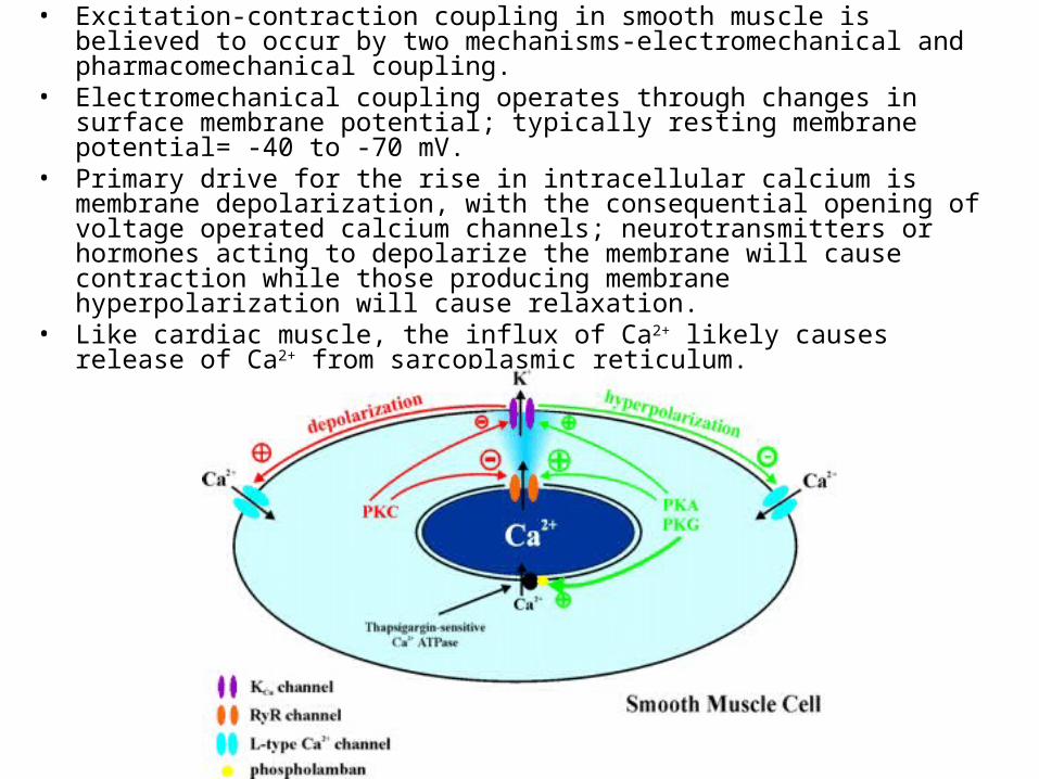
• Excitation-contraction coupling in smooth muscle is believed to occur by two mechanisms-electromechanical and pharmacomechanical coupling.
• Electromechanical coupling operates through changes in surface membrane potential; typically resting membrane potential= -40 to -70 mV.
• Primary drive for the rise in intracellular calcium is membrane depolarization, with the consequential opening of voltage operated calcium channels; neurotransmitters or hormones acting to depolarize the membrane will cause contraction while those producing membrane hyperpolarization will cause relaxation.
• Like cardiac muscle, the influx of Ca2+ likely causes release of Ca2+ from sarcoplasmic reticulum.
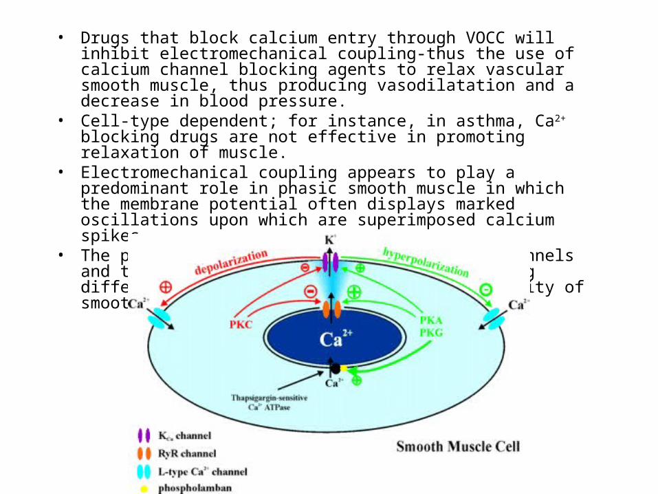
• Drugs that block calcium entry through VOCC will inhibit electromechanical coupling-thus the use of calcium channel blocking agents to relax vascular smooth muscle, thus producing vasodilatation and a decrease in blood pressure.
• Cell-type dependent; for instance, in asthma, Ca2+ blocking drugs are not effective in promoting relaxation of muscle.
• Electromechanical coupling appears to play a predominant role in phasic smooth muscle in which the membrane potential often displays marked oscillations upon which are superimposed calcium spikes
• The plasma membranes contain numerous ion channels and the distribution and properties vary among different tissues, contributing to the diversity of smooth muscle.
• Pharmacomechanical coupling- does not depend upon changes in membrane potential or calcium entry via the VOCC.
• The rise of intracellular Ca2+ is brought about by a combination of Ca2+ release from intracellular stores and Ca2+ entry through non-voltage gated channels, primarily receptor operated calcium channels or store operated Ca2+ channels
• Ca2+ signal often similar to that seen in many non-excitable cells, consisting of an initial rise in [Ca 2+]i followed by a smaller, but sustained increase dependent upon Ca2+ entry from the extracellular space.
• This secondary influx of Ca2+, in association with the process of Ca2+ sensitization whereby the contractile apparatus may be activated by near-resting levels of [Ca2+]i, allows muscles to maintain tone over prolonged periods in the presence of an agonist; occurs in tonic smooth muscle.
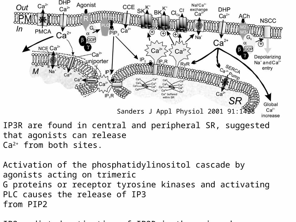
Sanders J Appl Physiol 2001 91:1438
IP3R are found in central and peripheral SR, suggested that agonists can releaseCa2+ from both sites.
Activation of the phosphatidylinositol cascade by agonists acting on trimericG proteins or receptor tyrosine kinases and activating PLC causes the release of IP3from PIP2
IP3 mediated activation of IP3R is the major pharmaco-mechanical coupling in SMC.Confirmed by specific inhibitors, contraction following photolytic release of caged IP3
• The relative importance of electromechanical or pharmacomechanical coupling for any given smooth muscle preparation can be estimated by determining the effects of inhibitors of VOCC’s on the contraction to agonists.
• For example, in guinea pig ileum, dihydropyridines such as nifedipine will virtually abolish all contractions, suggesting that electromechanical coupling predominates
• However, both mechanisms probably occur to some extent in all smooth muscle. In addition, the opening of ROCC and SOCC also produce membrane depolarization, thus activating electromechanical coupling.
• Approximately 20 years ago, it was hypothesized that receptor activation could lead to Ca2+ entry by a mechanism independent of membrane depolarization in smooth muscle
• Receptor operated currents have been described as non-selective cation currents rather than Ca2+ channel
• In the rabbit ear artery, externally applied ATP produced a rapid, transient depolarization of muscle, shown to result from activation of a non-selective cation conductance with significant Ca2+ permeability. Similar responses were reported to ATP in rat vas deferens, rabbit portal vein, and human saphenous veins.
• In addition to ATP, Noradrenaline, Acetylcholine, Histamine, Endothelin-1, Neurokinin A, Substance P, and Vasopressin have been shown to activate a receptor-operated cation current.
Store-operated calcium channels/currents• In the late 1980’s, Putney proposed the model for “capacitative
calcium entry” in which intracellular Ca2+ store depletion stimulated Ca2+ influx across the plasma membrane to maintain a raised [Ca2+]i in the face of prolonged agonist application and to aid in refilling of the stores on agonist withdrawal.
• It is not the Ca2+ released from the stores that activates SOCC. Thus, if the rise in [Ca2+]i is prevented by inclusion of a Ca2+ buffer, then the store operated current would still be present. It is the fact that the stores are empty of Ca2+ that drives the response by an as yet unknown mechanism.
• Many of the neurotransmitters which activate ROCC simultaneously activate phospholipase C, liberating IP3. Therefore, SOCC is activated due to IP3 mediated depletion of the sarcoplasmic reticulum.
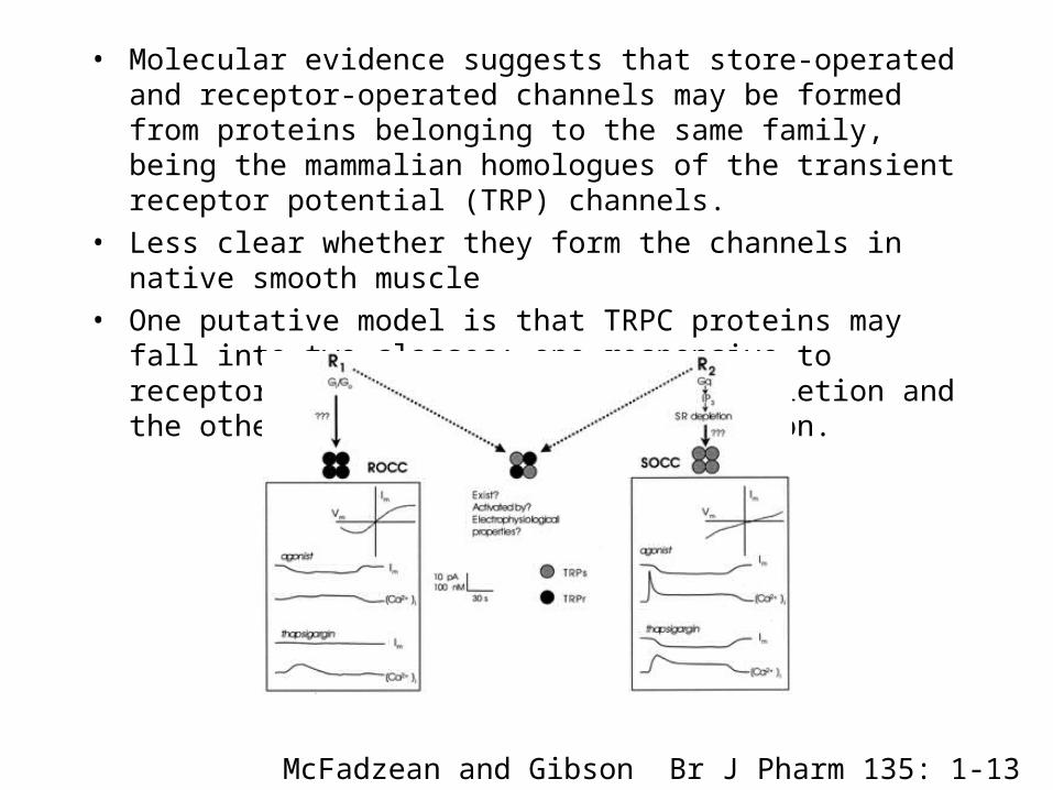
• Molecular evidence suggests that store-operated and receptor-operated channels may be formed from proteins belonging to the same family, being the mammalian homologues of the transient receptor potential (TRP) channels.
• Less clear whether they form the channels in native smooth muscle
• One putative model is that TRPC proteins may fall into two classes; one responsive to receptor activation but not store depletion and the other responsive to store depletion.
McFadzean and Gibson Br J Pharm 135: 1-13
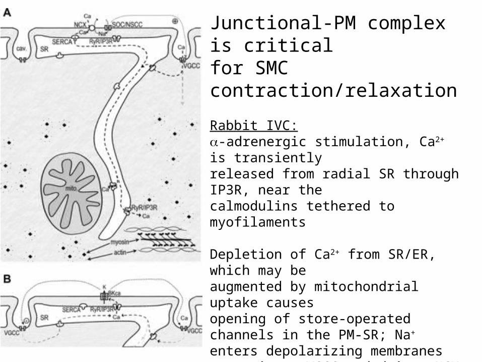
Junctional-PM complex is critical for SMC contraction/relaxation
Rabbit IVC:-adrenergic stimulation, Ca2+ is transientlyreleased from radial SR through IP3R, near thecalmodulins tethered to myofilaments
Depletion of Ca2+ from SR/ER, which may be augmented by mitochondrial uptake causes opening of store-operated channels in the PM-SR; Na+ enters depolarizing membranes to activate VGCC and drives NCX in reverse direction to supply extracellular Ca2+ to PM-SR junctional space, which is taken up by SERCA. As SR is refilled, IP3R are activated, to start the next wave of regenerative Ca2+ release.
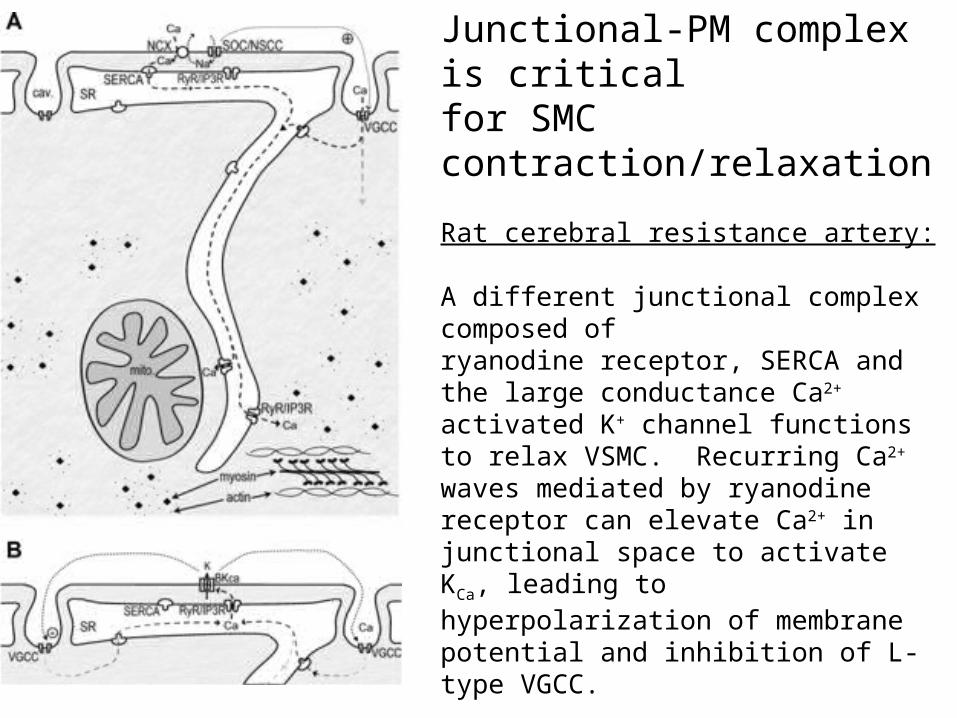
Junctional-PM complex is critical for SMC contraction/relaxation
Rat cerebral resistance artery:
A different junctional complex composed of ryanodine receptor, SERCA and the large conductance Ca2+ activated K+ channel functions to relax VSMC. Recurring Ca2+ waves mediated by ryanodine receptor can elevate Ca2+ in junctional space to activate KCa, leading to hyperpolarization of membrane potential and inhibition of L-type VGCC.
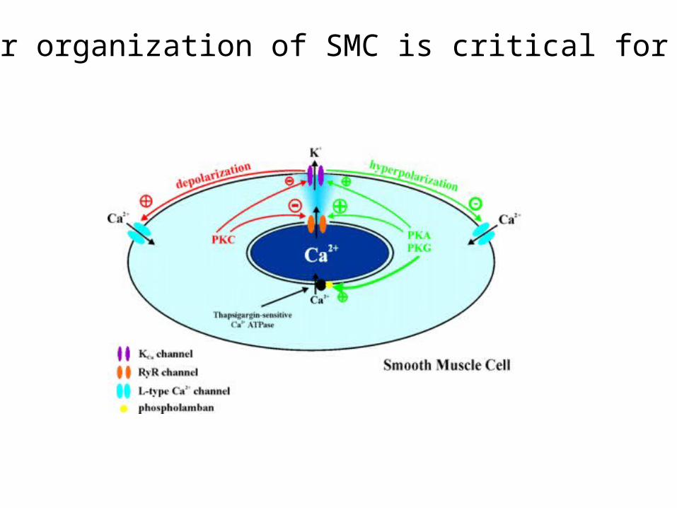
Molecular organization of SMC is critical for function
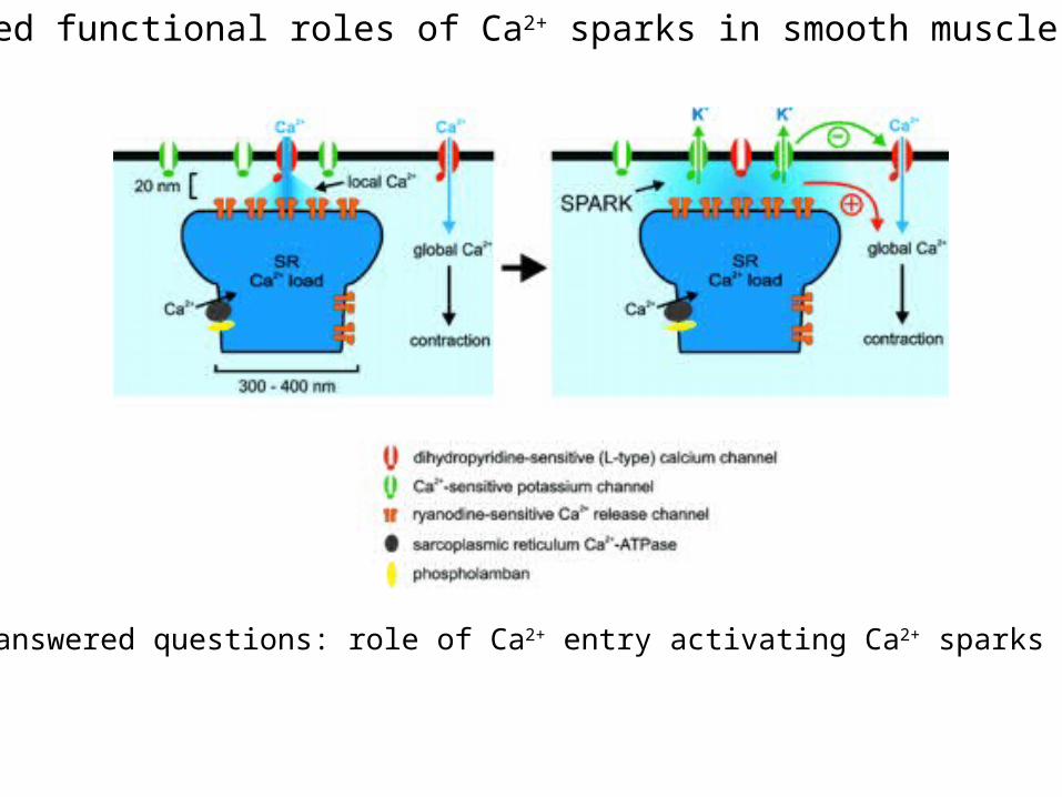
Proposed functional roles of Ca2+ sparks in smooth muscle cells
Unanswered questions: role of Ca2+ entry activating Ca2+ sparks
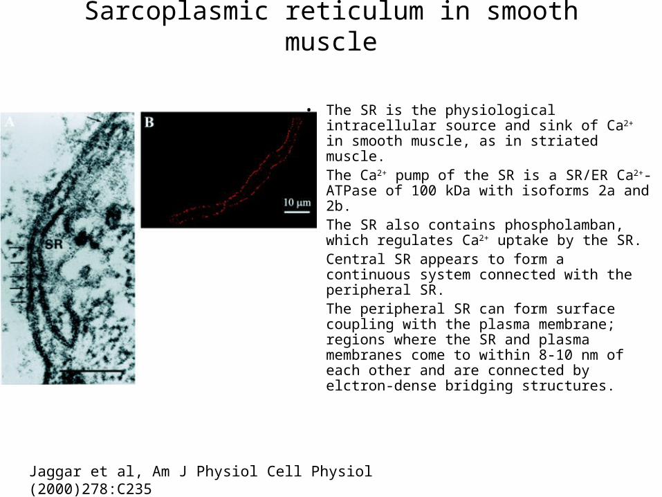
Sarcoplasmic reticulum in smooth muscle
• The SR is the physiological intracellular source and sink of Ca2+ in smooth muscle, as in striated muscle.
• The Ca2+ pump of the SR is a SR/ER Ca2+-ATPase of 100 kDa with isoforms 2a and 2b.
• The SR also contains phospholamban, which regulates Ca2+ uptake by the SR.
• Central SR appears to form a continuous system connected with the peripheral SR.
• The peripheral SR can form surface coupling with the plasma membrane; regions where the SR and plasma membranes come to within 8-10 nm of each other and are connected by elctron-dense bridging structures.
Jaggar et al, Am J Physiol Cell Physiol (2000)278:C235
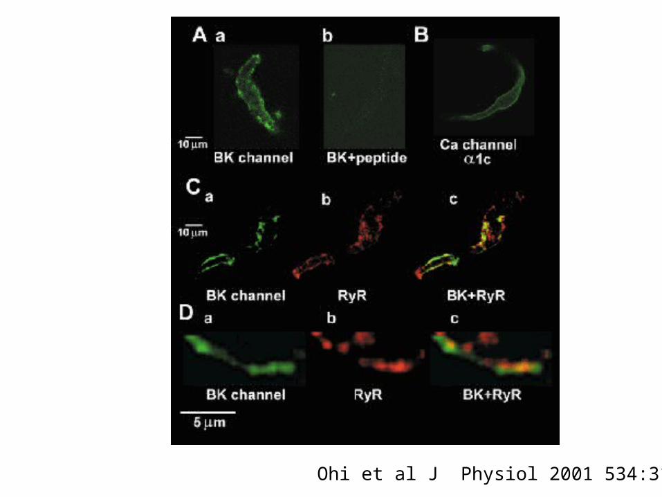
Ohi et al J Physiol 2001 534:313
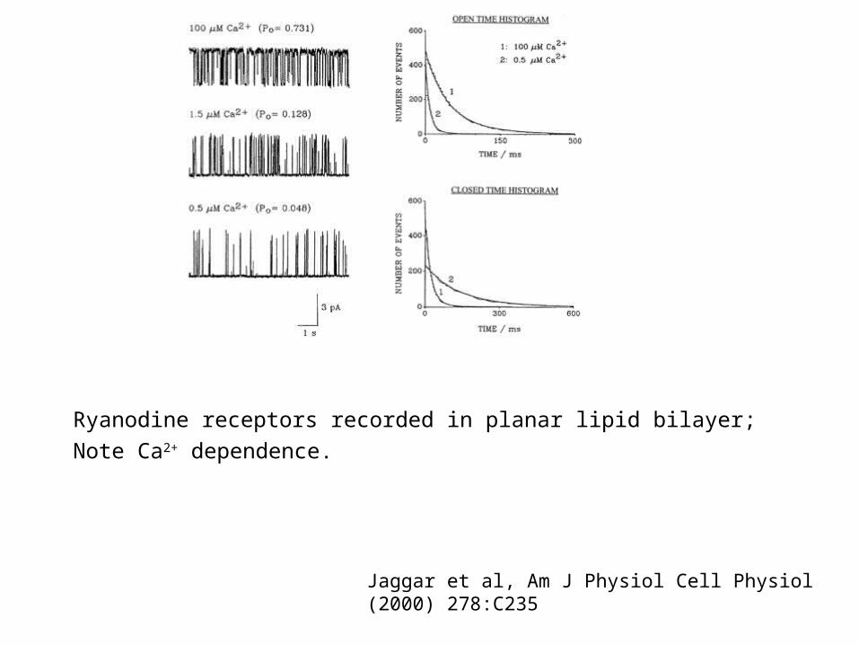
Ryanodine receptors recorded in planar lipid bilayer;
Note Ca2+ dependence.
Jaggar et al, Am J Physiol Cell Physiol (2000) 278:C235
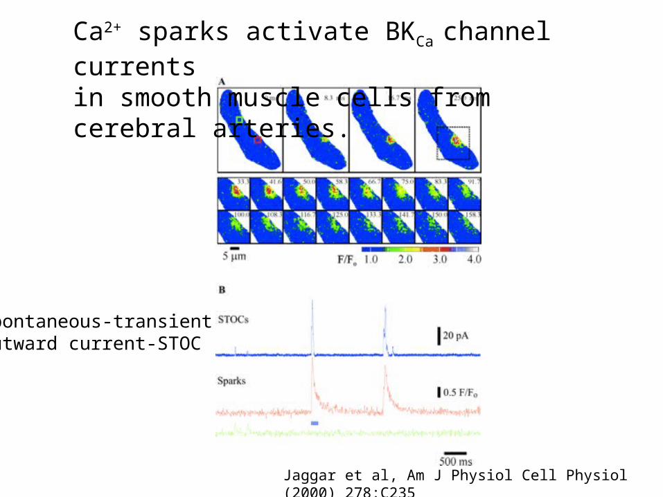
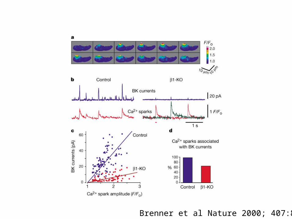
Ca2+ sparks activate BKCa channel currents in smooth muscle cells from cerebral arteries.
Jaggar et al, Am J Physiol Cell Physiol (2000) 278:C235
Spontaneous-transientoutward current-STOC
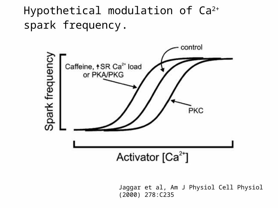
Hypothetical modulation of Ca2+ spark frequency.
Jaggar et al, Am J Physiol Cell Physiol (2000) 278:C235
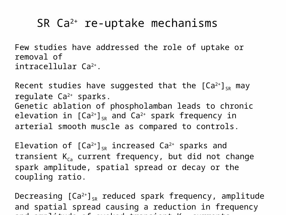
Few studies have addressed the role of uptake or removal of intracellular Ca2+.
Recent studies have suggested that the [Ca2+]SR may regulate Ca2+ sparks.Genetic ablation of phospholamban leads to chronic elevation in [Ca2+]SR and Ca2+ spark frequency in arterial smooth muscle as compared to controls.
Elevation of [Ca2+]SR increased Ca2+ sparks and transient KCa current frequency, but did not change spark amplitude, spatial spread or decay or the coupling ratio.
Decreasing [Ca2+]SR reduced spark frequency, amplitude and spatial spread causing a reduction in frequency and amplitude of evoked transient KCa currents, although the coupling ratio was not affected.
SR Ca2+ re-uptake mechanisms
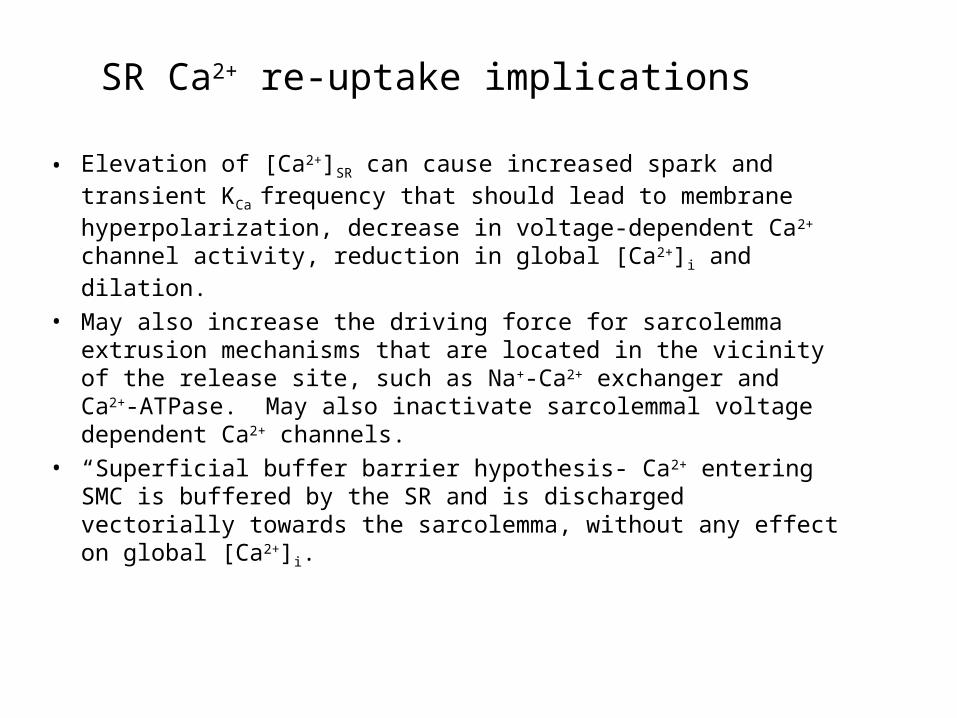
• Elevation of [Ca2+]SR can cause increased spark and transient KCa
frequency that should lead to membrane hyperpolarization, decrease in voltage-dependent Ca2+ channel activity, reduction in global [Ca2+]i and dilation.
• May also increase the driving force for sarcolemma extrusion mechanisms that are located in the vicinity of the release site, such as Na+-Ca2+ exchanger and Ca2+-ATPase. May also inactivate sarcolemmal voltage dependent Ca2+ channels.
• “Superficial buffer barrier hypothesis- Ca2+ entering SMC is buffered by the SR and is discharged vectorially towards the sarcolemma, without any effect on global [Ca2+]i.
SR Ca2+ re-uptake implications
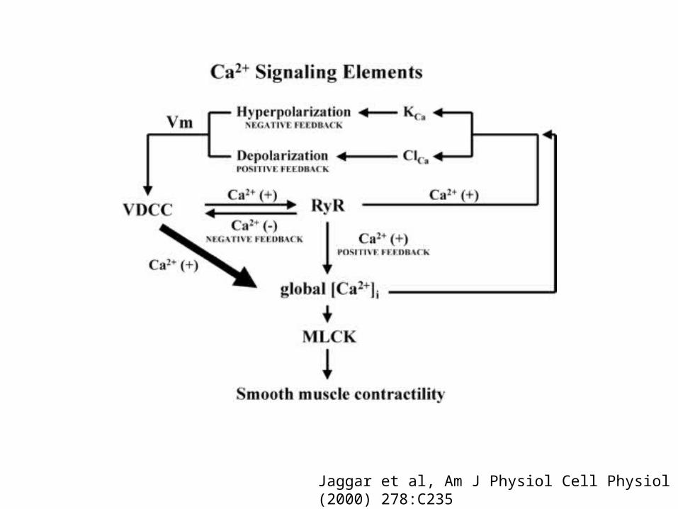
Jaggar et al, Am J Physiol Cell Physiol (2000) 278:C235
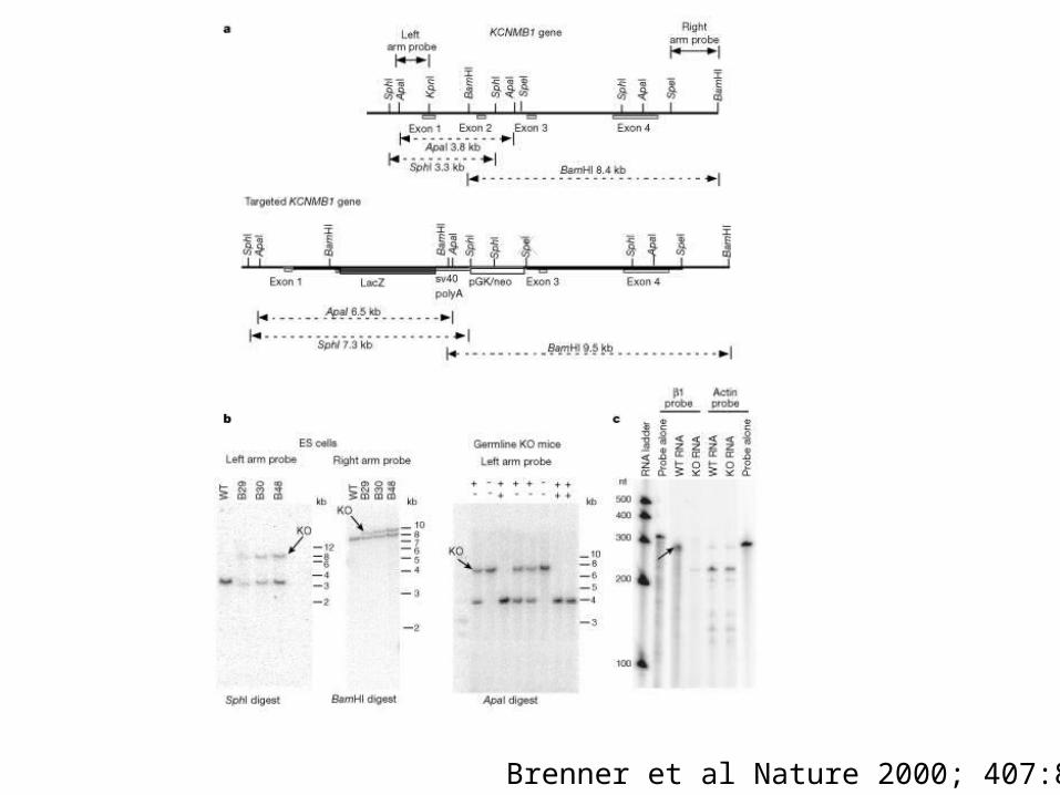
Brenner et al Nature 2000; 407:870
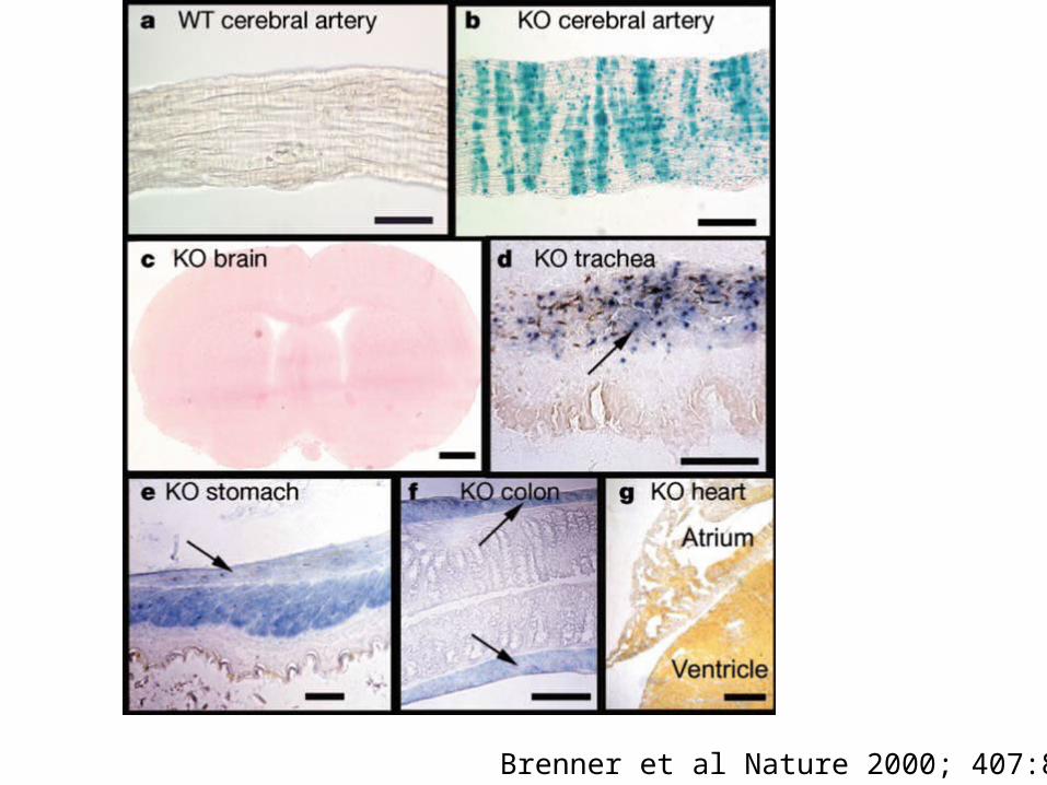
Brenner et al Nature 2000; 407:870
Brenner et al Nature 2000; 407:870
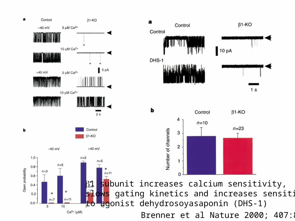
1 subunit increases calcium sensitivity,slows gating kinetics and increases sensitivityto agonist dehydrosoyasaponin (DHS-1)
Brenner et al Nature 2000; 407:870
• Myogenic tone refers to the ability of vascular smooth muscle to alter its state of contractility in response to changes in intraluminal pressure
• The vessel constricts in opposition to an increase in intravascular pressure and dilates when the pressure decreases
• Behavior observed in a variety of vascular tissues, including veins and conduit arteries, but especially prevalent in resistance vasculature.
• Classically described as being a Ca2+ dependent process where pressure evoked depolarization and Ca2+ entry through voltage gated Ca2+ channels play obligatory roles
• Consistent with a role for pressure-induced depolarization, blockers of voltage gated Ca2+ channels have been shown to reduce myogenic responses.
• Arteriolar SMC possess ion channels sensitive to cell membrane stretch that may be activated by vessel distension arising from an increase in intraluminal pressure.
• Have relative permeability: K+>Na+>Ca2+
• Ca2+ influx would be relatively small- generally believed that stretch activation of these channels mainly contributes to membrane depolarization with subsequent opening of voltage gated calcium channels.
• KCa currents have been shown to attenuate the stretch-induced changes in membrane potential and myogenic constriction.
• Mechanical perturbation of cell membranes may release factors that modulate the activity of such channels.
Brenner et al Nature 2000; 407:870
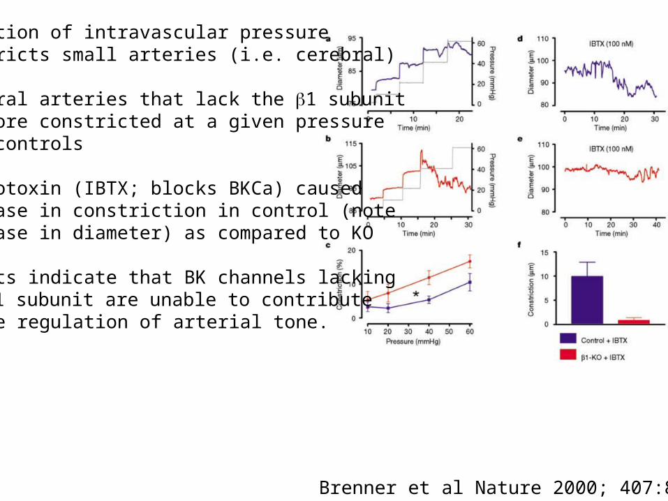
Elevation of intravascular pressureconstricts small arteries (i.e. cerebral)
Cerebral arteries that lack the 1 subunitare more constricted at a given pressurethan controls
Iberiotoxin (IBTX; blocks BKCa) causedincrease in constriction in control (note decrease in diameter) as compared to KO
Results indicate that BK channels lackingthe 1 subunit are unable to contributeto the regulation of arterial tone.
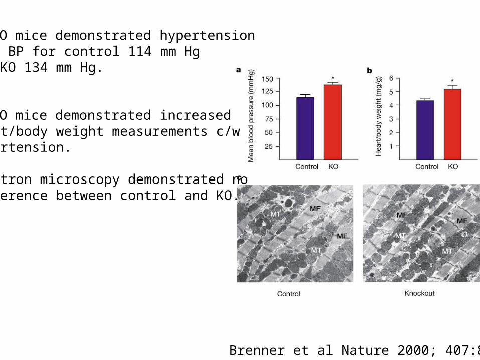
1-KO mice demonstrated hypertensionMean BP for control 114 mm Hgand KO 134 mm Hg.
1-KO mice demonstrated increasedheart/body weight measurements c/whypertension.
Electron microscopy demonstrated nodifference between control and KO.
Brenner et al Nature 2000; 407:870
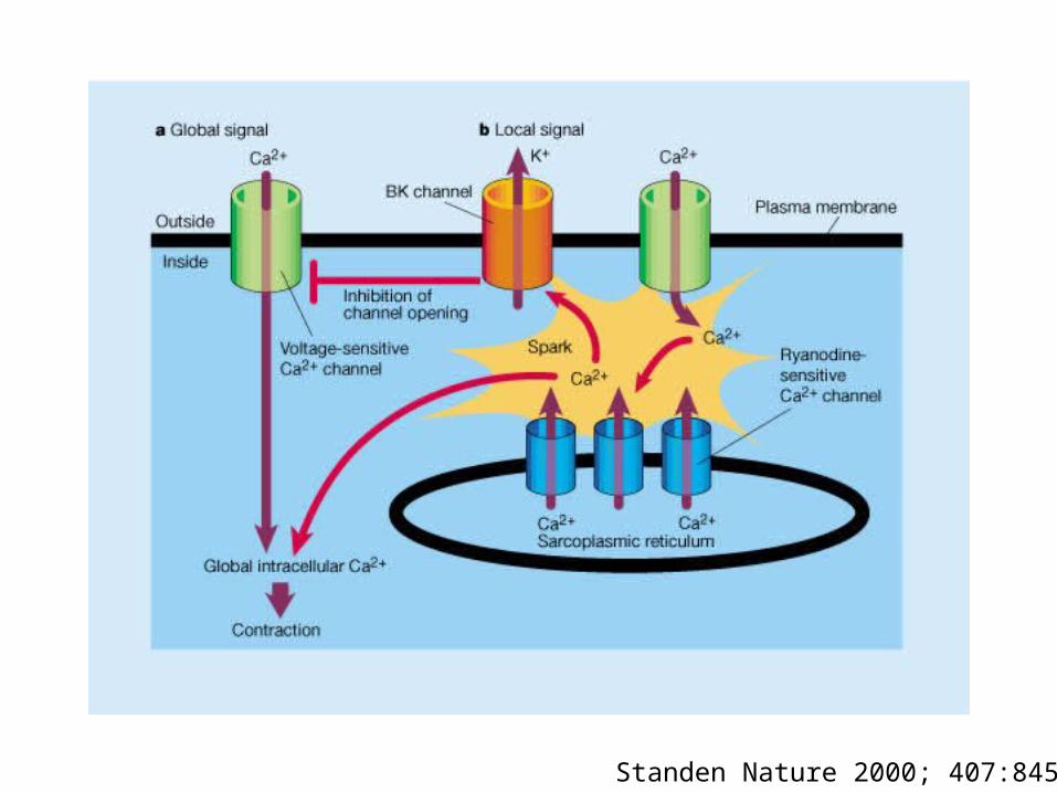
Standen Nature 2000; 407:845
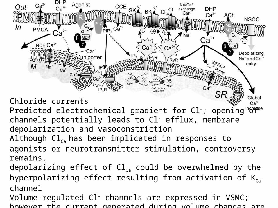
Chloride currentsPredicted electrochemical gradient for Cl-; opening of channels potentially leads to Cl- efflux, membrane depolarization and vasoconstrictionAlthough ClCa has been implicated in responses to agonists or neurotransmitter stimulation, controversy remains.depolarizing effect of ClCa could be overwhelmed by the hyperpolarizing effect resulting from activation of KCa channelVolume-regulated Cl- channels are expressed in VSMC; however the current generated during volume changes are not pharmacologically identified as Cl-; therefore, the role for Cl- channels in regulating myogenic tone requires further research.
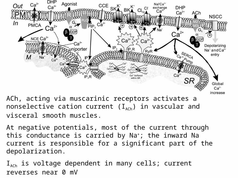
ACh, acting via muscarinic receptors activates a nonselective cation current (IACh) in vascular and visceral smooth muscles.
At negative potentials, most of the current through this conductance is carried by Na+; the inward Na current is responsible for a significant part of the depolarization.
IACh is voltage dependent in many cells; current reverses near 0 mV
IACh is regulated by G proteins, and activation of IACh is blocked by pertussis toxin
Unclear whether the conductance is a significant source of Ca2+
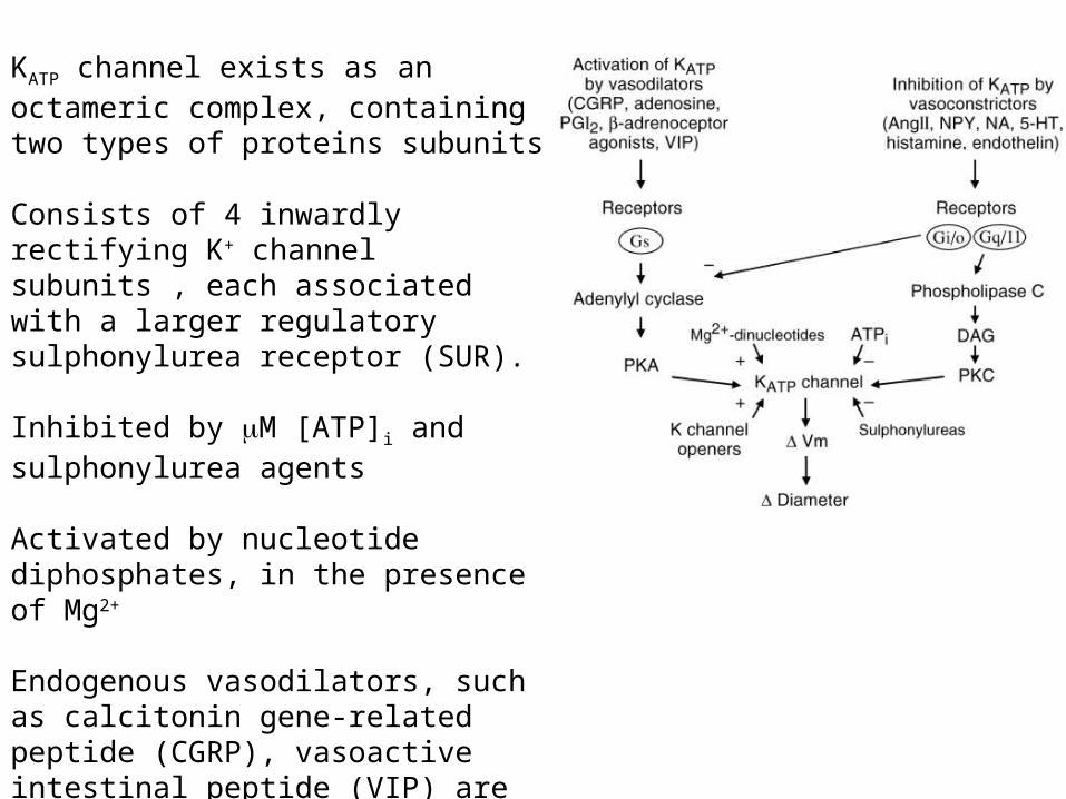
KATP channel exists as an octameric complex, containing two types of proteins subunits
Consists of 4 inwardly rectifying K+ channel subunits , each associated with a larger regulatory sulphonylurea receptor (SUR).
Inhibited by M [ATP]i and sulphonylurea agents
Activated by nucleotide diphosphates, in the presence of Mg2+
Endogenous vasodilators, such as calcitonin gene-related peptide (CGRP), vasoactive intestinal peptide (VIP) are mediated through PKA mediated activity of KATP channels.
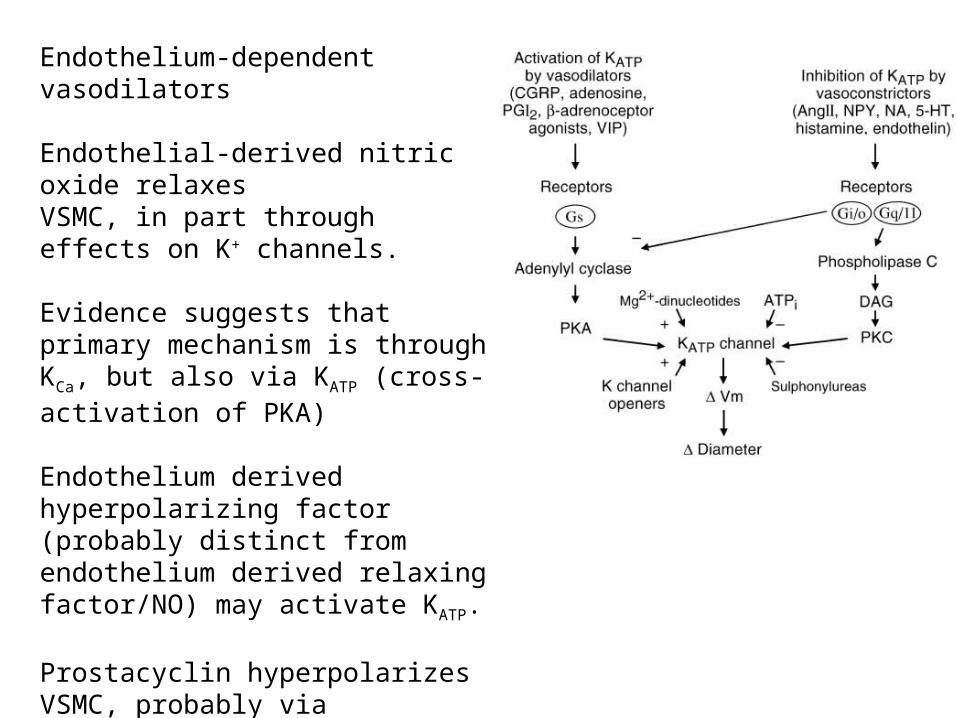
Endothelium-dependent vasodilators
Endothelial-derived nitric oxide relaxes VSMC, in part through effects on K+ channels.
Evidence suggests that primary mechanism is through KCa, but also via KATP (cross-activation of PKA)
Endothelium derived hyperpolarizing factor (probably distinct from endothelium derived relaxing factor/NO) may activate KATP.
Prostacyclin hyperpolarizes VSMC, probably via activation of KATP.
Adenosine activates KATP probably via activation of PKA
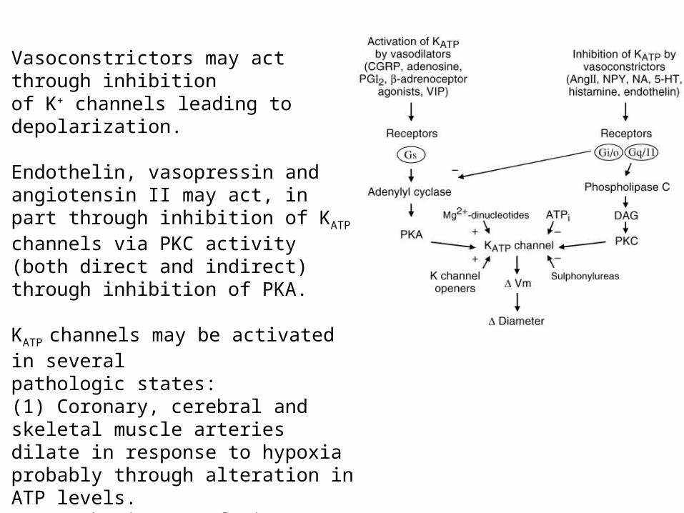
Vasoconstrictors may act through inhibitionof K+ channels leading to depolarization.
Endothelin, vasopressin and angiotensin II may act, in part through inhibition of KATP channels via PKC activity (both direct and indirect) through inhibition of PKA.
KATP channels may be activated in severalpathologic states:(1) Coronary, cerebral and skeletal muscle arteries dilate in response to hypoxia probably through alteration in ATP levels.(2) Ischemia/reperfusion: Reactive hyperemia may cause increased adenosine(3) Acidosis activates KATP
(4) Endotoxins and septic shock can activate KATP