waxing skin final
TRANSCRIPT

Clare Hargreaves-Norris
Structure of the skin
NVQ Level 2 Beauty Therapy
Clare Hargreaves-Norris
Layers of the skin
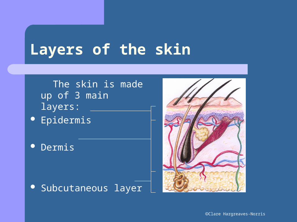
The skin is made up of 3 main layers:
Epidermis
Dermis
Subcutaneous layer
Clare Hargreaves-Norris
Epidermis
This is the outermost layer of the skin (the part that you can see), it does not have a blood supply of its own and is made up of 5 layers:
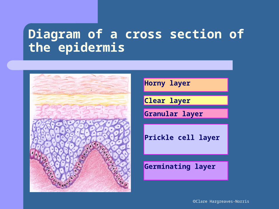
Horny layer – the outer layer of the skin, this is made up of scale like cells which are continuously shed (corn flakes).
Clear layer- this is made up of small transparent cells through which light can pass. This layer is only present in the palms of the hands and soles of the feet.
Granular layer – this layer is usually 1-3 layers thick. The cells have distinct granules and keratin is produced in this layer.
Prickle cell layer – this layer is 3-6 layers thick and the cells are constantly dividing.
Germinating layer – a single basal layer of cells, that contain the pigment melanin. The cells of the epidermis are produced in this layer and have a distinct nuclei. These cells divide continuously by a process known as mitosis.
Clare Hargreaves-Norris
Diagram of a cross section of the epidermis
Horny layer
Clear layer
Granular layer
Prickle cell layer
Germinating layer
Clare Hargreaves-Norris
Dermis
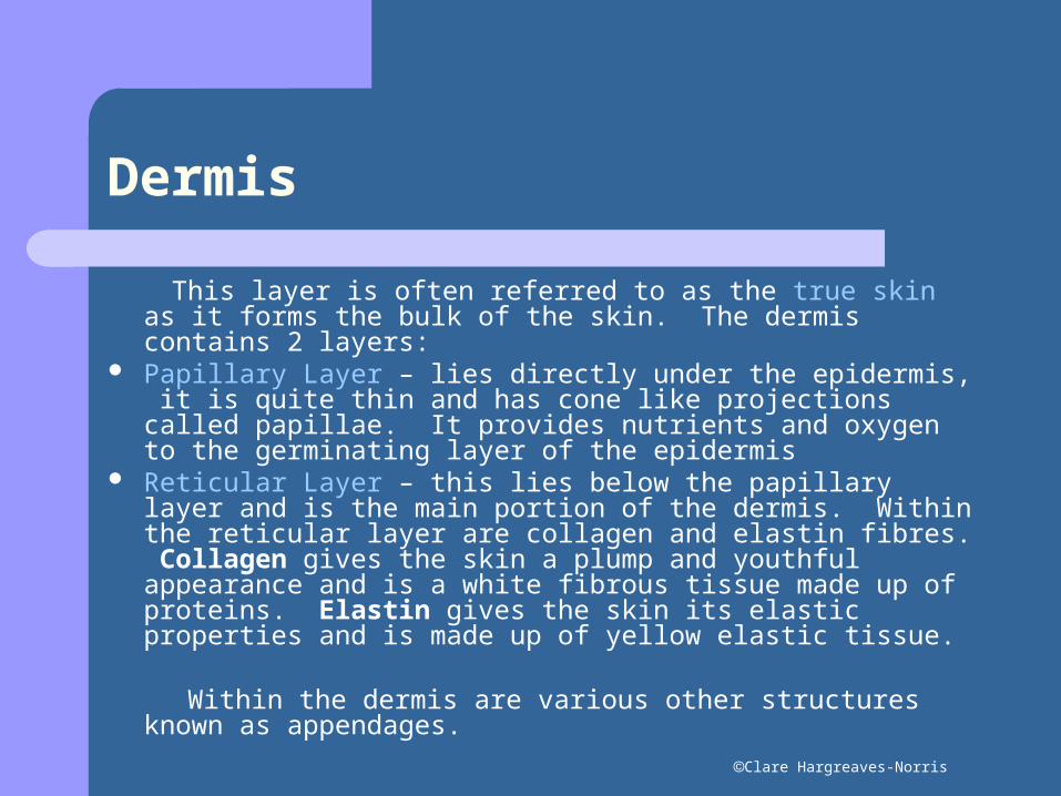
This layer is often referred to as the true skin as it forms the bulk of the skin. The dermis contains 2 layers:
Papillary Layer – lies directly under the epidermis, it is quite thin and has cone like projections called papillae. It provides nutrients and oxygen to the germinating layer of the epidermis
Reticular Layer – this lies below the papillary layer and is the main portion of the dermis. Within the reticular layer are collagen and elastin fibres. Collagen gives the skin a plump and youthful appearance and is a white fibrous tissue made up of proteins. Elastin gives the skin its elastic properties and is made up of yellow elastic tissue.
Within the dermis are various other structures known as appendages.
Clare Hargreaves-Norris
Subcutaneous layer
This is located under the dermis and is mainly made up of fat cells (adipose tissue). This fatty layer of the skin provides the plump contours of the body, protection, insulation, support and a food supply if needed.
Clare Hargreaves-Norris
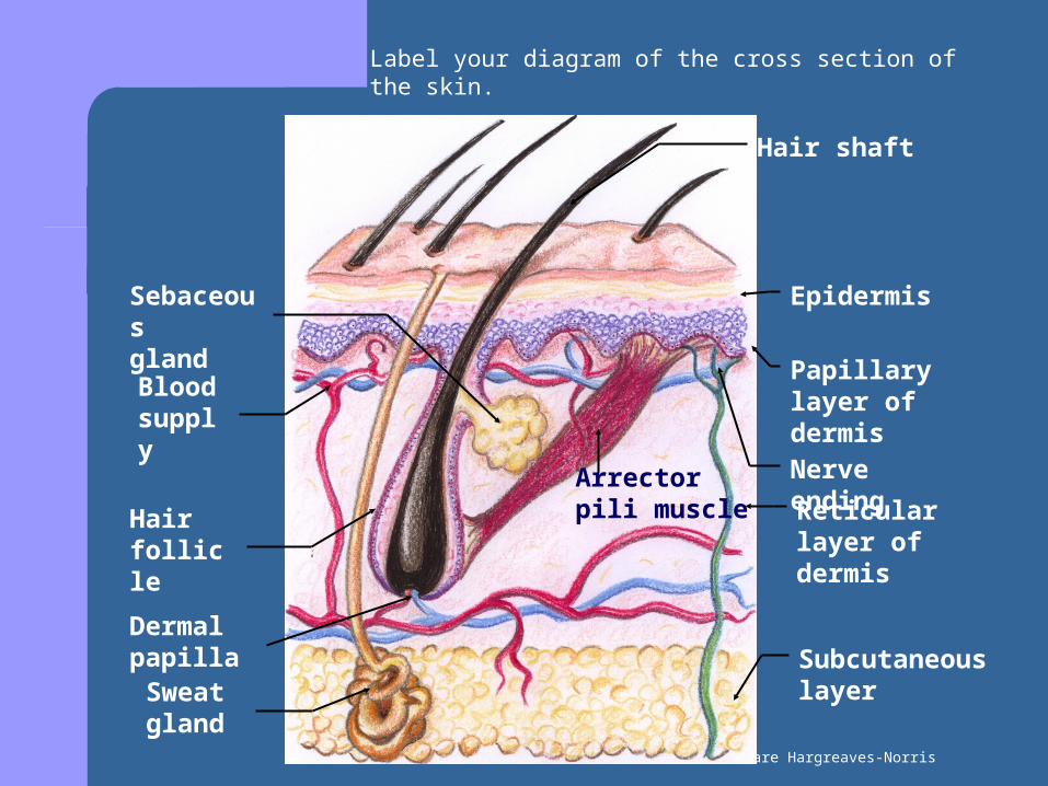
Papillary layer of dermis
Sebaceous gland
Nerve ending
Hair follicle
Arrector pili muscle
Subcutaneous layer
Reticular layer of dermis
Sweat gland
Dermal papilla
Blood supply
Hair shaft
Epidermis
Label your diagram of the cross section of the skin.
Clare Hargreaves-Norris
Lets take a closer look at the appendages of the skin.
Clare Hargreaves-Norris
Sweat glands
The sweat glands, which are sometimes referred to as the suderiferous glands, extend from the epidermis into the dermis and are found all over the body, however they are particularly numerous in the palms of the hands and the soles of the feet They regulate the body temperature by allowing sweat to evaporate from the body.
There are two different types of sweat glands: Eccrine glands – are found all over the body and
secrete a watery fluid. Apocrine glands – are found in the underarms, nipple
and groin area and become active at puberty. They secrete a thicker fluid that contains urea, fats, sugars and a small amount of protein. Body odour can occur when bacteria starts to break down the sweat.

Clare Hargreaves-Norris
Hair follicle
The hair follicle is an indentation of the epidermis with it’s walls forming a protective layer that houses the hair.
Clare Hargreaves-Norris
Hair shaft
The hair shaft is the portion of the hair that lies above the skins surface. It is the visible part that you see such as the eyebrows, eyelashes, body hair and the hair on the head.
Clare Hargreaves-Norris
Blood supply
Blood is supplied to the skin by small blood vessels known as blood capillaries. All parts of our body require a blood supply as it provides the vital oxygen and nutrients that tissues require in order to survive. The capillaries also remove waste products and toxins from the tissues. In addition, the capillaries also help to maintain the body temperature by dilating (widening) and constricting (narrowing).
Clare Hargreaves-Norris
Dermal papilla
The dermal papilla is the blood supply for the hair and its follicle. It provides food and oxygen, which are essential for the growth of the hair. The dermal papilla is a separate organ that serves the follicle; it is not part of the hair.
Clare Hargreaves-Norris
Arrector Pili Muscle
This muscle is attached to the hair follicle and it contracts when you are cold or frightened causing the hair to stand up on end. This action traps a layer of warm air around the body to keep the body warm. When the muscles contracts it pulls on the skin around the follicle opening therefore producing goose pimples
Clare Hargreaves-Norris
Sebaceous glands
These glands are found all over the body except for the palms of the hands and the soles of the feet and produce the natural oil of the skin - sebum. The sebaceous glands become more active at puberty due to the increased levels of androgens (male hormone) being produced. Men generally secrete more sebum then women, and you will usually note that the sebaceous glands become less active as we get older.
Sebum is bactericidal and fungicidal and so prevents against infection, it also provides protection and prevents the skin from drying out.

Clare Hargreaves-Norris
Nerve endings
Sensory nerve endings are found in the skin and detect changes in the environment such as heat, cold, touch, pain and pressure. There are different nerve endings that detect the different sensations. Sensory nerve endings send messages to our brain to let us know what we are feeling.
Motor nerves bring messages from our brain to bring about a response.