water balance in the eggs of the atlantic salmon salmo...
TRANSCRIPT

J. Exp. Biol. (1969), 50, 223-337 2 2 3With 6 text-figures
Printed in Great Britain
WATER BALANCE IN THE EGGS OF THEATLANTIC SALMON SALMO SALAR
BY W. T. W. POTTS AND P. P. RUDY, JR.*
Department of Biological Sciences, University of Lancaster,St Leonard's House, Lancaster, England
(Received 27 June 1968)
INTRODUCTION
The osmotic and ionic properties of the eggs of trout and salmon have attracted theinterest of physiologists for many years. The eggs are convenient biological materialand immense numbers of large, almost identical eggs can readily be obtained. Thevery slow development of the eggs raises in an acute form the ionic and osmoticproblems common to the eggs of all freshwater animals.
When shed into isotonic saline, eggs of salmon and trout maintain" an almost constantweight and a relatively high permeability to water. When shed into fresh water theeggs become almost impermeable to water but increase in weight by about i6%",during the first 2 hr., as the result of the formation of a perivitelline fluid beneath thechorion (Bogucki, 1930). Eggs in this impermeable state are described as 'water-hardened'. The decline in permeability to water is due to a change in the propertiesof the vitelline membrane (Krogh & Ussing, 1937), the outer chorion remainingpermeable to water even in the water-hardened condition (Gray, 1932). Krogh& Ussing (1937), as the result of experiments with deuterium oxide, decided that thepermeability of the vitelline membrane became immeasurably low. More recently,Kalman (1959), using tritiated water, showed that there was a small exchange of waterbetween the yolk and the surrounding media even in the water-hardened eggs of thetrout, Salmo gairdneri, but no estimate of the permeability was made. The formationof the perivitelline fluid is inhibited in a saline solution but the perivitelline fluid isformed when the eggs are shed into isotonic urea or glucose solutions (Bogucki, 1930).In this paper we describe the uptake and exchange of water in the eggs of Salmo salarwhen shed into a variety of media.
METHODS
Eggs were obtained from salmon removed from the River Lune at Broad Raine,Westmorland, in November 1966 and November 1967. Eggs and milt were strippedinto plastic bowls, previously rinsed in salmon Ringer. Every effort was taken to avoidcontamination of the eggs with river water which could initiate 'water-hardening' butit is possible that during the stripping small amounts of river water were occasionallyintroduced. The majority of experiments were carried out with fertilized eggs, but forcomparative purposes a few experiments were carried out with unfertilized eggs. Thetemperature of the river water at the time of the experiments was 3-5° C. The experi-
• Present address: Oregon Institute of Marine Biology, Charleston, Oregon 97420, U.S.A.
224 W. T. W. POTTS AND P. P. RUDY, JR.
ments were continued in a water-bath controlled at this temperature. A sample ofRiver Lune water collected the previous year contained 0-45 mM-Ca/1. and o-nmM-Mg/1. The salmon Ringer was modified from that of Holmes & Stott (i960) andcontained rather more calcium and less phosphate in order to bring the minor con-stituents more into line with the analysis of the coelomic fluid of 5. salar by Hayes,Darcy & Sullivan (1946).* The glucose solution contained 320 mM glucose/1. Experi-ments were also carried out in a variety of concentrations of sodium and calcium ions(Fig. 4). All experiments with tritiated water were carried out in solutions containing1 /iC. of tritium per ml. of solution.
Freshly fertilized eggs are contaminated with considerable volumes of coelomicfluid and semen. At the beginning of experiments involving dilute solutions the eggswere first rinsed rapidly in a large volume of a similar, non-tritiated solution. The eggswere then transferred to a plastic vessel containing the experimental radioactivesolution. Samples of fifty eggs were removed at intervals, blotted dry with tissue paperand transferred to weighed containers. Water was removed from the eggs for tritiumassay by a freeze-drying process described previously (Rudy, 1967). Activity wasmeasured with a Nuclear Enterprises Liquid Scintillation Counter using NS 220scintillation fluid. Water contents were obtained by drying the eggs to constant weightat ioo° C. Calcium and magnesium analyses were carried out by atomic absorptionspectroscopy.
Calculation of the exchange constant of water within the vitelline membrane
It is not practicable to calculate the exact permeability of the vitelline membrane inabsolute terms because the surface area of the vitelline membrane changes continuouslyduring development and it is difficult to estimate this area exactly. Estimates of thediameter of the yolk sac made with the chorion intact would be subject to error whichwould be further compounded when calculating the area. It is difficult to remove thechorion from the egg without breaking the vitelline membrane. Instead, the rateconstant of water exchange across the vitelline membrane has been calculated. To afirst approximation this will be proportional to the permeability of the vitellinemembrane.
After transfer to dilute solutions the eggs gain weight as the result of the formationof the perivitelline fluid. The water in the egg is distributed between the chorion, theperivitelline fluid and the yolk. In order to calculate the activity present in the yolk,the activity present in the chorion and in the perivitelline fluid must first be allowedfor. From the increase in the weight of the egg, the quantity of water in the perivitellinespace can be calculated. Determination of the water content of isolated chorions showthat the water content of the chorion alone is 3-0 mg., equivalent to 6% of the totalwater content of freshly stripped eggs. Freshly stripped eggs, removed from the loadingsolution before any significant perivitelline fluid had been formed, had a specificactivity of 5—10% that of the loading solution, probably corresponding to water in the
• The salmon Ringer was prepared by dissolving the following constituents in 1 1.: sodium chloride8-5 g.: potassium chloride 0-4 g.: calcium chloride 0-5 g.: disodium hydrogen phosphate 02 g.: sodiumdihydrogen phosphate 005 g.: sodium bicarbonate o-i g.: and magnesium sulphate C15 g. The finalconcentrations were 150 m-equiv. sodium/1., 151 -o m-equiv. chloride/1., 5-4m-equiv. potassium/1.,4-5 m-equiv. calcium/1., 1-25 m-equiv magnesium/1., 1-25 m-equiv. sulphate/1, and z-o m-equiv.phosphate/1. The total osmotic concentration was 330 m-osmole/1.

Water balance in the eggs of the Atlantic salmon Salmo salar 225
chorion, together with any pre-existing perivitelline fluid and surface contamination.Since the perivitelline fluid is formed by the uptake of water from the external media,and since when fully formed it continues to exchange both water and sodium rapidlywith the external medium (see below) it may be assumed that the specific activity ofthe water in both the chorion and the perivitelline fluid approximates to that of themedium. By using the following formula the specific activity of the water within theyolk can be calculated. If A is the specific activity of the loading solution, Wthe initialwater content of the egg in mg., excluding the chorion, w the increase in the weightof the egg in mg., B the specific activity of the yolk water, then
mean specific activity of egg water = - = .
It assumed that the water content of the yolk does not increase significantly during theexperiment. Calculations showed that net water influx into the yolk will be negligibleduring the first few hours. In the period immediately following the transfer to theradioactive solution by far the greater part of the activity will be confined to thechorion and the perivitelline fluid. Any error in the allowance made for these twocompartments will therefore produce a relatively large error in the calculated specificactivity of the yolk. This error will be largest in the first time interval when the specificactivity of the yolk is very low. However, estimates of egg weights are very precise.The average weights of single eggs from the three fishes used in these experimentswere 86-8., 93*6 and 83-9 mg., but the variation amongst eggs from individual fishwas very small, ca. 1 %. Similarly, in the trout, Salmo gcdrdneri, the average variationfrom the mean weight between eggs from different fishes was 10% but the averagevariation from the mean in eggs from a single fish was only 0*5 % (Manery & Irving,
1934)-From the change in the specific activity of the water in the yolk the exchange constant
for yolk water can be calculated for each time interval from the equation
where Bx and B% are the specific activity of the yolk on two occasions at time T apart.K is the fraction of yolk water exchanging per unit time. Once again any errors arecompounded in calculating K. The osmotic flux will be only a small fraction of thegross flux. K will be proportional to the permeability of the vitelline membrane foryolk of constant volume.
RESULTS
Each experiment was confined to eggs from a single fish. Figure 1 shows the changein water content of unfertilized eggs from one particular fish after stripping into riverwater. The initial mean weight of the eggs was 93-6 mg. Rapid water uptake wascompleted in the first 3 hr. This was followed by a very slow increase in the weight ofthe egg, due mainly to water entering the yolk. The chorion water remained constantand there was a slight decline in the perivitelline fluid, as the yolk expanded into theperivitelline space. The perivitelline space, together with the chorion was estimated inthe 50-day-old eggs by tritiated water exchange (see below). After 50 days the water
13 Exp. Biol. 50, 1

226 W. T. W. POTTS AND P. P. RUDY, JR.
content of the egg had increased by more than a third. No consistent differences in theproperties of fertilized and unfertilized eggs were detected in eggs up to a few daysold, but the following data refer to fertilized eggs unless otherwise stated.
160
140
120
100
80
60
40
-
-
• • * f '- jr"*""^ Perivitelline
^Chorion
Yolk water
Solids
• i i i i
-
-
_
-
2 3time (hr.)
5 50Days
Fig. i. Changes in the weight and the distribution of water in an unfertilized egg of Salmo salarwhen stripped into Lune river water. First 5 hr. 3 •4° C, later period io° C.
60Time (min.)
Fig. 2. Tritiated water exchange in eggs of Salmo salar. • , Freshly stripped fertilized eggs inLune river water. Oi Freshly stripped egg in salmon saline. A, Water-hardened eggs in Luneriver water. 3-4° C.
Water balance in the eggs of the Atlantic salmon Salmo salar 227
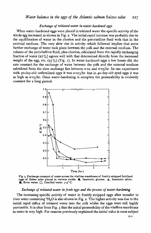
Exchange of tritiated water in water-hardened eggs
When water-hardened eggs were placed in tritiated water the specific activity of thewhole egg increased as shown in Fig. 2. The initial rapid increase was probably due tothe equilibration of water in the chorion and the perivitelline fluid with that in theexternal medium. The very slow rise in activity which followed implies that somefurther exchange of water took place between the yolk and the external medium. Thevolume of the perivitelline fluid, plus chorion, calculated from the rapidly exchangingfraction of water (27 %) agrees well with that determined directly from the increasedweight of the egg, etc. (25%) (Fig. 1). In water-hardened eggs a few hours old, therate constant,for the exchange of water between the yolk and the external mediumcalculated from the slow exchange lies between 0-02 and o-o^jhs. In one experimentwith 50-day-old unfertilized eggs it was o-o23/hr. but in 40-day-old eyed eggs it wasas high as o-o5/hr. Once water-hardening is complete the permeability is evidentlyconstant for a long period.
iO-5
0-3 -
0-2 -
0-1 -
Time (hr.)
Fig. 3. Exchange constant of water across the vitelline membrane of freshly stripped fertilizedeggs of Salmo salar placed in various media. # , Isosmotic glucose. A, Isosmotic saline.• , River water. D, Distilled water. 3-4° C.
Exchange of tritiated water in fresh eggs and the process of water-hardening
The increasing specific activity of water in freshly stripped eggs after transfer toriver water containing 3H2O is also shown in Fig. 2. The higher activity was due to theinitial rapid influx of tritiated water into the yolk whilst the eggs were'still highlypermeable. It is clear from Fig. 3 that the initial permeability of the vitelline membraneto water is very high. For reasons previously explained the initial value is most subject
228 W. T. W. POTTS AND P. P. RUDY, JR.
to error. However, even if an error is made in the initial assumptions this will havelittle effect on successive values of K which are based on the changes in weight and onthe specific activity of the whole egg. The permeability begins to decline almost atonce, and continues to decline for 7 or 8 hours. After that it remains virtually constantfor 50 days in unfertilized eggs.
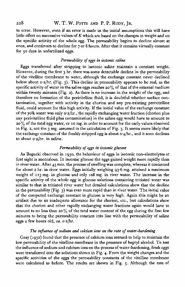
Permeability of eggs in isotonic saline
Eggs transferred after stripping to isotonic saline maintain a constant weight.However, during the first 3 hr. there was some detectable decline in the permeabilityof the vitelline membrane to water, although the exchange constant never declinedbelow about o-2/hr. (Fig. 3). This decline in permeability appears to be real, as thespecific activity of water in the saline eggs reaches 20 % of that of the external mediumwithin twenty minutes (Fig. 2). As there is no increase in the weight of the egg, andtherefore no formation of the perivitelline fluid, it is doubtful whether surface con-tamination, together with activity in the chorion and any pre-existing perivitellinefluid, could account for this high activity. If the initial value of the exchange constantof the yolk water was only o-2/hr., the rapidly exchanging water fraction (chorion plusany perivitelline fluid plus contamination) in the saline egg would have to amount to20% of the total egg water, or 10 mg. in order to account for the early values observedin Fig. 2, not the 3 mg. assumed in the calculation of Fig. 3. It seems more likely thatthe exchange constant of the freshly stripped egg is about o-4/hr., and it soon declinesto about o-2/hr. in saline.
Permeability of eggs in isotonic glucose
As Bogucki observed in 1930, the behaviour of eggs in isotonic non-electrolytes atfirst sight is anomalous. In isotonic glucose the eggs gained weight more rapidly thanin river water. After 45 min. the process of swelling was complete, whereas it continuedfor about 2 hr. in river water. Eggs initially weighing 93-6 mg. attained a maximumweight of 113 mg. in glucose and only 108 mg. in river water. The increase in thespecific activity of the whole egg in glucose solutions containing tritiated water wassimilar to that in tritiated river water but detailed calculations show that the declinein the permeability (Fig. 3) was even more rapid than in river water. The initial valueof the computed exchange constant in glucose is very high. Again this might be anartifact due to an inadequate allowance for the chorion, etc., but calculations showthat the chorion and other rapidly exchanging water fractions again would have toamount to no less than 20 % of the total water content of the egg during the first fewminutes to bring the permeability constant into line with the permeability of salineeggs a few hours old, ca. o-2/hr.
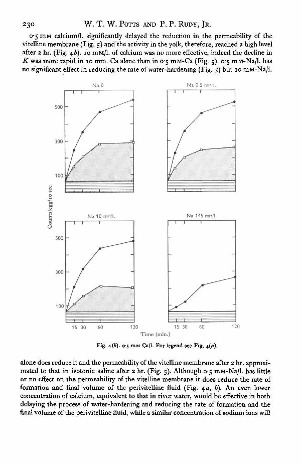
The influence of sodium and calcium ions on the rate of water-hardening
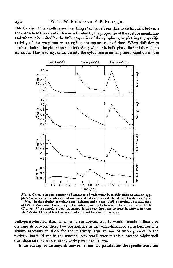
Gray (1932) found that the presence of calcium ions seemed to help to maintain thelow permeability of the vitelline membrane in the presence of heptyl alcohol. To testthe influence of sodium and calcium ions on the process of water-hardening, fresh eggswere transferred into the solutions shown in Fig. 4. From the weight changes and thespecific activities of the eggs the permeability constants of the vitelline membranewere calculated as before. The results are shown in Fig. 5. Although the rate of

Water balance in the eggs of the Atlantic salmon Salmo salar 229
increase in weight was similar in many of the dilute solutions, analyses showed thatthe behaviour of the eggs differed markedly in the different media. The formation ofthe perivitelline fluid was most rapid and most extensive in distilled water (Fig. 4a)but water-hardening, as shown by the decline in K, was also the most rapid in thismedium (Fig. 5). The activity in the yolk therefore remained low. In the presence of145 mM-Na/1. the formation of perivitelline fluid was completely inhibited and water-
soo
300
100
1 1
- /
NaO1
^ ^
f
-
Na 0 5 mM I.
Na 10 mMl. Na 145 mM/l.
500 -
300 -
100
15 30 60120 15 30Time (min.)
Fig. 4 (a). No Ca.
Fig. 4. Distribution of activity in the water compartments in eggs of Salmo salar when placedin solutions containing tritiated water and various concentrations of sodium and chloride iona.Activity in chorion g . Activity in perivitelline fluid @. Activity in yolk water D. 3 4° C.
hardening was delayed. The activity in the yolk reached a higher level after 2 hr. andwas continuing to increase. A low concentration of calcium, 0-5 mM/l., reduced boththe rate of formation and the final volume of the perivitelline fluid (Fig. 4*). A mediumwith 10 mM/l. of calcium delayed the formation of the perivitelline fluid even further,but 10 mM/l. of sodium alone had less effect (Fig. 4a).
23d W. T. W. POTTS AND P. P. RUDY, JR.
o*5 mM calcium/1, significantly delayed the reduction in the permeability of thevitelline membrane (Fig. 5) and the activity in the yolk, therefore, reached a high levelafter 2 hr. (Fig. 46). 10 mM/1. of calcium was no more effective, indeed the decline ini£was more rapid in 10 mm. Ca alone than in 0-5 mM-Ca (Fig. 5). 0-5 mM-Na/1. hasno significant effect in reducing the rate of water-hardening (Fig. 5) but 10 mM-Na/1.
Na 0 Na 0 5 mM. I.
Na 145 mM/l.
100
15 30 60 120 15 30 60 120
Time (min.)
Fig. 4(6). 0'5 mM Ca/1. For legend 8ee Fig. 4(0).
alone does reduce it and the permeability of the vitelline membrane after 2 hr. approxi-mated to that in isotonic saline after 2 hr. (Fig. 5). Although 0-5 mM-Na/1. has littleor no effect on the permeability of the vitelline membrane it does reduce the rate offormation and final volume of the perivitelline fluid (Fig. 4a, b). An even lowerconcentration of calcium, equivalent to that in river water, would be effective in bothdelaying the process of water-hardening and reducing the rate of formation and thefinal volume of the perivitelline fluid, while a similar concentration of sodium ions will
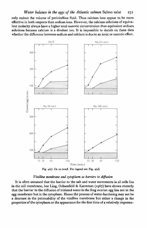
Water balance in the eggs of the Atlantic salmon Salmo salar 231
only reduce the volume of perivitelline fluid. Thus calcium ions appear to be moreeffective in both respects than sodium ions. However, the calcium solutions of equiva-lent molarity always have a higher total osmotic concentration than equivalent sodiumsolutions because calcium is a divalent ion. It is impossible to decide on these datawhether the difference between sodium and calcium is due to an ionic or osmotic effect.
Na 0 Na 0 5 mn I
500 -
Na 145 mM,
15 30 60 120 15 30 60 120
Time (min.)
Fig. 4(c). Ca 10 DIM/1. For legend see Fig. 4(a).
Vitelline membrane and cytoplasm as barriers to diffusion
It is often assumed that the barrier to the salt and water movements in all cells liesin the cell membrane, but Ling, Ochsenfeld & Karreman (1967) have shown recentlythat the barrier to the diffusion of tritiated water in the frog ovarian egg lies not in theegg membrane but in the cytoplasm. Hence the process of water-hardening may not bea decrease in the permeability of the vitelline membrane but either a change in theproperties of the cytoplasm or the appearance for the first time of a relatively imperme-
232 W. T. W. POTTS AND P. P. RUDY, JR.
able barrier at the vitelline surface. Ling et al. have been able to distinguish betweenthe case where the rate of diffusion is limited by the properties of the surface membraneand where it is limited by the bulk properties of the cytoplasm, by plotting the specificactivity of the cytoplasm water against the square root of time. When diffusion issurface-limited the plot shows an inflexion; when it is bulk-phase-limited there is noinflexion. That is to say, diffusion into the cytoplasm is initially more rapid when it is
Ca omM/1. Ca 0-5 ITIM/1. Caiomin/l.
1 0
^ 0 8
=5-06
* < M
0-2
12
10
<r- o-8
0-4
02
1 2
~ 1 0
* 0 8txS 0-6
0402
- . 0 6io-4^ 02
0-5 1-0 1-5 0-5 10 15Time (hr.)
0-5 1 0 1 5 2
Fig. 5. Changes in rate constant of exchange of yolk water in freshly stripped salmon eggsplaced in various concentrations of sodium and chloride ions calculated from the data in Fig. 4.
Note: In the solution containing zero calcium and 0-5 mM-Na/1, a fortuitous accumulationof small errors caused the activity in the yolk apparently to decrease between 30 min. and 1 h.(Fig. 4a). K has therefore been calculated in this case from the increase in activity between30 min. and 2 hr. and has been assumed constant between these times.
bulk-phase-limited than when it is surface-limited. It would remain difficult todistinguish between these two possibilities in the water-hardened state because it isalways necessary to allow for the relatively large volume of water present in theperi vitelline fluid and in the chorion. Any small error in this allowance might wellintroduce an inflection into the early part of the curve.
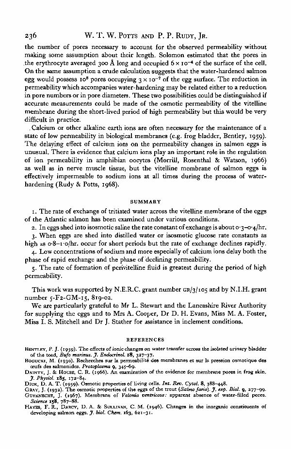
In an attempt to distinguish between these two possibilities the specific activities
Water balance in the eggs of the Atlantic salmon Salmo salar 233
of the egg water at different times were measured in eggs in saline (Fig. 6 a). The firstsection of the graph approximates to a straight line, and the slight rise at the first pointmay be due to surface contamination. However, the vitelline membrane lies beneaththe chorion some distance inside the surface of the egg. Diffusion into the egg in thefirst 2 or 3 min., mainly into the chorion, will therefore be bulk-phase-limited. Thechorion accounts for 6% of the whole egg water. When allowances are made for thischorionic water the influx curve of the tritiated water into the yolk shows a distinctinflexion, implying that the vitelline membrane is a barrier to diffusion (Fig. 6b),even in the freshly stripped egg.
20 0 4Time (min.)
Fig. 6. Increase of specific activity of water with time in freshly stripped eggs in saline.(a) Whole eggs • , (6) Yolk water O. 3-4° C.
DISCUSSION
Permeability of the viteUine membrane
Although Zotin (1965) suggests that the low rate of exchange of water in salmoneggs may be due to a peculiarity of the physical state of the water the present resultsare most easily interpreted in terms of a change in the permeability of the viteUinemembrane as originally suggested by Gray. It is difficult to assess the permeabilityof the egg when it is in the body cavity. The saline solution was intended to replace thecoelomic fluid of the fish. When the egg is transferred directly into the saline solution,the exchange constant of the egg is initially about o^/hr., but even in the saline after2 hr. it drops to about o-2/hr. This decline indicates that the saline is not exactlyequivalent to the coelomic fluids of the fish. When transferred to de-ionized water orinto salt-free glucose or dilute solutions of sodium or calcium ions the exchangeconstant (K) rises as high as i-o/hr., but then declines rapidly to about o-i or 0-2 after2 hr. and eventually to about 0025 after 10 hr. While it is possible that the exchangeconstant of eggs in the body cavity of the fish is normally as high as o-6-i-o/hr., the

234 w - T. W. POTTS AND P. P. RUDY, JR.
results of the experiment with calcium solutions suggest that this is not the case. Wheneggs are transferred into solutions containing little or no sodium but io mM of calcium(Fig. 5), the value of K during the first 15 min. is about 0-4—o-5/hr., but then rises to ahigh level of 1 -o/hr. before declining again. This suggests that the resting value of K isabout O'4/hr., and that transfer into dilute solutions causes an initial increase of thepermeability before it finally declines. Calcium ions appear to delay the onset of thestage of high permeability for a short time while high sodium concentrations delaythe onset indefinitely. If the activity within the chorion, external to the vitellinemembrane, is over-estimated this will produce a spuriously low value for K duringthe first period of time. While the low value of K during the first period of time incalcium solutions may be an artifact due to some error in estimating the activityexternal to the vitelline membrane, it is noteworthy that the formation of perivitellinefluid is also delayed by the presence of 10 mM of calcium (Fig. 4c). The formation ofthe perivitelline fluid is assessed unambiguously from the changes in weight of theegg (Fig. 1). The decline in permeability is most rapid in distilled water, or in salt-freesolutions of glucose, or in solutions containing little sodium and no calcium. Thepermeability of the vitelline membrane is apparently greatest in solutions containing0-5-10 mM-Ca/1. and 0-10 mM of Na/1. Similar high values of K may be reached incalcium-free solutions during the first few minutes but the permeability may declineso rapidly that the mean permeability during the 15 min. is lower. In solutions con-taining 10 mM-Ca/1. highest permeability occurs between 15 and 30 min. aftertransfer to the solution. For reasons discussed above it is unlikely that these highvalues are a product of spurious assumptions. There is some evidence of a decline inpermeability in solutions containing 145 mM sodium but low calcium. The mostconstant permeability is maintained in the solutions containing high concentrationsof both sodium and calcium, but the exchange constant is only about o-3/hr.
Although the permeability is'relatively high before laying and during the process ofwater-hardening, in absolute terms it is always remarkably low, and falls to almostnegligible values once the egg is water-hardened. From the surface area of the vitellinemembrane, and from K, it can be calculated that the permeability of the egg beforeshedding is probably about 0-06 to /43//£2/sec, and even at its highest it does not riseabove o-2/i3//i2/sec* In water-hardened eggs the permeability is < 0-004/J3//i2/sec.In comparison the permeability of the surface of Amoeba is o-35//43//j2/sec. (Presscott& Zeuthen, 1953). The surface of the alder fly larva, Sialis lutaria, has a permeabilityof between 0-04 and 0-05 /*3//i2/sec. (Shaw, 1955). In contrast, ovarian eggs of thefrog have a permeability of as high as 89 /j3//t2/sec, but this declines to 1-2 /£3//£2/sec.before laying (Hevesy, Hofer & Krogh, 1935). In Valonia ventricosa permeability is2-4 /i3//i2/sec. (Gutknecht, 1967). The very low permeability of the salmon egg is nodoubt related to its prolonged development in fresh water. Even this low permeabilityto water results in a slow but continuous swelling of the yolk during development.In the unfertilized egg this is accompanied both by an increase in total volume of theegg, but also by a reduction in the volume of perivitelline fluid. In the course of 50days the volume of the water in the yolk increases by about 36 mg. or 0-072 mg. perday. Water-hardened eggs during the period contain on average 70 mg. of water whichexchanges with a rate constant of o-025/hr. The total water exchange is therefore
• See Appendix.

Water balance in the eggs of the Atlantic Salmon Salmo salar 235
42 mg./day. The estimated osmotic influx, calculated from the total water flux and theosmotic concentration of the yolk, is 0-019 mg. water/day. The real osmotic influx is0-72 mg./day or nearly four times greater than that calculated from the diffusion flux.This discrepancy probably indicates the presence of pores through which water flowsinto the yolk (Koefoed-Johnsen & Ussing, 1953), although some part of the discrep-ancy may be due to the presence of unstirred layers at the surface (Dainty & House,1966). In the fertilized egg the permeability to water increases again in the eyed stage,about 40 days, but is still very low in absolute terms: K is only o-O5/hr. This is twicethat for water-hardened unfertilized eggs of the same age but still very low by com-parison with most freshwater organisms. This increase may be due at least in part tothe increased surface area of the embryo rather than to increased permeability per se.
Formation of the perivitelline fluid
In the water-hardened egg the perivitelline fluid forms 22% of the weight. All thewater in the perivitelline fluid together with the capsule water exchanges within a fewminutes with the external medium (Fig. 2). Formation of the perivitelline fluid isalmost complete within 1 hr. of shedding into distilled water or into solutions con-taining little sodium; but in solutions of 145 mM-Na/1., approximately isotonic withthe body fluid, the formation of perivitelline fluid is completely suppressed, and theeggs then maintain a constant weight. However, when the eggs are shed into salt-freeisosmotic glucose solutions, the formation of the perivitelline fluid takes place morerapidly than in river water. Bogucki observed many years ago that perivitelline fluidappeared when eggs were shed into isosmotic solutions of urea or glucose. Theglucose solution is equivalent to distilled water for both perivitelline fluid formationand permeability reduction. Even 0-5 mM/1. of sodium or calcium slightly reduces therate of formation of the perivitelline fluid, while 10 mM calcium/1, markedly reduces it.Just as calcium delays the onset of the phase of high permeability, calcium also delaysthe onset of the formation of perivitelline fluid (Fig. 4c). The parallel between forma-tion of perivitelline fluid and the state of high permeability cannot be an artifact of theassumptions in the calculations. If the volume of perivitelline fluid were under-estimated at any time, this would produce a spuriously high value for the activitywithin the yolk water and hence of the permeability and vice versa.
The process of perivitelline fluid formation is obscure but the fluid exerts an appreci-able osmotic pressure and contains some substance which concentrates sodium ions(Rudy & Potts, 1968). Bogucki (1930) suggested that the yolk liberated a colloidalsubstance into the perivitelline space at the time of shedding which then took upwater to form the perivitelline fluid. In FunduUis heteroclitus the formation of theperivitelline fluid coincides with the disappearance of a layer of small clear vesiclesfrom immediately below the vitelline membrane (Kao & Chambers, 1954). It islikely that the phase of high permeability in Salmo salar is related to the release of theperivitelline colloid which must temporarily disrupt the integrity of the vitellinemembrane. Electron microscopy might elucidate this point.
From the ratio of the osmotic to the diffusional influx (Solomon, 1959) the dia-meter of the pores may be estimated at 6-3 A, similar to that in the erythrocyte.However, the osmotic permeability of the erythrocyte to water is 125 ^ /^ / sec .(Dick 1959), implying a much higher density of pores. It is not possible to calculate
236 W. T. W. POTTS AND P. P. RUDY, JR.
the number of pores necessary to account for the observed permeability withoutmaking some assumption about their length. Solomon estimated that the pores inthe erythrocyte averaged 300 A long and occupied 6 x io~* of the surface of the cell.On the same assumption a crude calculation suggests that the water-hardened salmonegg would possess io8 pores occupying 3 x io~7 of the egg surface. The reduction inpermeability which accompanies water-hardening may be related either to a reductionin pore numbers or in pore diameters. These two possibilities could be distinguished ifaccurate measurements could be made of the osmotic permeability of the vitellinemembrane during the short-lived period of high permeability but this would be verydifficult in practice.
. Calcium or other alkaline earth ions are often necessary for the maintenance of astate of low permeability in biological membranes (e.g. frog bladder, Bentley, 1959).The delaying effect of calcium ions on the permeability changes in salmon eggs isunusual. There is evidence that calcium ions play an important role in the regulationof ion permeability in amphibian oocytes (Morrill, Rosenthal & Watson, 1966)as well as in nerve muscle tissue, but the vitelline membrane of salmon eggs iseffectively impermeable to sodium ions at all times during the process of water-hardening (Rudy & Potts, 1968).
SUMMARY
1. The rate of exchange of tritiated water across the vitelline membrane of the eggsof the Atlantic salmon has been examined under various conditions.
2. In eggs shed into isosmotic saline the rate constant of exchange is about 0'3-0-4/hr.3. When eggs are shed into distilled water or isosmotic glucose rate constants as
high as o-8-i-o/hr. occur for short periods but the rate of exchange declines rapidly.4. Low concentrations of sodium and more especially of calcium ions delay both the
phase of rapid exchange and the phase of declining permeability.5. The rate of formation of perivitelline fluid is greatest during the period of high
permeability.
This work was supported by N.E.R.C. grant number GR/3/105 and by N.I.H. grantnumber 5-F2-GM-15, 819-02.
We are particularly grateful to Mr L. Stewart and the Lancashire River Authorityfor supplying the eggs and to Mrs A. Cooper, Dr D. H. Evans, Miss M. A. Foster,Miss I. S. Mitchell and Dr J. Stather for assistance in inclement conditions.
REFERENCES
BENTLEY, P. J. (1959). The effects of ionic changes on water transfer across the isolated urinary bladderof the toad, Bufo marinus. J. Endocrinol. 18, 327-37.
BOGUCKI, M. (1930). Recherches sur la permeabilite des membranes et sur la pression osmotique desoeufs des salmonides. Protoplasma 9, 345-69.
DAINTY, J. & HOUSE, C. R. (1966). An examination of the evidence for membrane pores in frog skin.J. Phytiol. 185, 172-84.
DICK, D. A. T. (1959). Osmotic properties of living cells. Int. Rev. Cytol. 8, 388-448.GRAY, J. (1932). The osmotic properties of the eggs of the trout (Sahno fario). J. exp. Biol. 9, 277-09.GUTKNECHT, J. (1967). Membrane of Valonia ventricosa: apparent absence of water-filled pores.
Science 158, 787-88.HAYES, F. R., DARCY, D. A. & SULLIVAN, C. M. (1946). Changes in the inorganic constituents of
developing salmon eggs. J. biol. Chem. 163, 621-31.
Water balance in the eggs of the Atlantic salmon Salmo salar 237HEVESEY, G., HOFER, E. & KROGH, A. (1935). The permeability of the akin of frogs to water as deter-
mined by D,0 and HtO. Skand. Arch. Phytiol. 7a, 109-214.HOLMES, W. N. and STOTT, G. H. (i960). Studies of the respiratory rates of excretory tissues in the
cutthroat trout, Salmo clarkii. Phytiol. Zool. 33, 9-20.KALMAN, S. M. (1959). Sodium and water exchange in the trout egg.J. cell. comp. Phytiol. 54, 155-62.KAO, C. Y. & CHAMBERS, R. (1954). The internal hydrostatic pressure of the fundulus egg. I. The
activated egg. J. exp. Biol. 31, 139-49.KOEFOED-JOHNSEN, V. & UssiNG, H. H. (1953). The contribution of diffusion flow to the passage of
DtO through living membranes. Ada Phytiol. Scand. a8, 60-76.KROGH, A. & USSING, H. H. (1937). A note on the permeability of trout eggs to D,O and H,O. J. exp.
Biol. 14, 35-37.LING, G. N., OCHSENFELD, M. M. & KARREMAN, G. (1967). Is the cell membrane a universal rate
limiting barrier to the movement of water between the living cell and its surrounding medium?J. gen. Phytiol. 50, 1807-20.
MANERY, J. F. & IRVING, L. (1934). Water changes in trout eggs at the time of laying. J. cell comp.phytiol. 5, 457-63.
MORRILL, G. A., ROSENTHAL, J. & WATSON, D. E. (1966). Membrane permeability changes in amphibianeggs at ovulatioru J. cell Phytiol. 67, 375—82.
PRESCOTT, D. M. & ZEUTHEN, E. (1953). Water diffusion and filtration across cell surfaces. Acta phytiol.Scand. 38, 77-94.
RUDY, P. P. (1967). Water permeability in selected decapod Crustacea. Comp. Biochem. Phytiol. 2a, 581-9.RUDY, P. P. & POTTS, W. T. W. (1968). Sodium balance in the eggs of the Atlantic salmon, Salmo talar.
J. exp. Biol. (in the Press).SHAW, J. (1955). The permeability and structure of the cuticle of the aquatic larva oiSialU lutaria.J. exp.
Biol. 32, 330-52.SOLOMON, A. K. (1959). Equivalent pore dimensions in cellular membranes. Proc. Nat. Biophytict.
Conf. 1st p. 314. Columbus, Ohio.ZOTTN, A. I. (1965). The uptake and movement of water in embryos. Symp. Soc. Exp. Biol. 19, 365-84-
APPENDIX
The absolute permeability of the vitelline membrane may be estimated as follows:It is assumed that the egg, without the chorion, weighs 90 mg., has a density of i-o
and contains 50 mg. of water.The radius (r) of the vitelline membrane is then 2-78 mm., as $ 77T3 = 90.The area of the vitelline membrane (477T2) is then 97-6 mm.2.If the rate constant of water exchange in freshly stripped eggs is o-ifh"1 then the
r, 1 M O-4 X CO
water flux/sq. mm./hr. = .Hence water flux in
0-4x50 1000/i3m2/sec. = • x —— = 0-057.r "^ ' 97-6 3600 Dl
Similarly, if the rate constant for water hardened eggs = O-O25A"1 the permeability= 0-003 5 /^8//t2/sec- a nd if the rate constant rises to i-oA"1 during the period of highpermeability then the absolute permeability on these assumptions = o-i4/w
s//t2/sec.