· david walker 3 mrcp, frca, fficm 1critical care department, ... therefore the decrease in...
TRANSCRIPT
1
Title
CRITICAL CARE ECHO ROUNDS: Haemodynamic instability
Authors
Ashraf Roshdy1
MBBch, MSc, MD, MRCP
Nadia Francisco2
MSc
Alejandro Rendon3 MSc
Stuart Gillon4 MbChB, MRCP, FFICM, PGDip*
David Walker3 MRCP, FRCA, FFICM
1Critical Care Department, Alexandria University, Alexandria, Egypt
2National Heart and Lung Institute (NHLI), Imperial College London, London, UK
3University College of London Hospitals, London, UK
4Critical Care Unit, King’s College Hospital, London, UK
(*S Gillon, PGDip in Perioperative and Critical Care Echocardiography)
Corresponding Author
Ashraf Roshdy MBBch, MSc, MD, MRCP
Address: Critical Care Department, Alexandria University, Faculty of Medicine, Alazarita
Alexandria, Egypt.
Email: [email protected]
Telephone: +201225194490
Page 1 of 21
2
Short title
Role of echocardiography in shock state
Key words:
Echocardiography, Echo, Critical, Intensive, Hemodynamic, Shock
Abstract
The use of echocardiography, whilst well established in cardiology, is a relatively new concept in
critical care medicine. However, in recent years echocardiography’s potential as both a diagnostic
tool and a form of advanced monitoring in the critically ill patient has been increasingly recognised.
In this series of Critical Care Echo Rounds, we will explore the role of echocardiography in critical
illness, beginning here with haemodynamic instability. We discuss the pathophysiology of the shock
state, the techniques available to manage haemodynamic compromise, and the unique role which
echocardiography plays in this complex process.
Case:
A 69 year old female presents to the emergency department with a
fever, confusion and pain on urinating . Her blood pressure on arrival was 70/40,
heart rate of 117. Despite three litres of intravenous fluid she remained
hypotensive. A central venous catheter was inserted, Noradrenaline
commenced and she was admitted to the intensive care unit for management
of her shock state. At six hours post admission she was on high dose of
Noradrenaline (0.7 mcg/kg/min) but blood pressure remained problematic. An
echocardiogram was requested to better determine her haemodynamic state.
Page 2 of 21

3
Shock state
Shock is defined as acute circulatory failure with inadequate or inappropriately distributed
tissue perfusion resulting in generalised cellular hypoxia.(1)
Originally a French term, “Choc” was
translated into English as description of a collapse state following trauma but its usage has expanded
to cover a syndrome of inadequate tissue perfusion and oxygenation.(2)
Management of the shock state constitutes one of the biggest challenges in critical care
medicine. Classically, shock is classified into four broad aetiological categories: hypovolaemic,
cardiogenic, obstructive and distributive (table 1). Whilst this provides a useful means of
determining the principal underlying mechanism it is somewhat of an oversimplification. Multiple
mechanisms may co-exist (as is often the case in, for example, severe sepsis), furthermore,
interventions undertaken to correct one aspect of pathophysiology often have detrimental
consequences on other haemodynamic parameters (for example the introduction of inodilator
agents to improve myocardial contractility often precipitates hypotension via vasodilation). Modern
intensive care management of shock therefore involves real time identification and correction of the
underlying pathophysiological derangement coupled with continuous titration of multiple
haemodynamic variables (table 2) with a view to optimising oxygen delivery and utilisation.
To guide this physiological manipulation of contractility, flow and resistance, numerous
forms of haemodynamic monitoring have been developed including the pulmonary artery catheter,
oesophageal Doppler and various forms of arterial wave form analysis. Yet despite the wealth of
information which these devices provide, none offers the diagnostic capability nor the subtle
appreciation of cardiovascular performance of echocardiography. Echocardiography plays a role in
diagnosis of the underlying aetiology whilst allowing evaluation of other pathophysiological
parameters contributing to the hemodynamic instability (e.g. fluid status). To this end, echo is
integral to the optimal management of the shocked patient.
Page 3 of 21
4
Pragmatically, the approach to echo in shock differs from a standard echo lab study. Firstly,
the purpose is not to perform a comprehensive formal examination but rather to obtain a real time
appreciation of cardiovascular function. Secondly, the dynamic nature of critical illness requires
serial studies to observe progress and the effect of any intervention on cardiovascular physiology.
Finally, study findings are useless if not communicated directly and immediately with the clinical
team as this allows immediate intervention and real time observation of the effect. The dynamic
nature of both critical illness and its management makes a delayed data communication poorly
representative of the patient’s rapidly changing condition.
In the aforementioned case, the contribution of heart failure, relative hypovolaemia and
peripheral vasodilatation is not known. An echo in addition to other haemodynamic monitoring is
fundamental in guiding the treating physician.
A systematic approach to the shocked patient requires the team to answer four pertinent
questions (Figure 1):
1- Is there evidence of major obstruction to blood flow?
2- Will the patient respond appropriately to fluid resuscitation?
3- Is the systemic vascular resistance low necessitating a vasopressor?
4- Is there evidence of myocardial dysfunction necessitating an inotrope?
Role of Echocardiography in haemodynamic management
1- Obstruction:
Echocardiography is the gold standard means of diagnosing cardiac tamponade. A small
acutely accumulating fluid in the pericardium can have more haemodynamic effect than a large
chronic one. Echo allows visualisation and quantification of pericardial effusion and determination of
the impact upon physiology – i.e. is there evidence of tamponade. Moreover, it has a major role in
guiding emergency pericardiocentesis. (3)
Echo findings suggestive of tamponade are: respiratory
Page 4 of 21
5
variability of the mitral and tricuspid maximum E velocity (by more than 25% and 40% respectively);
diastolic right atrial collapse; and diastolic right ventricular collapse (Video 2). Typically the inferior
vena cava is dilated, with reduced respiratory variation (<50%). It should be noted that cardiac
tamponade can present also by left ventricular (LV) dysfunction rather than the right ventricle (RV),
either due to localised pericardial effusion or circumferential effusion in patients with pulmonary
hypertension. (4)
An acute rise in RV afterload (acute corpulmonale (ACP)) may precipitate or aggravate
shock. Classically, ACP is the consequence of pulmonary embolism which obstructs pulmonary
blood flow. However, the profound oxygenation and ventilation problems - and destruction of lung
architecture - associated with severe lung disease (e.g. acute respiratory distress syndrome (ARDS))
may also give rise to elevations in pulmonary artery pressures and increased RV afterload,
particularly if associated with high pressure mechanical ventilation. (5,6)
It is rare for the right
ventricular systolic pressure (RVSP) to increase beyond 60 mmHg unless there is underlying pre-
existent pulmonary hypertension. ACP is represented on echo as RV dilatation (most commonly
identified by measuring RV diameter from the parasternal long axis view (PLAX), right ventricular
outflow (RVOT) and Apical four chamber view (AP4C) views on TTE or as an RV:LV area ratio >0.6 on
transoesophageal Echocardiogram (TOE). (6)
Other signs include D-shaped left ventricle (Shift of
interventricular septum (IVS) from the centre of the RV) on parasternal short axis and rarely used a
shortened pulmonary acceleration time (less than 105 ms in Parasternal short axis view (PSAX) or RV
outflow view) (Video 1). The timing of the IVS shift (D shaped LV) is beneficial in the differentiating
RV volume overload (e.g. TR) from pressure overload (IVS shift is maximum at end-diastole in RV
volume overload while in pressure overload it is marked both in diastole and most marked at end-
systole).(7)
Pulmonary artery pressures may be quantified by means of the velocity of the tricuspid
regurgitation jet.
Page 5 of 21
6
Pressure is the product of multiplying flow by resistance (Pressure = Flow X Resistance),
therefore the decrease in pulmonary flow as in massive pulmonary embolism associated with shock
state can lead to underestimation of echo derived RVSP. To overcome this, measurement of the
pulmonary vascular resistance (PVR) may be more helpful. Classically, right heart catheterisation
with measurements of the cardiac output (CO) and the pulmonary artery pressures are used to
calculate the PVR, and this technique remains the gold standard. Non-invasive means of measuring
PVR using the transthoracic Echocardiography (TTE) have been suggested. (8)
One method utilises the
ratio between the tricuspid regurge (TR) velocity to time velocity integral (TVI) measured in the
RVOT:
PVR = 10 X TR Velocity/TVIrvot
A TRV/TVIrvot ratio < 0.2 has a sensitivity and specificity of 70% and 94% respectively to
detect PVR < 2 Woods Unit (which equates to normal pulmonary vascular resistance).(9)
Another
method suggests an index integrating the pre-ejection period (time between the onset of TR and the
onset of pulmonary flow), Pulmonary Acceleration time (AcT) and total systolic time (the sum of the
pre-ejection period and pulmonary ejection time).(10)
A third study suggested an index of pulmonary
artery systolic pressure (PASP) to heart rate (HR) times the RVOT time TVI. The benefit of the latter is
the inclusion of the heart rate and the right atrial pressure (RAP). (11)
These methods have not
however been validated in the critically ill. In our experience, acquiring optimum Echo windows to
accurately obtain those measurements is challenging in an ICU patient.
Obstruction to flow may also occur due to stenosis. This may be related to a long standing,
fixed valvular or membranous lesion (e.g. aortic stenosis) exacerbated by superimposed critical
illness, in which case echo is the diagnostic modality of choice. Alternatively and more commonly,
the obstruction may be dynamic: a relatively under recognised phenomenon in the critically ill.
Dynamic obstruction occurs most commonly in the left ventricular outflow tract (LVOT). In the
outpatient setting, LVOT obstruction (LVOTO) is typically associated with hypertrophic
cardiomyopathy but can occur in structurally normal hearts or in cases of left ventricular
Page 6 of 21
7
hypertrophy (LVH) in the context of hypovolaemia and inappropriate inotrope use. (12)
Its incidence
increases post cardiac surgery and myocardial infarction. (13,14)
Clinically, LVOTO appears as a low
cardiac output state refractory to inotropes. Echo is the only available tool which allows its definitive
identification. LVOTO is frequently the result of systolic anterior motion (SAM) of the mitral valve
which can be identified in 2D or by placing the M-mode cursor across the mitral valve leaflets in the
PLAX. Continuous wave Doppler (CW) interrogation of the LVOT from the AP5C typically shows a
dagger shape, late peaking waveform; when haemodynamically significant it is associated with a
high peak gradient. Pulsed wave (PW) can be used to track the gradients from LVOT towards the
apex.
2- Fluid management: Hypovolaemia and Fluid Responsiveness:
Exclusion of obstructive shock may be followed by assessment of volume status and fluid
responsiveness. The administration of fluid increases left ventricular end diastolic volume, thereby
increasing myocardial stretch, thus improving myocardial performance as described by the Frank-
Starling law. This improvement occurs only up to a point, beyond which further fluid may be
detrimental to cardiac performance (approximately half of the ICU patients do not respond to fluid
challenge). (15)
When considering fluid status, it is important to appreciate the distinction between
the terms hypovolaemia and fluid responsiveness. Hypovolaemia is defined as a decrease in
circulating volume; fluid responsiveness is defined as ≥ 15% increase in CO or stroke volume (SV)
after fluid administration.
(http://pact.esicm.org/media/HaemMon%20and%20Mgt%208%20April%202013%20final.pdf as
accessed on 1 March 2014). Whilst an overtly hypovolaemic patient will almost certainly be fluid
responsive, a patient need not to be overtly hypovolaemic to demonstrate increase cardiac output in
response to fluid. Therefore, even in the absence of overt hypovolaemia (e.g. euvolaemic or even
hypervolaemic patients can be fluid responsive), it is common practice to assess for fluid
responsiveness, with a view to optimise filling status and in so doing improve CO and oxygen
Page 7 of 21
8
delivery. Moreover, the fluid responsiveness is more simple and practical to test in comparison to a
real intravascular hypovolaemia.
The role of echo in fluid management is therefore first to diagnose overt hypovolaemia as a
precipitant of the shock state, and second to identify those patients in whom fluid will improve CO
and optimise haemodynamics, regardless of the primary pathology. Hypovolaemia is represented on
echo as a small, hyperdynamic left ventricle: LV end diastolic and end systolic volumes are
decreased, as is inferior vena cava diameter. Such patients are, by definition, fluid responsive.
In determining fluid responsiveness in an apparently normovolaemic patient, a number of
echo techniques may be employed. (16)
• Stroke volume variation (SVV): changes in intra-thoracic pressure throughout
the respiratory cycle impacts upon cardiac filling: left sided venous return is less during
inspiration (as blood is sequestered in the pulmonary vessels) and greater during expiration,
this is reflected by variation in the stroke volume measured throughout the respiratory
cycle. SVV is more marked in the presence of inadequate hypovolaemia; an SVV > 9.5% is
predictive of fluid responsiveness.(17)
Variation of the LVOT VTI is the easiest mean of
identifying SVV; when variation alone is sought, the LVOT diameter is not required. (18)
• Inferior vena cava (IVC) respiratory variation: IVC size and variation is
commonly used in ultrasound to estimate right atrial pressure. Only one small study (n=40)
evaluated this measurement in spontaneously ventilated patients and showed sensitivity of
70% and specificity of 80% of fluid responsiveness in those with IVC variability >40%.(19)
However, the use of IVC variability as a marker of fluid responsiveness has been better
validated in ventilated patients. In this group, IVC variation is more subtle; a threshold of
12% change in diameter (Max diameter – Minimum diameter/Mean diameter) is widely used
as a predictor of fluid responsiveness. (20)
Page 8 of 21
9
There are however several limitations to techniques based upon IVC respiratory variation:
inconsistent respiratory tidal volumes, cardiac arrhythmia, low lung compliance (as in ARDS), raised
intra-abdominal pressure and open chest all render these techniques less useful. (21)
An alternative test of fluid responsiveness is to observe the impact of a fluid bolus on SV and
CO. This may be achieved by means of the passive leg raising (PLR) test: raising the supine patient’s
legs approximately 300 ml of blood is transferred from the legs to the thorax. The effect of this
‘virtual’ and reversible fluid challenge on stroke volume may be assessed by measuring LVOT TVI
before and after the PLR; an increase of >10% is suggestive of fluid responsiveness. (22)
The technique
involves moving the patient from a semi-recumbent 45 degree position to supine whilst
simultaneously raising the legs to 45 degrees. (23)
The fluid responsiveness should be assessed
preferably within a minute of performing the test. Increased intra-abdominal pressure and starting
the test from a horizontal position limits the test value. (23)
Fluid administration should continue in shocked patients till the resolution of the shock
state, patients become fluid unresponsive or when they reach a raised left atrial pressure (LAP)
risking the development of pulmonary oedema especially if they are not mechanically ventilated.
Need for inotropy:
If obstructive shock and severe valvular dysfunction have been excluded and filling status
optimised, low cardiac output could be due to myocardial dysfunction. Myocardial dysfunction may
be addressed in a number of ways (table 2).
Cardiac dysfunction is common in critical illness and importantly even in the absence of a
history of cardiac pathology; the incidence of LV systolic dysfunction in severe sepsis is reported to
be as high as 60%.(24)
Cardiac dysfunction commonly co-exists with other mechanisms of shock.
Page 9 of 21
10
Consequently, echocardiographic assessment of myocardial function is increasingly utilised in the
critically ill even if a cardiac pathology is felt clinically to be unlikely.
Takotsubo cardiomyopathy – a condition associated with excess circulating catecholamines
and which manifests as non-ischaemic wall motion abnormalities – has a higher incidence within the
intensive care; (25)
the increasing use of echo in critical illness has increased the detection of this
condition. The ability to diagnose Takotsubo as a contributing factor in hemodynamic instability is a
further example of the unique role of echocardiography over other cardiac monitors within the ICU.
In the outpatient environment, quantification of systolic function most commonly takes the
form of LV ejection fraction (EF). The dynamic loading conditions encountered in critical illness make
the EF less useful. Of greater importance in critical care is the cardiac index (CI) (which represents
the end point of overall cardiac function). CI is most commonly determined by multiplying heart rate
by the product of LVOT TVI and LVOT cross sectional area: this determines cardiac output which is
subsequently divided by body surface area to give the standardised cardiac index. Frequent echo
studies with repeated quantification of CI and LV function allows both the effectiveness of
interventions to be assessed and therapy to be titrated. In cases of low CO/CI and signs of
hypoperfusion, the detection of decreased LV contractility should orient the treating physician to
start inotropic support (Pharmacological or Mechanical). On the other hand, when interpreting the
echo study, the existence of any inotropic support should be considered as it affects the EF, CO and
CI. Serial studies should give a dynamic picture about both the pathophysiological changes and the
inotrope effect on the cardiac function.
3- Need for Vasopressors:
Persistent hypotension despite exclusion of significant cardiac pathology and optimisation of
cardiac physiology suggests distributive shock (Fig.2). Blood pressure is determined by both cardiac
output and systemic vascular resistance (SVR):
Mean arterial blood pressure = CO X SVR
Page 10 of 21
11
Hence inappropriate reduction in SVR (as commonly occurs in severe sepsis, anaphylaxis
and with many sedative drugs) leads to hypotension. In pure distributive shock the LV is
hyperdynamic with supranormal EF and CO. Management involves the use of vasoconstrictors, the
most commonly used agent being noradrenaline.
Commonly distributive shock coexists with both hypovolaemia and cardiac dysfunction. The
routine use of echocardiography is therefore a key to ensure the optimum balance of fluids,
inotropes, and vasoconstrictors.
Effect of inotropes, vasopressors and mechanical ventilation on the echo data:
Interpretation of echo in the haemodynamically unstable critical care patient must take into
account a number of factors not encountered in the outpatient setting. Firstly the degree of current
haemodynamic support must be considered: in the patient receiving no inotropic support a CO of
four litres may be acceptable; the same CO in a patient receiving high dose adrenaline is very
different prospect.
The conflicting effects of haemodynamic interventions must be considered. In general,
inodilators increase EF but decrease SVR; vasoconstrictors increase SVR and in so doing, increase LV
afterload; fluids improve LV function to a point but are detrimental in excess. Furthermore, most
inotropes are chronotropic as well leading to increase in cardiac oxygen consumption. In an
increasingly aged ICU population with underlying coronary artery disease, this can precipitate
myocardial ischaemia. Repeated assessment to allow regular titration of therapy is the key.
Mechanical ventilation adds additional complexity, increasing right ventricular afterload.
Mechanical ventilation can be titrated in response to echo findings if RV dysfunction is evident.
Limitations of Echocardiography in the ICU
Page 11 of 21
12
• Environmental factors within the intensive care make the physical act of echo
challenging. Transoesophageal echo is often a feasible alternative in the frequently sedated ICU
population.
• Echo cannot provide continuous monitoring. Repeated scanning places a significant
burden on resources. This may be ameliorated to some extent by training critical care staff in basic
echo and utilised focused scanning. Recent technological advances can offer in the future a solution
(e.g. disposable TOE probes)
• Despite the use of standardised measurements, there is undoubtedly a degree of
subjectivity and operator variability in critical care echocardiography.
• Finally, if not appropriately decontaminated, the echo probe can be a source of
infection transmission in the ICU.
Summary
Echocardiography is becoming a standard of care on the ICU. We believe echo to be
essential in the diagnostic workup of shock. We believe it constitutes the best tool to assess the
haemodynamic state as a whole, thus guiding the array of available haemodynamic interventions. An
understanding of critical illness and its management is a key to maximising the benefit of echo in the
ICU.
Conflict of interest
No conflict of interest.
Funding
This article did not receive any specific grant from any funding agency in the public, commercial or
not-for-profit sector
References:
Page 12 of 21
13
1- Graham CA, Parke TRJ. Critical care in the emergency department: shock and circulatory support.
Emerg Med J 2005 22 17–21.
2- Clarke J: Translation from the French original of H.F. Le Dran (1737). A Treatise, or Reflections Drawn
from Practice on Gun-Shot Wounds. London, UK, 1743
3- Gwinnutt CL, Driscoll PA (2003). Trauma Resuscitation: The Team Approach (2nd ed.). Oxford: BIOS.
ISBN 1-85996-009-X
4- Mars T, Mikolavcic H, Salobir B, Podbregar M. Echocardiography of isolated subacute left heart
tamponade in a patient with cor pulmonale and circumferential pericardial effusion. Cardiovasc
Ultrasound 2010 14 8-27.
5- Price LC, Wort SJ, Finney SJ, Marino PS, Brett SJ. Pulmonary vascular and right ventricular dysfunction
in adult critical care: current and emerging options for management: a systematic literature review.
Critical Care 2010 14 R169.
6- Vieillard-Baron A, Schmitt JM, Roch Augarde R, Fellahi JL, Prin S, Page B, Beauchet A, Jardin F. Acute
cor pulmonale in acute respiratory distress syndrome submitted to protective ventilation: Incidence,
clinical implications, and prognosis. Crit Care Med 2001 29(8) 1551-1555.
7- Rudski LG, Lai WW, Afilalo J, Hua L, Handschumacher MD, Chandrasekaran K, Solomon SD, Louie EK,
Schiller NB. Guidelines for the echocardiographic assessment of the right heart in adults: a report
from the American Society of Echocardiography endorsed by the European Association of
Echocardiography, a registered branch of the European Society of Cardiology, and the Canadian
Society of Echocardiography. J Am Soc Echocardiogr 2010 23 685-713.
8- Milan A, Magnino C, Veglio F. Echocardiographic indexes for the non-invasive evaluation of pulmonary
hemodynamics. J Am Soc Echocardiogr. 2010 23(3) 225-239
9- Abbas AE, Fortuin FD, Schiller NB, Appleton CP, Moreno CA, Lester SJ. A simple method for
noninvasive estimation of pulmonary vascular resistance. J Am Coll Cardiol. 2003 19; 41(6) 1021-1027.
10- Scapellato F, Temporelli PL, Eleuteri E, Corra` U, Imparato A, Giannuzzi P. Accurate noninvasive
estimation of pulmonary vascular resistance by Doppler echocardiography in patients with chronic
failure heart failure. J Am Coll Cardiol 2001 37 1813-1819.
Page 13 of 21
14
11- Haddad F, Zamanian R, Beraud AS, Schnittger I, Feinstein J, Peterson T, Phil Yang, Ramona Doyle and
David Rosenthal. A Novel non-invasive method of estimating pulmonary vascular resistance in
patients with pulmonary arterial hypertension. J Am Soc Echocardiogr 2009 22 523-529.
12- Brown JM, Murtha W, Fraser J, Khoury V. Dynamic Left Ventricular Outflow Tract Obstruction in
Critically Ill Patients. Critical Care and Resuscitation 2002 4 170-172.
13- Panduranga P, Maddali MM, Mukhaini MK and Valliattu J. Dynamic left ventricular outflow tract
obstruction complicating aortic valve replacement: A hidden malefactor revisited. Saudi J Anaesth.
2010 4(2) 99–101.
14- Chockalingam A, Tejwani L, Aggarwal K, Dellsperger KC. Dynamic Left Ventricular Outflow Tract
Obstruction in Acute Myocardial Infarction With Shock: Cause, Effect, and Coincidence. Circulation
2007 116 e110-e113
15- Michard F and Teboul JL. Predicting Fluid Responsiveness in ICU Patients. A Critical Analysis of the
Evidence. Chest 2002 121 2000–2008.
16- Justin C. Mandeville and Claire L. Colebourn. Can Transthoracic Echocardiography be used to Predict
Fluid Responsiveness in the Critically Ill Patient? A Systematic Review. Critical Care Research and
Practice Volume 2012, Article ID 513480, 9 pages.
17- Berkenstadt H, Margalit N, Hadani M, Friedman Z, Segal E, Villa Y, Perel A. Stroke volume variation as
a predictor of fluid responsiveness in patients undergoing brain surgery. Anesth Analg 2001 92 984-
989.
18- Feissel M, Michard F, Mangin I, Ruyer O, Faller JP, Teboul JL: Respiratory changes in aortic blood
velocity as an indicator of fluid responsiveness in ventilated patients with septic shock. Chest 2001
119 867-873.
19- Muller L, Bobbia X, Toumi M, Louart G, Molinari N, Ragonnet B, Quintard H, Leone M, Zoric L, Lefrant
JY et al. Respiratory variations of inferior vena cava diameter to predict fluid responsiveness in
spontaneously breathing patients with acute circulatory failure: need for a cautious use. Critical Care
2012, 16:R188. http://ccforum.com/content/16/5/R188
20- Feissel M, Michard F, Faller JP and Teboul JL. The respiratory variation in inferior vena cava diameter
as a guide to fluid therapy. Intensive Care Medicine 2004 30 1834-1837.
Page 14 of 21
15
21- Monnet X, Teboul JL: Assessment of volume responsiveness during mechanical ventilation: recent
advances. Critical Care 2013 17 217.
22- Monnet X, Rienzo M, Osman D, Anguel N, Richard C, Pinsky MR, Teboul JL: Passive leg raising predicts
fluid responsiveness in the critically ill. Crit Care Med 2006 34 1402-1407.
23- Marik PE, Monnet X, Teboul JL. Hemodynamic parameters to guide fluid therapy. Ann Intensive Care.
2011 1(1) 1.
24- Repessé X, Charron C, and Vieillard-Baron A. Evaluation of left ventricular systolic function revisited in
septic shock. Critical Care 2013 17 164.
25- Akashi YJ, Goldstein DS, Barbaro G and Ueyama T. Takotsubo Cardiomyopathy: A New Form of Acute,
Reversible Heart Failure. Circulation 2008 118 2754-2762.
Legends for figures and videos
Figure 1 Proposed algorithm of simplified step-wise hemodynamic approach
Figure 2 (A) subcostal view of the IVC. Note the measurement during inspiration and expiration. (B) Apical 5-
chamber view with PWD in the LVOT. The measured LVOT VTI at the aortic valve level can be used for
calculation of the Cardiac output.
Video 1 PSAX showing dilated and impaired systolic function of the LV.
Video 2 Apical 4-chamber view showing pericardial effusion with RA collapse (considered an
echocardiographic early sign of cardiac tamponade).
Page 15 of 21
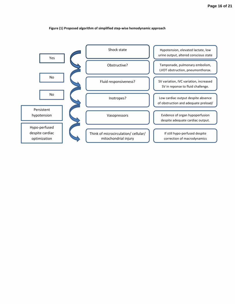
Figure (1) Proposed algorithm of simplified step-wise hemodynamic approach
Shock state
Obstructive?
Fluid responsiveness?
Inotropes?
Vasopressors
Think of microcirculation/ cellular/
mitochondrial injury
Yes
No
No
Hypotension, elevated lactate, low
urine output, altered conscious state
Tamponade, pulmonary embolism,
LVOT obstruction, pneumonthorax.
SV variation, IVC variation, increased
SV in reponse to fluid challenge.
Low cardiac output despite absence
of obstruction and adequate preload/
Evidence of organ hypoperfusion
despite adequate cardiac output.
If still hypo-perfused despite
correction of macrodynamics
Persistent
hypotension
Hypo-perfused
despite cardiac
optimization
Page 16 of 21
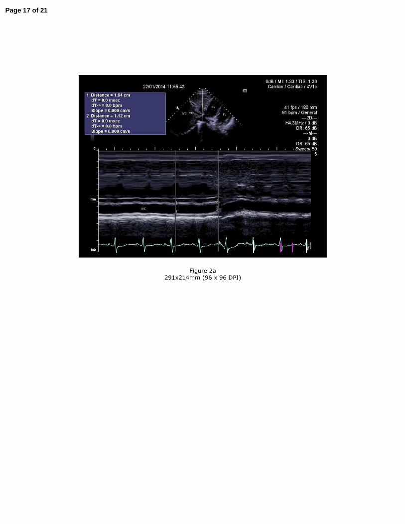
Figure 2a
291x214mm (96 x 96 DPI)
Page 17 of 21

Figure 2b
291x214mm (96 x 96 DPI)
Page 18 of 21
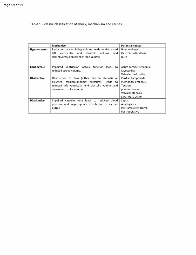
Table 1 – classic classification of shock, mechanism and causes
Mechanism Potential causes
Hypovolaemic Reduction in circulating volume leads to decreased
left ventricular end diastolic volume and
subsequently decreased stroke volume.
Haemorrhage
Gastrointestinal loss
Burn
Cardiogenic Impaired ventricular systolic function leads to
reduced stroke volume.
Acute cardiac Ischaemia
Myocarditis
Valvular dysfunction
Obstructive Obstruction to flow (either due to stenosis or
elevated cardiopulmonary pressures) leads to
reduced left ventricular end diastolic volume and
decreased stroke volume.
Cardiac Tamponade
Pulmonary embolus
Tension
pneumothorax
Valvular stenosis
LVOT obstruction
Distributive Impaired vascular tone leads to reduced blood
pressure and inappropriate distribution of cardiac
output
Sepsis
Anaphylaxis
Post-arrest syndrome
Post-operative
Page 19 of 21
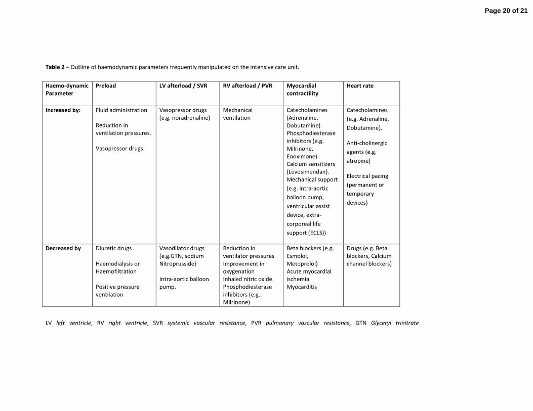
Table 2 – Outline of haemodynamic parameters frequently manipulated on the intensive care unit.
LV left ventricle, RV right ventricle, SVR systemic vascular resistance, PVR pulmonary vascular resistance, GTN Glyceryl trinitrate
Haemo-dynamic
Parameter
Preload LV afterload / SVR RV afterload / PVR Myocardial
contractility
Heart rate
Increased by: Fluid administration
Reduction in
ventilation pressures.
Vasopressor drugs
Vasopressor drugs
(e.g. noradrenaline)
Mechanical
ventilation
Catecholamines
(Adrenaline,
Dobutamine)
Phosphodiesterase
inhibitors (e.g.
Milrinone,
Enoximone).
Calcium sensitizers
(Levosimendan).
Mechanical support
(e.g. intra-aortic
balloon pump,
ventricular assist
device, extra-
corporeal life
support (ECLS))
Catecholamines
(e.g. Adrenaline,
Dobutamine).
Anti-cholinergic
agents (e.g.
atropine)
Electrical pacing
(permanent or
temporary
devices)
Decreased by Diuretic drugs
Haemodialysis or
Haemofiltration
Positive pressure
ventilation
Vasodilator drugs
(e.g.GTN, sodium
Nitroprusside)
Intra-aortic balloon
pump.
Reduction in
ventilator pressures
Improvement in
oxygenation
Inhaled nitric oxide.
Phosphodiesterase
inhibitors (e.g.
Milrinone)
Beta blockers (e.g.
Esmolol,
Metoprolol)
Acute myocardial
ischemia
Myocarditis
Drugs (e.g. Beta
blockers, Calcium
channel blockers)
Page 20 of 21

Page 21 of 21