virtual histology of transgenic mouse embryos for …...virtual histology of transgenic mouse...
TRANSCRIPT
Virtual Histology of Transgenic MouseEmbryos for High-Throughput PhenotypingJohn T. Johnson
1[, Mark S. Hansen
2[, Isabel Wu
3, Lindsey J. Healy
1, Christopher R. Johnson
1, Greg M. Jones
1,
Mario R. Capecchi4,5
, Charles Keller3*
1 Scientific Computing and Imaging Institute, University of Utah, Salt Lake City, Utah, United States of America, 2 Division of Pediatric Hematology-Oncology, Department of
Pediatrics, University of Utah, Salt Lake City, Utah, United States of America, 3 Department of Cellular & Structural Biology and Department of Pediatrics, Children’s Cancer
Research Institute, The University of Texas Health Science Center, San Antonio, Texas, United States of America, 4 Department of Human Genetics, University of Utah, Salt
Lake City, Utah, United States of America, 5 Howard Hughes Medical Institute, University of Utah, Salt Lake City, Utah, United States of America
A bold new effort to disrupt every gene in the mouse genome necessitates systematic, interdisciplinary approaches toanalyzing patterning defects in the mouse embryo. We present a novel, rapid, and inexpensive method for obtaininghigh-resolution virtual histology for phenotypic assessment of mouse embryos. Using osmium tetroxide todifferentially stain tissues followed by volumetric X-ray computed tomography to image whole embryos, isometricresolutions of 27 lm or 8 lm were achieved with scan times of 2 h or 12 h, respectively, using mid-gestation E9.5–E12.5embryos. The datasets generated by this method are immediately amenable to state-of-the-art computationalmethods of organ patterning analysis. This technique to assess embryo anatomy represents a significant improvementin resolution, time, and expense for the quantitative, three-dimensional analysis of developmental patterning defectsattributed to genetically engineered mutations and chemically induced embryotoxicity.
Citation: Johnson JT, Hansen MS, Wu I, Healy LJ, Johnson CR, et al. (2006) Virtual histology of transgenic mouse embryos for high-throughput phenotyping. PLoS Genet 2(4):e61. DOI: 10.1371/journal.pgen.0020061
Introduction
Gene targeting in mice allows unprecedented insight intothe function of genes and their roles in patterning themammalian embryo [1]. A full understanding of mammaliandevelopment by this means, using the gene-targeting ap-proach for every one of the approximately 25,000 or moremouse genes, may seem like a daunting task. Nevertheless,more than 10% of known mouse genes have already beendisrupted by gene targeting. Moreover, the National Institutesof Health is leading an effort to create a collection of mouselines with disruption of every known gene [2]. The challengelaid before developmental biologists will be to systemicallyanalyze morphological phenotypes and, where possible,determine the quantitative contribution of each gene towardspatterning of the embryo. Tools for this type of ‘‘phenomic’’analysis must include rapid, inexpensive, and accessible high-throughput methods of high-resolution anatomical imaging(addressed here) as well as stage-specific, statistically-averagedwild-type morphological atlases that can be used to discernnormal variation from mutant phenotype [3].
The introduction of magnetic resonance microscopy(MRM) represented an important time savings over classicalhistology in the screening of E6.5–E19 mouse embryos formutant morphological phenotypes [4] (reviewed by Schneiderand Bhattacharya [5] and Tyszka et al. [6]). However, thespecialized and expensive equipment required for such highfield magnetic resonance scans is not widely available.Furthermore, scans at useful resolutions (12–43 lm, butgenerally 25 lm) require 9–14 h of instrument time [7,8] at acost of approximately US$200 per hour.
We introduce a new method of obtaining virtual histologyusing X-ray microscopic computed tomography (microCT).This technique permits mid-gestation mouse embryos to bescanned at up to 8-lm resolution in comparable or less time,
and at a fraction of the expense of MRM. This new method,which employs osmium tetroxide to differentially staintissues, will facilitate the quantitative, three-dimensionalanalysis of mouse developmental defects for researchers ata broad range of institutions at relatively little expense—andat resolutions and throughput comparable to or exceedingmagnetic resonance methods.
Results
Virtual Histology by Computed Tomography Is a Powerful
Method for Imaging Embryo AnatomyFormalin-fixed wild-type E11.5 embryos whose cell mem-
branes were stained in a 1% solution of osmium tetroxidewere imaged by volumetric computed tomography (CT) at 8-lm isometric resolution. External surface features of thescanned embryos were represented as isosurfaces (Figure 1A),demonstrating a level of detail comparable to a dissectionmicroscope. Internal structures could be visualized by a semi-transparent maximum intensity projection of the entireembryo, similar to a plain radiograph (Figure 1B). In orderto compare the spatial resolution of traditional optical
Editor: Wayne Frankel, The Jackson Laboratory, United States of America
Received January 23, 2006; Accepted March 13, 2006; Published April 28, 2006
DOI: 10.1371/journal.pgen.0020061
Copyright: � 2006 Johnson et al. This is an open-access article distributed underthe terms of the Creative Commons Attribution License, which permits unrestricteduse, distribution, and reproduction in any medium, provided the original authorand source are credited.
Abbreviations: CT, computed tomography; microCT, microscopic computedtomography; MRM, magnetic resonance microscopy
* To whom correspondence should be addressed. E-mail: [email protected]
[ These authors contributed equally to this work.
PLoS Genetics | www.plosgenetics.org April 2006 | Volume 2 | Issue 4 | e610471

histology to microCT-based virtual histology, paraffin-em-bedded 4.5-lm sections were stained with hematoxylin andeosin and visualized at 23 magnification (Figure 1C–1E).Sagittal, coronal, and axial sections by microCT-based virtualhistology were comparable in the delineation of internalfeatures (organs and tissues) to paraffin sections at the samemagnification. Anatomical landmarks as small as the dorsalroot ganglia, the neural tube, and the anterior cardinal veincould be easily discerned. Because osmium staining increaseswith lipid content of tissues, large differences in densities areobserved between tissues (normalized CT attenuation valuesfor tissue ranged from 5,785 to 32,767 Hounsfield units).
Virtual Histology is Most Amenable to Mid-GestationEmbryos
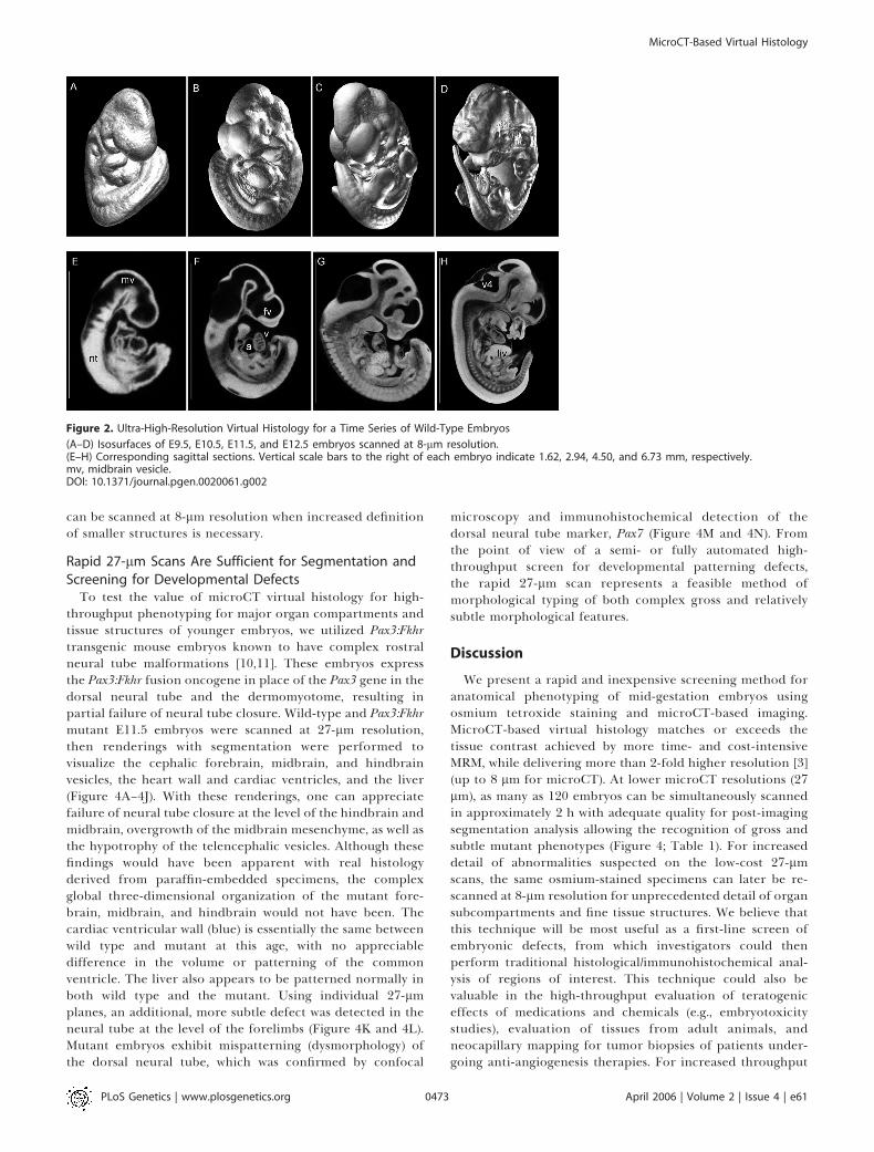
Unlike MRM, which does not necessarily require staining,the osmium tetroxide stain for microCT-based virtualhistology is best suited to gestational ages with limitedepidermal layers (unless the epidermis is manually removedwith a #2 forceps or protease digested prior to staining [9]). Inour experience, mid-gestation whole embryos (E8–E13.5) thatlack significant epidermal development are most appropriatefor this method, although skinned embryos up to E19 can besatisfactorily stained and imaged as well (unpublished data).Figure 2 demonstrates isosurfaces (Figure 2A–2D) and sagittalcross-sections (Figure 2E–2H) of a time series of E9.5, E10.5,E11.5, and E12.5 embryos, respectively, scanned at 8-lmisometric resolution. At these resolutions, features such as thedeveloping brain vesicles, neural tube, heart chambers, andliver can be clearly delineated. Due to the increased lipidcontent of the liver, attenuation of osmium-stained hepatictissue results in the highest opacity and brightness.
Rapid 27-lm Resolution Scans Can Be Performed on aSmall Animal CT Scanner
Lower spatial resolutions, comparable to most typicalmagnetic resonance methods (25–27 lm) can be achieved
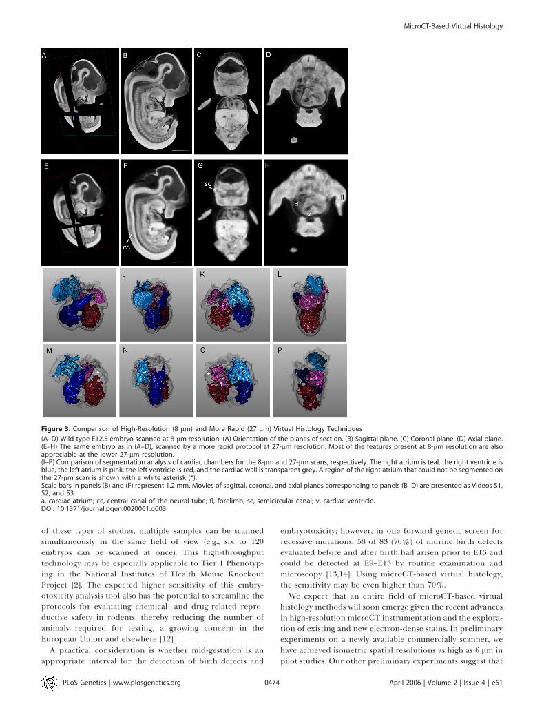
with only a 2-h acquisition time on a small animal CTscanner. These live-animal scanners are more likely to beavailable at academic imaging core facilities. Employing a GEeXplore RS small animal scanner, we performed scans ofwild-type E12.5 embryos at 27-lm isometric resolution inapproximately 2 h. A comparison of 8-lm and 27-lm scans ofthe same embryo is shown in Figure 3. Although 8-lmsections (Figure 3A–3D) display considerably higher spatialresolution, the 27-lm sections (Figure 3E–3H) were nonethe-less adequate to distinguish features such as the semicircularcanal, the neural tube central canal, and the cardiacchambers. From the perspective of high-throughput pheno-typing, the resolution of these 27-lm microCT scans were inthe range of MRM, but at a nearly 6-fold time savings and a300-fold cost savings. Furthermore, these 2 h, 27-lmresolution scans were adequate to perform high-qualitysegmentation analysis of major organ compartments, aprerequisite for computer-based, automated phenotyping(Figure 3M–3P). A caveat is that the small lumens within someorgans, such as the right atrium of the heart, are less wellsegmented in the rapid scan than by the higher resolutionscan (Figure 3I–3L). However, as shown in this figure, thesame osmium-stained embryo scanned at 27-lm resolution
Figure 1. Comparison of Virtual and Paraffin Histology of an E11.5
Embryo Scanned at 8 lm
(A) Isosurface renderings of the CT-scanned embryo.(B) Maximum intensity projection of the same embryo, with overlyingplaces of section.(C–E) Sagittal, coronal, and axial sections of an E11.5 littermate.(F–H) Sagittal, coronal, and axial computed tomography sections of theembryo in panels (A) and (B), corresponding to the planes of section inpanels (C–E). At low-power magnification, virtual and true paraffinhistology demonstrate a similar level of detail. Scale bar indicates 400 lm.a, cardiac atrium; cv, cardinal vein; drg, dorsal root ganglia; fv, forebrainvesicle; liv, liver; nt, neural tube; v, cardiac ventricle; v4, fourth ventricle.DOI: 10.1371/journal.pgen.0020061.g001
PLoS Genetics | www.plosgenetics.org April 2006 | Volume 2 | Issue 4 | e610472
MicroCT-Based Virtual Histology
Synopsis
Developmental biology is entering the digital age, thanks toadvancements in imaging technologies, instrumentation, andsoftware. These advancements are converging with discoveries indevelopmental biology to deliver unprecedented insight into howhuman development is impacted by the products we use, theenvironment we live in, and our genetic composition. Industrialsocieties are becoming increasingly concerned with the exposure ofwomen and their unborn fetuses to pharmaceuticals and common-place household chemicals. In addition, understanding geneticcauses of birth defects is now possible through the isolation ofspecific genes, which can be efficiently disrupted in embryos, andsubsequently observed for birth defects. Such studies of embry-otoxicity typically involve the use of mouse embryos. However,evaluation of mouse embryos in the past has involved expensiveand cumbersome external inspection and thin sectioning for viewunder the microscope. As such, developmental biologists haveeagerly anticipated the advent of tools that would allow them toroutinely assess the complex and dynamic organization of embryosusing techniques that are fast and inexpensive. In this article, theauthors introduce a rapid, high-quality, and inexpensive techniquefor the three-dimensional visualization of mouse embryos using X-ray computed tomography that is ideally suited for researchers inpharmaceutical, industrial, and academic laboratories.
can be scanned at 8-lm resolution when increased definitionof smaller structures is necessary.
Rapid 27-lm Scans Are Sufficient for Segmentation andScreening for Developmental Defects
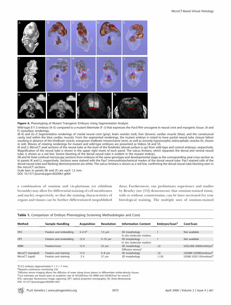
To test the value of microCT virtual histology for high-throughput phenotyping for major organ compartments andtissue structures of younger embryos, we utilized Pax3:Fkhrtransgenic mouse embryos known to have complex rostralneural tube malformations [10,11]. These embryos expressthe Pax3:Fkhr fusion oncogene in place of the Pax3 gene in thedorsal neural tube and the dermomyotome, resulting inpartial failure of neural tube closure. Wild-type and Pax3:Fkhrmutant E11.5 embryos were scanned at 27-lm resolution,then renderings with segmentation were performed tovisualize the cephalic forebrain, midbrain, and hindbrainvesicles, the heart wall and cardiac ventricles, and the liver(Figure 4A–4J). With these renderings, one can appreciatefailure of neural tube closure at the level of the hindbrain andmidbrain, overgrowth of the midbrain mesenchyme, as well asthe hypotrophy of the telencephalic vesicles. Although thesefindings would have been apparent with real histologyderived from paraffin-embedded specimens, the complexglobal three-dimensional organization of the mutant fore-brain, midbrain, and hindbrain would not have been. Thecardiac ventricular wall (blue) is essentially the same betweenwild type and mutant at this age, with no appreciabledifference in the volume or patterning of the commonventricle. The liver also appears to be patterned normally inboth wild type and the mutant. Using individual 27-lmplanes, an additional, more subtle defect was detected in theneural tube at the level of the forelimbs (Figure 4K and 4L).Mutant embryos exhibit mispatterning (dysmorphology) ofthe dorsal neural tube, which was confirmed by confocal
microscopy and immunohistochemical detection of thedorsal neural tube marker, Pax7 (Figure 4M and 4N). Fromthe point of view of a semi- or fully automated high-throughput screen for developmental patterning defects,the rapid 27-lm scan represents a feasible method ofmorphological typing of both complex gross and relativelysubtle morphological features.
Discussion
We present a rapid and inexpensive screening method foranatomical phenotyping of mid-gestation embryos usingosmium tetroxide staining and microCT-based imaging.MicroCT-based virtual histology matches or exceeds thetissue contrast achieved by more time- and cost-intensiveMRM, while delivering more than 2-fold higher resolution [3](up to 8 lm for microCT). At lower microCT resolutions (27lm), as many as 120 embryos can be simultaneously scannedin approximately 2 h with adequate quality for post-imagingsegmentation analysis allowing the recognition of gross andsubtle mutant phenotypes (Figure 4; Table 1). For increaseddetail of abnormalities suspected on the low-cost 27-lmscans, the same osmium-stained specimens can later be re-scanned at 8-lm resolution for unprecedented detail of organsubcompartments and fine tissue structures. We believe thatthis technique will be most useful as a first-line screen ofembryonic defects, from which investigators could thenperform traditional histological/immunohistochemical anal-ysis of regions of interest. This technique could also bevaluable in the high-throughput evaluation of teratogeniceffects of medications and chemicals (e.g., embryotoxicitystudies), evaluation of tissues from adult animals, andneocapillary mapping for tumor biopsies of patients under-going anti-angiogenesis therapies. For increased throughput
Figure 2. Ultra-High-Resolution Virtual Histology for a Time Series of Wild-Type Embryos
(A–D) Isosurfaces of E9.5, E10.5, E11.5, and E12.5 embryos scanned at 8-lm resolution.(E–H) Corresponding sagittal sections. Vertical scale bars to the right of each embryo indicate 1.62, 2.94, 4.50, and 6.73 mm, respectively.mv, midbrain vesicle.DOI: 10.1371/journal.pgen.0020061.g002
PLoS Genetics | www.plosgenetics.org April 2006 | Volume 2 | Issue 4 | e610473
MicroCT-Based Virtual Histology
of these types of studies, multiple samples can be scannedsimultaneously in the same field of view (e.g., six to 120embryos can be scanned at once). This high-throughputtechnology may be especially applicable to Tier 1 Phenotyp-ing in the National Institutes of Health Mouse KnockoutProject [2]. The expected higher sensitivity of this embry-otoxicity analysis tool also has the potential to streamline theprotocols for evaluating chemical- and drug-related repro-ductive safety in rodents, thereby reducing the number ofanimals required for testing, a growing concern in theEuropean Union and elsewhere [12].
A practical consideration is whether mid-gestation is anappropriate interval for the detection of birth defects and
embryotoxicity; however, in one forward genetic screen forrecessive mutations, 58 of 83 (70%) of murine birth defectsevaluated before and after birth had arisen prior to E13 andcould be detected at E9–E13 by routine examination andmicroscopy [13,14]. Using microCT-based virtual histology,the sensitivity may be even higher than 70%.We expect that an entire field of microCT-based virtual
histology methods will soon emerge given the recent advancesin high-resolution microCT instrumentation and the explora-tion of existing and new electron-dense stains. In preliminaryexperiments on a newly available commercially scanner, wehave achieved isometric spatial resolutions as high as 6 lm inpilot studies. Our other preliminary experiments suggest that
Figure 3. Comparison of High-Resolution (8 lm) and More Rapid (27 lm) Virtual Histology Techniques
(A–D) Wild-type E12.5 embryo scanned at 8-lm resolution. (A) Orientation of the planes of section. (B) Sagittal plane. (C) Coronal plane. (D) Axial plane.(E–H) The same embryo as in (A–D), scanned by a more rapid protocol at 27-lm resolution. Most of the features present at 8-lm resolution are alsoappreciable at the lower 27-lm resolution.(I–P) Comparison of segmentation analysis of cardiac chambers for the 8-lm and 27-lm scans, respectively. The right atrium is teal, the right ventricle isblue, the left atrium is pink, the left ventricle is red, and the cardiac wall is transparent grey. A region of the right atrium that could not be segmented onthe 27-lm scan is shown with a white asterisk (*).Scale bars in panels (B) and (F) represent 1.2 mm. Movies of sagittal, coronal, and axial planes corresponding to panels (B–D) are presented as Videos S1,S2, and S3.a, cardiac atrium; cc, central canal of the neural tube; fl, forelimb; sc, semicircular canal; v, cardiac ventricle.DOI: 10.1371/journal.pgen.0020061.g003
PLoS Genetics | www.plosgenetics.org April 2006 | Volume 2 | Issue 4 | e610474
MicroCT-Based Virtual Histology
a combination of osmium and cis-platinum (or ethidiumbromide) may allow for differential staining of cell membranesand nuclei, respectively, so that the staining characteristics oforgans and tissues can be further differentiated (unpublished
data). Furthermore, our preliminary experience and studiesby Bentley (see [15]) demonstrate that osmium-stained tissue,with or without counterstains, can be later sectioned for truehistological staining. The multiple uses of osmium-stained
Figure 4. Phenotyping of Mutant Transgenic Embryos Using Segmentation Analysis
Wild-type E11.5 embryo (A–E) compared to a mutant littermate (F–J) that expresses the Pax3:Fkhr oncogene in neural crest and myogenic tissue. (A andF) Isosurface renderings.(B–E) and (G–J) Segmentation renderings of cranial neural crest (grey), brain vesicles (red), liver (brown), cardiac muscle (blue), and the conotruncalcavity (red within the blue cardiac muscle). From the segmented renderings, the mutant embryo is noted to have partial neural tube closure failureresulting in absence of the hindbrain vesicle, overgrown midbrain mesenchyme (mm), as well as severely hypomorphic telencephalic vesicles (fv, shownin red). Movies of rotating renderings for mutant and wild-type embryos are presented as Videos S4 and S5.(K and L) MicroCT axial sections of the neural tube at the level of the forelimbs (dorsal surface is up) from wild-type and control embryos, respectively.Magnification of the neural tube is shown in the upper right insets of each panel. The sulcus limitans, which separates the dorsal and ventral neuraltube, is shown as a red line. Severe blunting of the dorsal neural tube is evident in the mutant embryo.(M and N) Axial confocal microscopy sections from embryos of the same genotype and developmental stage as the corresponding axial cross-section asin panels (K and L), respectively. Sections were stained with the Pax7 immunohistochemical marker of the dorsal neural tube. Pax7-stained cells of thedorsal neural tube and flanking dermomyotome are white. The sulcus limitans is shown as a red line, confirming the dorsal neural tube blunting seen inthe microCT sections.Scale bars in panels (B) and (F) are each 1.2 mm.DOI: 10.1371/journal.pgen.0020061.g004
Table 1. Comparison of Embryo Phenotyping Screening Methodologies and Costs
Method Sample Handling Acquisition Resolution Information Content Embryos/Scana Cost/Scan
EFIC Fixation and embedding 3–4 hb 1.5 lm 3D morphology 1 Not available
In situ molecular markers
OPT Fixation and embedding 12 h 5–10 lm 3D morphology 1 Not available
In situ molecular markers
MRM Fixation/none 12 h 25 lm 3D morphology �6 US$2,400 ($600/embryo)d
Diffusion tensorsc
MicroCT (standard) Fixation and staining 12 h 6–8 lm 3D morphology �6 US$480 (US$80/embryo)d
MicroCT (rapid) Fixation and staining 2 h 27 lm 3D morphology �120 US$80 (US$1.50/embryo)d
aE12.5 embryos (approximately 5 3 5 3 7 mm).bRequires continuous monitoring [16].cDiffusion tensor imaging allows the diffusion of water along tissue planes to differentiate similar-density tissues.dCost estimates are based upon an academic rate of US$200/hour for MRM and US$40/hour for microCT.EFIC, episcopic fluorescence image capturing; OPT, optical projection tomography; 3D, three-dimensional.DOI: 10.1371/journal.pgen.0020061.t001
PLoS Genetics | www.plosgenetics.org April 2006 | Volume 2 | Issue 4 | e610475
MicroCT-Based Virtual Histology
tissues will therefore speed the transition from microCT-based screens to episcopic [16] and microscopic histologicalverification of suspected morphological phenotypes.
In order to achieve high-throughput phenotyping, orphenomics, the computing methods for analysis must besemi-automated or fully automated. Magnetic resonanceimaging of postnatal mouse brains (as well as human brains)has led to significant advances in semi- or fully automateddeformable shape mapping whereby specimens of differentsizes can be compared for the eventual detection of statisticallydifferent features of morphology [17]. MicroCT-based virtualhistology datasets are ideally suited to this type of analysis.
MicroCT-based virtual histology is not intended to replacethe generally more versatile magnetic resonance methods(for a review of MRM, see references [3,5,7]), but is instead auseful adjunct for anatomical imaging. A comparison of CTand magnetic resonance methods, applications, and costs aregiven in Table 1. Included for comparison are the low-throughput but exceptional resolution methods of episcopicfluorescence image capturing [16,18] and optical projectiontomography [19]. Magnetic resonance imaging allows theanalysis of a variety of tissue properties to be interrogated,depending upon the pulse sequence, and post-processingparameters such as diffusion tensors [17] allow the organ-ization of tissues to visualized and modeled in unprecedentedways. Instead, microCT-based virtual histology offers apotentially higher resolution mode of morphometrics thatis simple to implement, relatively inexpensive, and morerapid than comparable methods of phenotyping embryoanatomy.
Materials and Methods
Sample preparation. The Pax3:Fkhr transgenic mice have beenpreviously described [10,11]. Embryos were harvested at E9.5–E12.5gestational ages, then fixed in 10%buffered formalin overnight at 4 8C.Hematoxylin and eosin–stained paraffin sections were prepared usingestablished methods [10], then visualized at 23 magnification on aNikon Eclipse 80i microscope (Nikon, Tokyo, Japan). Immunohisto-chemistry with the Pax7 monoclonal antibody (DevelopmentalHybridoma Studies Bank, Iowa City, Iowa, United States) wasperformed as previously described [10]. For microCT-based virtualhistology, formalin-fixed embryos were stained using a beta version ofa commercially available kit (Numira Biosciences, San Antonio, Texas,United States http://www.numirabio.com). In brief, embryos werestained to saturation overnight in a solution of 0.1 M sodiumcacodylate (pH 7.2), 1% glutaraldehyde, and 1% osmium tetroxiderocking at room temperature.We note that osmium tetroxide requirescareful handling that is possible in most molecular biology laborato-ries (for the international safety and handling information, see http://www.cdc.gov/niosh/ipcsneng/neng0528.html). Embryos were thenwashed for 30 min in 0.1 M sodium cacodylate buffer, and twice morefor 30 min in phosphate-buffered saline. Samples were thentransitioned by a series of gradients to 100% ethanol prior toscanning. For each 25%, 50%, 75%, and 100% ethanol gradientequilibration, embryos were incubated for 15–30 min, or until theembryos lost buoyancy. Prior to imaging, embryos were placed in 2-mlpolypropylene microfuge tubes.
Imaging. High-resolution volumetric CT of embryos was per-formed at 8-lm3 isometric voxel resolution using an eXplore LocusSP MicroCT specimen scanner (GE Healthcare, London, Ontario,Canada). This volumetric scanner employs a 3,500 3 1,750 CCD(charge-coupled device) detector for Feldkamp cone-beam recon-struction, and is similar in performance to other commerciallyavailable in vitro scanners under $300,000 that are commonlyoperated at regional core facilities (http://ccri.uthscsa.edu/ImagingFacility.html). In this study, the platform-independentparameters of current, voltage, and exposure time were kept constantat 100 lA, 80 kVP, and 4,000 ms, respectively. For each scan, 900evenly spaced views were averaged from eight frames/view, filtered by
0.2-mm aluminum. At 8-lm resolution, the field of view of thisinstrument is 15 3 15 3 15 mm. Each scan took approximately 12 h.Cost of this method is approximate US$40 per hour (US$480 perscan). Images were reconstructed with the manufacturer’s proprietaryEVSBeam software.
More rapid volumetric CT scans of embryos performed at 27-lm3
isometric voxel resolution using an eXplore Locus RS small animalMicroCT scanner (GE Healthcare). Like the specimen scanner, thislive-animal volumetric scanner employs a 3,50031,750 CCD detectorfor Feldkamp cone-beam reconstruction and is similar in perform-ance to other commercially available in vivo scanners under $300,000that are commonly operated at regional core facilities (http://ccri.uthscsa.edu/ImagingFacility.html). In this study, the platform-inde-pendent parameters of current, voltage, and exposure time were keptconstant at 450 mA, 80 kVP, and 2000 ms, respectively. For each scan,450 evenly spaced views were averaged from six frames/view. At 27-lm resolution, the field of view of this instrument is 45345345 mm.Each scan took 2 h and 4 min. Cost of this method is also approximateUS$40 per hour (US$80 per scan). Images were reconstructed withthe manufacturer’s proprietary EVSBeam software. The 600 MBthree-dimensional dataset and the raw data for the scan weretransferred to a single DVD (approximate total, 4.5 GB). Preliminaryvisualizations (unpublished data) and virtual histology sections weregenerated in real time with the freely-available MicroView program(http://microview.sf.net).
Isosurface generation. Isosurfaces renderings of the CT datasetswere generated using a combination of the open-source Teemutilities [20] and the open-source, platform-independent BioImage[21] rendering program. Both sets of software are available online(http://software.sci.utah.edu). Sub-sampling of the microCT volumes,which are significantly larger than the available memory on currentgraphics cards, was also performed using the Teem libraries. The finalsub-sampled volumes were 255 3 255 3 255 mm and equally spacedusing a nearest neighbor approach. Pre-computing of the gradientand Hessian data was also performed using the Teem libraries. Thecombined process of sub-sampling and generation of gradient andHessian data produced two data volumes, which were stored togetherwith the original CT values in a ‘‘VGH’’ (value, gradient, Hessian)volume. The VGH volumes were subsequently used to createisosurfaces in real time with the BioImage software package.
Segmentation. Image volume segmentations were done using aWatershed algorithm [22] provided in the National Library ofMedicine Insight Segmentation and Registration Toolkit (ITK;http://www.itk.org). Watershed segmentation is a region-growingalgorithm in which user-defined seed points are positioned in areasof interest, and a statistical analysis is made of the gradientmagnitude in the local neighborhood to find the standard deviation.The structure is then found as the perimeters of the seed pointsextend through the continuous surrounding area, without crossingpoints where the gradient magnitude is beyond one standarddeviation of the mean. The resulting structures are saved to disk asseparate volumes. Segmentations were done on a computer equippedwith 2.5 GHz Intel Pentium III processor and 2 GB of RAM. Theresulting volumes were then combined into one volume by markingeach segmentation as a unique value, and then merging it into theoriginal again, such that the original value of existing segmentation isoverwriten with the new marked value.
Volume renderings. The volume renderings of wild-type andPax3:Fkhr mutant embryo datasets were created using direct volumerendering techniques using the in-house software package, Nenners[23]. Nenners reads a volume and casts n2 geometric rays through thevolume per pixel. For each ray that intersects with the volume,equally spaced samples of length Dt are acquired along the respectiveray. To ensure that renderings capture the fine detail of these high-resolution datasets Dt�||vi � vi � 1||, where vi is the position of ith
voxel,the data are sampled at least once per cubic voxel. Samples areacquired by convolution using separable kernels (page 197 in [24]).Cubic B-splines were used to interpolate both the CT value andgradient [25]. Color and opacity were defined at each sample pointusing a two-dimensional transfer function, with gradient magnitudeand X-ray density defining the domain [26,27]. This was done in afront-to-back fashion, such that when the opacity of a ray becomesequal to or greater than 1, the ray is terminated early and the next rayis computed. Bounding volumes were also used to speed this processsuch that rays that have not yet or will not hit the volume were notsampled. The gradient was also used to approximate the Phongshading models used [28]. For Figure 3, a curvature-based transferfunction [28] and depth queuing was also applied to the volumerenderings. The transfer function is based on the dot product of theangle of an incident ray with the surface normal. If the dot product
PLoS Genetics | www.plosgenetics.org April 2006 | Volume 2 | Issue 4 | e610476
MicroCT-Based Virtual Histology
was less than some constant k, 0 , k , 1, then the surface was markedblack to emphasis surfaces perpendicular to the viewing direction.Furthermore, a depth queue to shade interpolated samples was used togive a greater feeling of depth and to seperate foreground frombackground. Parameters of camera position, viewing direction, andlighting relative tothe specimenwerealsopassedtodefine theviewof therendering. The softwarewas runon anSGIOnyx 3800 (SiliconGraphics,Mountain View, California, United States) at 80 s per rendering.
Supporting Information
Video S1. Sagittal Sections of a Wild-Type E12.5 Embryo at 8-lmResolution
Found at DOI: 10.1371/journal.pgen.0020061.sv001 (1.2 MB WMV).
Video S2. Coronal Sections of a Wild-Type E12.5 Embryo at 8-lmResolution
Found at DOI: 10.1371/journal.pgen.0020061.sv002 (1.3 MB WMV).
Video S3. Axial Sections of a Wild-Type E12.5 Embryo at 8-lmResolution
Found at DOI: 10.1371/journal.pgen.0020061.sv003 (1.7 MB WMV).
Video S4. Rendering of a Segmented Pax3:Fkhr Mutant E11.5 Embryo
This embryo corresponds to Figure 4F–4J.
Found at DOI: 10.1371/journal.pgen.0020061.sv004 (3.6 MB MOV).
Video S5. Rendering of a Segmented Wild-Type E11.5 Embryo
This embryo corresponds to Figure 4A–4E.
Found at DOI: 10.1371/journal.pgen.0020061.sv005 (3.6 MB MOV).
Accession Numbers
The National Center for Biotechnology Information (NCBI) (http://www.ncbi.nlm.nih.gov) accession number for the Pax3:Fkhr fusiononcogene is AY743239, and for Pax7 is NM_011039.
Acknowledgments
The Pax7 antibody developed by Atsushi Kawakami was obtainedfrom Developmental Studies Hybridoma Bank (University of Iowa).We thank Paul Picot, Mike Cole, and Tim Morgan at General ElectricHealthcare for scanning services, Steve Fullwood at Nikon forassistance with digital microscopy, Gordon Kindlmann for consulta-tion on rendering methods, and Gary Gaufo for thoughtful commentsduring manuscript preparation.
Author contributions. CK conceived and designed the experi-ments. MSH, IW, and CK performed the experiments. JTJ, LJH, GMJ,MRC, and CK analyzed the data. CRJ and GMJ contributed reagents/materials/analysis tools. JTJ and CK wrote the paper.
Funding. This work was supported in part by a National Institutesof Health National Center for Research Resources Center award toCRJ (P41RR12553) and an Anatomical & Molecular Imaging InitiativeAward to CK by the Children’s Cancer Research Institute.
Competing interests. CK is the co-founder of a biotechnologycompany named Numira Biosciences, which may pursue thecommercialization of the patent-pending technology in this manu-script. The intent of our manuscript, however, is to communicate thismethod so that it may be reproduced and utilized freely at academicinstitutions. &
References1. Capecchi MR (2005) Gene targeting in mice: Functional analysis of the
mammalian genome for the twenty-first century. Nat Rev Genet 6: 507–512.2. Austin CP, Battey JF, Bradley A, Bucan M, Capecchi M, et al. (2004) The
knockout mouse project. Nat Genet 36: 921–924.3. Jacobs RE, Ahrens ET, Dickinson ME, Laidlaw D (1999) Towards a
microMRI atlas of mouse development. Comput Med Imaging Graph 23:15–24.
4. Smith BR, Johnson GA, Groman EV, Linney E (1994) Magnetic resonancemicroscopy of mouse embryos. Proc Natl Acad Sci U S A 91: 3530–3533.
5. Schneider JE, Bhattacharya S (2004) Making the mouse embryo trans-parent: Identifying developmental malformations using magnetic reso-nance imaging. Birth Defects Res C Embryo Today 72: 241–249.
6. Tyszka JM, Fraser SE, Jacobs RE (2005) Magnetic resonance microscopy:Recent advances and applications. Curr Opin Biotechnol 16: 93–99.
7. Schneider JE, Bamforth SD, Farthing CR, Clarke K, Neubauer S, et al. (2003)High-resolution imaging of normal anatomy, and neural and adrenalmalformations in mouse embryos using magnetic resonance microscopy. JAnat 202: 239–247.
8. Smith BR, Linney E, Huff DS, Johnson GA (1996) Magnetic resonancemicroscopy of embryos. Comput Med Imaging Graph 20: 483–490.
9. Andras Nagy MG, Vintersten Kristina, Behringer Richard, editors (2003)Manipulating the mouse embryo, a laboratory manual. 3rd edition. ColdSpringHarbor (NewYork):ColdSpringHarborLaboratoryPress.pp680–681.
10. Keller C, Hansen MS, Coffin CM, Capecchi MR (2004) Pax3:Fkhr interfereswith embryonic Pax3 and Pax7 function: Implications for alveolarrhabdomyosarcoma cell of origin. Genes Dev 18: 2608–2613.
11. Keller C, Arenkiel BR, Coffin CM, El-Bardeesy N, DePinho RA, et al. (2004)Alveolar rhabdomyosarcomas in conditional Pax3:Fkhr mice: Cooperativityof Ink4a/ARF and Trp53 loss of function. Genes Dev 18: 2614–2626.
12. Abbott A (2005) Deal on toxicity law fails to appease. Nature 438: 406.13. Kasarskis A, Manova K, Anderson KV (1998) A phenotype-based screen for
embryonic lethal mutations in the mouse. Proc Natl Acad Sci U S A 95:7485–7490.
14. Anderson KV, Lacy E, Niswander L, Timmer JR (2002) The Sloan-KetteringInstitute Mouse Project. New York: Sloan-Kettering Institute. Available:http://mouse.ski.mskcc.org. Accessed 28 March 2006.
15. Ritman EL (2004) Micro-computed tomography—Current status anddevelopments. Annu Rev Biomed Eng 6: 185–208.
16. Rosenthal J, Mangal V, Walker D, Bennett M, Mohun TJ, et al. (2004) Rapidhigh resolution three dimensional reconstruction of embryos withepiscopic fluorescence image capture. Birth Defects Res C Embryo Today72: 213–223.
17. Zhang J, Miller MI, Plachez C, Richards LJ, Yarowsky P, et al. (2005)Mapping postnatal mouse brain development with diffusion tensormicroimaging. Neuroimage 26: 1042–1051.
18. Weninger WJ, Mohun T (2002) Phenotyping transgenic embryos: A rapid 3-D screening method based on episcopic fluorescence image capturing. NatGenet 30: 59–65.
19. Sharpe J, Ahlgren U, Perry P, Hill B, Ross A, et al. (2002) Optical projectiontomography as a tool for 3D microscopy and gene expression studies.Science 296: 541–545.
20. Kindlmann G (2005) Teem, version 1.8.0 [computer program]. Available:http://teem.sourceforge.net. Accessed 24 March 2006.
21. Scientific Computing and Imaging Institute (SCI) (2002) BioPSE: Problemsolving environment for modeling, simulation, image processing, andvisualization for biomedical computing applications. Available: http://software.sci.utah.edu/biopse.html. Accessed 27 March 2006.
22. Cates JE, Whitaker RT, Jones GM (2005) Case study: An evaluation of user-assisted hierarchical watershed segmentation. Med Image Anal 9: 566–578.
23. Johnson TN (2005) Nenners [computer program]. Available: http://www.sci.utah.edu/;tjohnson/nenners. Accessed 28 March 2006.
24. Gonzalez RC, Woods RC (2002) Digital image processing. 2nd edition.Upper Saddle River (New Jersey): Prentice Hall. 793 p.
25. Kindlmann GL, Whitaker R, Tasdizen T, Moller T (2003) Curvature-basedtransfer functions for direct volume rendering: Methods and applications.Proc IEEE Visualization: 513–520.
26. Levoy M (1988) Display of surfaces from volume data. IEEE Comput GraphAppl 8: 29–37.
27. Kniss JM, Kindlmann GL, Hansen CD (2005) Multidimentional transferfunctions for volume rendering. In: Hansen CD, Johnson CR, editors. Thevisualization handbook. San Diego: Academic Press. pp. 189–210.
28. Gooch A, Gooch B, Shirly P, Cohen E. (1998) A non-photorealistic lightingmodel For automatic technical illustration. Proceedings of ACM SIG-GRAPH 1998. New York: ACM Press. pp. 447–452.
PLoS Genetics | www.plosgenetics.org April 2006 | Volume 2 | Issue 4 | e610477
MicroCT-Based Virtual Histology