video-assisted thoracic surgery (vats) systematic ...cdn.intechweb.org/pdfs/28643.pdf ·...
TRANSCRIPT

14
Video-Assisted Thoracic Surgery (VATS) Systematic Mediastinal Nodal Dissection
Khalid Amer The Cardiovascular & Thoracic Unit, Southampton General Hospital, Southampton,
United Kingdom
1. Introduction
VATS lobectomy and other major pulmonary resections (VMPR) are growing in popularity.
One of the main criticisms against minimal access in lung cancer surgery is that mediastinal
nodes could be difficult to assess. It was shown by different authorities that VATS complete
nodal dissection is feasible and does not differ from that performed by an open thoracotomy
[1]. Despite conflicting reports, there is an international agreement that nodal dissection
does not influence the disease free or the overall survival in lung cancer. However; proper
staging of Non Small Cell Lung Cancer (NSCLC) enables standardization of decision on
treatment and evaluation of such treatment comparing it to different centres around the
world. Recent publications have shown a significant statistical gain in 5 year survival if
stage IIa and higher were treated by adjuvant chemotherapy [2, 3]. It is therefore absolutely
mandatory to get the staging right in early lung cancer; otherwise patients would be denied
a significant chance of cure. In a pressurised service where commissioning is governed by
patients waiting times, targets and cost effectiveness, surgeons might feel reluctant to extend
the operating time to perform Systematic Nodal Dissection (SND). The risk of improper
mediastinal staging in our view is by far greater than extending the duration of the
operation. The long-term results of stage migration lead to faulty comparison, and might
dictate the wrong management, ending in completely erroneous survival statistics.
Oncological randomised controlled trials rely on final histological staging, and therefore it is
mandatory to obtain correct staging to avoid erroneous survival statistics in such trials. In
our view the only contraindication to SND would be technical difficulty with dissection in
the presence of severe adhesions
The way we stage lung cancer has changed over the years. The TNM6 classification [4] is now superseded by the IASLC new TNM7 classification [5]. Fortunately the naming and significance of nodal stations has not changed substantially. Precarinal nodes #3 for a left sided tumour is now considered an N3 stage. Precarinal nodes for a right sided tumour are designated as #4 (there are no #3R), and these are regarded as N2 disease.
2. Definitions
There are different protocols for staging the mediastinum in search of metastasis in N2 nodes. These include:
www.intechopen.com

Topics in Thoracic Surgery 248
• Selective node sampling: the surgeon decides which node looked diseased and
randomly removes that node (chance node).
• Sentinel nodal sampling: at operation the primary tumour is injected with 99Technitium
tracer, and a Geiger counter is used to identify the sentinel hilar nodes which are
dissected. If frozen section confirmed absence of metastases, the rest of nodal dissection
is omitted (decision node).
• Systematic nodal sampling: one or two nodes sampled from each zonal station
(selective).
• Systematic Nodal dissection: at least 2 nodes from each field or station, and at least 3
fields are dissected (total of at least 6 nodes). Must always include subcarinal nodes
(universally accepted) [6].
• Lobe-specific nodal sampling: oriented towards the different lymphatic drainage of
different lobes e.g. for a right upper lobe tumour, the fields to harvest would be #2-4.
Subcarinal lymphadenectomy is not always necessary for tumours of the right upper
lobe and left upper trisegmentectomy (selective) [7].
• Extended nodal dissection: by definition means bilateral dissection of nodes (no
consensus on extent).
Each of these protocols has points of strengths as well as weaknesses. In general the more
the number of harvested nodes, the more likely it is to reveal normal looking nodes with
metastatic tumour cells.
• Skip metastases: when stations N2 are involved in the absence of N1 involvement, or
N3 involvement in the absence of either N1-N2 nodes. The importance of this
phenomenon is not fully understood [8].
• Micrometastases: The prognosis of cancer patients is largely determined by the
occurrence of distant metastases. The presence of clinically occult few malignant cells
within nodal tissue, bone marrow and pleural fluid, and the clinical relevance of
circulating tumour cells are still debatable. The importance of such nodal involvement
is not fully understood, as it does not inevitably lead to disease dissemination and
disease progression [9, 10].
3. Invasive v non-invasive staging
The tools of staging the mediastinum in NSCLC are either invasive in nature such that
histological confirmation of nodal involvement is sought, or non-invasive, whereby an
imaging technique is used to infer involvement of nodes by secondary metastasis. Invasive
procedures such as mediastinoscopy, mediastinotomy, EBUS-TBNA (Endo Bronchial Ultra
Sound - Trans Bronchial Fine Needle Aspirate) and EUS (Endoscopic or trans oesophageal
Ultra Sound guided Trucut biopsy or FNA) are still developing. These investigations have
limitations in terms of tissue yield, safety profile and cost. At best these are sampling
techniques, aimed at sampling specific nodes which have been highlighted by other non-
invasive techniques. None of these procedures can claim radical dissection of mediastinal
nodes. However, recently VAMLA (Video Assisted Mediastinal Lymph Adenectomy) [11]
has claimed bilateral mediastinal clearance of nodes, yet there are still issues with reaching
stations #5L & #6L [figure 1] and the distant stations #8 & #9. To enhance the yield of nodes
VATS was added to VAMLA to achieve radicality of nodal dissection [12]. On the other
www.intechopen.com

Video-Assisted Thoracic Surgery Systematic Mediastinal Nodal Dissection 249
hand TEMLA (Transcervical Extended Mediastinal Lymph Adenectomy) was introduced in
2004, which involved a collar incision in the neck, elevation of the sternal manubrium with a
special retractor, and claims bilateral dissection of all mediastinal nodes apart from #8 & #9
[13]. The choice of using any of the above mentioned techniques depends on the philosophy
of nodal sampling versus radical adenectomy.
Non-invasive preoperative techniques have largely concentrated on CT and PET. Whilst Computed Tomography (CT) can give great anatomical details of the mediastinum and other chest anatomy, it cannot differentiate benign from malignant tissue. Positron Emission Tomography (PET) on the other hand was claimed to make that biological distinction.
Fig. 1. Station #5L node out of reach of the mediastinoscope (blue circle).
4. The Role of PET
Great hopes were pinned on PET as it seemed to be the most convenient non-invasive
staging tool for a fast tract keyhole surgery such as VMPR designed for early lung cancer.
PET was expected to identify nodal disease in CT negative and normal looking
mediastinum. Recent meta analysis reporting the PET/CT mediastinal staging in patients
with NSCLC found the median sensitivity to be 85% (range 67% to 91%) and specificity of
90% (range 82% to 96%) [14]. Gilles et al and Plathow et al summarised the current views
about the elevated glucose metabolism in cancers [15,16]. Tumour cells adapt to hypoxia by
upregulation of glucose Transporter (GLUTs) and increased activity of Hexokinase. The
GLUT is the first energy-independent glucose transporter across the cell membrane down
the concentration gradient. Tumours increase their level of energy production by engaging
in glycolysis, which is a relatively inefficient way to produce energy compared to aerobic
oxidation (2 ATP molecules versus 30 ATPs). The toxic acidic tumour microenvironment
results in death of normal tissue while tumour cells evade apoptosis by maintaining normal
www.intechopen.com
Topics in Thoracic Surgery 250
intracellular pH. It is thought that this process give the tumour cells a competitive
advantage for local growth, ultimately leading to invasion of basement membrane and
distant metastases. Primary tumours and their nodal secondaries express high GLUT1
upregulation, which in turn is tied to 18F-FDG accumulation in the tumour cell, and hence
directly related to SUVmax (Maximum Standard Uptake Value). GLUT expression is tied to
tumour cell type and differentiation. Squamous cell carcinoma exhibit over expression of
GLUT1 whereas adenocarcinoma does not. One of the serious disappointments of PET
scanning in lung cancer is the low uptake of carcinoids, adenocarcinoma and
bronchioloalveolar carcinomas, in some series up to 40%. This tumour biological behaviour
explains why PET is blinded to adenocarcinoma, Bronchiolo-alveolar carcinoma (BAC) and
carcinoids tumours. For the same reason the importance of the SUVmax (>3.5) as a surrogate
value for malignancy has been played down. Another important snag about the uptake of
the FDG metabolite is the mass of active tissue. A node under 1cm in diameter is unlikely to
show up as a hot spot on PET even if it was completely replaced by secondary malignant
tissue. Al-Sarraf et al found that integrated CT/PET images had reduced sensitivity for non-
enlarged <1cm nodes (40%) [17]. Clinicians should be aware of this fact when interpreting
the results, and histological confirmation should be sought on CT positive (>1.0cm in its
shortest diameter) or PET positive nodes. The international literature seems to suggest that
the rate of unexpected (occult) N2 disease in c-N0-1 to be 10%. It is likely that the role of PET
will continue to evolve with further clinical studies using other new tracers such as the
thymidine analogue 3'-deoxy-3'-[18F]fluorothymidine, which more specifically targets
proliferative activity of malignant lesions and can differentiate them from the false-positive
inflammatory lesions, as seen with FDG [18]. It should not be forgotten that one of the very
useful functions of routinely performed PET in early lung cancer is to exclude obvious
metastases to liver, adrenal, bone etc that would have otherwise precluded curative
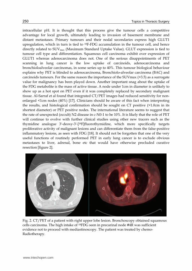
resection [figure 2].
Fig. 2. CT/PET of a patient with right upper lobe lesion. Bronchoscopy obtained squamous cells carcinoma. The high intake of 18FDG seen in precarinal node #4R was sufficient evidence not to proceed with mediastinoscopy. The patient was treated by chemo-Radiotherapy.
www.intechopen.com
Video-Assisted Thoracic Surgery Systematic Mediastinal Nodal Dissection 251
5. Preoperative v postoperative staging
The significance of preoperative as opposed to postoperative staging in resectable early lung cancer is tied to what the clinician wants to do with the information. There might be little disagreement about the N1 disease, but controversy surrounds N2 disease. In our opinion For a CT negative and PET negative mediastinum, no further investigation is needed, and patients should proceed to resectional operation + SND. Further multidisciplinary management should be based on SND staging. This is in line with the latest published British Thoracic Society (BTS) guidelines [19]. The dilemma arises when there is histological evidence of single station N2 disease preoperatively. The choices being (1) avoid surgery all together and opt for chemo-radiotherapy (2) induce chemotherapy before surgery, or (3) make a run for surgery while the tumour is operable and follow that by adjuvant chemotherapy / radiotherapy. The first approach is advocated by Albain et al (2009) who showed that lobectomy will add little to Chemo-radiotherapy for patients with stage IIIa (N2) non-small-cell lung cancer, at the expense of higher mortality (evidence level 1b) [20]. The second approach is supported by the S9900 trial follow up published in 2010 which continues to show that the best treatment for N2 resectable lung cancer would be induction chemotherapy followed by surgery (evidence level 1b) [21]. Rocco et al (2010) is supportive of the third approach, concluding that standard treatment of initially resectable stage IIIa NSCLC remains surgery followed by adjuvant chemotherapy (evidence level 2a) [22]. The subject remains controversial, and patients should be involved in decision taking. Surgery is known to give local control and reduce local recurrence, whereas chemotherapy is a systemic treatment designed to reduce disease progression and distant metastases. Currently we rely on CT/PET, mediastinoscopy or EBUS to direct the patient to one form of treatment or prevent unnecessary operation. However, Lim et al conducted a systematic review of all the published meta-analysis of randomised trials in preoperative versus postoperative chemotherapy in patients with resectable lung cancer (evidence level 1a) [23]. They concluded that in patients with resectable lung cancer, there was no difference in overall and disease-free survival between the timing of administration of chemotherapy (postoperative versus preoperative). Clearly this sends a strong message that earnest preoperative investigation of the mediastinum in PET negative resectable early lung cancer might be unnecessary. Myers et al specifically considered the cost effectiveness of routine mediastinoscopy in CT-negative, PET-negative patients with stage I lung cancer [24]. They concluded that routine mediastinoscopy would add an average 0.01 years (3.65 days) of life at a cost of $201,918 per life-year gained. Therefore they do not recommend routine mediastinoscopy in PET-negative patients. Our practice advocates neoadjuvant chemotherapy followed by surgical VMPR-SND followed by adjuvant chemotherapy based on proper SND staging, provided nodal involvement remains single station or single zonal. Multizonal involvement is best served by chemo-radiotherapy, as it is regarded as systemic disease. Surgery alone will not have an impact on the 5 years survival, but might have a palliative effect on local recurrence, and might be considered for instance to control haemoptysis, or continued sepsis precluding the start of other modalities of treatment such as chemotherapy.
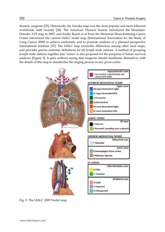
6. Where are these mediastinal nodes?
Although nodal mapping has been there for a long time, it seems that there is considerable
discordance in nomenclature and designation of nodal stations between Asian and European
www.intechopen.com
Topics in Thoracic Surgery 252
thoracic surgeons [25]. Historically the Naruke map was the most popular and most followed
worldwide until recently [26]. The American Thoracic Society introduced the Mountain-
Dressler ATS map in 2007, and finally Rusch et al from the Memorial Sloan-Kettering Cancer
Centre introduced the current IASLC nodal map (International Association for the Study of
Lung Cancer 2009) to achieve uniformity and to promote analyses of a planned prospective
international database [27]. The IASLC map reconciles differences among other used maps,
and provides precise anatomic definitions for all lymph node stations. A method of grouping
lymph node stations together into "zones" is also proposed for the purposes of future survival
analyses [Figure 3]. It goes without saying that surgeons should familiarise themselves with
the details of this map to standardise the staging process in any given centre.
Fig. 3. The IASLC 2009 Nodal map.
www.intechopen.com
Video-Assisted Thoracic Surgery Systematic Mediastinal Nodal Dissection 253
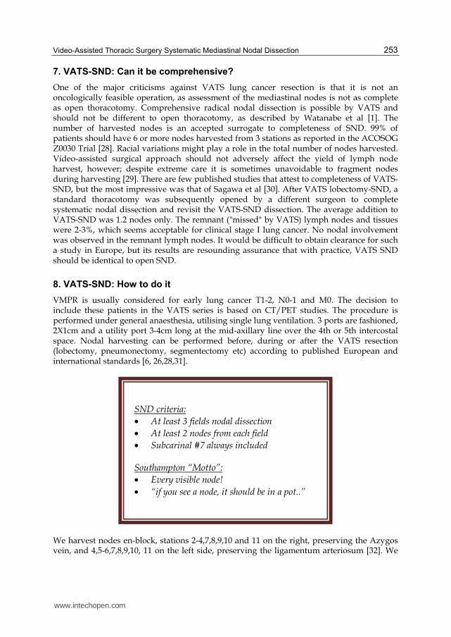
7. VATS-SND: Can it be comprehensive?
One of the major criticisms against VATS lung cancer resection is that it is not an oncologically feasible operation, as assessment of the mediastinal nodes is not as complete as open thoracotomy. Comprehensive radical nodal dissection is possible by VATS and should not be different to open thoracotomy, as described by Watanabe et al [1]. The number of harvested nodes is an accepted surrogate to completeness of SND. 99% of patients should have 6 or more nodes harvested from 3 stations as reported in the ACOSOG Z0030 Trial [28]. Racial variations might play a role in the total number of nodes harvested. Video-assisted surgical approach should not adversely affect the yield of lymph node harvest, however; despite extreme care it is sometimes unavoidable to fragment nodes during harvesting [29]. There are few published studies that attest to completeness of VATS-SND, but the most impressive was that of Sagawa et al [30]. After VATS lobectomy-SND, a standard thoracotomy was subsequently opened by a different surgeon to complete systematic nodal dissection and revisit the VATS-SND dissection. The average addition to VATS-SND was 1.2 nodes only. The remnant ("missed" by VATS) lymph nodes and tissues were 2-3%, which seems acceptable for clinical stage I lung cancer. No nodal involvement was observed in the remnant lymph nodes. It would be difficult to obtain clearance for such a study in Europe, but its results are resounding assurance that with practice, VATS SND should be identical to open SND.
8. VATS-SND: How to do it
VMPR is usually considered for early lung cancer T1-2, N0-1 and M0. The decision to include these patients in the VATS series is based on CT/PET studies. The procedure is performed under general anaesthesia, utilising single lung ventilation. 3 ports are fashioned, 2X1cm and a utility port 3-4cm long at the mid-axillary line over the 4th or 5th intercostal space. Nodal harvesting can be performed before, during or after the VATS resection (lobectomy, pneumonectomy, segmentectomy etc) according to published European and international standards [6, 26,28,31].
We harvest nodes en-block, stations 2-4,7,8,9,10 and 11 on the right, preserving the Azygos vein, and 4,5-6,7,8,9,10, 11 on the left side, preserving the ligamentum arteriosum [32]. We
SND criteria:
• At least 3 fields nodal dissection
• At least 2 nodes from each field
• Subcarinal #7 always included
Southampton “Motto”:
• Every visible node!
• “if you see a node, it should be in a pot..”
www.intechopen.com
Topics in Thoracic Surgery 254
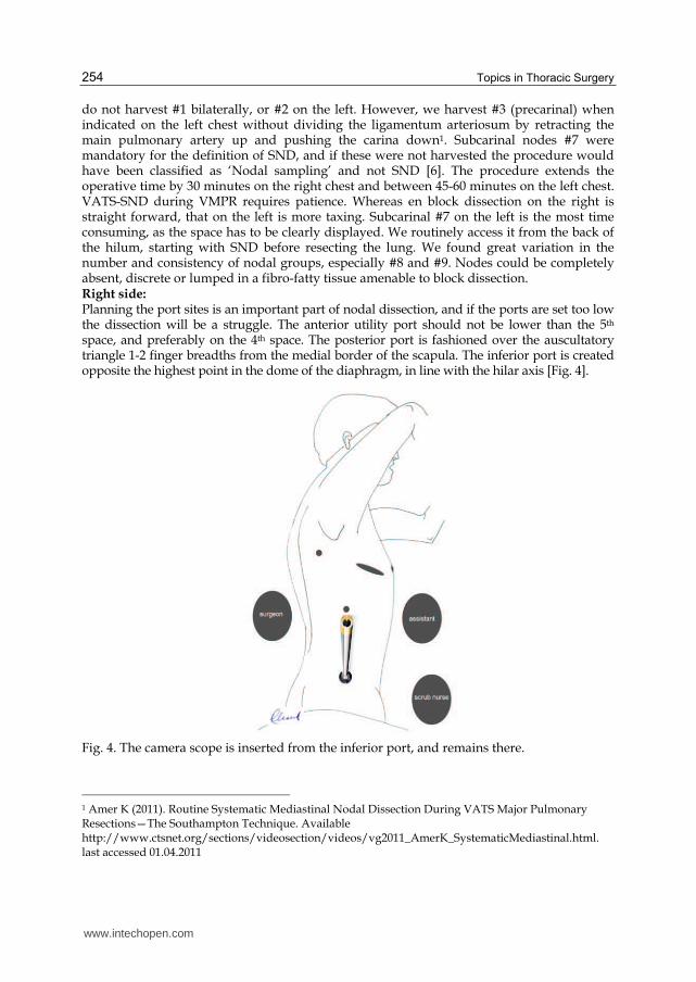
do not harvest #1 bilaterally, or #2 on the left. However, we harvest #3 (precarinal) when indicated on the left chest without dividing the ligamentum arteriosum by retracting the main pulmonary artery up and pushing the carina down1. Subcarinal nodes #7 were mandatory for the definition of SND, and if these were not harvested the procedure would have been classified as ‘Nodal sampling’ and not SND [6]. The procedure extends the operative time by 30 minutes on the right chest and between 45-60 minutes on the left chest. VATS-SND during VMPR requires patience. Whereas en block dissection on the right is straight forward, that on the left is more taxing. Subcarinal #7 on the left is the most time consuming, as the space has to be clearly displayed. We routinely access it from the back of the hilum, starting with SND before resecting the lung. We found great variation in the number and consistency of nodal groups, especially #8 and #9. Nodes could be completely absent, discrete or lumped in a fibro-fatty tissue amenable to block dissection. Right side: Planning the port sites is an important part of nodal dissection, and if the ports are set too low the dissection will be a struggle. The anterior utility port should not be lower than the 5th space, and preferably on the 4th space. The posterior port is fashioned over the auscultatory triangle 1-2 finger breadths from the medial border of the scapula. The inferior port is created opposite the highest point in the dome of the diaphragm, in line with the hilar axis [Fig. 4].
Fig. 4. The camera scope is inserted from the inferior port, and remains there.
1 Amer K (2011). Routine Systematic Mediastinal Nodal Dissection During VATS Major Pulmonary Resections—The Southampton Technique. Available http://www.ctsnet.org/sections/videosection/videos/vg2011_AmerK_SystematicMediastinal.html. last accessed 01.04.2011
www.intechopen.com
Video-Assisted Thoracic Surgery Systematic Mediastinal Nodal Dissection 255
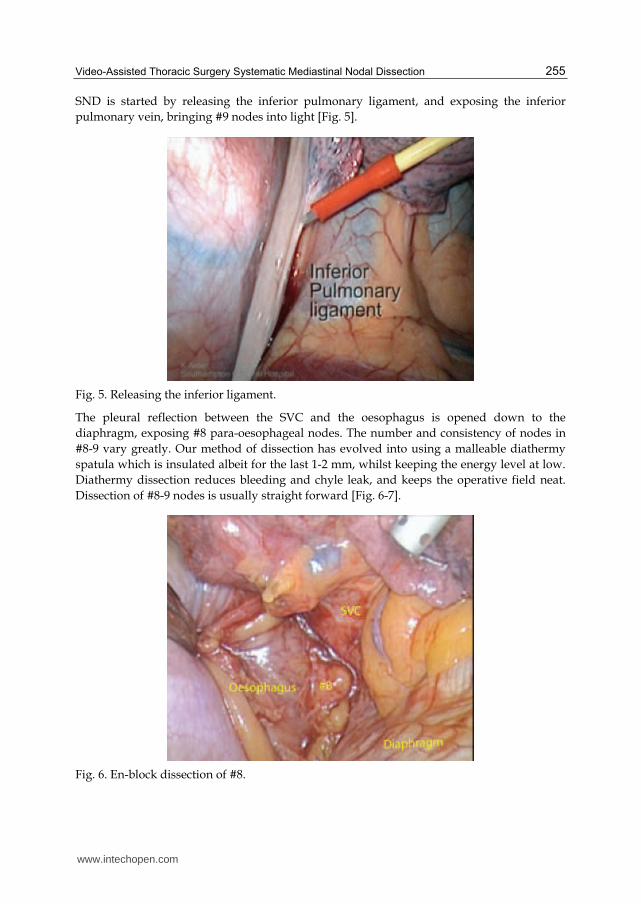
SND is started by releasing the inferior pulmonary ligament, and exposing the inferior
pulmonary vein, bringing #9 nodes into light [Fig. 5].
Fig. 5. Releasing the inferior ligament.
The pleural reflection between the SVC and the oesophagus is opened down to the
diaphragm, exposing #8 para-oesophageal nodes. The number and consistency of nodes in
#8-9 vary greatly. Our method of dissection has evolved into using a malleable diathermy
spatula which is insulated albeit for the last 1-2 mm, whilst keeping the energy level at low.
Diathermy dissection reduces bleeding and chyle leak, and keeps the operative field neat.
Dissection of #8-9 nodes is usually straight forward [Fig. 6-7].
Fig. 6. En-block dissection of #8.
www.intechopen.com

Topics in Thoracic Surgery 256
Fig. 7. Schematic location of #8 & #9.
The subcarinal nodes on the right side are found between the right main bronchus and the
oesophagus. The lung is retracted anteriorly and the pleural reflection at the back of the
hilum is opened from the inferior ligament to the concavity of the Azygos vein, medial to
the vagus nerve. All vagal bronchial branches could be cut with impunity [Fig. 8-10].
Fig. 8. Vagal bronchial twigs.
www.intechopen.com

Video-Assisted Thoracic Surgery Systematic Mediastinal Nodal Dissection 257
Fig. 9. Schematic location of #7.
Fig. 10. The thoracic duct surface anatomy.
The right main bronchus is identified and followed proximally until the left main bronchus is seen and identified. The subcarinal nodes are dissected off their blood supply, and for convenience of retrieval a Polythene bag could be used. This is not always necessary. Careful labelling of nodes is to be practiced here as para-oesophageal #8 and para-bronchial #10 nodes could easily be mistaken as #7. Care must be taken not to dig holes in the membranous part of the bronchus or the delicate oesophagus. One should not worry much about thoracic duct injury in this location, as the duct is tucked away from harm’s way by the oesophagus [Fig. 10]. At the end of this dissection the right main bronchus, the left main bronchus and the subcarinal space should be well on display [Fig. 11].
www.intechopen.com

Topics in Thoracic Surgery 258
Fig. 11. The right subcarinal space.
Dissection of the parabronchial nodes #2-4 lies within the superior triangle. This triangle is
bound by the Vagus and Phrenic nerves, and based on the Azygos vein [Fig. 12].
Fig. 12. The right superior triangle.
The pleura is opened like a trap door, just lateral to the SVC and just above the Azygos vein
[Fig. 13]. The Vagus nerve sould be found plastered to the inside flap of the pleura.
Retraction of the pleura using a Prolene stitch opens the triangle and helps in dissection
[Fig. 14].
www.intechopen.com

Video-Assisted Thoracic Surgery Systematic Mediastinal Nodal Dissection 259
Fig. 13. Trap door to #2-4.
Fig. 14. Retraction of Vagus nerve.
Station #2-4 nodes in the para and pre-tracheal groups exist in a fibro-fatty block that could be dissected en block most of the times. Low energy diathermy is used as before. The dissection is started by pushing the SVC away from the block. One should be aware of the existence of at least one constant vein draining directly from the block to the SVC [Fig. 15]. These veins should be controlled by metal ligaclips or ultrasonic device before proceeding. If they are accidently cut they have the propensity to retract and disappear, making control of the bleeding difficult.
www.intechopen.com

Topics in Thoracic Surgery 260
Fig. 15. Direct draining vein from block to SVC.
Further deeper dissection high in the triangle between the SVC and the block identifies the main stem trachea [Fig. 16]. Once the apex of the block is brought down, the dissection becomes easier.
Fig. 16. Identifying the main stem trachea.
Next the lateral part of the block is separated from the vagus nerve. The block is then lifted off the tracheal, and the retrocaval part is freed. Large lymphatic channels could be seen here, and differentiated from nerves by their lobular contour and loss of sheen. Again the block is delivered out of the chest in a Polythene bag for convenience of retrieval only [Fig. 17]. Small discrete nodes are retrieved directly on a surgical instrument.
www.intechopen.com

Video-Assisted Thoracic Surgery Systematic Mediastinal Nodal Dissection 261
Fig. 17. Retrieval in a Polythene bag of #2-4.
The bed of the superior triangle is made of the arch of the aorta and the right bracheocephalic artery and the main stem trachea [Fig. 18]. The recurrent laryngeal nerve descends into the thoracic inlet parallel to the vagus on the lateral side of the carotid artery. It makes a quick exit out of the chest as it loops around the origin of the right subclavian artery, soon after it enters the thoracic inlet. It continues its course cephalad towards the trachea-oesophageal groove in the neck. This point of looping is approximately 1 cm from the aortic arch, and corresponds to the length of the brachiochephalic trunk [Fig. 18]. It lies at the apex of the superior triangle, and diathermy should be used with extreme caution in this area, especially when the highest #2 nodes are attempted.
Fig. 18. Location of the right recurrent laryngeal nerve.
www.intechopen.com
Topics in Thoracic Surgery 262
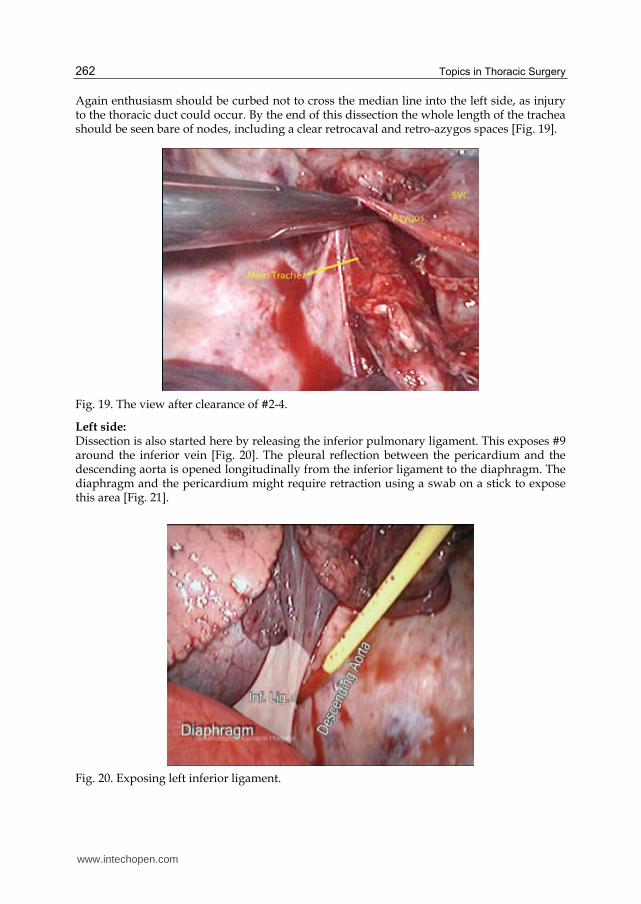
Again enthusiasm should be curbed not to cross the median line into the left side, as injury to the thoracic duct could occur. By the end of this dissection the whole length of the trachea should be seen bare of nodes, including a clear retrocaval and retro-azygos spaces [Fig. 19].
Fig. 19. The view after clearance of #2-4.
Left side: Dissection is also started here by releasing the inferior pulmonary ligament. This exposes #9 around the inferior vein [Fig. 20]. The pleural reflection between the pericardium and the descending aorta is opened longitudinally from the inferior ligament to the diaphragm. The diaphragm and the pericardium might require retraction using a swab on a stick to expose this area [Fig. 21].
Fig. 20. Exposing left inferior ligament.
www.intechopen.com

Video-Assisted Thoracic Surgery Systematic Mediastinal Nodal Dissection 263
Fig. 21. Pleural landmarks for opening #8 	.
Fig. 22. En-block dissection of left #8.
This exposes #8 nodes which again could be absent, discrete or forming a fibro-fatty block.
Care is taken not to injure the vagus, oesophagus, and other organs which are usually not
there but could be there, such as a hiatus hernia [Fig. 22].
Dissection of the subcarinal #7 nodes on the left side is time-consuming, and require a
prepared plan of action, good suction and detailed mastery of the surrounding anatomy. On
retracting the lung anteriorly two nerves and a vein are noted to cross the arch of the aorta.
The Phrenic nerve passes anterior to the hilum, whereas the Vagus passes posterior to the
hilum. The superior intercostal vein draining the upper 3-4 spaces traverses the upper part
of the aorta, crossing the origins of the left subclavian and carotid arteries and drain straight
into the innominate vein [Fig. 23].
www.intechopen.com
Topics in Thoracic Surgery 264
Fig. 23. The hilum watershed between Phrenic and Vagus nerves.
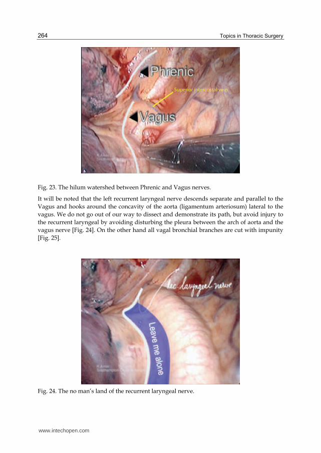
It will be noted that the left recurrent laryngeal nerve descends separate and parallel to the
Vagus and hooks around the concavity of the aorta (ligamentum arteriosum) lateral to the
vagus. We do not go out of our way to dissect and demonstrate its path, but avoid injury to
the recurrent laryngeal by avoiding disturbing the pleura between the arch of aorta and the
vagus nerve [Fig. 24]. On the other hand all vagal bronchial branches are cut with impunity
[Fig. 25].
Fig. 24. The no man’s land of the recurrent laryngeal nerve.
www.intechopen.com
Video-Assisted Thoracic Surgery Systematic Mediastinal Nodal Dissection 265
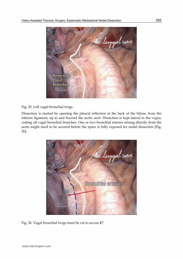
Fig. 25. Left vagal bronchial twigs.
Dissection is started by opening the pleural reflection at the back of the hilum, from the
inferior ligament, up to and beyond the aortic arch. Dissection is kept lateral to the vagus,
cutting all vagal bronchial branches. One or two bronchial arteries arising directly from the
aorta might need to be secured before the space is fully exposed for nodal dissection [Fig.
26].
Fig. 26. Vagal bronchial twigs must be cut to access #7.
www.intechopen.com
Topics in Thoracic Surgery 266
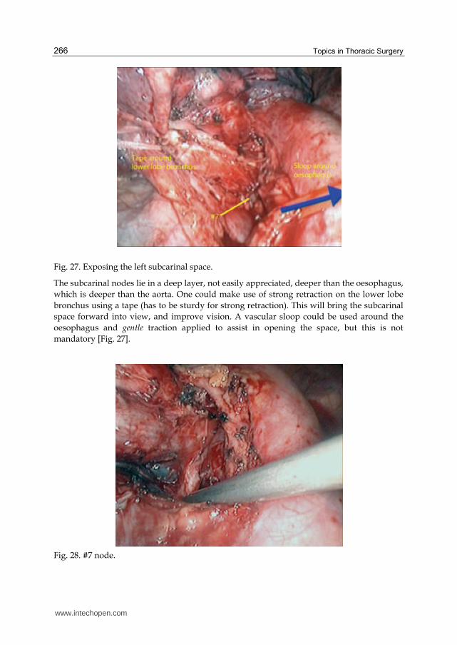
Fig. 27. Exposing the left subcarinal space.
The subcarinal nodes lie in a deep layer, not easily appreciated, deeper than the oesophagus,
which is deeper than the aorta. One could make use of strong retraction on the lower lobe
bronchus using a tape (has to be sturdy for strong retraction). This will bring the subcarinal
space forward into view, and improve vision. A vascular sloop could be used around the
oesophagus and gentle traction applied to assist in opening the space, but this is not
mandatory [Fig. 27].
Fig. 28. #7 node.
www.intechopen.com

Video-Assisted Thoracic Surgery Systematic Mediastinal Nodal Dissection 267
The principle here is to follow the lower lobe bronchus proximally, as it leads us to the
subcarinal space. Pinpoint diathermy dissection of the nodes off their blood supply is
performed, taking care not to dig holes in the membranous part of the bronchus. The right
main bronchus and the subcarinal space should be well on display by the end of this
dissection [Fig. 28-29].
Fig. 29. Anatomy of the subcarinal space.
Nodes that are clearly related to the inferior vein, lower lobe bronchus or the main
pulmonary artery are labelled as #10. However; the most lateral of the aorto-pulmonary
group are labelled as #4, and these are at a deeper level than #10.
Fig. 30. Exposing pretracheal #3 nodes.
www.intechopen.com

Topics in Thoracic Surgery 268
The precarinal #3 nodes could be accessed from the left side if required. The plane of
dissection lies below the pulmonary artery, and hence there is no need to dissect and cut
the ligamentum arteriosum. The main pulmonary artery is freed from the bronchus and a
sloop passed around it. The space under the artery is dissected, and explored by pushing
the carina down and the artery up. This manoeuvre exposed the main stem trachea [Fig.
30]. Pretracheal nodes are identified and dissected. Minimal use of diathermy is
recommended in this position, as this is the likely position to injure the recurrent
laryngeal nerve.
Fig. 31. The left superior triangle.
Dissection of #5 (preaortic) and #6 (sub-aortic and aorto-pulmonary) nodes should be
attempted en block. These nodes exist in a triangle similar to the right side, bound by the
vagus and Phrenic nerves and the arch of the aorta [Fig. 31]. The fibro-fatty block is lifted off
the main pulmonary artery into the aorto-pulmonary space, medial to the vagus nerve. The
phrenic nerve is identified and slung using a vascular sloop to avoid harming it. The nodal
block is dissected up to the origin of the left subclavian artery, and the block delivered out of
the chest.
9. Complications of SND
VATS-SND is safe, and does not add to the morbidity or mortality of the originally planned operation. However there are some complications the surgeon should be aware of: Major complications:
• Vascular injury; SVC, Aortic arch, Azygos vein etc.
• Bronchial injury; usually the membranous part of major bronchi, especially dissecting around the subcarinal space
• Recurrent laryngeal nerve injury; on the right the danger arises when diathermy is used around the origin of the subclavian artery, and on the left when dissecting #3 (precarinal) at the space between main pulmonary artery and main stem trachea.
www.intechopen.com
Video-Assisted Thoracic Surgery Systematic Mediastinal Nodal Dissection 269
• Chyle leak; is rare and usually occurs if dissection involved large lymphatic ducts, mobilisation of oesophagus or in the presence of abnormal anatomical course of the thoracic duct.
• Port-site seedling, which is rare (0.5%) and seems to happen irrespective of whether the nodes were retrieved in a polythene bag or not [32,33].
Minor complications: • Increased postoperative tube drainage. • Irritant cough due to diathermy close to the main bronchi. • Temporary odynophagia (painful swallowing) due to mobilisation of the oesophagus.
10. VATS nodal sampling v dissection
The current evidence suggests that complete mediastinal lymph node dissection is associated with improved survival compared with node sampling in patients with stage I–IIIa NSCLC undergoing resection [34].
11. SND and immune response
It was reported that Systematic lymphadenectomy added to major lung resection performed by open thoracotomy does not increase postoperative humoral immune response in uncomplicated cases [35]. However; there are no studies in the literature that looked into the VMPR-SND and the role of SND in postoperative inflammatory response.
12. Conclusion
VATS Systematic Nodal Dissection during VATS major pulmonary resections is feasible and safe. It should be performed routinely even when nodal involvement is unlikely, as 10% of patients in clinical stage N0-1 will have N2 disease. Multidisciplinary adjuvant treatment of lung cancer should be based on SND staging.
13. References
[1] Watanabe A, Koyanagi T, Ohsawa H, Mawatari T, Nakashima S, Takahashi N, Sato H, Abe T. Systematic node dissection by VATS is not inferior to that through an open thoracotomy: A comparative clinicopathologic retrospective study. Surgery 2005;138:510-517.
[2] Arriagada R, Bergman B, Dunant A, Le Chevalier T, Pignon JP, Vansteenkiste J; Cisplatin-Based Adjuvant Chemotherapy in Patients with Completely Resected Non–Small-Cell Lung Cancer. N Engl J Med. 2004;350:351-60.
[3] Douillard JY, Rosell R, De Lena M, Carpagnano F, Ramlau R, Gonzáles-Larriba JL, Grodzki T, Pereira JR, Le Groumellec A, Lorusso V, Clary C, Torres AJ, Dahabreh J, Souquet PJ, Astudillo J, Fournel P, Artal-Cortes A, Jassem J, Koubkova L, His P, Riggi M, Hurteloup P. Adjuvant vinorelbine plus cisplatin versus observation in patients with completely resected stage IB-IIIA non-small-cell lung cancer (Adjuvant Navelbine International Trialist Association [ANITA]): a randomised controlled trial: Lancet Oncol. 2006;7:719-27.
[4] Union Internationale Contre le Cancer. TNM classification of malignant tumours, Wiley-Liss, New York (1997) 93–7.
www.intechopen.com
Topics in Thoracic Surgery 270
[5] Goldstraw P, Crowley J, Chansky K, Giroux DJ, Groome PA, Rami-Porta R, Postmus PE, Rusch V, Sobin L. The IASLC Lung Cancer Staging Project: proposals for the revision of the TNM stage groupings in the forthcoming (seventh) edition of the TNM classification of malignant tumours. J Thorac Oncol 2007;2:706-714.
[6] Lardinois D, De Leyn P, Van Schil P, Rami Porta R, Waller D, Passlick B, Zielinski M, Lerut T, Weder W. ESTS guidelines for intraoperative lymph node staging in non-small cell lung cance. Eur J Cardiothorac Surg 2006;30:787-792.
[7] Asamura H, Nakayama H, Kondo H, Tsuchiya R, Naruke T. Lobe-specific extent of systematic lymph node dissection for non-small cell lung carcinomas according to a retrospective study of metastasis and prognosis. J Thorac Cardiovasc Surg. 1999 Jun;117(6):1102-11.
[8] Misthos P, Sepsas E, Athanassiadi K, Kakaris S, Skottis I. Skip metastases: analysis of their clinical significance and prognosis in the IIIA stage of non-small cell lung cancer Eur J Cardiothorac Surg 2004;25:502-508
[9] Ono T, Minamiya Y, Ito M, Saito H, Motoyama S, Nanjo H, Ogawa J. Sentinel node mapping and micrometastasis in patients with clinical stage IA non-small cell lung cancer. Interact Cardiovasc Thorac Surg. 2009 Oct;9(4):659-61.
[10] Riethdorf S, Wikman H, Pantel K. Review: Biological relevance of disseminated tumor cells in cancer patients. Int J Cancer. 2008 Nov 1;123(9):1991-2006
[11] Witte B, Hürtgen M. Video-assisted mediastinoscopic lymphadenectomy (VAMLA). J Thorac Oncol. 2007 Apr;2(4):367-9.
[12] Witte B, Messerschmidt A, Hillebrand H, Gross S, Wolf M, Kriegel E, Neumeister W, Hürtgen M. Combined videothoracoscopic and videomediastinoscopic approach improves radicality of minimally invasive mediastinal lymphadenectomy for early stage lung carcinoma. Eur J Cardiothorac Surg. 2009 Feb;35(2):343-7. Epub 2008 Dec 16.
[13] Zieliński M. Technical pitfalls of transcervical extended mediastinal lymphadenectomy-how to avoid them and to manage intraoperative complications. Semin Thorac Cardiovasc Surg. 2010 Autumn;22(3):236-43.
[14] Van Tinteren H, Hoekstra OS, Smit EF, van den Bergh JH, Schreurs AJ, Stallaert RA, van Velthoven PC,Comans EF, DiepenhorstFW, VerboomP, van Mourik JC, Postmus PE, Boers M, Teule GJ. Effectiveness of PET in the preoperative assessment of patients with suspected non-small cell lung cancer: The PLUS multicenter randomised trial. Lancet 2002;359:1388-92.
[15] Gillies RJ, Robey I, Gatenby RA. Causes and consequences of increased glucose metabolism of cancers. J Nucl Med. 2008;49:24S-42S.
[16] Plathow C, Weber WA. Tumor cell metabolism imaging. J Nucl Med. 2008;49:43S-63S. [17] Al-Sarraf N, Gately K, Lucey J, Wilson L, McGovern E, Young V. Lymph node staging
by means of positron emission tomography is less accurate in non-small cell lung cancer patients with enlarged lymph nodes: Analysis of 1145 lymph nodes. Lung Cancer 2008;60:62-68
[18] Delphine L. Chen and Farrokh Dehdashti. Advances in Positron Emission Tomographic Imaging of Lung Cancer. J Thorac Cardiovasc Surg. 2004;127:1093-9.
[19] Eric Lim, David Baldwin, Michael Beckles, John Duffy, James Entwisle, Corinne Faivre-Finn, Keith Kerr, Alistair Macfie, Jim McGuigan, Simon Padley, Sanjay Popat, Nicholas Screaton, Michael Snee, David Waller, Chris Warburton, Thida Win.
www.intechopen.com
Video-Assisted Thoracic Surgery Systematic Mediastinal Nodal Dissection 271
Guidelines on the radical management of patients with lung cancer. Thorax 2010;65:iii1–iii27
[20] Albain KS, Swann RS, Rusch VW, Turrisi AT 3rd, Shepherd FA, Smith C, Chen Y, Livingston RB, Feins RH, Gandara DR, Fry WA, Darling G, Johnson DH, Green MR, Miller RC, Ley J, Sause WT, Cox JD. Radiotherapy plus chemotherapy with or without surgical resection for stage III non-small-cell lung cancer: a phase III randomised controlled trial. Lancet 2009;374:379-386
[21] Pisters KM, Vallières E, Crowley JJ, Franklin WA, Bunn PA Jr, Ginsberg RJ, Putnam JB Jr, Chansky K, Gandara D. Surgery with or without preoperative paclitaxel and carboplatin in early-stage non-small-cell lung cancer: Southwest Oncology Group Trial S9900, an intergroup, randomized, phase III trial. J Clin Oncol. 2010;28:1843-9.
[22] Rocco G, Perrone F, Rossi A, Gridelli C. Surgical management of non-small cell lung cancer with mediastinal lymphadenopathy. Clinical Oncology 2010;22:325-333
[23] Lim E, Harris G, Patel A, Adachi I, Edmonds L, Song F. Preoperative versus postoperative chemotherapy in patients with resectable non-small cell lung cancer: systematic review and indirect comparison meta-analysis of randomized trials. J Thorac Oncol. 2009;4:1380-8
[24] Meyers BF, Haddad F, Siegel BA, Zoole JB, Battafarano RJ, Veeramachaneni N, Cooper JD and Patterson GA. Cost effectiveness of routine mediastinoscopy in CT and PET-screened patients with stage I lung cancer. J Thorac Cardiovasc Surg 2006;131:822-829.
[25] Watanabe S, Ladas G, Goldstraw P. Inter-observer variability in systematic nodal dissection: comparison of European and Japanese nodal designation. Ann Thorac Surg. 2002 Jan;73(1):245-8
[26] Naruke T, Tsuchiya R, Kondo H, Nakayama Haruhiko, Asamura H. Lymph node sampling in lung cancer: how should it be done? Eur J Cardiothorac Surg 1999;16:17-24.
[27] Rusch VW, Asamura H, Watanabe H, Giroux DJ, Rami-Porta R, Goldstraw P; Members of IASLC Staging Committee.The IASLC lung cancer staging project: a proposal for a new international lymph node map in the forthcoming seventh edition of the TNM classification for lung cancer. J Thorac Oncol. 2009 May;4(5):568-77.
[28] Darling GE, Allen MS, Decker PA, Ballman K, Malthaner RA, Inculet RI, Jones DR, McKenna RJ, Landreneau RJ, Putnam JB Jr. Number of Lymph Nodes Harvested from a Mediastinal Lymphadenectomy: Results of the Randomized, Prospective ACOSOG Z0030 Trial. Chest. 2010 Sep 9. [Epub ahead of print]
[29] Gossot, D (2010). Atlas of endoscopic Major Pulmonary Resections. Paris: Springer-Verlag. 22-32.
[30] Sagawa M, Sato M, Sakurada A, Matsumura Y, Endo C, Handa M, Kondo T. A prospective trial of systematic nodal dissection for lung cancer by video-assisted thoracic surgery: can it be perfect? Ann Thorac Surg 2002;73:900-4.
[31] Watanabe A, Koyanagi T, Obama T, Ohsawa H, Mawatari T, Takahashi N, Ichimiya Y. Assessment of node dissection for clinical stage I primary lung cancer by VATS. Eur J Cardiothorac Surg. 2005;27:745-752.
[32] Amer K, Khan AZ, Singh N, Addis B, Jogai S, Harden S, Peebles C, Brown I. Video-assisted thoracic surgery systematic mediastinal nodal dissection and stage migration: impact on clinical pathway. Eur J Cardiothorac Surg. 2011 Apr 13.
www.intechopen.com
Topics in Thoracic Surgery 272
[33] McKenna RJ, Houck W, Fuller CB. Video-assisted thoracic surgery lobectomy: experience with 1,100 cases. Ann Thorac Surg. 2006;81(2):421-5.
[34] Scott WJ, Allen MS, Darling G, Meyers B, Decker PA, Putnam JB, McKenna RW, Landrenau RJ, Jones DR, Inculet RI, Malthaner RA. Video-assisted thoracic surgery versus open lobectomy for lung cancer: a secondary analysis of data from the American College of Surgeons Oncology Group Z0030 randomized clinical trial. J Thorac Cardiovasc Surg. 2010 Apr;139(4):976-81
[35] Szczesny TJ, Słotwiński R, Szczygieł B, Stankiewicz A, Zaleska M, Kopacz M, Olesińska-Grodź A. Systematic mediastinal lymphadenectomy does not increase postoperative immune response after major lung resections. Eur J Cardiothorac Surg. 2007 Dec;32(6):868-72. Epub 2007 Oct 17.
www.intechopen.com

Topics in Thoracic SurgeryEdited by Prof. Paulo Cardoso
ISBN 978-953-51-0010-2Hard cover, 486 pagesPublisher InTechPublished online 15, February, 2012Published in print edition February, 2012
InTech EuropeUniversity Campus STeP Ri Slavka Krautzeka 83/A 51000 Rijeka, Croatia Phone: +385 (51) 770 447 Fax: +385 (51) 686 166www.intechopen.com
InTech ChinaUnit 405, Office Block, Hotel Equatorial Shanghai No.65, Yan An Road (West), Shanghai, 200040, China
Phone: +86-21-62489820 Fax: +86-21-62489821
Thoracic Surgery congregates topics and articles from many renowned authors around the world coveringseveral different topics. Unlike the usual textbooks, Thoracic Surgery is a conglomerate of different topics fromPre-operative Assessment, to Pulmonary Resection for Lung Cancer, chest wall procedures, lung cancertopics featuring aspects of VATS major pulmonary resections along with traditional topics such as Pancoasttumors and recurrence patterns of stage I lung disease, hyperhidrosis, bronchiectasis, lung transplantationand much more. This Open Access format is a novel method of sharing thoracic surgical information providedby authors worldwide and it is made accessible to everyone in an expedite way and with an excellentpublishing quality.
How to referenceIn order to correctly reference this scholarly work, feel free to copy and paste the following:
Khalid Amer (2012). Video-Assisted Thoracic Surgery (VATS) Systematic Mediastinal Nodal Dissection, Topicsin Thoracic Surgery, Prof. Paulo Cardoso (Ed.), ISBN: 978-953-51-0010-2, InTech, Available from:http://www.intechopen.com/books/topics-in-thoracic-surgery/video-assisted-thoracic-surgery-vats-systematic-mediastinal-nodal-dissection