vickifaye g. abdellah, sed, edd, rn, f aan curriculum vitae name: vicki larrainc simmons degree and...
TRANSCRIPT
THE PREVALENCE OF BACTERIAL CONT AMINA nON IN THE VENTILATOR BELLOWS OF ANESTHESIA MACHINES
1.
by
Vicki L. Simmons, BSN LT,USN,NC
Presented to the Graduate School of Nursing Faculty of the Unifonned Services University of the Health Sciences
in Partial Fulfillment of the Requirements
for the Degree of MASTER OF SCIENCE in NURSING
UNIFORMED SERVICES UNIVERSITY OF THE HEALTH SCIENCES
July 1997 .
Committee Chair
Committee Member e McCarthy, CRNA, Ph.
~~ Committee Member Eugene Levine, Ph.D.
Dean and Professor, Graduate School of Nursing Faye G. Abdellah, SeD, EdD, RN, F AAN
CURRICULUM VITAE
Name: Vicki Larrai nc S immons
Degree and date to be conferred: Master of Science in Nursing 1997
Secondary Educat ion: Valdosta High School, Valdosta, Ga. Ju ne 6, 1977
Collegiate instit ut ions aucnded Dales Degree Date of Degree
Abraham Baldwin Agricu ltural College George Washington University
6177 - 3178 5178-5179 6/88 - 8/88 San Diego City College
Point Lorna Nazarene College University of Californi a, San Diego Extension Complex Uniformed Services Uni versity of the Health Sciences
Major: Nurse Anesthesia
Professional pos itions held :
Graduate Student Nurse Anesthes ia
Assistant Div ision Officer, PACU Evening Charge Nurse , PACU Staff Nurse, PACU Staff Nurse , Same Day Surgery Night Charge Nurse, Medical Ward Staff Nurse, Medical Ward
9/88 - 6/91 BSN 617191
9/92 - 5/93
6/95 - Present
Uniformed Services University of the Health Sciences. Bethesda. Md. Naval Medical Center, San Diego, Ca. Naval Medica l Center, San Diego, Ca. Naval Medical Center, San Diego, Ca. Naval Medical Center, San Diego, Ca. Naval Medical Center, San Diego, Ca. Nava l Medical Center, San Diego, Ca.
(ii)
DISCLAIMER STATEMENT Department of Defense
"This work was sUPPol1cd by the Uniformed Services University of the Health Sciences.
The opinions or assertions contained here in arc the pri vate opinions of the author and arc
not to be construed as official or rcnecting the views of the Department of Defense or the
Uni formed Services University of the Health Sciences."
(i ii )
COPYRIGHT STATEMENT
The author hereby certifies that the usc of an y copyrighted material in the thesis titled:
THE PREVALENCE OF BACTERIAL CONT AMIN ATION IN THE
VENTILATOR BELLOWS OF ANESTHESIA MACHINES
beyond brief excerpts is with the permiss ion of the copyright owner, and will save and
hold harmless the Uni formed Services University of the Health Sciences from any
damage which may arise from such copyright violations.
(iv)
ABSTRACT
The anesthes ia breathing circuit has been inconclusively implicated as a source of
postoperative, hospital -acquired respiratory infections that foll ow surgery. Little
investigation of the prevalence of bacterial contamination in the anesthesia vent ilator
bellows has been donc. The purpose of thi s study was to determine the prevalence of
bacterial contamination in the ventilator be llows of anesthesia machines. A prospective
study of 12 anesthesia machines at a major medical facility was conducted to determine
whether ventilator bellows are bacterially contaminated and to quanti fy and identify any
bacterial organisms present. Two sets of samples were collected. Twelve machines were
sampled in the morning and 10 machines were sampled in the afternoon after surgery was
completed. Us ing sterile culturette bacterial swabs and aseptic technique, samples were
collected by swabbing the internal diameter of the ventil ator bellows. All of the morning
samples and 80% of the afternoon samples were negati ve for bacterial growth. Two
(20%) of the afternoon samples had 3+ growth or greater than 100 colony formi ng units
of Staphylococcus epidermidis indicating external contamination during collection or
laboratory processi ng. From these findings, the ven til ator be llows does nol appear to
harbor pathogenic microorganisms. These data support thai the present methodology of
cleaning and sterili zation of the anesthesia machine at thi s facility is effect ive.
(v)
THE PREVALENCE OF BACTERIAL CONTAMINATION IN THE VENTILATOR BELLOWS OF ANESTHESIA MACHINES
by
Vicki Larraine Si mmons, BSN
THESIS
Presented to the Graduate School of Nursing Faculty of
the Uniformed Services University of the Health Sciences
in Partial Fulfi llment
of (he Requirements
for the Degree of
MASTER OF SCIENCE DEGREE
UNIFORMED SERVICES UNIVERSITY OFTHE HEALTH SCIENCES
October, 1997
(vi)
DEDICATION
To my husband and children I dedicate the creation of thi s thes is. Without the ir
love, encouragement , and support the attainme nt of a dream and the creat ion of th is thes is
would not have been poss ible.
To my mother and father , I dedicate thi s paper and thank you for instilling wi lh in
me a strong work ethic and the desi re for knowledge and for your many years of
encouragement and support.
(vi i)
ACKNOWLEDGMENT
The assistance, gu idance and support of numerous people have contributed to
making possib le the attainme nt o f thi s degree. I am espec ially grateful to Dr. W. Patrick
Monaghan chairperson , and the members of the thes is advisory committee, Dr. E. Jane
McCarthy, and Dr. Eugene Levine. Their guidance, knowledge, patience, and suppo rt
was invaluable.
Gratefu l appreciation is also ex tended to HMC Salvador and to HM I Royg, the
laboratory personnel responsible for the laboratory processing and testing of the samples,
who gave wi ll ingly of the ir time during thi s study.
(vii i)
TABLE OF CONTENTS
Chapter One _____________________ _
Introduct ion ____________________ _
Statement of Problem _________________ _
Research Questions _________________ _
Definition of Terms ____ ______________ _
Li mitations of Study _________________ _
Chapter Two _____________ _______ __ _
Theoretical Framework ________________ _
Literature Review _ _________________ _
Chapter Three ______________________ _
Me' hodology ______________ _ ___ _
Chapter Four ______________ _____ _ __ _
Results ___________________ _ _
Chapter Five _____________________ _ _
Conclusions ____________ ________ _
References ________________ _______ _
(ix)
5
6
6
8
9
9
12
18
18
2 1
21
25
25
27
LIST OF FIGURES
Figure 1. The Number of Morn ing and Aftern oon Samples that were Pos iti ve and
Negati ve for Bacterial Growth in 12 Anesthesia Ventilator Bellows ....... .. . 23
Figure 2. The Age in Months of the 12 Anesthesia Ven til ator Bellows that were
Sampled in thi s Study .. ... ... .. .. ..... ....... .... .. .. ............ .. .. ... ... .......... .... ... ... . 24
(x)
CHAPTER ONE: INTRODUCTION
The prevalence of bacterial contami nation in various components of the
anesthesia mach ine may be related to hospi tal-acqui red in fect ions. The anesthes ia
ventilator bellows is a reservoir that receives the exhaled gases from the patient via the
exhalation chamber. AI the completion of exhalation, it redi rects those same gases
through the carbon diox ide absorber which arc mixed with gases from the common gas
out let, and ult imately back to the palient via the inhalat ion chamber and the patient's
breathing circuit. The ventil ator bellows port ion of the anesthesia machine was stud ied
because it is a major component of the anesthes ia breathing circui t and is often
overlooked.
Nosocomial infections are major sources of morbidity and mortality among
hospitalized pat ients. "Nosocomial" is a medica l te rm for "hos pi tal-associated" and is
used to describe infections that arise during hospit alization as a complication of another
ill ness (Ryan, 1994). As a result o f these in fect ions, hospi tal costs are increased, hospi tal
stays are prolonged, and pati ents are thereby placed at higher risk for morbidity and
mortali ty. Nosocomial in fec tions are reputed to extend hospi talization an average of 4
days per infection, and nearl y 4% of all nosocomial in fec tions resulted in death (Smith, et
aI, 1996). The rates of nosocomial infections for most hospitals in the United States arc
estimated to range between 3 and 5%. These pe rcentages represent millions of hospi lal
acquired cases annually since more than 40 mi llion persons are hospitali zed in the United
States each year (Ryan, 1994).
Infectious hazards appear to be inherent in the hospital environment. The
infectious agents responsible for nosocomial infections ari se from various sources
2
including the patents' own normal bacterial nora. In addition to any
immunocompromising disease or therapy, the hospital may impose additional risks by
treatments that breach the normal defense barriers. Surgery, urinary or intravenous
catheters, and invasive diagnostic procedures all may provide normal nonpathogenic
bacterial or viral flora with access 10 usually sterile s ites (Ryan, 1994). Once introduced
to these protected o r immunological pri vi leged areas the normal barriers are breached and
acute infection may result .
In conj unct ion with these hospi tal generated ri sks, more opportuni stic infections
are occurring due to the increasing elderl y pat ient component of the population. The
modern pract ice of med ic ine is keeping ind ividuals alive longer through surgical
procedures and powerful drugs that affect immune status. As a consequence, organisms
that were once considered harmless are now feared as opportuni stic pathogens. The
geriatric population and patients who are immu nocompromised therefore are specificall y
at risk.
Nosocomial infections are also known to result from the phenomenon of cross
infect ion, or "the transmission from onc patie nt to another" (Ryan . 1994, p. 824).
Transmission is usuall y by direct contact , by hosp ital personnel fo r example, but , airborne
transmission is also possible. Much of the success of modern medicine is re lated to
medical devices that support or monitor basic body functions. Neve rtheless, by their
very nature, invasive dev ices such as catheters and vent ilators catTy the ri sk of
nosocomial infec tions because they bypass normal defense barriers, provid ing organ isms
access to normally steril e fluid s and ti ssues. The ri sk of infect ion is associaled wi th the
degree of patient debilitation and various factors concern ing the design and management
of these devices.
Medical devices that arc most frequently associated with nosocomial infections
are urinary and vascular catheters, and ventilators (Ryan, 1994). According to Ryan
(1994, p. 825), "machines that assist or con trol respiration by pumping air directly into
the trachea have a great potential for infection if the aerosol they deliver becomes
contaminated." Lower respiratory tract infections account for 14 to 18% of all
nosocomial infec tions, the third largest type of such infections.
Infection control is paramount to the prevention of nosocomial infections and the
problems associated with them. Asepsis, the central concept of infection control, is
defined by Ryan (1994, p. 827) as the "prevention of contact between microorganisms
and susceptible sites" using methods of steri li zation and di sinfection. Ryan (1994, p.
827) also states that "The surgical su ite and operating room represent the most contro ll ed
and rigid application of aseptic principles."
3
lnhalat ional anesthesia equipment has been suspect since the 1950s as a potential
source for organisms that cause nosocomial infect ions. This primary concern of the ri sk
of cross- infection has led to the evolution of modern anesthesia practice and equipment
over the past 40 years. Disposable endotracheal tubes and c ircuits, bacterial filters, and a
variety of standard infect ion corllrol practices arc in use today as the result of ongoing
attempts to reduce or eliminate the potential ri sk of transmiss ion of organisms to patients.
Numerous studies have been reponed which attempt to clearly establi sh or refute
any connection between anesthesia equipmenl and postoperative nosocomial infect ions.
It is quite difficuh to prove any direct cause and effect relationship between anesthesia
machines and subsequent nosocomial infec tions. Although new methods and equipment
were consequent ly deve loped and have become the standard of pract ice, the controversy
as to anesthes ia's ro le in nosocomial infect ions con tinues to be debated. The possible
introduction of bac teria from the anesthes ia circle system may undoubtedl y increase the
patient's risk for developing a lower respi ratory tracl infec tion. Accord ing to Smith ,
Wygant, McGrory and Silka ( 1996, p. 153), "A relationship between contamination of
anesthes ia equipment and infect ion of the patient must be considered because as many as
40 out of 10,000 patients receivi ng general anesthesia may suffer postoperati vely from
hospi tal.acqu ired pneumonia." However, most anesthesia providers have been reluctant
in recogn izing that contaminated anesthesia equ ipment and venti lators may be a
sign ificant source of cross-infection because of the difficult y in pinpo int ing the ac tual
source of postoperati ve pulmonary infections (Shiotani , Nicholcs, Ball inger, and Shaw,
1971). This may be due in part to the diffi culty in recognizing and tracing the actual
epidemiology and fomites invo lved in the nosocomial infection. Clin ical repon s which
attribute postoperat ive infect ion to contaminated anesthesia equipment are rare and often
inconclusive (Garibaldi . Britt, Webster. and Pace, 198 1).
Carbon diox ide absorbers, inspiratory and expi ratory val ves, and ventil ator
bellows are integra l components of the anesthesia machine and complete the connec tion
to the patient's breathi ng ci rcui t. Because bacte rial fi lte rs and disposable ci rcu its arc in
routine use, these components of the anesthesia machine which are located more distan t
from the pat ient . and yet prev iously identi fied to be potcnlial sources of organisms.
receive less attent ion in routi ne cleaning and decontamination pract ices performed by
most anesthesia depart ments (Dryde n. 1975). These areas arc ofte n not rout inely cleaned
or decontaminated between uses on patients because they pose mechan ical problems in
4
5
handling and may nOI readily tolerate the usual dccolllaminalion methods available
(Dryden, 1975). The typical busy operating room schedule does not usually allow for the
time necessary to perform th is level of cleani ng between cases. The prevailing altitude
appears to be thallhese arcas pose little threat to the patient (Dryden, 1975). Some
studies have shown that organ isms can travel from the exp iratory limb, through the
carbon d iox ide absorber and to the ventil ator. Organi sms have been cultured from these
machines although most of these studies predate Ihe advent and routine use of d isposable
equipment.
Statement of the Problem
Litt le investi gation of the prevalence of bacteri al contamination in the anesthesia
ventilator bellows has been reported. The pri mary focus of infect ion control in anesthesia
appears to center around the patient's breathi ng c ircuit. However, most patients receivi ng
general anesthesia arc routinely ven tilated mechanicall y lI sing anesthesia vent ilators.
Now that disposab le equipment is in rOlltine lise, the poten ti al risk of in fec tion from sites
within the anesthesia machine that are more di stan t from the patien t needs to be full y
assessed to determi ne whet her the anesthesia machine still remains a potent ial threat to
the patien t or whether it can be assumed to be safe fo r patient use.
Some nosocomial in fections are suspected [0 be related to bac terial organi sms that
may be present with in the anesthes ia machine. Sterili zation/d isinfection procedures for
anesthesia equipment arc cstablished to mini mize the potent ial transfer of these
organi sms to patients. The anesthesia ventil ator bellows, which is considered to be a
major component of the patient's breathing circuit , has not been clearly examined for its
6
potential risk in harboring organi sms that may be involved in cross-infection. Therefore.
the prevalence of bacterial contamination in the vCIHilator bellows of anesthesia machines
considered to rCildy for patient use is the focus of thi s study.
Data obtained from thi s study will hopeful ly heighten the awareness among
anesthesia providers of the prevalence of bacterial contaminat ion in the anesthesia
ve ntilator be llows. This study will further documcnllhe presence of bacterial organ isms
and eva luate the current infection contro l practi ces utili zed wi thin the anesthesia
profess ion. Ulti mately, changes may be made that wi ll reduce post-operat ive morbidity
and mon ality related to nosocomial pu lmonary infect ions and. thereby. reduce health
eare costs.
Research Questions
1. Is the anesthes ia ventil ator be llows contaminated?
2. What is the quantity of bacteri al organisms present in the anesthesia bellows?
3. What are the major bacterial organisms identified within [he anesthes ia ventil ator
bellows?
Defi nition of Terms
Nosocomial Infection: Medical term for "hospi tal-acquired" used to describe infections
that arise during hosp it ali zation (Ryan, 1 994).
Cross- Infection: The transmiss ion of an infec tion from one patient to anmher (Ryan ,
1994).
Aseps is: The prevention of contact between microorganisms and suscept ible sites
through infection control methods such as sterili zation and disinfection (Ryan,
1994).
Sterilization: "The complete killing, or removal, of all li ving organisms ... " (Ryan, 1994,
p. 171 ).
Disinfection: "The destruction of pathogenic microorganisms by processes that fail to
meet the c riteria for steri li zati on" (Ryan, 1994, p. 171 ).
Pasteurization: "The usc of heat at a temperature suffi cient to inactivate important
pathogenic organisms ... , but at a temperature below that needed to ensure
steri li zation" (Ryan, 1994, p. 17 1).
7
Normal Bacterial Flora: Nonpathogenic organisms that usuall y res ide in or on the human
body withou t causing disease (Ryan, 1994).
Opportunistic Pathogens: Normal bacteri al fl ora that become pathogenic when given the
opportuni ty during altered physiological stales of the host (Ryan, 1994).
Anesthes ia Ventilator Bellows: A component of the anesthesia machine that acts as a
reservoir for expi red ai r and mechanicall y provides ve ntil ation for the
patient (Ehrcnwerth & Eiscnkrafl, 1993).
Anesthesia Breath ing C ircuit: The interface between the patient and the anesthesia
machine which provides the gas pathway. The basic components of a circle
system consist of an inspiratory and ex piratory limb, each with a unid irectional
valve, and a reservo ir bag or counterlung moving reciprocally with the pat ient's
lungs. Other components include a carbon dioxide absorber, a fresh-gas infl ow
site. and a pop-off va lve for venti ng excess gas (Ehrcnwcrth & Eisenkraft. 1993).
Thioglyco latc "Thio" Broth Tubes: A general-purpose liquid culture media containi ng
8
nutrients designed to sat isfy the growth requirements of bacteria to permit
isolation and propagation. Specimens may be added direc tl y to the media and the
presence of bacteri a is detected by the appearance of turb id ity within the media
after an incubation period (Ryan , 1994).
Limitations of the Study
The samples for th is study were collected at one institution for ease in specimen
co llection and submiss ion to the laboratory. The sample size was limited by the num ber
of anesthesia machines avail able at thi s faci lity. Viral and fungal contamination were
excluded to narrow the focus on bacterial contamination of the anesthesia venti lator
bellows.
Since the number and length of the scheduled surgeri es varied, the amount of time
that elapsed after surgery ended and specimens were collected also varied. Consequentl y,
every effort was made to coll ect the evening samples as soon after surgery ended as was
possible. Another related limitation was that the number of general anesthet ics de li vered
in each of the operating rooms varied depending on the type of surgery and the Iype of
anesthesia chosen by the anesthes ia provider. Also, anesthesia machines in use at lhe
time of spec imen coll ection were exeluded in order to maintain the integrity of the other
samples co ll ected.
CHAPTER TWO: THEORETICAL FRAMEWORK
The approach to infection control has been examined for decades with the goal in
hospitals of eliminating or reducing the incidence of nosocomial infection. The central
concept in infection control is to prevent or remove threatening microbes before they can
reach the patient. Aseptic techniques, standardized medical and nursing procedures, as
well as disinfection and sterilization methods have been developed and are performed
with infection con trol as one of the primary goa ls.
Some types of medical equipment are routine ly reused from patient to patient and
pose a threat as a vehicle for nosocomial infections. Spaulding, Cundy and Turner,
recognizing this threat, developed a framework that di vided medical devices into three
categories based on the risk of infection associated with thei r clinical use. Subsequently,
sterili zation or disinfection procedures were establi shed for each category (as c ited in
Rendell-Baker, \993).
This classification system for medical devices is currently recognized as the
cleaning guidelines for a variety of medical equipment, including that used to provide
anesthesia. The first category consists of "critical items" which include devices that
penetrate the sk in or are in contact with steril e areas of the body, such as sp inal needles or
surgical instruments, and require sterili zat ion. Next arc the "semi criti cal items" which
include devices thal are in contact with mucous membranes, such as endoscopes or
tracheal tubes, and require high-level di sin fection or may be sterilized. The third
category, "noncritical items", includes devices that are in contact with intact sk in and
body surfaces, such as blood pressure cuffs, and require intermediate or low
leve ldisinfection (Rende ll -Baker, 1993).
9
10
Ant isept ics and d isinfectant s are chemicals used to kill germs. Antiseptics,
however, can be used on li ving tissues where as di sinfectants can not. Disinfection is
further subdi vided into three leve ls based on the degree of its bacterial kill ability. High
level disinfection ki ll s bacteria, fungi, and vi ruses, but large numbers of endaspores may
not be killed. If sufficient con tact time is allowed, most of these disinfectants can
produce steri li zation. Intermediate-Icvel dis infection kill s bacteria, tubercu losis
organisms, fungi, and most viruses, but no t bacteria endospores. Low-level di sinfect ion
ki ll s most bacteria and some fungi and viruses, but does not kill tuberculosis organisms or
cndesperes (Rendell ·Baker, 1993).
Sterilizat ion kill s all bacteria, including endospores, fungi, and viruses. It can be
accomplished by steam or gas autoclaving. Steam is the preferred method for equipment
that can tolerate the heat and steam such as laryngoscope blades, and has the quickest
turn·around time. Ethylene oxide gas is useful for items that cannot tolerate high
temperatures and steam such as items made of plastic or rubber. This method requires al
least a 24-hour turn-around time to eliminate any residual ethylene oxide gas (Rendell
Baker, 1993),
Most pieces of anesthes ia equipment do not need to be sterile. The majority of
items used in anesthesia fa ll into the semicrilical category and require high-level
disinfection . They include fiberoptic endoscopes and ce rt ain reusable items, such as
rubber face masks, breathing circuits, tracheal tubes and temperature probes. Hard
surfaces that become contami nated need an intermediate-level disinfec tion (Rcndc1l-
Baker, 1993).
Pasteuri zat ion, now widely used to di sinfect plastic and rubber anesthes ia and
II
respiratory equipment, kill s most organisms, including mycobacteria, but docs not dest roy
spores. It can be used when clean rather than sterile equipment is acceptab le. After
blood, secretions, and other contaminants arc removed, the equipment is subjected to hot
water at 77 degrees Cen tigrade for 30 minutes. The anesthesia ventilator bellows, for
example , can be effectively cleaned by thi s method (Rendell -Baker, 1993).
Prior to choosi ng any of these methods, all reusable anesthesia items must first be
cleaned and decontaminated by washing them with a detergent or disinfectant to remove
all blood, mucus, and foreign material. Next , they arc rinsed and dried, and are then
considered to be decontaminated. Since these items are reasonably free of transmitting
infection, they are rendered safe to handle (Rendell -Baker, 1993).
The Assoc iation of Operating Room Nurses (AORN) has published recommended
practi ces for the cleaning and processing of anesthesia equipment. In 1985, the AORN
stated , "Any apparatus that harbors microorganisms, ... is a hazard in the operating room
(p. 627)." They recognize that anesthesia equipment has the potential to be a vec tor in the
transmission of microorganisms and that when patients arc anesthetized or ventilated, the
ri sk of cross-infection can be minimized by proper and regular cleaning, disi nfection or
steril ization (AORN, 1985, 199 1 & 1994), According to the AORN (1985, p. 625),
"Anesthes ia ventilators should be considered an extension of the breathi ng bag in the
anesthes ia ci rcuit and can become contaminated." Their specific recommendat ion for the
ventilator is that the venti lator bellows should be cleaned fo llowed by high- level
disinfection or sterilizat ion according to an estab li shed rout ine since patient exposure
causes microbial buildup (AORN, 1991 ).
The American Association of Nurse Anestheti sts (AANA) has also developed
infection control guide lines for anesthesia ( 1989). No spec ific guideline was li sted for
the cleaning or disinfection of the ventil ator bellows. The Association did address
external cleaning of the anesthesia venti lator and the usc of bacterial filters for the
protection against inadvertent contamination. The AANA (1989, p. 301) al so made a
generalized statement stating that , "Special consideration shou ld be given to
manufacturer's instructio ns, especiall y concerning the appropriate time interval for
effective disinfection o r sterili zatio n. "
Spalding et ai 's framework for categori zing medical equ ipment and its cleaning
guide lines are clearly de li neated. Consequently, adherence to these guidelines should
result in the ncar elimination of nosocomial infections associated with all types of
medical equipment. However, the incidence of nosocomial infections, including those
potentially related to inhalational anesthes ia equipment, continues to persist.
Literature Review
The relationship of anesthesia to the hazards of nosocomial infections is
complicated and varied. The design and construction of much of the equipment used in
anesthesia is such that many items are not readily cleaned or sterilized. Whi le the
anesthesia techniques employed provide obvious and often invaluable benefits, they
neveflheless increase the ri sk of in fection for patients.
In the past, a perfunctory rinse of the breath ing tube and mask was the extent of
the anesthes ia providers' attempt at cleanliness. There was little interest in routine
sterilization of the breathing system because there had been few reports of cross
contamination re lated to anesthesia equipment (Thomas, 1968). It was not until the late
12
1960s, when cardiac and other surgeons reported outbreaks of postoperative chest
infections arising from sources in the anesthesia ventilator or breathing bag and tubing,
that it was rea lized that a real problem might ex ist.
13
Di sposable breathing circuits were introduced into routine pract ice throughout
many of the developed countries in 1968. Prior to their use and eventual replacement of
reusable rubber breath ing c ircuits, several studies concluded that microorganisms pass
from the patient to other parts of the anesthesia system such as the mask, endotracheal
lube, breathing c ircuit, and reservoir bag. These studies also suggested that without
proper sanitization of the anesthesia breathing system, that organisms may be
di sseminated to subsequem patiems (Joseph, 1952; Gross, 1955; Stark, Green , & Pask,
1962; Pandit, Mehta, & Agarwal, 1967).
Russell ( 1968) actually determined the route of travel of speci fic bacteria in the
anesthesia machine and the disease-producing potential of the organisms iso lated. Not
onl y did he culture the breathing circuit and soda lime canister, he also included the
ventilator. Bacteri al contamination was found in all areas, with the greatest degree and
incidence in those portions nearest the patient.
A second part of Russe ll's study involved sterili zing the removable parts of the
anesthesia machine with ethylene oxide followed by the routine administrat ion of
anesthesia. Afterwards, several parts were again found to be colonized with the same
organisms isolated from the patient's upper respi ratory tract such as Staphylococcus
aurells and Pseudomonas aertlgirlOsa. He concluded th ai most parts of the anesthesia
system do become contaminated with microorganisms.
Another similar study performed by Thomas (1968) supported the opinion that
14
organisms con taminating the anesthesia system originate from the pat ient. He was also
able to demonstrate the movement of organ isms found in the unslcrili zed ventil ator used
in his study to other areas of the system that had been sterili zed, the reservoir bag and
expiratory tubing . Th is finding further increased the concern about the ri sks of cross
infection to the pat ient.
The soda-lime used in the carbon d iox ide absorber has been thought lO potentially
offcr protection from cross- in fect ion for the patient during the admin istratio n of
anesthesia. However, th is lOO is controvers ial. Accord ing to a study pe rformed by
Dryden (1969, p. 942), "not all organi sms are Slopped by the mechanical or chemical
action of the absorbent ." Murphy, Fitzgeorge, and Barrett ( 1991 ) predicted and
demonstrated that soda-lime does exert a potent bacteriocidal effec t on nonspori ng
organi sms. They did state, however, that spores may be more resistant to the highl y
alkaline medium than the parent o rgan ism and, subsequentl y, advocated the continuation
of the policy for sterilization o r di sinfection o f no n-di sposable anesthesia compo nents.
Another device designed to reduce the ri sk of cross- in fection is the bacterial filter
whic h is placed in the anesthesia breathing circuit. The purpose of thi s filter is to protect
the pat ient from the organi sms that may be present wi th in the anesthesia mac hine and .
sometimes, to protect the machine from the o rganisms in the patient's ai rway, depending
on its placement with in the breath ing ci rcu it. Currentl y, at least one bacte rial fi ller is
placed in the patient'S breathing circuit and on the inspirato ry side to protect the pati en t.
In 1981. there were two studies pe rformed wi th sim ilar outcomes to eval uate
whether bacterial filters reduce and/or prevent postoperat ive pulmonary in fect ions.
Fee ley, Hamilton. Xavier, Moyers. and Eger (1981) examined whether the steril e
15
di sposable breathing circuit wi th a bacterial filter over the clean reusable breathing
ci rcuit without a bacterial filter would reduce the incidence of postoperative pulmonary
infection. They found the overall infec ti on rate to be 3.5% with no significant d ifference
in the infection rales between the two groups .
Garibaldi, Britt , Webster, and Pace ( 198 1) performed thei r study by compari ng the
incidence rate of postoperati ve pneumonia between groups using di sposable breathing
circuits with bacterial rilters and d isposable breath ing circuits without bacterial fi lters.
The study showed an infection rate of 17% and concluded that the bacterial fitters d id not
reduce the incidence of infect ion rales. They reported in thei r study that the routine use of
bacterial filte rs for thi s purpose is not cost-effective .
Albrecht and Dryden (1974) reported a retrospective study of 220 patient chal1s
after an ex te nsive fi ve-year cleanup regimen was instituted at thei r faci lity. After
obtaining positi ve cultures from all parts of the breathing ci rcuit, they instituted the
cleaning of endotracheal tubes, masks, breathing tu bes and bags wi th disinfectan ts. Later,
they began to gas steri li ze the breathing circuits between every case using ethylene ox ide,
but thi s still left the soda- lime cani ster and valve asse mbl y contaminated.
The next step in Albrecht and Dryden's cleanu p regimen included the use of
disposable absorber and val ve systems whi le the rest of the ci rcuit was sterili zed. This
insu red clean circuits and absorbers for every patent. Finall y, anesthesia ventilators and
laryngoscopcs were added.
A random rev iew of the chart s of patient s who had had noninfected abdominal
surgica l procedures during the institution of the total cleanup regimen revealed a dramatic
drop in postoperati ve pu lmonary infections from 23% before the cleanup to 6% after.
16
The infection rate remained high (26%) until the d isposable absorber was employed.
Albrecht and Dryden (1974) acknowledged that not all variables of their study were
amenab le to control, but claimed that the only change was in the complete cleaning of the
breathing circuit between each anesthetic casco
Dryden reported in 1989 that an additional step in hi s facility's cleanup reg imen
had been added. Along with s ingle-use absorbers and breathing circuits, they we re ll sing
ventilators that allow simple mechanical separation of the bellows from the ve ntil ator
drive system and patient ventilator components that tolerate flash steam autoclaving. The
rubber ven tilator bellows were now being cleaned by flash sterilization.
Effective sterili zat ion of the bellows required that the bellow fo lds be separated to
permit an effective bacteria kill. Through trial and error, Dryden was able to determine
an appropriate exposure time which provided for an effective bacteria kill wi thout
damage to the ven ti lator components. After adding the ventil ator to the cleanup reg imen,
Dryden (1989) reported an incidence of nosocomial pulmonary in fections at zero percent.
Details of Dryden's study were not given in hi s 1989 report. He did however
describe the events that occurred after an unplanned lapse in the cleanup protocol for
several days. Accord ing to Dryden, "Seven of eight ventilator units became contaminated
with organisms that included Pseudomonas, Staphylococcus aI/reus, mold, yeast, fungus,
and Flavobacterium (1989, p. 1123)." There was also a suspected relationship between a
Pseudomonas organism found on a ventil ator and a burn patient. The on ly sources of this
organi sm identified by the laboratory during thi s time were from the burn patien t and the
vent il ator bellows used during the patient's surgery.
In summary, the literature review is inconclusive as to whether contaminated
17
anesthesia systems are potential sources of bacteria that lead to postoperative nosocomial
infections. For the most part , the studies clearl y report that organisms are present
throughout the anesthesia circle system and that the contamination may often o ri gi nate
from the anesthetized patient. The controversy continues as to the pathogenici ty of these
organisms found within the anesthesia system and exposed to subseque nt patients.
Use of di sposable breathing circuits and bacterial filters has not produced an y
notable difference in the overt percentage of postoperati ve infection rates. It is accepted
that the bacteriocidal dfects of soda-lime does not appear to guarantee protection from
cross-infection from patient to patient. Cleaning, di sinfection , and sterilization
procedures were deve loped to minimize organisms on reusable anesthesia equipment.
Lack of adherence to these protocols was reponed by some authors and was suspected by
others.
Whether the anesthesia venti lator bellows is a source of bacteria! contamination to
anesthet ized patients has not been clearly documented. Studies have focused on the
anesthesia breathing circu it , including the carbon dioxide absorber. Albrecht and Dryden
did di scuss the venti lator bel lows as a source of contamination, but studies have not been
donc. The purpose of thi s study was to identify the prevalence of bacteri al growth in the
ventilator bellows of anesthesia machines cons idered to be safe for patient use. In
addition, the prevalence of bacterial organisms present in the morning pri or to the start of
the day was compared with the prevalence in the evening after anesthesia had been
repeated ly admini stered to determine if repeated use corre lated with the level of
contamination.
CHAPTER THREE: METHODOLOGY
A major medical facility located in the SLaLe of Mary land was chosen as the study
site. This faci lity has a busy operat ing room schedule five days a week and is open to
pClform surg ical procedures 24 hours a day, seven days a week . There arc 14 operating
rooms located in the main operating room suite and two additional operat ing rooms
located in labor and delivery. Permission from the facility was obtained prior to
spec imen collection. Actual patient consent was not required since the protocols fo r
protection of human ri gh ts were not affected in this slUdy.
The study sample consisted of 10- 12 anesthesia machines routinely used by th is
facility . The anesthes ia machines located in labor and delivery and in two of the rooms in
the main operating room suite were automatically exc luded from the study due to the
infrequency of general anesthesia being administered there. Two sets of samples were
collected on the same day during the normal work week from the same anesthesia
machines. These samples were co llected at different times.
The first set of samples was collected in the early morning hours prior to the
beginning of the work day for case of coll ec tion and to prevent any interruption to the
operating room schedule . Samples co llected at thi s ti me were reflective of anesthesia
machines that are in their best "ready for patient use" status. A second set was co ll ected
from the same anesthesia machines as soon as possible after the last case of the day was
fini shed. These samples were reflective of anesthesia mac hines that have been repeated ly
used throughout the day on multip le patients. Aside from the usual replacement of the
disposable breathing circuit between pat ients, no other cleaning or disinfection of the
anesthesia machine was performed.
18
19
Using sterile culturette bacterial swabs, the anesthesia machine be llows was
di sassembled , exposed and sampled. The cu lture samples were collected asepticall y and
transported to the microbiology laboratory at the Unifot111ed Services Uni versity of the
Health Sciences within the allowable two-four hours of specimen collection. All cultures
were collected by the investigator to eliminate an y vari ation in sample collection. The
external housing of the ventil ator was removed, and then using aseptic technique, the
emire circumference of the internal li p at the base of the ventilator bellows was swabbed
two limes in one continuous mot ion. For identification purposes, each swab was assigned
a number between 1 and 12 that corresponded to the identification numbers assigned to
the anesthesia mach ines. Each specimen al so received the letter "A" or the letter "P" to
further different iate the morning from the evening samples.
At the laboratory, all swabs were immediately placed into thioglycolate broth
tubes by laboratory personnel. The thioglycolate lubes were assigned identification
numbers and letters to allow trac ing to those given to the corresponding swabs. The tubes
were incubated for 24 hours in 5- 10% carbon dioxide and 90-95% oxygen at 35-37
degrees centigrade.
The thiogl ycollate tubes were visuall y checked for the presence of bacterial
growth by observing for turbidity at the end of 24 hours of incubation. If growth was
visible in the thioglycollate tubes, subcultures from the tubes onto blood agar plates were
performed and incubated under aerob ic and anaerobic condi tions. A count of the number
of bacteri al colony forming un its (CFUs) after incubat ion was completed and recorded by
the microbiology technici ans. Colony counts were ran ked according to the fo ll owing
scheme used by duMoulin and Saubermann (1977): I to 9 CFUs was designated 1+;
20
10 to 99 CFUs was designated 2+; morc than 100 CFUs was designated 3+. If no growth
was noted with in the th ioglycoll atc lubes, the tubes were reincubalcd for an additional 24
and/or 48 hours and reassessed fo r growth after each additional 24 hours of incubation.
Any growth observed walO fu rther worked-up 10 identify the m.ljor organisms presenl. An
unused ste rile cuhurene swab, blood agar plate and thioglycollate tube was incubated
along with the samples to test for contamination and to act as cont rols.
Stat istica l summarization of these data showed the prevalence of bacterial
contamination within the anesthesia machines considered 10 be rcady fo r patient usc. The
prevalence of contaminat ion in the morning was compared with that in the evening to
de tennine the effect of repeated use on the level of con tami nat ion present. The ex te nt of
the growth was evaluated based on the colony counts. Finall y, all cultures exhibiting
growth were further tested to identify the bacterial organisms present .
CHAPTER FOUR: RESULTS
The 22 samples in this study were all collected from Ohmeda Modulus II Plus
anesthesia machines that were used (0 provide general inhalational anesthesia on the day
of sample colleclion. Twelve anesthesia machines were sampled in the morning before
the start of any cases, and 10 of those same machines we re sampled aga in late r that same
day after the cases were finished. Two anesthesia machines had (0 be excluded from the
afternoon sampling because surgery was still in progress at the time of sample co llect ion.
All samples were collected by the investigator usi ng aseptic technique as
previously described. Less than one hour was required to collect each set of samples, and
all samples were promplly de li vered to the microbiology laboratory at the Uniformed
Services Uni versity of the Health Sciences wi thin two hours of the ir collection. Upon
arriva l at the laboratory, qualified, experienced laboratory personnel immediately placed
the samples in thioglycollate broth tubes that were labe lled wi th each sample's
corresponding identification number, and then incubated. Negative controls for
contami nants were simu ltaneously processed and incubated with the samples. The
samples we re evaluated after 24, 48, and 72 hours of incubat ion fo r any signs of bacterial
growth. Appropriate subsequent tests were performed as needed 10 isolate and identify
any organisms that exhibited growth.
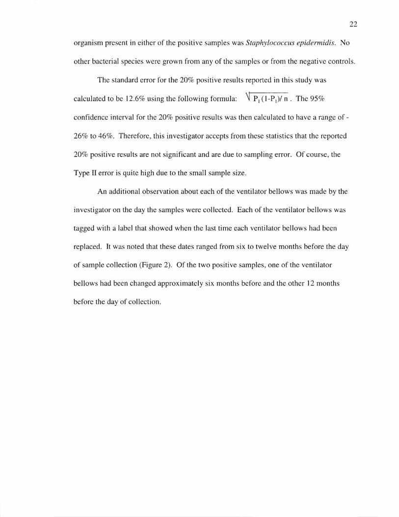
Of the J 2 samples co llec ted in the morn ing. [00% of them were reported to have
no visible evidence of bacterial growth. Of the 10 afte rnoon samples. two or 20% of
them were later reported positive for bac terial growth (Figure I). The index ind icated
that 3+ or greater than 100 colony forming un its (CFUs) were counted for each of these
positi ve samples. After addi tional tests were performed for identification, the only
21
22
organism presen t in e ither of the positive samples was Sraphylococclts epidermidis. No
other bacterial species were grown from any orthe samples or from the negative con tro ls .
The standard error for the 20% positive results reported in this study was
ca lculated to be 12.6% using the fol lowing formula: ~ PI (I -PI)! n. The 95%
confidence interval for the 20% positive resu lts was then calculated to have a range of -
26% to 46%. Therefore, this investigator accepts from lhese statistics that the reported
20% positive results are not significant and are due to sampling error. Of course, the
Type U error is quite high due to the small sample size.
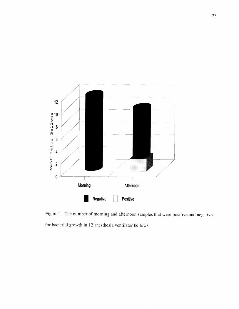
An additional observation about each of the venti lator bellows was made by the
investigator on the day the samples were co llected. Each of the ventilator bellows was
tagged with a label that showed when the last time each ventilator bellows had been
replaced. It was noted that these dates ranged from six to twe lve months before the day
of sample collection (Figure 2). Of the two positivc samples, one of the ve ntilator
bellows had been changed approximately six months before and the other 12 months
before the day of collect ion.
23
11
• 10 l 0 M M 8 • • " 6 0 ~ • 4 M
"" ~ c 1 • >
0
Morning Afternoon
• Negative U Positive
Figure I . T he number of morn ing and afternoon samples that were positi ve and negative
for bacterial growth in 12 anesthesia ventilator bellows.
U 6 Months
• 12 Months
~ 9 Months
24
Figure 2. The age in months of the 12 anesthesia ventilator be llows that were sampled in
th is study.
CHAPTER FIVE: CONCLUS ION
The findings of thi s study support the opin ion that the ventil alOr bel lows does not
harbor microorganisms and docs not present as a loci fo r nosocomial agents . No
pathogenic organisms as measured by tu rbid ity were grown from the 22 samples that
were collected . The two samples wh ich were positive for growth of Staphylococcus
epidermidis indicates that some external contamination of the samples may have occurred
during sample coll ection by the investi gator o r during the placement of the culturelle
swabs into the Ihioglycollate broth by laboratory personne l. Staphylococcus epidermidis
is a known skin contaminant and not a normal flora found in the respiratory tract, and it is
unlikely that it would be found in the ventilator bellows. These data support that the
anesthesia ven tilator bellows in thi s study were protected from palienllo patient
respi ratory contamination.
The focus of thi s study was to identify whether the ventilato r bellows of
anesthesia machines may contri bute to postoperative nosocomial in fections by serving as
a potenti al source for bacteria l contamination to patients. This study supports the work of
Murphy, et al ( 199 I) who stated that soda lime exerts a bacte riocidal effect on nonsporing
organisms. This study implies that stc ri lizat ion of the vcntil ator be llows, an import am
component of the cleanup regimen inst it uted by Dryden ( 1989), may not be a necessary
step in precluding the risk of cross contamination. Fina ll y, these data support th at the
presen t methodology of cleaning and sterili zation of anesthesia mach ines at thi s fac il ity is
effecti ve.
Gencralization of these findings is limitcd by the collection of the samples at one
institut ion and the d iminutive size of the sample. Recomme ndations from th is study to
25
26
fully document the potential risks of cross contamination from anesthesia c ircle systems
include repeating this study with a larger sample size co llected at multiple institu tions.
Another recomme ndat ion is to expand the number of sites sampled within each
anesthesia circle system to include the carbon dioxide absorbe r, the inspiratory and
expiratory valves, as well as the venti lator bellows at multip le institutions. Ways to
identify and e liminate the risks of cross-i nfecti on between anesthesia systems and
surgical patients must be further investigated to reduce patient morbidity and mortality
and to avoid unnecessary hospital costs.
REFERENCES
American Association of Nurse Anestheti sts, ( 1989). AANA develops infection
control guidelines for anesthesia. AANA Journal. 57. (4), 299-302 .
Association of Operating Room Nurses, (1994). Proposed recommended
practi ces for cleaning and processing anesthesia equipment. AORN Journal, 60, (3), 487-
489.
Association of Operating Room Nurses, ( 1985). Recommended practices:
Cleaning and processing anesthes ia equipment. AORN Journal. 4 1. (3), 625-63 1.
Assoc iation of Operating Room Nurses, (199 1). Recommended practices:
Cleaning and processing anesthesia equipment. AORN Journal, 53, (3), 775-777.
Albrecht, W. H. & Dryden, G. E. ( 1974). Five-year experience with the
development of an individually clean anesthes ia system. Anesthes ia and Analgesia. 53.
(1),24-28 .
Dryden, G. E. ( 1989). Flash steril izat ion of anesthesia bellows reduces
postoperative infection rate. AORN Journal , 49, (4), 11 23- 11 25.
Dryden, G. E. ( 1969). Risk of contamination from the anesthesia circle absorber:
An evaluation. Anesthes ia and Analges ia, 48, (6), 939-943.
Dryden, G. E. ( 1975). Uncleaned anesthesia equ ipment. lAMA, 233, ( 12) , 1297-
1298.
DuMoulin, G. C. & Saubermann , A. 1., (1977). The anesthes ia machine and
circle system are not li kely to be sources of bacterial contamination. Anesthes iology. 47.
353-358.
Feeley, T. W., Hamilton, W. K., Xavie r, B. , Moyers, l ., & Eger ll . E. I. (198 1).
27
Sterile anesthes ia breath ing circuits do not prevent poslOperalive pulmonary infec tion.
Anesthesiology, 54, 369-372.
Garibaldi, R. A. , Britt , M. R. , Webste r, c., & Pace, N. L. (198 1). Failure of
bacterial fillers to reduce the incidence of pneumonia after inhalation anesthesia.
Anesthesiology. 54. 364-368.
28
Gross, G. L. ( 1955). Decontamination of anesthesia apparatus. Anesthesio logy .
.l.Q, 903-909.
Joseph, 1. M. ( 1952). Disease transmission by ineffic ientl y san iti zed
anestheti zing apparatus. 1.A.M.A.. 149, 1196-1198.
Murphy, P. M. , Filzgeorgc, R. 8., & Barren, R. F. ( 199 J). Viability and
d istribution of bacteria after passage through a circle anaesthetic system. British Journal
of Anaesthes ia. 66, 300-304.
Pandi t, S. K., Mehta, S., & Agarwal, S. C. (1967). Ri sk of cross-infection from
inhalation anaesthetic equipment. British Journal of Anaesthesia, 39. 838-844.
Rendell-Baker, L (1993). Maintenance, cleaning, and steril ization of anesthesia
equipment. In J. Ehrenwerth & J . B. Eisenhaft (Eds.), Anesthesia equipment: Princ iples
and applications (pp. 492-510). 51. Louis: Mosby.
Russe ll. J. P. ( 1968). The Sterili zat ion dilemma: Where wi ll it end? Laboratory
aspects. Anesthesia and Analges ia, 47, (6), 653-656.
Ryan, K. J. ( 1994). Nosocomial infections and hospital inrection control. In K.
J. Ryan (Ed.), Sherris medical microbiology: An introduction to infectious di seases (pp.
823-830). Norwalk, 8: Appleton & Lange.
Shiotani, G. M. , Nicholes, P., Ballinger. C. M. , & Shaw, L.. ( 1971). Prevention
29
of contamination of the circ le system and ventil ators with a new disposable filter.
Anesthes ia and Analgesia. SO. (5), 844-854.
Smith, c., Wygant, M. E., McGrory, R, & Sil ka, M. , (1996). An evaluation of
one and two airflow filters in preventing the movement of bac teria through the anesthes ia
circle system. AANA Journal. 64, (2), 153- 156.
Stark, D. C. C , Green, C. A., & Pask, E. A. , ( 1962). Anaesthetic machines and
cross- infection. Anaesthes ia. 17. ( 1), 12-20.
Thomas, E. T . ( 1968). The sterili zati on dil emma: W here will it end? C li nical
aspects. Anesthesia and Analgesia. 47, (6), 657-662.