vegf regulates pcb 104-mediated stimulation of permeability and transmigration of breast cancer...
TRANSCRIPT

www.elsevier.com/locate/yexcr
Experimental Cell Research 296 (2004) 231–244
VEGF regulates PCB 104-mediated stimulation of permeability
and transmigration of breast cancer cells in human microvascular
endothelial cells
Sung Yong Eum,a Yong Woo Lee,a Bernhard Hennig,b and Michal Toboreka,*
aDepartment of Surgery, University of Kentucky, Lexington, KY 40536, USAbCollege of Agriculture, University of Kentucky, Lexington, KY 40536, USA
Received 15 September 2003; received in revised form 23 January 2004
Available online 16 March 2004
Abstract
Polychlorinated biphenyl (PCB) congeners, a group of worldwide, persistent environmental contaminants, are known to cause
carcinogenesis and tumor promotion, and may also affect the development of cancer metastasis. Because vascular endothelial cells create a
selective barrier to the passage of cancer cells, we hypothesize that specific PCB congeners can disrupt endothelial integrity and increase the
transendothelial migration of tumor cells. To examine this hypothesis, we elucidated the effects of 2,2V,4,6,6V-pentachlorobiphenyl (PCB 104), a
representative of highly ortho-substituted non-coplanar PCB congeners, on the endothelial permeability and transendothelial migration of
MDA-MB-231 breast cancer cells. Exposure of human microvascular endothelial cell 1 (HMEC-1) to PCB 104 induced endothelial
hyperpermeability and markedly increased transendothelial migration of MDA-MB-231 cells. These effects were associated with
overexpression of vascular endothelial growth factor (VEGF). PCB 104-mediated elevation of VEGF expression was induced by
phosphatidylinositol 3-kinase (PI3K) but not affected by co-treatments with antioxidants or the NF-nB inhibitor SN50. In addition, the PI3K-
dependent pathway was involved in PCB 104-induced activation of AP-1, a transcription factor implicated in the regulation of VEGF gene
expression. The VEGF receptor (KDR/Flk-1) antagonist SU1498 and the PI3K inhibitor LY294002 inhibited PCB 104-induced
hyperpermeability. These results indicate that PCB 104 may contribute to tumor metastasis by inducing VEGF overexpression that stimulates
endothelial hyperpermeability and transendothelial migration of cancer cells.
D 2004 Elsevier Inc. All rights reserved.
Keywords: PCB 104; VEGF; Metastasis; Endothelial cells; Permeability; Breast cancer
Introduction continuous monolayer which functions as a selective barrier
During the formation of blood-borne metastasis, tumor
cells disseminate from the primary tumor to secondary sites
in various organs. Because vascular endothelial cells form a
0014-4827/$ - see front matter D 2004 Elsevier Inc. All rights reserved.
doi:10.1016/j.yexcr.2004.01.030
Abbreviations: BSA, bovine serum albumin; FBS, fetal bovine serum;
HBSS, Hank’s balanced salt solution; EGCG, epigallocatechin-3-gallate;
HMEC-1, human microvascular endothelial cell 1; PCB, polychlorinated
biphenyl; PCB 104, 2,2V,4,6,6V-pentachlorobiphenyl; PDTC, pyrrolidine
dithiocarbamate; PI3K, phosphatidylinositol 3-kinase; PMA, phorbol-12-
myristate-13-acetate; VEGF, vascular endothelial growth factor.
* Corresponding author. Molecular Neuroscience and Vascular Biology
Laboratory, Division of Neurosurgery, Department of Surgery, University
of Kentucky Medical Center, 800 Rose Street, Lexington, KY 40536. Fax:
+1-859-323-2705.
E-mail address: [email protected] (M. Toborek).
to the passage of cancer cells from blood stream to the
underlying tissues, endothelial dysfunction has a significant
influence on the fate of circulating cancer cells in the blood
vessel [1–3]. Specifically, an increase in endothelial perme-
ability can accelerate metastatic process through the facili-
tated transmigration of cancer cells across the microvascular
endothelial monolayer [4–6].
Endothelial hyperpermeability can be induced either by
intracellular or extracellular stimuli such as reactive oxygen
species (ROS), cytokines, and growth factors [7–9]. It
appears that one of the most important factors involved in
the regulation of endothelial permeability is vascular endo-
thelial growth factor (VEGF) [9,10]. Evidence indicates that
VEGF can disrupt endothelial integrity and increase perme-

S.Y. Eum et al. / Experimental Cell Research 296 (2004) 231–244232
ability across the endothelial monolayers in vivo or in vitro
and promote transendothelial migration of leukocytes and
cancer cells, including breast cancer cells [10–12].
Polychlorinated biphenyls (PCBs) are a class of poly-
chlorinated aromatic hydrocarbons composed of 209 discrete
congeners. Due to their high lipophilicity and structural
stability, PCBs are among the most extensively investigated
persistent environmental pollutants that bioaccumulate in the
food chain and are concentrated in fatty tissues [13]. In
animals and humans, chronic exposure to PCBs produces a
variety of effects including neurotoxicity, pro-inflammatory
effects, carcinogenesis as well as tumor-promoting effects
[14–17]. Indeed, both in vitro and in vivo evidences dem-
onstrated that selected PCBs have carcinogenesis and tumor-
promoting activity by inducing oxidative damage in the liver
[16,17]. In addition, our group and others have reported that
specific PCBs, such as PCB 77, may increase endothelial cell
permeability, increase oxidative stress, decrease cellular
antioxidants, and activate redox-regulated transcription fac-
tors, such as nuclear factor-nB (NF-nB) [15,18].Among different PCBs, highly ortho-chlorinated PCB
congeners such as 2,2V,4,6,6V-pentachlorobiphenyl (PCB
104) appear to be of particular interest. For example, this
group of PCBs through their estrogenic activity can increase
proliferation of breast cancer cells [19]. Our recent study
showed that PCB 104 can increase the adhesion of human
leukemia cells (THP-1) to human umbilical vein endothelial
cells (HUVEC) through upregulation of adhesion molecules
[20] as well as induce apoptosis of human microvascular
endothelial cells (HMEC) [21]. However, the detailed mech-
anisms of PCB 104-induced effects on vascular endothelium
and transendothelial migration of cancer cells remain unclear.
Because the vascular endothelium plays a regulatory role
in transendothelial migration of cancer cells during meta-
static process, we hypothesize that ortho-substituted non-
coplanar PCBs can facilitate transendothelial migration of
cancer cells through disruption of vascular endothelial in-
tegrity. Results of the present study indicate that PCB 104
can increase endothelial permeability and transendothelial
migration of breast cancer cells through the phosphatidyli-
nositol 3-kinase (PI3K)-mediated upregulation of VEGF.
Materials and methods
Reagents
2,2V,4,6,6V-Pentachlorobiphenyl (PCB 104, >99% pure)
was purchased from AccuStandard (New Haven, CT) and
dissolved in dimethyl sulfoxide (DMSO). Levels of DMSO
in the experimental medium were less than 0.1% and did not
affect endothelial cell metabolism. LY294002, wortmannin,
SU1498, SN50, and SN50M were purchased from Calbio-
chem (La Jolla, CA). HIF-1a antibody was purchased from
Novus Biologicals (Littleton, CO) and antibodies against
phospho-Akt (Ser 473) and phospho-c-Jun were obtained
from Cell Signaling (Beverly, MS). All other chemicals and
reagents including pyrrolidine dithiocarbamate (PDTC) and
epigallocatechin-3-gallate (EGCG) were purchased from
Sigma (St. Louis, MO).
Cell cultures and PCB treatment
Human microvascular endothelial cells (HMEC-1) were a
generous gift from Dr. Eric Smart (University of Kentucky
Medical Center, Lexington, KY). HMEC-1 are an immor-
talized cell line obtained by transformation of human micro-
vascular endothelial cells with the SV40 large T antigen.
These cells retain endothelial cell phenotype and functional
characteristics [22]. HMEC-1 were cultured in MCDB 131
medium (GibcoBRL; Rockville, MD) enriched with 10%
fetal bovine serum (FBS), 2 mM L-glutamine (Sigma), 50
units/ml penicillin, 50 Ag/ml streptomycin (GibcoBRL), 1
Ag/ml hydrocortisone (Sigma), and 0.01 Ag/ml epidermal
growth factor (EGF) (Roche; Indianapolis, IN) in 5% CO2
atmosphere at 37jC. Before each experiment, the cells were
serum-starved in experimental medium containing 1% FBS
without EGF for 18 h.
The MDA-MB-231 cells (a metastatic breast cancer cell
line) were purchased from the American Type Culture
Collection (Manassas, VA) and cultured in suspension in
RPMI 1640 medium (GibcoBRL) supplemented with 10%
fetal bovine serum, 50 units/ml penicillin, and 50 Ag/ml
streptomycin.
Serum concentration of PCBs can reach approximately 3
AM in people exposed to these toxicants [23,24]; however,
local levels of PCBs in extracellular space are not known.
Therefore, in the present study, cells were treated with a
range of PCB 104 concentrations such as 2, 5, 10, or 15
AM. Similar experimental design was used in our previ-
ously published manuscripts [20,21]. In a selected experi-
ment, HMEC-1 were pretreated with inhibitors of specific
signaling pathways for 30 min before adding PCB 104. The
inhibitors were then maintained in the media throughout the
PCB 104 exposure. Stock solution of PCB 104 was
prepared in DMSO and the same amounts of DMSO as
in PCB-treated cells were added to control cultures. Basic
composition of experimental medium was the same as that
of growth medium, except for serum concentration which
was lowered to 1%. In selected experiments, phorbol-12-
myristate-13-acetate (PMA; Sigma) at a concentration of 10
nM was used as positive control.
Permeability assay
HMEC-1 (1 � 105 cells) were seeded on fibronectin-
coated Transwell polycarbonate filters (12-mm diameter,
0.4-Am pore size, Corning Costar) and allowed to grow to
confluence. Fresh complete medium was replaced every 2
days. To confirm confluence of endothelial monolayer,
transendothelial electrical resistance (TEER) across the
inserts was measured daily using Millicell-ERS voltohm-
S.Y. Eum et al. / Experimental Cell R
meter (Millipore, Bedford, MA). The resistance increased
progressively and reached a plateau at day 4 after seeding,
indicating the complete formation of the monolayer. There-
fore, all experiments were performed 5–6 days after seeding.
To determine its effects on endothelial permeability, PCB
104 with or without pretreatment of inhibitors was added for
24 h to both the lower and the upper compartment of
Transwell system. After the cultures were rinsed twice with
Kreb-Ringer Glucose (KRG) solution, 1.5 ml of KRG
solution was added into the lower chambers of Transwell,
and 0.5 ml of FITC-dextran 40 (FD-40, 1 mg/ml in KRG
solution) was loaded into the upper chambers. The systems
were incubated for 1 h at 37jC in a humidified atmosphere
(5% CO2) and the assay was stopped by removing the upper
chambers. Aliquots (0.5 ml) from the lower chambers were
transferred to new well of 24-well plate and fluorescence of
FD-40 was determined with a microplate spectrofluorometer
(Molecular Devices SPECTRA-max GeminiXS) using 483
nm as excitation and 517 nm as emission wavelengths.
Relative permeability was expressed by the ratio of FITC-
dextran transported into the lower chamber compared to
untreated-control group. All assays were performed at least
in triplicate.
Cell adhesion assay
Adhesion of MDA-MB-231 cells to HMEC-1 was
assessed according to the method of Braut-Boucher et al.
[25] with modifications. Briefly, HMEC-1 cells were grown
to confluence on fibronectin-coated wells of 24-well plates.
The cultures were treated with PCB 104 for 24 h and before
the adhesion assay they were washed three times with
Hank’s balanced salt solution (HBSS) containing 1% bovine
serum albumin (BSA) (HBSS–BSA).
The MDA-MB-231 cells were suspended in the amount
of 1.0 � 106 cells/ml HBSS–BSA and labeled with 5 AMcalcein-AM (Calbiochem) by 30-min incubation at 37jCfollowed by three washings with HBSS–BSA. The labeled
MDA-MB-231 cells were then incubated with PCB 104-
treated HMEC-1 for 30 min at 37jC. Cultures were
carefully washed three times with HBSS–BSA to remove
nonadherent cells. The adherence was quantified by fluo-
rescence measurements of the attached calcein-labeled
MDA-MB-231 cells using excitation of 490 nm and emis-
sion of 517 nm. The percentage of MDA-MB-231 cells
adherent to the endothelial monolayer was calculated using
the formula:
% adhesion ¼ ðFa � FbÞ=ðFt � FbÞ � 100
where Fa is the fluorescence of the adherent MDA-MB-
231 cells, Ft is the fluorescence of the total calcein-
labeled MDA-MB-231 cells added, and Fb is the back-
ground fluorescence of wells. The results are expressed as
the percentage of adhesion values determined in control
cultures.
Transendothelial cell migration assay
HMEC-1 were seeded in the amount of 5.0 � 104
cells and grown to confluence on fibronectin-coated
Transwell polycarbonate filters (6.5-mm diameter, 8.0-
Am pore size, Corning Costar). The medium was replaced
every 2 days and confluent cultures were exposed to PCB
104 as described for the permeability assay. HMEC-1
were then washed twice with migration medium (serum-
free MCDB 131 containing 1% BSA) and the calcein-
labeled MDA-MB-231 cells (4.0 � 104 cells) suspended
in 100 Al of the same medium were added to the mo-
nolayer of HMEC-1 (i.e., to the upper chamber of the
Transwell system). After incubation for 10 h, cells were
fixed with 4% formaldehyde and washed extensively with
PBS. To remove nonmigrating cells, cells on the upper
face of the filter were gently scraped using a cotton swab
and the migrating tumor cells were observed under
fluorescent microscope (Nikon eclipse E600, Nikon,
NY). Migrating cells were counted from five random
fields using the �200 magnification. All assays were
performed in triplicate.
Conventional and real-time RT-PCR
Total RNAwas prepared from PCB 104-treated HMEC-1
using TRI reagent (Sigma) according to the manufacturer’s
instructions. First strand cDNA was generated from 1 Ag of
the total RNA using the Reverse Transcription System kit
(Promega, Madison, WI) and either oligo dT15 primers (for
conventional RT-PCR) or random hexamer primers (for real-
time RT-PCR). To measure VEGF mRNA expression by
conventional RT-PCR, cDNA was amplified by PCR using
VEGF-specific primers (sense, 5V-CTA CCT CCA CCATGC
CAA GT-3V, and antisense, 5V-TCT CTC CTATGT GCT GG
CCT-3V, to yield a 311 bp product for VEGF). VEGF-specificprimers were designed using the Primer3 software (White-
head Institute, Cambridge, MA). In addition, h-actin expres-
sion was assessed as a housekeeping gene (sense, 5V-AGCACA ATG AAG ATC AAG AT-3V, antisense, 5V-TGT AAC
GCA ACTAAG TCATA-3V, to yield a 188 bp product) [26].The PCR reaction mixture consisted of Taq PCR Master Mix
(Qiagen, Valencia, CA), 2 Al of cDNA, and 20 pmol of
primer pairs in a total volume of 50 Al. PCR was initiated
with a hot start (94jC, 5 min) and continued for 25 cycles
(VEGF) or 21 cycles (h-actin) of denaturation (94jC, 45 s),
annealing (60jC, 45 s), and extension (72jC, 1 min) before
the final extension (72jC, 7 min). PCR products were
separated by 2% agarose gel electrophoresis, stained with
SYBR Gold (Molecular Probes, Eugene, OR) solution for 1
h, and visualized by phosphoimage analysis (FLA-5000,
Fuji, Stamford, CT).
VEGFmRNA expression was confirmed by real-time RT-
PCR using the ABI Prism 7000 Sequence Detection System
(Applied Biosystems, Foster City, CA). PCR amplification
was performed using TaqMan Universal PCR Master Mix
esearch 296 (2004) 231–244 233

S.Y. Eum et al. / Experimental Cell R234
(Applied Biosystems) according to the manufacturer’s
instructions. Commercially available predeveloped primer
pair and TaqMan probe (Applied Biosystems) were used to
determine VEGF mRNA levels. PCR cycles consisted of an
initial denaturation step at 95jC for 10 min, followed by
95jC for 15 s and 60jC for 60 s (for up to 45 cycles). PCR
amplification of 18S RNA (a housekeeping gene) was
performed for each sample to normalize VEGF mRNA
levels. A standard curve was generated by plotting the
threshold cycle (Ct) vs. the log concentration of the serial
dilutions of the cDNA from the sample obtained from
HMEC-1 treated with 10 AMPCB 104 for 12 h. Each sample
was analyzed three times.
Electrophoretic mobility shift assay (EMSA)
Nuclear extracts were prepared as describe earlier [21]
with minor modifications. The cells were suspended in 1 ml
of lysis buffer (10 mM Tris–HCl, pH 8.0, 60 mM KCl, 1
mM EDTA, 1 mM dithiothreitol, 100 AM phenylmethylsul-
fonyl fluoride, 0.1% NP-40), lysed for 5 min on ice, and
centrifuged at 600 � g for 4 min at 4jC to collect nuclei.
Then, the nuclear pellets were washed with 1 ml of lysis
buffer without NP-40, and then lysed in 50 Al of nuclearextract buffer (20 mM Tris–HCl, pH 8.0, 420 mM NaCl,
1.5 mM MgCl2, 0.2 mM EDTA, 25% glycerol) for 10 min
on ice, and centrifuged at 12,000 � g for 15 min at 4jC.Nuclear extracts were stored in �80jC freezer before use.
Protein concentrations of isolated nuclear extracts were
determined using commercial Bradford regent (Sigma).
Double-stranded oligonucleotides containing the consensus
sequences of the binding sites for transcription factors NF-
nB and AP-1 were end-labeled with [g-32P]-ATP using
bacteriophage T4 polynucleotide kinase (Promega) accord-
ing to the manufacturer’s instructions. Unincorporated
nucleotides were removed by gel filtration chromatography
using mini Quick Spin Oligo Columns (Boehringer Man-
heim Corporation; Indianapolis, IN). Binding reactions were
performed with 4 Ag (NF-nB) or 2 Ag (AP-1) of nuclear
protein extracts in a 20 Al volume of reaction mixture (10
mM Tris–Cl, pH 7.5, 50 mM NaCl, 1 mM EDTA, 0.1 mM
dithiothreitol, 10% glycerol, and 2 Ag of poly[dI-dC]). After
adding the reagents, the mixture was incubated for 25 min at
room temperature. Then, 40,000 cpm of 32P-labeled specific
oligonucleotide probe was added, and the binding mixture
was incubated for 25 min at room temperature. In antibody
supershift experiments, nuclear protein extracts were incu-
bated for 25 min at room temperature with 2 Ag of specific
antibody before adding the 32P-labeled specific oligonucle-
otide probe. Resultant protein–DNA complexes were ana-
lyzed on a non-denaturing 5% polyacrylamide gel using
0.25� TBE buffer (50 mM Tris–HCl, 45 mM boric acid,
0.5 mM EDTA, pH 8.4) for 3 h at 150 V. The gel was
transferred to Whatman 3MM paper, dried on a gel dryer,
and exposed to an X-ray film at �80jC with an intensifying
screen.
Measurement of intracellular reactive oxygen species (ROS)
Intracellular ROS levels were determined by the 2V,7V-dichlorofluorescein (DCF) method as described earlier [20]
with minor modifications. Briefly, confluent HMEC-1 cul-
tured on fibronectin-coated 24-well plates were incubated
with PCB 104 for 30 min followed by incubation with 20 AM2V,7V-dichlorodihydrofluorescein diacetate (H2DCF-DA) for
30 min at 37jC. In selected experiments, cultures were
pretreated for 30 min with antioxidants before exposure to
PCB 104. At the end of incubation, cultures were rinsed
twice with HBSS and 0.5 ml of HBSS was added into each
well. The relative fluorescence was assessed using micro-
plate spectrofluorometer (SPECTRA-max GeminiXS, Mo-
lecular Devices, Sunnyvale, CA). The excitation and
emission wavelengths were 485 and 530 nm, respectively.
Results were expressed in relative fluorescence units (RFU).
Cell viability assays (MTT assay)
MTT [3-(4,5-dimethylthiazol-2-yl)-2,5-diphenyltetrazo-
lium bromide] conversion assay was performed to assess
cell viability. Briefly, confluent HMEC-1 cultures were
treated with PCB 104 for up to 48 h. In selected experiments,
inhibitors of specific signaling pathways were added 30 min
before PCB 104 treatment and then maintained in media
throughout the PCB 104 exposure. After incubation, the cells
were rinsed two times with PBS and incubated with MTT
solution (1 mg/ml in experimental medium) for 4 h at 37jC.The insoluble colored formazan salts were dissolved in
DMSO and the absorbance was assessed at 570 nm. Results
represent the mean and standard deviation of quadruplicate
determinations.
Western blotting
Confluent HMEC-1 seeded onto 100-mm plates, were
treated with PCB 104, washed with cold PBS, and lysed
with lysis buffer (1% Nonidet P-40, 20 mM Tris–HCl [pH
7.6], 1 mM EDTA, 0.5 mM EGTA, 10 mM MgCl2, 1 mM
Na3VO4, 2 mM dithiothreitol, 1 Ag/ml of aprotinin, 1 Ag/ml of leupeptin, and 1 mM phenylmethylsulfonyl fluo-
ride). The cell lysates were incubated for 20 min at 4jCfollowed by centrifugation at 12,000 � g at 4jC. Samples
(30 Ag of protein/lane) were electrophoresed in 10% SDS-
PAGE and transferred onto a Hybond-ECL membrane
(Amersham Biosciences, Piscataway, NJ). The membrane
was blocked for 1 h with 5% (w/v) nonfat dry milk in
Tris-buffered saline containing 0.1% (v/v) Tween 20
(TBS-T) and incubated overnight at 4jC with anti-phos-
pho-Akt or anti-Akt antibody. The membrane was then
washed with TBS-T and incubated for 2 h with horserad-
ish peroxidase-conjugated secondary antibody. After wash-
ing three times with TBS, immunoreactive protein bands
were visualized with the enhanced chemiluminescence
system (Amersham).
esearch 296 (2004) 231–244
Cell Research 296 (2004) 231–244 235
Enzyme-linked immunosorbent assay (ELISA)
Medium protein levels of VEGF were quantified by
using Quantikine human VEGF immunoassay (R&D Sys-
tems, Minneapolis, MN) according to the manufacturer’s
instructions. This assay recognizes the soluble isoforms
VEGF121 and VEGF165. Briefly, HMEC-1 were cultured
in fibronectin-coated wells of 6-well plate until conflu-
ence. To determine VEGF protein, HMEC-1 were treated
with 10 AM PCB 104 for 12 or 36 h. The media were
collected, centrifuged, and the supernatants used for
ELISA. In addition, HMEC-1 were lysed with 1 N NaOH
and protein amounts were determined per well using the
Bradford assay (Sigma) to normalize the amounts of
VEGF released.
Statistical analysis
Results are expressed as means F SD. Data were statis-
tically analyzed using one-way ANOVA followed by Stu-
S.Y. Eum et al. / Experimental
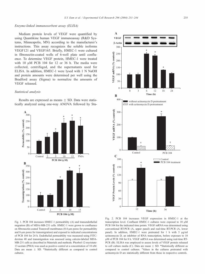
Fig. 1. PCB 104 increases HMEC-1 permeability (A) and transendothelial
migration (B) of MDA-MB-231 cells. HMEC-1 were grown to confluence
on fibronectin-coated Transwell membranes (0.4-Am pores for permeability
and 8-Am pores for transmigration) and exposed to indicated concentrations
of PCB 104 for 24 h. Endothelial permeability was measured using FITC-
dextran 40 and transmigration was assessed using calcein-labeled MDA-
MB-231 cells as described in Materials and methods. Phorbol-12-myristate-
13-acetate (PMA) was used as positive control at a concentration of 10 nM.
Data are mean F SD. *Statistically different as compared to control
cultures.
Fig. 2. PCB 104 increases VEGF expression in HMEC-1 at the
transcription level. Confluent HMEC-1 cultures were exposed to 10 AMPCB 104 for the indicated time points. VEGF mRNAwas determined using
conventional RT-PCR (A, upper panel) and real-time RT-PCR (A, lower
panel). In addition, HMEC-1 were pretreated for 1 h with 5 Ag/ml
actinomycin D, an inhibitor of RNA transcription, before exposure to 10
AM of PCB 104 for 8 h. VEGF mRNAwas determined using real-time RT-
PCR (B). ELISA was employed to assess levels of VEGF protein released
to cell culture media (C). Data are mean F SD. *Statistically different as
compared to control cultures. yValues in the cultures pretreated with
actinomycin D are statistically different from those in respective controls.

S.Y. Eum et al. / Experimental Cell Research 296 (2004) 231–244236
dent’s t test. Statistical probability of P < 0.05 was considered
significant.
Fig. 4. PI3K mediates PCB 104-induced VEGF expression in HMEC-1.
Confluent HMEC-1 cultures were pretreated with the indicated concen-
trations of LY294002 or wortmannin (inhibitors of PI3K) for 30 min before
exposure to 10 AM PCB 104 for 12 h. VEGF mRNAwas determined using
conventional RT-PCR (A, upper panel) and real-time RT-PCR (A, lower
panel). ELISA was employed to assess levels of VEGF protein released to
cell culture media (B). Data are mean F SD. *Statistically different as
compared to control cultures. yLevels in the groups PCB 104 plus LY294002
Results
PCB 104 increases HMEC-1 permeability and
transendothelial migration of MDA-MB-231 cells
The effects of PCB 104 on HMEC-1 permeability were
assessed using FITC-dextran 40 (FD-40) and the fibronectin-
coated Transwell system with 0.4-Am pore size. Exposure of
HMEC-1 to PCB 104 for 24 h resulted in a dose-dependent
increase in permeability (Fig. 1A). A prominent disruption of
HMEC-1 integrity was observed in cultures treated with 10
AM PCB 104. An increase in PCB 104 concentration to 15
AM did not result in further elevation of FD-40 transfer
across HMEC-1 monolayers. In these experiments, 10 nM
PMA was used as a positive control.
An increase in endothelial permeability may result in
elevated transendothelial migration of cancer cells. There-
fore, the effects of PCB 104 on the transmigration of MDA-
MB-231 cells across HMEC-1 monolayers were also eval-
uated in the present study. Calcein-labeled MDA-MB-231
cells and the Transwell system with 8-Am pore size were
used in these experiments. To exclude the direct effects of
PCB 104 on cancer cells, HMEC-1 cultures were washed
twice with migration medium (serum-free MCDB 131 con-
taining 1% BSA) before adding MDA-MB-231 cells. As
illustrated in Fig. 1B, PCB 104 treatment induced a dose-
dependent increase in the migration of MDA-MB-231 cells
across the HMEC-1 monolayers. The effective concentration
of PCB 104 was 5 AM and a further increase in PCB 104
level did not potentate these effects.
Because 10 AM PCB 104 effectively increased endothe-
lial permeability and transmigration of tumor cells, this
concentration was selected for further experiments which
were focused on the mechanisms of PCB 104-induced
disruption of endothelial integrity. In addition, treatment
with 10 AM PCB 104 for up to 48 h did not show cytotoxic
effects as measured by the MTT conversion assay (data not
shown).
Fig. 3. PCB 104 stimulates PI3K activity in HMEC-1 without activation of
HIF-1 transcription factor. Confluent HMEC-1 cultures were treated with
10 AM PCB 104 for the indicated time points. Activation of PI3K was
determined by Western blot in whole cell extracts using antibodies against
phosphorylated Akt (p-Akt). Levels of HIF-1a were assessed by Western
blot in nuclear extracts of control and PCB 104-treated HMEC-1.
or PCB 104 plus wortmannin are statistically different as compared to those
in cultures treated with PCB 104 alone. PCB, PCB 104.
PCB 104 increases VEGF expression at the transcription
level through the PI3K-dependent mechanism
VEGF is one of the most important endothelial-derived
agents regulating vascular permeability. Therefore, VEGF
expression was determined both at the mRNA and protein
levels in HMEC-1 exposed to PCB 104 (Fig. 2). As
illustrated in Fig. 2A, VEGF mRNA reached maximum
levels in HMEC-1 treated with 10 AM PCB 104 for 6 and 12
h and returned to basal values after a 24-h PCB 104
exposure.

Fig. 5. PCB 104-mediated PI3K activity stimulates AP-1 DNA binding and
c-Jun phosphorylation in HMEC-1. Nuclear extracts were isolated from
HMEC-1 treated with 10 AM PCB 104 for the indicated time (A). HMEC-1
were pretreated with the PI3K inhibitors LY294002 (3 AM) or wortmannin
(1 AM) for 30 min followed by 10 AM PCB 104 for 3 h (B). AP-1 DNA
binding activity was analyzed by EMSA. Competition study was performed
by the addition of excess unlabeled AP-1 oligonucleotides. Supershift
assays were performed with 2 Ag of antibodies against c-Jun (c-Jun Ab) or
phosphorylated c-Jun (p-c-Jun Ab). In addition, levels of phosphorylated c-
Jun (p-c-Jun) were assessed by Western blot (WB). Ctl, control; PCB, PCB
104; LY, LY294002; Wort, wortmannin.
S.Y. Eum et al. / Experimental Cell Research 296 (2004) 231–244 237
To determine whether PCB 104 induces VEGF mRNA
expression at the transcriptional level, confluent HMEC-1’s
were pretreated for 1 h with actinomycin D, the inhibitor of
RNA transcription, and then incubated with PCB 104 for 8 h.
PCB 104-mediated VEGF mRNA levels were completely
abolished by actinomycin D (Fig. 2B).
PCB 104-mediated increase in VEGF mRNA expression
was associated with elevated VEGF protein levels. As shown
in Fig. 2C, a 12-h exposure to PCB 104 significantly
increased VEGF protein in cell culture media. However,
these values returned to control levels in HMEC-1 treated
with PCB 104 for 36 h.
Evidence suggests that the phosphatidylinositol 3-kinase
(PI3K) pathway may regulate VEGF expression via activa-
tion of Akt kinase and stimulation of hypoxia inducible
factor-1 (HIF-1) [42,43]. Therefore, the possibility that PCB
104 can stimulate these pathways was also examined in the
Fig. 6. PCB 104 increases cellular oxidative stress and NF-nB DNA binding
activity. DCF fluorescence was measured after 30 min of treatment with the
indicated concentrations of PCB 104 (A). To study the effects of antioxidants
on PCB 104-mediated oxidative stress, HMEC-1 were pretreated for 30 min
with antioxidants PDTCor EGCGat the indicated concentrations (B). NF-nBDNA binding activity was analyzed by EMSA using nuclear extracts
prepared fromHMEC-1 treatedwith 10 AMof PCB104 for the indicated time
(C). Competition study was performed by the addition of excess unlabeled
NF-nB oligonucleotides. Supershift assay was performed with 2 Ag of
antibodies against p65 or p50, the components of NF-nB complex. Data are
meanF SD. *Statistically different as compared to control cultures. yLevels
in the groups PCB 104 plus EGCG or PCB 104 plus PDTC are statistically
different as compared to those in cultures treated with PCB 104 alone. PDTC,
pyrrolidine dithiocarbamate; EGCG, epigallocatechin-3-gallate.
present study. As shown in Fig. 3, treatment with PCB 104
for 15 or 30 min markedly increased phosphorylation of Akt
kinase, the cellular target of PI3K. On the other hand,
exposure to PCB 104 did not stimulate HIF-1. As indicated
in Fig. 3, a 15- to 180-min treatment with PCB 104 did not

S.Y. Eum et al. / Experimental Cell Research 296 (2004) 231–244238
affect protein levels of HIF-1a, the major subunit of HIF-1,
in nuclear extracts of HMEC-1.
To support the regulatory role of PI3K in PCB 104-
induced VEGF mRNA overexpression, pretreatment of
HMEC-1 with LY294002 and wortmannin, specific inhib-
itors of PI3K activation, markedly and in dose-dependent
manners decreased PCB 104-mediated stimulation of VEGF
mRNA levels as determined by conventional and real-time
RT-PCR (Fig. 4A, upper and lower panel, respectively).
Consistent with these results, LY294002 and wortmannin
Fig. 7. Oxidative stress-responsive pathways are not involved in PCB 104-
induced overexpression of VEGF in HMEC-1. Confluent HMEC-1 cultures
were pretreated for 30 min with the indicated concentrations of antioxidants
PDTC or EGCG or with 50 Ag/ml of SN50 (the inhibitor of NF-nB nuclear
translocation) before exposure to 10 AM PCB 104. SN50M is a negative
control for the SN50 peptide. VEGF mRNA was determined using
conventional RT-PCR (upper panels) and real-time RT-PCR (lower panels)
after 12-h incubation of PCB 104. Data are mean F SD. *Statistically
different as compared to control cultures. PCB, PCB 104; PDTC,
pyrrolidine dithiocarbamate; EGCG, epigallocatechin-3-gallate.
Fig. 8. VEGF and PI3K regulate PCB 104-induced elevation of HMEC-1
permeability. HMEC-1 were grown to confluence on fibronectin-coated
Transwell membranes (0.4-Am pores) and pretreated with the indicated
concentrations of SU1498 (A) or with LY294002 (B) for 30 min before
exposure to 10 AM PCB 104 for 24 h. HMEC-1 permeability was
determined as described in Materials and methods. Data are mean F SD.
*Statistically different as compared to control cultures. yLevels in the
groups PCB 104 plus SU1498 or PCB 104 plus LY294002 are statistically
different as compared to those in the PCB 104 group.
also effectively inhibited PCB 104-induced production of
VEGF protein (Fig. 4B).
PCB 104-mediated PI3K activation stimulates
phosphorylation of c-Jun and AP-1 DNA binding
The human VEGF promoter contains four potential
binding sites of AP-1 [47] which play a key regulatory role
in VEGF expression [43–46]. Therefore, we assessed AP-1
DNA binding activity and c-Jun phosphorylation in nuclear
extracts of HMEC-1 exposed to PCB 104. As illustrated in
Fig. 5A, PCB 104 markedly stimulated AP-1 DNA binding.
Based on a supershift assay, c-Jun was identified as the major
component of PCB 104-induced AP-1 complex. The in-
crease in AP-1 DNA binding was accompanied by elevated
levels of phosphorylated c-Jun (Fig. 5A). To illustrate the
role of PI3K signaling in these effects, treatment with

S.Y. Eum et al. / Experimental Cell Research 296 (2004) 231–244 239
LY294002 and wortmannin markedly blocked PCB 104-
mediated activation of AP-1 DNA binding and c-Jun phos-
phorylation (Fig. 5B).
PCB 104-mediated overexpression of VEGF is oxidative
stress-independent
The PI3K signaling pathway is known to be regulated by
cellular redox status. In addition, PCB congeners were
Fig. 9. VEGF and PI3K regulate PCB 104-induced elevation of transendothelial m
grown to confluence on fibronectin-coated Transwell membranes (8-Am pores) and
10 AM PCB 104 and these inhibitors for 24 h. Transmigration of the calcein-lab
methods. Data are mean F SD. *Statistically different as compared to control c
SU1498 are statistically different as compared to those in cultures treated with PC
calcein-labeled MDA-MB-231 cells on the lower site of the Transwell membran
experimental groups as described in A. Photomicrographs were taken using a flu
shown to increase intracellular ROS levels in several types
of cells and tissues [15,20]. Therefore, a series of experi-
ments was performed to examine the possibility that oxida-
tive stress and nuclear factor-nB (NF-nB) could be involved
in PCB 104-mediated VEGF expression. NF-nB is a down-
stream target of PI3K signaling and a redox-responsive
transcription factor.
Exposure of HMEC-1 to PCB 104 induced cellular
oxidative stress (Figs. 6A and 6B) and increased DNA-
igration of MDA-MB-231 cells across HMEC-1 monolayers. HMEC-1 were
pretreated with LY294002 (1 AM) and SU1498 (50 AM) for 30 min prior to
eled MDA-MB-231 cells was quantified (A) as described in Materials and
ultures. yLevels in the groups PCB 104 plus LY294002 or PCB 104 plus
B 104 alone. (B) Shows the representative fluorescent images of migrating
es. Photomicrographs are representative of comparisons between different
orescent microscope with a �20 objective.
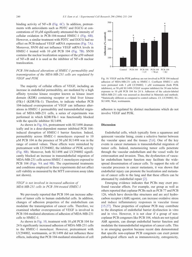
Fig. 10. VEGF and the PI3K pathway are not involved in PCB 104-induced
adhesion of MDA-MB-231 cells to HMEC-1. Confluent HMEC-1 cells
were pretreated with 3 AM LY294002, 1 AM wortmannin (both PI3K
inhibitors), or 50 AM SU1498 (VEGF receptor inhibitor) for 30 min before
exposure to 10 AM PCB 104 for 24 h. Adhesion of the calcein-labeled
MDA-MB-231 cells was assessed as described in Materials and methods.
*Statistically different as compared to control cultures. LY, LY294002; SU,
SU1498; Wort, wortmannin.
S.Y. Eum et al. / Experimental Cell Research 296 (2004) 231–244240
binding activity of NF-nB (Fig. 6C). In addition, pretreat-
ments with antioxidants such as PDTC and EGCG at con-
centrations of 10 AM significantly attenuated the intensity of
cellular oxidation in PCB-104-treated HMEC-1 (Fig. 6B).
However, a similar treatment with PDTC and EGCG had no
effect on PCB-induced VEGF mRNA expression (Fig. 7A).
Moreover, SN50 did not influence VEGF mRNA levels in
HMEC-1 treated with 10 AM PCB 104 (Fig. 7B). SN50
contains the nuclear localization sequence of the p50 subunit
of NF-nB and it is used as the inhibitor of NF-nB nuclear
translocation.
PCB 104-induced alterations of HMEC-1 permeability and
transmigration of the MDA-MB-231 cells are regulated by
VEGF and PI3K
The majority of cellular effects of VEGF, including an
increase in endothelial permeability, are mediated by a high
affinity tyrosine kinase receptor known as kinase insert
domain (KDR) containing receptor or fetal liver kinase
(Flk)-1 (KDR/Flk-1). Therefore, to indicate whether PCB
104-induced overexpression of VEGF can influence alter-
ations in HMEC-1 permeability and transendothelial migra-
tion of MDA-MB-231 cells, a series of experiments was
performed in which KDR/Flk-1 was functionally blocked
with the specific inhibitor SU1498.
As shown in Fig. 8A, pretreatment with SU1498 dramat-
ically and in a dose-dependent manner inhibited PCB 104-
induced disruption of HMEC-1 barrier function. Indeed,
permeability across HMEC-1 monolayers exposed to 10
AM PCB 104 in the presence of 50 AM SU1498 was in the
range of control values. These effects were mimicked by
pretreatment with LY294002, the inhibitor of PI3K activity
(Fig. 8B). Moreover, both SU1498 and LY294002 signifi-
cantly blocked an increase in transendothelial migration of
MDA-MB-231 cells across HMEC-1 monolayers exposed to
PCB 104 (Figs. 9A and 9B). The experimental treatments
and conditions employed in these experiments did not affect
cell viability as measured by the MTTconversion assay (data
not shown).
VEGF is not involved in increased adhesion of
MDA-MB-231 cells in PCB 104-treated HMEC-1
We previously reported that PCB 104 can increase adhe-
sion of tumor cells to human endothelial cells. In addition,
changes of adhesion properties of the endothelium can
modulate the transmigration of cancer cells. Therefore, we
examined whether overexpression of VEGF is involved in
PCB 104-mediated alterations of adhesion of MDA-MB-231
cells to HMEC-1.
As shown in Fig. 10, treatment with 10 AM PCB 104 for
24 h significantly increased adhesion of MDA-MB-231 cells
to the HMEC-1 monolayer. However, pretreatment with
LY294002, wortmannin, or SU1498 did not influence these
effects, indicating that PCB 104-mediated stimulation of cell
adhesion is regulated by distinct mechanisms which do not
involve VEGF and PI3K.
Discussion
Endothelial cells, which typically form a squamous and
quiescent vascular lining, create a selective barrier between
the vascular space and the tissues [5,6]. One of the key
events in cancer metastasis is transendothelial migration of
tumor cells. Indeed, metastasizing tumor cells penetrate
through the vascular endothelium and the vessel walls for
extravasation and invasion. Thus, the dysfunction of vascu-
lar endothelium barrier function may facilitate the wide-
spread dissemination of cancer cells. To support the role of
vascular processes in cancer metastasis, it was shown that
endothelial injury can promote the localization and metasta-
sis of cancer cells to the lung and that these effects can be
attenuated by endothelial repair [2].
Emerging evidence indicates that PCBs may exert pro-
found vascular effects. For example, our group as well as
others reported that coplanar PCBs such as PCB 77 and PCB
126, which have dioxin-like structures and are aryl hydro-
carbon receptor (AhR) agonist, can increase oxidative stress
and induce inflammatory responses in vascular tissue
[15,27]. These properties of coplanar PCB may contribute
to the disruption of endothelial barrier dysfunction in vitro
and in vivo. However, it is not clear if a group of non-
coplanar PCB congeners like PCB 104, which are not typical
AhR agonists, can disrupt endothelial barrier function and
modulate the transendothelial migration of cancer cells. This
is an emerging question because recent data demonstrated
that specific non-coplanar PCB congeners can exert potent
pathological effects such as immunotoxicity, estrogenicity,

Fig. 11. Proposed diagram of PCB 104-mediated intracellular signaling
leading to increased permeability and transendothelial migration of cancer
cells. AP-1, activator protein-1; NF-nB, nuclear factor-nB; PCB 104,
2,2V,4,6,6V-pentachlorobiphenyl; PI3K, phosphatidylinositol 3-kinase; ROS,reactive oxygen species; VEGF, vascular endothelial growth factor.
S.Y. Eum et al. / Experimental Cell Research 296 (2004) 231–244 241
and tumor-promoting activity. It was reported that PCB 104
can cause immunosuppression through induction of apopto-
sis of human monocytic cells [28]. In addition, non-coplanar
PCBs, including PCB 104, significantly increased MCF-7
breast cancer cell proliferation; the effect which could be
inhibited by co-treatment with an estrogen antagonist
hydroxytamoxifen [17]. Recent study from our group dem-
onstrated that PCB 104 upregulated expression of adhesion
molecules and increased the adhesion of leukemia cells to
human endothelial cells [20]. In addition, results of the
present study indicate that PCB 104 can markedly disrupt
integrity of the vascular endothelium and increase transmi-
gration of breast tumor cells.
Several vascular mechanisms can be responsible for the
facilitation of extravasation and dissemination of tumor cells.
For example, upregulation of specific adhesion molecules on
the endothelial surface can increase transendothelial migra-
tion of cancer cells through the increased adhesion of cancer
cells to endothelial cells [1,5]. In addition, transmigration of
cancer cells can be augmented by disruption of cell junctions
[6,10]. Finally, several soluble factors such as VEGF can
induce the disruption of endothelial integrity, which in turn
may directly enhance penetration of tumor cells and facilitate
establishment of cancer metastasis [9,10]. Microvascular
hyperpermeability in proximity to metastatic tumors can also
accelerate plasma protein extravasation to stimulate tumor
growth [29].
Results of the present study clearly indicate that exposure
of HMEC-1 to PCB 104 can increase permeability and
transendothelial migration of cancer cells through overex-
pression of VEGF. Two critical facts support this statement:
(1) exposure to PCB 104 increased VEGF expression both at
the mRNA and protein levels (Figs. 2A and 2B), and (2)
SU1498, the antagonist of the VEGF receptor (KDR/Flk-1),
significantly inhibited PCB 104-induced hyperpermeability
of HMEC-1 and transendothelial migration of breast cancer
cells (Figs. 8 and 9, respectively).
VEGF, also known as vascular permeability factor
(VPF), has potent vascular permeable activity specific to
vascular endothelial cells implicated in endothelial barrier
dysfunction and also plays an essential role in promoting
new blood vessel formation (angiogenesis) during tumor
development [30,31]. Indeed, inhibition of VEGF function
can effectively prevent tumor growth through incomplete
blood vessel formation. Thus, VEGF has been recognized as
a primary mediator of metastasis formation in a number of
human cancers. Although the detailed mechanisms of
VEGF-mediated stimulation of hyperpermeability are not
known, evidence indicates that the rearrangement and al-
tered expression of endothelial junctional proteins can be
involved in this process [12,32,33]. It was also reported that
VEGF can induce proliferation and invasion of VEGF
receptor-expressing breast cancer cells [34]. Thus, it is
possible that VEGF secreted by PCB 104-exposed endothe-
lial cells can affect transendothelial migration of cancer cells
both through increased permeability of endothelial mono-
layers and the alteration of invasive properties of tumor
cells.
Recent evidence indicated that VEGF can increase ex-
pression of adhesion molecules such as E-selectin, intercel-
lular adhesion molecule 1 (ICAM-1), and vascular cell
adhesion molecule 1 (VCAM-1) in human endothelial cells.
These molecules can increase the adhesiveness and trans-
endothelial migration of tumor cells [35,36]. However,
results of the present study indicated that VEGF is not
involved in PCB 104-induced elevated adhesiveness of
MDA-MB-231 cells to HMEC-1. Indeed, neither functional
inhibition of the VEGF receptor nor inhibition of the PI3K
signaling did affect adhesion of MDA-MB-231 breast tumor
cells to HMEC-1 monolayers (Fig. 10). Thus, PCB 104-
induced stimulation of transendothelial migration and adhe-
sion of tumor cells to the vascular endothelium appear to be
regulated by distinct mechanisms (Fig. 11). To support this
notion, it was reported that tumor cells have higher affinity
to extracellular matrix (ECM) components, such as fibro-
nectin, than to the surface of an intact endothelial monolayer
[4]. Indeed, a blockage of the adhesion of tumor cells to
ECM components significantly prohibited metastatic forma-
tion [37].
In the present study, we identified that the PI3K signaling
pathway is involved in PCB 104-induced VEGF expression.
First, we provided evidence that the PI3K pathway is
activated in PCB 104-treated HMEC-1 (Fig. 3); and second,
we demonstrated that PI3K inhibitors such as LY294002 or
wortmannin can block the effects of PCB 104 on VEGF

S.Y. Eum et al. / Experimental Cell Research 296 (2004) 231–244242
expression (Fig. 4), endothelial permeability (Fig. 8B), and
transmigration of MDA-MB-231 cells (Fig. 9). These find-
ings support recent literature data. For example, it was
reported that PI3K plays a role in increased VEGF produc-
tion induced by several stimuli, including oxidative stress
and platelet-derived growth factor [38,39]. In addition, it was
shown that expression of constitutively active PI3K or Akt is
associated with an increased steady state of VEGF mRNA
levels in endothelial cells and fibroblasts [40].
Our data indicate that the induction of VEGF mRNA
level by PCB 104 can be completely blocked by pretreat-
ment with actinomycin D, indicating the transcriptional
regulation of this process (Fig. 2B). Therefore, to clarify
the molecular mechanisms of VEGF mRNA overexpres-
sion, we assessed the role of oxidative stress and redox-
regulated transcription factors such as HIF-1, AP-1, and NF-
nB in PCB 104-induced VEGF expression.
Literature data suggest that reactive oxygen species
(ROS) can stimulate VEGF expression in vascular endothe-
lial cells, retina pigment epithelial cells, and keratinocytes
[38,41]. We reported that PCB congeners can induce acti-
vation of NF-nB, a representative transcription factor of the
PI3K pathway, through increased intracellular ROS level
[15]. Therefore, in the present study, we tested the possibil-
ity that PCB 104-mediated ROS can induce PI3K activation
and VEGF expression. Unexpectedly, both antioxidants
employed in the present study, PDTC and EGCG, had no
effects on PCB 104-induced changes of PI3K and VEGF
expression (Fig. 7A). In addition, SN50, the inhibitor of NF-
nB nuclear translocation, did not affect VEGF mRNA
expression in HMEC-1 exposed to PCB 104 (Fig. 7B).
Thus, it appears that PCB 104-induced overexpression of
VEGF in HMEC-1 does not require an increase in oxidative
stress to induce the PI3K signaling pathway and VEGF
expression (Fig. 11).
HIF-1 is a transcription factor which may regulate VEGF
gene transcription through the binding to the hypoxia-
responsive element (HRE) on the VEGF promoter [42–
44]. HIF-1 consists of two subunits, HIF-1a and HIF-1h;however, its transcriptional activity primarily depends on
HIF-1a levels translocated into the nucleus [42]. Therefore,
we determined HIF-1a protein levels in nuclear extracts
isolated from HMEC-1 exposed to PCB 104. As shown in
Fig. 3, the nuclear content of HIF-1a was not changed by
PCB 104 exposure, indicating that the HIF-1 signaling
pathway is not involved in PCB 104-induced VEGF ex-
pression in HMEC-1.
The promoter of the human VEGF gene contains four
potential AP-1 binding sites [47]. Moreover, AP-1, and
especially its subunit c-Jun, can regulate VEGF gene
transcription with or without functional cooperation with
HIF-1 [43–46]. For example, insulin-like growth factor 1
(IGF-1) can stimulate VEGF synthesis through the Akt-
dependent pathway via activation of AP-1 and HIF-1a [44].
In contrast, lead-mediated induction of VEGF expression in
human astrocytes is regulated by the AP-1-dependent sig-
naling pathway, independent of HIF-1 activation [45].
Similarly, our data indicate that PCB 104 can enhance
phosphorylation of c-Jun and AP-1 DNA binding activity
through the PI3K pathways (Fig. 5) without affecting HIF-
1a protein levels (Fig. 3). These findings suggest that PCB
104 may stimulate VEGF gene transcription in HMEC-1
through the PI3K–Akt pathway via AP-1 activation and
independently of HIF-1 (Fig. 11).
In conclusion, the present study indicates that exposure of
HMEC-1 to PCB 104 can lead to increased endothelial
permeability and transendothelial migration of breast tumor
cells. It appears that overexpression of VEGF via the PI3K-
mediated mechanism can be the underlying mechanisms of
these effects (Fig. 11). These results suggest that highly
ortho-substituted non-coplanar PCB congeners such as PCB
104 can induce vascular alterations that may promote the
development of blood-borne metastases.
Acknowledgments
Supported in part by grants from NIH/NIEHS (P42 ES
07380), the Department of Defense (DAMD17-99-1-9247),
and NRICGP/USDA (00-35200-9101).
References
[1] F.W. Orr, H.H. Wang, R.M. Lafrenie, S. Scherbarth, D.M. Nance,
Interactions between cancer cells and the endothelium in metastasis,
J. Pathol. 190 (2000) 310–329.
[2] R. Lafrenie, S.G. Shaughnessy, F.W. Orr, Cancer cell interactions with
injured or activated endothelium, Cancer Metastasis Rev. 11 (1992)
377–388.
[3] N. Jahroudi, J.S. Greenberger, The role of endothelial cells in tumor
invasion and metastasis, J. Neuro-Oncol. 23 (1995) 99–108.
[4] R.H. Kramer, R. Gonzalez, G.L. Nicolson, Metastatic tumor cells
adhere preferentially to the extracellular matrix underlying vascular
endothelial cells, Int. J. Cancer 26 (1980) 639–645.
[5] L.L. Rubin, Endothelial cells: adhesion and tight junctions, Curr.
Opin. Cell Biol. 4 (1992) 830–833.
[6] H. Satoh, Y. Zhong, H. Isomura, M. Saitoh, K. Enomoto, N. Sawada,
M. Mori, Localization of 7H6 tight junction-associated antigen along
the cell border of vascular endothelial cells correlates with paracellu-
lar barrier function against ions, large molecules, and cancer cells,
Exp. Cell Res. 222 (1996) 269–274.
[7] H. Lum, K.A. Roebuck, Oxidant stress and endothelial cell dysfunc-
tion, Am. J. Physiol.: Cell Physiol. 280 (2001) C719–C741.
[8] J.M. Anderson, M.S. Balda, A.S. Fanning, The structure and regula-
tion of tight junctions, Curr. Opin. Cell Biol. 5 (1993) 772–778.
[9] S. Nakamori, H. Okamoto, T. Kusama, K. Shinkai, M. Mukai, H.
Ohigashi, O. Ishikawa, H. Furukawa, S. Imaoka, H. Akedo, Increased
endothelial cell retraction and tumor cell invasion by soluble factors
derived from pancreatic cancer cells, Ann. Surg. Oncol. 4 (1997)
361–368.
[10] T.H. Lee, H.K. Avraham, S. Jiang, S. Avraham, Vascular endothelial
growth factor modulates the transendothelial migration of MDA-MB-
231 breast cancer cells through regulation of brain microvascular
endothelial cell permeability, J. Biol. Chem. 278 (2003) 5277–5284.
[11] S. Esser, M.G. Lampugnani, M. Corada, E. Dejana, W. Risau, Vas-
cular endothelial growth factor induces VE-cadherin tyrosine phos-

S.Y. Eum et al. / Experimental Cell Research 296 (2004) 231–244 243
phorylation in endothelial cells, J. Cell Sci. 111 (Pt. 13) (1998)
1853–1865.
[12] C.G. Kevil, D.K. Payne, E. Mire, J.S. Alexander, Vascular permeabil-
ity factor/vascular endothelial cell growth factor-mediated permeabil-
ity occurs through disorganization of endothelial junctional proteins,
J. Biol. Chem. 273 (1998) 15099–15103.
[13] R.D. Kimbrough, Polychlorinated biphenyls (PCBs) and human
health: an update, Crit. Rev. Toxicol. 25 (1995) 133–163.
[14] P.R. Kodavanti, T.R. Ward, J.D. McKinney, H.A. Tilson, Increased
[3H]phorbol ester binding in rat cerebellar granule cells by polychlori-
nated biphenyl mixtures and congeners: structure–activity relation-
ships, Toxicol. Appl. Pharmacol. 130 (1995) 140–148.
[15] B. Hennig, P. Meerarani, R. Slim, M. Toborek, A. Daugherty, A.E.
Silverstone, L.W. Robertson, Proinflammatory properties of coplanar
PCBs: in vitro and in vivo evidence, Toxicol. Appl. Pharmacol. 181
(2002) 174–183.
[16] E.M. Silberhorn, H.P. Glauert, L.W. Robertson, Carcinogenicity of
polyhalogenated biphenyls: PCBs and PBBs, Crit. Rev. Toxicol. 20
(1990) 440–496.
[17] P.L. Andersson, A. Blom, A. Johannisson, M. Pesonen, M. Tysklind,
A.H. Berg, P.E. Olsson, L. Norrgren, Assessment of PCBs and hy-
droxylated PCBs as potential xenoestrogens: in vitro studies based on
MCF-7 cell proliferation and induction of vitellogenin in primary
culture of rainbow trout hepatocytes, Arch. Environ. Contam. Toxi-
col. 37 (1999) 145–150.
[18] M. Toborek, S.W. Barger, M.P. Mattson, P. Espandiari, L.W.
Robertson, B. Hennig, Exposure to polychlorinated biphenyls
causes endothelial cell dysfunction, J. Biochem. Toxicol. 10 (1995)
219–226.
[19] M.R. Fielden, I. Chen, B. Chittim, S.H. Safe, T.R. Zacharewski,
Examination of the estrogenicity of 2,4,6,2V,6V-pentachlorobiphenyl(PCB 104), its hydroxylated metabolite 2,4,6,2V,6V-pentachloro-4-biphenylol (HO-PCB 104), and a further chlorinated derivative,
2,4,6,2V,4V,6V-hexachlorobiphenyl (PCB 155), Environ. Health Per-
spect. 105 (1997) 1238–1248.
[20] W. Choi, S.Y. Eum, Y.W. Lee, B. Hennig, L.W. Robertson, M.
Toborek, PCB 104-induced proinflammatory reactions in human vas-
cular endothelial cells: relationship to cancer metastasis and athero-
genesis, Toxicol. Sci. 75 (2003) 47–56.
[21] Y.W. Lee, H.J. Park, K.W. Son, B. Hennig, L.W. Robertson, M.
Toborek, 2,2V,4,6,6V-Pentachlorobiphenyl (PCB 104) induces apopto-
sis of human microvascular endothelial cells through the caspase-
dependent activation of CREB, Toxicol. Appl. Pharmacol. 189
(2003) 1–10.
[22] A.L. True, A. Rahman, A.B. Malik, Activation of NF-kappaB in-
duced by H2O2 and TNF-alpha and its effects on ICAM-1 expression
in endothelial cells, Am. J. Physiol.: Lung Cell. Mol. Physiol. 279
(2000) L302–L311.
[23] M. Wassermann, D. Wassermann, S. Cucos, H.J. Miller, World PCBs
map: storage and effects in man and his biologic environment in the
1970s, Ann. N. Y. Acad. Sci. 320 (1979) 69–124.
[24] A.A. Jensen, Background levels in humans, in: R.D. Kimbrough,
A.A. Jensen (Eds.), Halogenated Biphenyls, Terphenyls, Naphtha-
lenes, Dibenzodioxines and Related Products, Elsevier, San Diego,
1989, pp. 348–364.
[25] F. Braut-Boucher, J. Pichon, P. Rat, M. Adolphe, M. Aubery, J. Font,
A non-isotopic, highly sensitive, fluorimetric, cell – cell adhesion
microplate assay using calcein AM-labeled lymphocytes, J. Immunol.
Methods 178 (1995) 41–51.
[26] A. Ballester, A. Velasco, R. Tobena, S. Alemany, Cot kinase activates
tumor necrosis factor-alpha gene expression in a cyclosporin A-resis-
tant manner, J. Biol. Chem. 273 (1998) 14099–14106.
[27] J.J. Schlezinger, R.D. White, J.J. Stegeman, Oxidative inactivation of
cytochrome P-450 1A (CYP1A) stimulated by 3,3V,4,4V-tetrachlorobi-phenyl: production of reactive oxygen by vertebrate CYP1As, Mol.
Pharmacol. 56 (1999) 588–597.
[28] K.J. Shin, S.S. Bae, Y.A. Hwang, J.K. Seo, S.H. Ryu, P.G. Suh,
2,2V,4,6,6V-Pentachlorobiphenyl induces apoptosis in human monocyt-
ic cells, Toxicol. Appl. Pharmacol. 169 (2000) 1–7.
[29] D.M. McDonald, P. Baluk, Significance of blood vessel leakiness in
cancer, Cancer Res. 62 (2002) 5381–5385.
[30] G. Neufeld, T. Cohen, S. Gengrinovitch, Z. Poltorak, Vascular endo-
thelial growth factor (VEGF) and its receptors, FASEB J. 13 (1999)
9–22.
[31] I. Zachary, G. Gliki, Signaling transduction mechanisms mediating
biological actions of the vascular endothelial growth factor family,
Cardiovasc. Res. 49 (2001) 568–581.
[32] B.P. Eliceiri, R. Paul, P.L. Schwartzberg, J.D. Hood, J. Leng, D.A.
Cheresh, Selective requirement for Src kinases during VEGF-in-
duced angiogenesis and vascular permeability, Mol. Cell 4 (1999)
915–924.
[33] W. Wang, W.L. Dentler, R.T. Borchardt, VEGF increases BMEC
monolayer permeability by affecting occludin expression and tight
junction assembly, Am. J. Physiol.: Heart Circ. Physiol. 280 (2001)
H434–H440.
[34] D.J. Price, T. Miralem, S. Jiang, R. Steinberg, H. Avraham, Role of
vascular endothelial growth factor in the stimulation of cellular inva-
sion and signaling of breast cancer cells, Cell Growth Differ. 12 (2001)
129–135.
[35] P. Brodt, L. Fallavollita, R.S. Bresalier, S. Meterissian, C.R. Norton,
B.A. Wolitzky, Liver endothelial E-selectin mediates carcinoma cell
adhesion and promotes liver metastasis, Int. J. Cancer 71 (1997)
612–619.
[36] J. Laferriere, F. Houle, M.M. Taher, K. Valerie, J. Huot, Transendo-
thelial migration of colon carcinoma cells requires expression of E-
selectin by endothelial cells and activation of stress-activated protein
kinase-2 (SAPK2/p38) in the tumor cells, J. Biol. Chem. 276 (2001)
33762–33772.
[37] M. Yi, E. Ruoslahti, A fibronectin fragment inhibits tumor growth,
angiogenesis, and metastasis, Proc. Natl. Acad. Sci. U. S. A. 98
(2001) 620–624.
[38] I. Kosmidou, A. Xagorari, C. Roussos, A. Papapetropoulos, Reactive
oxygen species stimulate VEGF production from C(2)C(12) skeletal
myotubes through a PI3K/Akt pathway, Am. J. Physiol.: Lung Cell.
Mol. Physiol. 280 (2001) L585–L592.
[39] D. Wang, H.J. Huang, A. Kazlauskas, W.K. Cavenee, Induction of
vascular endothelial growth factor expression in endothelial cells by
platelet-derived growth factor through the activation of phosphatidy-
linositol 3-kinase, Cancer Res. 59 (1999) 1464–1472.
[40] B.H. Jiang, J.Z. Zheng, M. Aoki, P.K. Vogt, Phosphatidylinositol 3-
kinase signaling mediates angiogenesis and expression of vascular
endothelial growth factor in endothelial cells, Proc. Natl. Acad. Sci.
U. S. A. 97 (2000) 1749–1753.
[41] M. Kuroki, E.E. Voest, S. Amano, L.V. Beerepoot, S. Takashima, M.
Tolentino, R.Y. Kim, R.M. Rohan, K.A. Colby, K.T. Yeo, A.P. Ada-
mis, Reactive oxygen intermediates increase vascular endothelial
growth factor expression in vitro and in vivo, J. Clin. Invest. 98
(1996) 1667–1675.
[42] H. Zhong, K. Chiles, D. Feldser, E. Laughner, C. Hanrahan, M.M.
Georgescu, J.W. Simons, G.L. Semenza, Modulation of hypoxia-in-
ducible factor 1alpha expression by the epidermal growth factor/phos-
phatidylinositol 3-kinase/PTEN/AKT/FRAP pathway in human
prostate cancer cells: implications for tumor angiogenesis and thera-
peutics, Cancer Res. 60 (2000) 1541–1545.
[43] K. Salnikow, T. Kluz, M. Costa, D. Piquemal, Z.N. Demidenko, K.
Xie, M.V. Blagosklonny, The regulation of hypoxic genes by calcium
involves c-Jun/AP-1, which cooperates with hypoxia-inducible factor
1 in response to hypoxia, Mol. Cell Biol. 22 (2002) 1734–1741.
[44] V. Poulaki, C.S. Mitsiades, C. McMullan, D. Sykoutri, G. Fanourakis,
V. Kotoula, S. Tseleni-Balafouta, D.A. Koutras, N. Mitsiades, Regu-
lation of vascular endothelial growth factor expression by insulin-like
growth factor I in thyroid carcinomas, J. Clin. Endocrinol. Metab. 88
(2003) 5392–5398.
[45] M.A. Hossain, C.M. Bouton, J. Pevsner, J. Laterra, Induction of

S.Y. Eum et al. / Experimental Cell Research 296 (2004) 231–244244
vascular endothelial growth factor in human astrocytes by lead. In-
volvement of a protein kinase C/activator protein-1 complex-depen-
dent and hypoxia-inducible factor 1-independent signaling pathway,
J. Biol. Chem. 275 (2000) 27874–27882.
[46] C. Michiels, E. Minet, G. Michel, D. Mottet, J.P. Piret, M. Raes,
HIF-1 and AP-1 cooperate to increase gene expression in hypoxia:
role of MAP kinases, IUBMB Life 52 (2001) 49–53.
[47] E. Tischer, R. Mitchell, T. Hartman, M. Silva, D. Gospodarowicz, J.C.
Fiddes, J.A. Abraham, The human gene for vascular endothelial
growth factor, J. Biol. Chem. 266 (1991) 11947–11954.