vascular endothelial growth factor receptor-1...
TRANSCRIPT
Vascular Endothelial Growth Factor Receptor-1 Activation
Mediates Epithelial to Mesenchymal Transition in
Human Pancreatic Carcinoma Cells
Anthony D. Yang,1E. Ramsay Camp,
1Fan Fan,
2Lanlan Shen,
3Michael J. Gray,
2
Wenbiao Liu,2Ray Somcio,
2Todd W. Bauer,
1Yan Wu,
4Daniel J. Hicklin,
4
and Lee M. Ellis1,2
Departments of 1Surgical Oncology, 2Cancer Biology, and 3Leukemia, The University of Texas M.D. Anderson Cancer Center,Houston, Texas; and 4ImClone Systems, Inc., New York, New York
Abstract
Our laboratory has shown that vascular endothelial growthfactor receptor-1 (VEGFR-1) expression on human pancreaticcancer cell lines mediates cell migration and invasion. Becauseepithelial to mesenchymal transition (EMT) also plays a role incell motility by altering the cell phenotype and morphology, wehypothesized that VEGFR-1 activation induces molecularalterations that mediate EMT. Our treatment of the humanpancreatic cancer cell line L3.6pl with the VEGFR-1 ligandsVEGF-A and VEGF-B led to morphologic changes characteristicof EMT, including loss of polarity, increased intercellularseparation, and the presence of pseudopodia. Immunofluores-cent staining with antibodies to E-cadherin and B-cateninshowed that VEGFR-1 activation led to translocation ofE-cadherin and B-catenin from their usual cell membrane–bound location to the cytoplasm and nucleus, respectively.Western blotting showed that VEGFR-1 activation led todecreased expression of the epithelial markers E-cadherinand plakoglobin, increased expression of the mesenchymalmarkers vimentin and N-cadherin, and increased nuclearexpression of B-catenin. Pretreatment of tumor cells with aVEGFR-1 blocking antibody inhibited the VEGFR-1-inducedimmunohistochemical and molecular changes in E-cadherin.VEGFR-1 activation led to an increase in expression ofthe EMT-associated transcription factors Snail, Twist, andSlug. The changes mediated by VEGFR-1 in this pancreaticcarcinoma cell line are highly consistent with the changescharacteristic of EMT. Given our previous finding of VEGFR-1-mediated tumor cell invasion and migration in pancreaticcarcinoma cells, we hypothesize that VEGFR-1 plays a role intumor progression in pancreatic cancer through the inductionof EMT. (Cancer Res 2006; 66(1): 46-51)
Introduction
Pancreatic cancer is an aggressive disease, with few patientssurviving 2 years after diagnosis. Current therapies for locallyadvanced or metastatic disease have little effect on the naturalhistory of this malignancy. Understanding the molecular mecha-nisms underlying the progression of pancreatic cancer may provideinsight for the development of novel antineoplastic therapies.
Vascular endothelial growth factor receptor-1 (VEGFR-1 or Flt-1),one of three tyrosine kinase receptors for vascular endothelialgrowth factor (VEGF), is a cell membrane–bound tyrosine kinasereceptor that binds VEGF (or VEGF-A). VEGFR-1 also serves as thesole tyrosine kinase receptor for VEGF-B and placental growthfactor. VEGFR-1 was initially thought to play a minor role in VEGF-mediated signal transduction. However, knockout studies targetingVEGFR-1 have implicated VEGFR-1 as a critical mediator of bothdevelopmental and physiologic angiogenesis (1). Although VEGFR-1was originally described as being exclusively expressed onendothelial cells, our laboratory recently published data describingVEGFR-1 expression on tumor cells in several pancreatic andcolorectal cancer cell lines (2, 3). In these two tumor systems,VEGFR-1 was found to be functional and its activation led toincreased invasion and migration.The epithelial to mesenchymal transition (EMT) is a process
initially observed in embryonic development in which cells loseepithelial characteristics and gain mesenchymal properties such asincreased motility and invasion (4). Growth factors includinghepatocyte growth factor, transforming growth factor-h, andepidermal growth factor have been found to induce EMT (5).Recent research suggests that EMT is also important in tumorprogression (4, 6). Bates et al. previously established a link betweenVEGFR-1 and EMT in colon carcinoma cell organoids (7). We haveextended this observation to human pancreatic cancer cells whereour studies were intended to comprehensively analyze themolecular and morphologic changes associated with EMTfollowing VEGFR-1 activation in human pancreatic cancer cells.In this study, we hypothesized that VEGFR-1 increases the
invasion and migration of pancreatic cancer cells by mediatingEMT. To test this hypothesis, cells from human pancreatic cancercell lines were treated with the VEGFR-1 ligands, VEGF-A andVEGF-B. Gross cell morphology, immunohistochemical localizationof the cellular components involved in EMT, and expression ofthe molecular markers of EMT were assessed for changes afterVEGFR-1 activation. Finally, to determine if VEGFR-1 activationwas sufficient to induce EMT, a monoclonal blocking antibody toVEGFR-1 was used to determine if the observed EMT changescould be inhibited.
Materials and Methods
Cell lines and culture conditions. The L3.6pl metastatic pancreatic
cancer cell line derived from the FG human pancreatic cancer cell line was
kindly provided by I. J. Fidler, DVM, Ph.D. (The University of Texas M.D.Anderson Cancer Center, Houston, TX). The PANC-1 human pancreatic
cancer cell line was obtained from the American Type Culture Collection
(Manassas, VA). Cells were cultured and maintained in minimal essential
Requests for reprints: Lee M. Ellis, Department of Surgical Oncology, Unit 444,The University of Texas M.D. Anderson Cancer Center, P.O. Box 301402, Houston, TX77230-1402. Phone: 713-792-6926; Fax: 713-792-4689; E-mail: [email protected].
I2006 American Association for Cancer Research.doi:10.1158/0008-5472.CAN-05-3086
Cancer Res 2006; 66: (1). January 1, 2006 46 www.aacrjournals.org
Priority Report
Research. on July 6, 2018. © 2006 American Association for Cancercancerres.aacrjournals.org Downloaded from
medium (MEM) supplemented with 10% fetal bovine serum (FBS),penicillin-streptomycin (Flow Laboratories, Rockville, MD), vitamins,
sodium pyruvate, L-glutamine, and nonessential amino acids (Life
Technologies, Inc., Grand Island, NY). Cells were incubated in 5% CO2 +
95% air at 37jC. In vitro experiments were done at 50% to 70% cellconfluence. Cells were used at passages 3 to 15 after their receipt from the
supplier, and were confirmed to be free of Mycoplasma . Visual evaluations
of cells were done on either small clusters of cells or at the edges of large
groups of cells, given that cells surrounded by other cells in large clusterswould likely be unable to migrate or invade in two-dimensional cultures.
Reagents. Recombinant human VEGF-A165 and VEGF-B167 were
purchased from R&D Systems, Inc. (Minneapolis, MN) and used at
concentrations of 10 and 50 ng/mL, respectively, for all experiments. Themonoclonal VEGFR-1 blocking antibody 18F1 was provided by ImClone
Systems (New York, NY). In the VEGFR-1 blocking experiments, cells were
pretreated with 18F1 1 hour before being treated with VEGF-B. HumanKLH-K IgG (ImClone Systems), a nonspecific human antibody, was used as a
control. To focus these studies, we used only VEGF-B because this is a more
specific ligand for VEGFR-1 than VEGF-A. In addition, our laboratory has
shown that VEGFR-2 (KDR) is not expressed in pancreatic cancer cells (2).Antibodies used for immunofluorescent and Western blot analyses were
as follows: rabbit anti-actin (Sigma-Aldrich, Co., St. Louis, MO), rabbit anti-
ZO-1 (Invitrogen Corp., Carlsbad, CA), mouse anti-E-cadherin (Invitrogen),
rabbit anti-h-catenin (Chemicon International, Inc., Temecula, CA), mouse
anti-plakoglobin (Chemicon International), mouse anti-N-cadherin (Invi-
trogen), goat anti-vimentin (Upstate Chemical, Inc., Temecula, CA), goat
anti-Snail (Santa Cruz Biotechnology, Inc., Santa Cruz, CA), rabbit anti-
Twist (Santa Cruz Biotechnology), goat anti-Slug (Santa Cruz Biotechnol-
ogy), rabbit anti-lamin B1 (Active Motif, Carlsbad, CA), rabbit anti-Cu/Zn
superoxide dismutase (Stressgen, Inc., Vancouver, BC, Canada).Morphologic analysis. Cells were grown to 40% to 50% confluence in
MEM + 10% FBS and were then serum-deprived overnight in MEM + 1%
FBS. Cells were then treated with VEGF-A and VEGF-B in MEM + 1% FBS
and visualized with a Nikon light microscope at 10� magnification andimaged every 24 hours with digital photography. Two observers blinded to
the treatment conditions assessed the images for the presence or absence of
morphologic characteristics consistent with EMT: spindle-shaped cells (loss
of polarity), increases in intercellular separation, and pseudopodia. Theobservers ranked images in order of increasing presence of morphologic
changes consistent with EMT.
Fluorescent immunohistochemistry. L3.6pl cells were grown on poly-L-lysine-coated glass coverslips (BD Biosciences, San Jose, CA) to 40% to
50% confluence, serum-deprived overnight (MEM + 1% FBS), and then
treated with VEGF-A or VEGF-B in the presence or absence of pretreatment
for 1 hour with VEGFR-1 blocking antibody or nonspecific control IgG. Afterbeing fixed with acetone, cells were permeabilized in 0.5% Triton X-100
(Sigma-Aldrich), and blocked with normal horse and goat serum in PBS.
Cells were incubated with primary antibodies (E-cadherin or h-catenin)overnight at 4jC. Slides were then washed and incubated with theappropriate FITC-conjugated secondary antibody. Cells were then incubat-
ed with Hoechst 33342 (Invitrogen) for nuclear staining and mounted with
propyl gallate under glass coverslips. For actin staining, cells were plated onglass coverslips and fixed, incubated with Alexa Fluor 488 phalloidin
(Molecular Probes, Inc., Eugene, OR) overnight at 4jC, and then mounted
with propyl gallate. Cells were then visualized for immunofluorescence with
a laser scanning Olympus microscope at �20 magnification or a ConfocalLSM510 microscope (Carl Zeiss Microimaging, Inc., Thornwood, NY) at �63
magnification.
Western blot analysis. L3.6pl cells at 50% to 70% confluence were
treated with VEGF-A or VEGF-B in the presence or absence of VEGFR-1-blocking antibody after overnight serum deprivation in MEM + 1% FBS. For
whole cell protein extraction, cells were lysed with radioimmunoprecipita-
tion assay buffer ‘‘B’’ protein lysis buffer. Nuclear protein was extracted with
a commercially available kit (ActiveMotif). The isolated protein wasquantified by a commercially available modified Bradford assay (Bio-Rad
Laboratories, Hercules, CA). Western blot protein samples were prepared by
boiling isolated proteins with denaturing sample buffer. The protein was
then separated by SDS-PAGE on a 10% polyacrylamide gel and transferredto a polyvinylidene difluoride membrane (Millipore Corp., Billerica, MA).
The membranes were then blocked with 5% nonfat dry milk in TBS
and 0.1% Tween 20 for 1 hour and probed with the appropriate primary
antibody overnight at 4jC. The membranes were then washed and incu-bated with the appropriate horseradish peroxidase–conjugated secondary
antibody (Amersham Biosciences, Piscataway, NJ) for 1 hour at room
temperature. Membranes were then washed and protein bands were
visualized by using a commercially available enhanced chemiluminescencekit (Amersham Biosciences). To verify the accuracy of loading of protein
isolated from whole-cell lysate, membranes were incubated in stripping
solution for 30 minutes at 65jC, washed, and reprobed with anti-h-actinantibody as a loading control. The purity and loading accuracy of nuclearlysates was confirmed by probing of isolated nuclear fractions with anti-
lamin B1 antibody (exclusively nuclear protein) and anti-Cu/Zn superoxide
dismutase antibody (exclusively cytoplasmic protein). Confirmation ofexpression level changes of E-cadherin following VEGF-A and VEGF-B
stimulation was done in a second pancreatic cancer cell line, PANC-1, in an
identical manner.
Statistical analysis. Statistical analysis was done with InStat StatisticalSoftware V2.03 (GraphPad Software, San Diego, CA). Statistical significance
was defined as P < 0.05.
Results
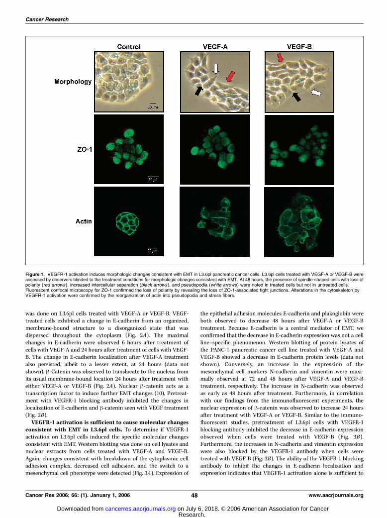
VEGFR-1 activation induces morphologic changes consis-tent with EMT in L3.6pl cells. The first indication that EMTinduced by VEGFR-1 activation might mediate the increase wepreviously observed in migration and invasion in pancreatic cancercells were changes in the morphology of L3.6pl cells that had beentreated with VEGF-A and VEGF-B. At 48 hours after the initiationof treatment, blinded investigators observed differences in thegross appearance of VEGF-treated cells as compared withuntreated cells. The phenotypic changes observed included lossof cell polarity causing a spindle-cell morphology, increasedintercellular separation signifying loss of intercellular adhesion,and increased formation of pseudopodia observed emanating fromthe cell membrane (Fig. 1). We confirmed the loss of polarity byimmunofluorescent staining for ZO-1. The loss/decrease of ZO-1following VEGF-A and VEGF-B treatment indicates that VEGFR-1activation leads to loss of tight junctions associated withorganizing membrane polarity (8). In addition, VEGFR-1 activationled to reorganization of actin from a predominantly membrane-bound location to newly formed pseudopodia that we observed vialight microscopy. Actin was also observed to form stress fibersthroughout the cytoplasm following treatment with VEGF-A andVEGF-B. These changes are typical of cells with a mesenchymalphenotype rather than the usual epithelial phenotype of L3.6plcells, indicating that the cells were undergoing EMT after VEGFR-1activation. Approximately 30% of the cells in culture were observedto be undergoing changes consistent with EMT by both directvisualization and fluorescent microscopy. Of note, all morphologicstudies were done on plastic culture containers, whereas allimmunofluorescent studies were done on glass slides, which mayexplain the subtle differences in the appearance betweenmorphologic and immunohistochemical studies.VEGFR-1 activation induces changes in the localization of
cellular EMT markers in L3.6pl cells. One of the hallmarks ofEMT is the breakdown of the cytoplasmic-cell adhesion complex(9). The breakdown of this complex causes a change in thelocalization of E-cadherin and h-catenin from their usualmembrane-bound location. To determine if activation of VEGFR-1led to changes consistent with EMT, immunofluorescent staining
VEGFR-1 Induces EMT in Pancreatic Cancer
www.aacrjournals.org 47 Cancer Res 2006; 66: (1). January 1, 2006
Research. on July 6, 2018. © 2006 American Association for Cancercancerres.aacrjournals.org Downloaded from
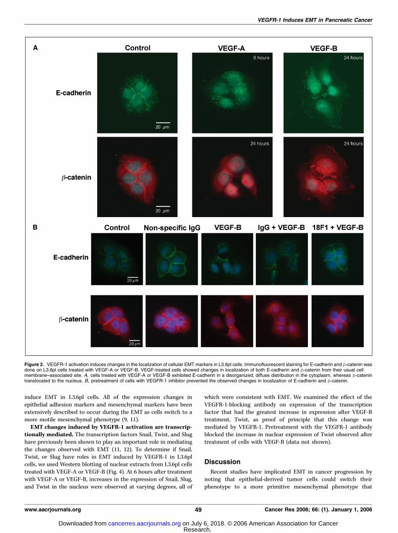
was done on L3.6pl cells treated with VEGF-A or VEGF-B. VEGF-treated cells exhibited a change in E-cadherin from an organized,membrane-bound structure to a disorganized state that wasdispersed throughout the cytoplasm (Fig. 2A). The maximalchanges in E-cadherin were observed 6 hours after treatment ofcells with VEGF-A and 24 hours after treatment of cells with VEGF-B. The change in E-cadherin localization after VEGF-A treatmentalso persisted, albeit to a lesser extent, at 24 hours (data notshown). h-Catenin was observed to translocate to the nucleus fromits usual membrane-bound location 24 hours after treatment witheither VEGF-A or VEGF-B (Fig. 2A). Nuclear h-catenin acts as atranscription factor to induce further EMT changes (10). Pretreat-ment with VEGFR-1 blocking antibody inhibited the changes inlocalization of E-cadherin and h-catenin seen with VEGF treatment(Fig. 2B).VEGFR-1 activation is sufficient to cause molecular changes
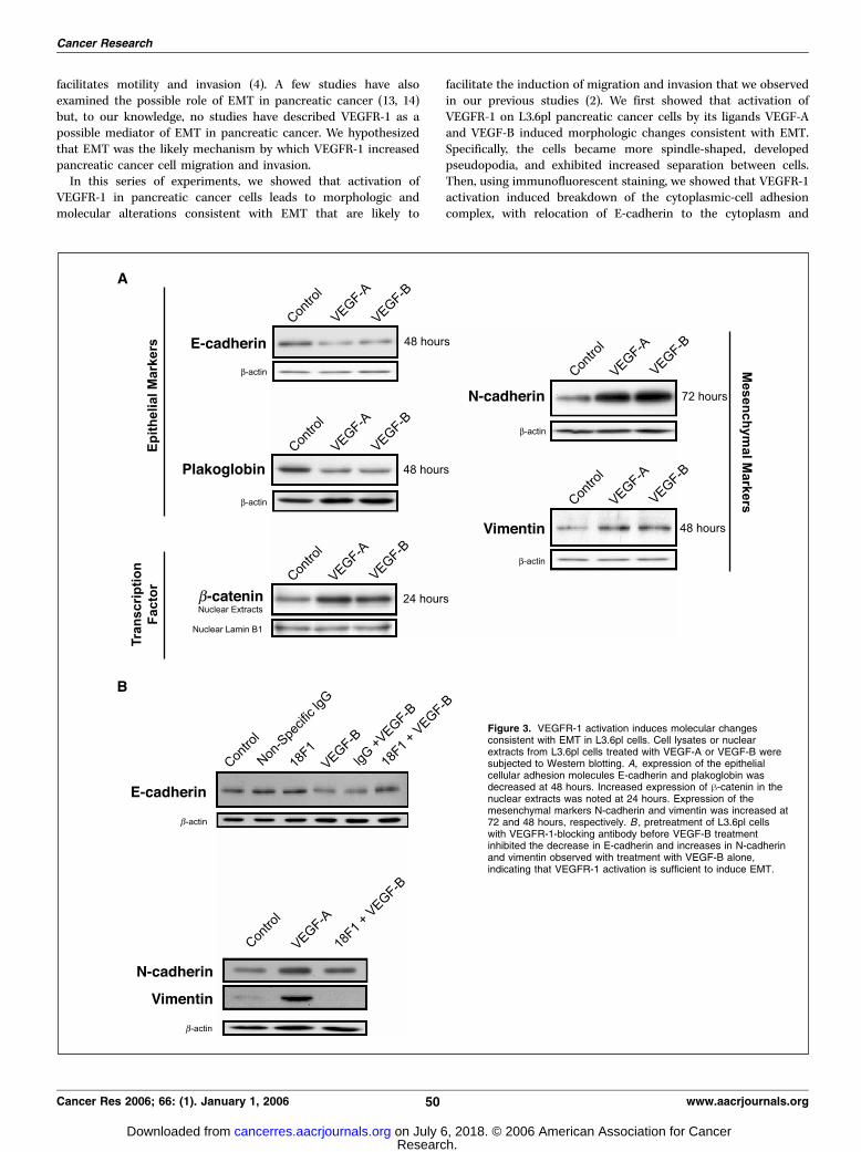
consistent with EMT in L3.6pl cells. To determine if VEGFR-1activation on L3.6pl cells induced the specific molecular changesconsistent with EMT, Western blotting was done on cell lysates andnuclear extracts from cells treated with VEGF-A and VEGF-B.Again, changes consistent with breakdown of the cytoplasmic celladhesion complex, decreased cell adhesion, and the switch to amesenchymal cell phenotype were detected (Fig. 3A). Expression of
the epithelial adhesion molecules E-cadherin and plakoglobin wereboth observed to decrease 48 hours after VEGF-A or VEGF-Btreatment. Because E-cadherin is a central mediator of EMT, weconfirmed that the decrease in E-cadherin expression was not a cellline–specific phenomenon. Western blotting of protein lysates ofthe PANC-1 pancreatic cancer cell line treated with VEGF-A andVEGF-B showed a decrease in E-cadherin protein levels (data notshown). Conversely, an increase in the expression of themesenchymal cell markers N-cadherin and vimentin were maxi-mally observed at 72 and 48 hours after VEGF-A and VEGF-Btreatment, respectively. The increase in N-cadherin was observedas early as 48 hours after treatment. Furthermore, in correlationwith our findings from the immunofluorescent experiments, thenuclear expression of h-catenin was observed to increase 24 hoursafter treatment with VEGF-A or VEGF-B. Similar to the immuno-fluorescent studies, pretreatment of L3.6pl cells with VEGFR-1blocking antibody inhibited the decrease in E-cadherin expressionobserved when cells were treated with VEGF-B (Fig. 3B ).Furthermore, the increases in N-cadherin and vimentin expressionwere also blocked by the VEGFR-1 antibody when cells weretreated with VEGF-B (Fig. 3B). The ability of the VEGFR-1 blockingantibody to inhibit the changes in E-cadherin localization andexpression indicates that VEGFR-1 activation alone is sufficient to
Figure 1. VEGFR-1 activation induces morphologic changes consistent with EMT in L3.6pl pancreatic cancer cells. L3.6pl cells treated with VEGF-A or VEGF-B wereassessed by observers blinded to the treatment conditions for morphologic changes consistent with EMT. At 48 hours, the presence of spindle-shaped cells with loss ofpolarity (red arrows ), increased intercellular separation (black arrows), and pseudopodia (white arrows ) were noted in treated cells but not in untreated cells.Fluorescent confocal microscopy for ZO-1 confirmed the loss of polarity by revealing the loss of ZO-1-associated tight junctions. Alterations in the cytoskeleton byVEGFR-1 activation were confirmed by the reorganization of actin into pseudopodia and stress fibers.
Cancer Research
Cancer Res 2006; 66: (1). January 1, 2006 48 www.aacrjournals.org
Research. on July 6, 2018. © 2006 American Association for Cancercancerres.aacrjournals.org Downloaded from
induce EMT in L3.6pl cells. All of the expression changes inepithelial adhesion markers and mesenchymal markers have beenextensively described to occur during the EMT as cells switch to amore motile mesenchymal phenotype (9, 11).EMT changes induced by VEGFR-1 activation are transcrip-
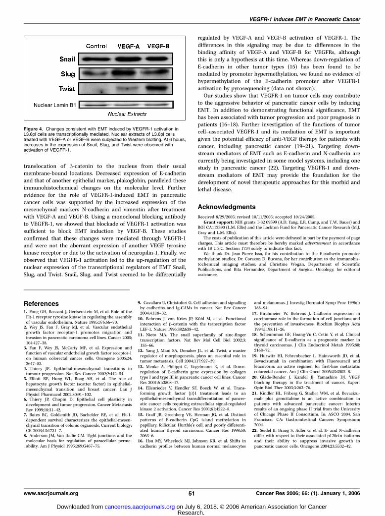
tionally mediated. The transcription factors Snail, Twist, and Slughave previously been shown to play an important role in mediatingthe changes observed with EMT (11, 12). To determine if Snail,Twist, or Slug have roles in EMT induced by VEGFR-1 in L3.6plcells, we used Western blotting of nuclear extracts from L3.6pl cellstreated with VEGF-A or VEGF-B (Fig. 4). At 6 hours after treatmentwith VEGF-A or VEGF-B, increases in the expression of Snail, Slug,and Twist in the nucleus were observed at varying degrees, all of
which were consistent with EMT. We examined the effect of theVEGFR-1-blocking antibody on expression of the transcriptionfactor that had the greatest increase in expression after VEGF-Btreatment, Twist, as proof of principle that this change wasmediated by VEGFR-1. Pretreatment with the VEGFR-1 antibodyblocked the increase in nuclear expression of Twist observed aftertreatment of cells with VEGF-B (data not shown).
Discussion
Recent studies have implicated EMT in cancer progression bynoting that epithelial-derived tumor cells could switch theirphenotype to a more primitive mesenchymal phenotype that
Figure 2. VEGFR-1 activation induces changes in the localization of cellular EMT markers in L3.6pl cells. Immunofluorescent staining for E-cadherin and h-catenin wasdone on L3.6pl cells treated with VEGF-A or VEGF-B. VEGF-treated cells showed changes in localization of both E-cadherin and h-catenin from their usual cellmembrane–associated site. A, cells treated with VEGF-A or VEGF-B exhibited E-cadherin in a disorganized, diffuse distribution in the cytoplasm, whereas h-catenintranslocated to the nucleus. B, pretreatment of cells with VEGFR-1 inhibitor prevented the observed changes in localization of E-cadherin and h-catenin.
VEGFR-1 Induces EMT in Pancreatic Cancer
www.aacrjournals.org 49 Cancer Res 2006; 66: (1). January 1, 2006
Research. on July 6, 2018. © 2006 American Association for Cancercancerres.aacrjournals.org Downloaded from
facilitates motility and invasion (4). A few studies have alsoexamined the possible role of EMT in pancreatic cancer (13, 14)but, to our knowledge, no studies have described VEGFR-1 as apossible mediator of EMT in pancreatic cancer. We hypothesizedthat EMT was the likely mechanism by which VEGFR-1 increasedpancreatic cancer cell migration and invasion.In this series of experiments, we showed that activation of
VEGFR-1 in pancreatic cancer cells leads to morphologic andmolecular alterations consistent with EMT that are likely to
facilitate the induction of migration and invasion that we observedin our previous studies (2). We first showed that activation ofVEGFR-1 on L3.6pl pancreatic cancer cells by its ligands VEGF-Aand VEGF-B induced morphologic changes consistent with EMT.Specifically, the cells became more spindle-shaped, developedpseudopodia, and exhibited increased separation between cells.Then, using immunofluorescent staining, we showed that VEGFR-1activation induced breakdown of the cytoplasmic-cell adhesioncomplex, with relocation of E-cadherin to the cytoplasm and
Figure 3. VEGFR-1 activation induces molecular changesconsistent with EMT in L3.6pl cells. Cell lysates or nuclearextracts from L3.6pl cells treated with VEGF-A or VEGF-B weresubjected to Western blotting. A, expression of the epithelialcellular adhesion molecules E-cadherin and plakoglobin wasdecreased at 48 hours. Increased expression of h-catenin in thenuclear extracts was noted at 24 hours. Expression of themesenchymal markers N-cadherin and vimentin was increased at72 and 48 hours, respectively. B , pretreatment of L3.6pl cellswith VEGFR-1-blocking antibody before VEGF-B treatmentinhibited the decrease in E-cadherin and increases in N-cadherinand vimentin observed with treatment with VEGF-B alone,indicating that VEGFR-1 activation is sufficient to induce EMT.
Cancer Research
Cancer Res 2006; 66: (1). January 1, 2006 50 www.aacrjournals.org
Research. on July 6, 2018. © 2006 American Association for Cancercancerres.aacrjournals.org Downloaded from
translocation of h-catenin to the nucleus from their usualmembrane-bound locations. Decreased expression of E-cadherinand that of another epithelial marker, plakoglobin, paralleled theseimmunohistochemical changes on the molecular level. Furtherevidence for the role of VEGFR-1-induced EMT in pancreaticcancer cells was supported by the increased expression of themesenchymal markers N-cadherin and vimentin after treatmentwith VEGF-A and VEGF-B. Using a monoclonal blocking antibodyto VEGFR-1, we showed that blockade of VEGFR-1 activation wassufficient to block EMT induction by VEGF-B. These studiesconfirmed that these changes were mediated through VEGFR-1and were not the aberrant expression of another VEGF tyrosinekinase receptor or due to the activation of neuropilin-1. Finally, weobserved that VEGFR-1 activation led to the up-regulation of thenuclear expression of the transcriptional regulators of EMT Snail,Slug, and Twist. Snail, Slug, and Twist seemed to be differentially
regulated by VEGF-A and VEGF-B activation of VEGFR-1. Thedifferences in this signaling may be due to differences in thebinding affinity of VEGF-A and VEGF-B for VEGFRs, althoughthis is only a hypothesis at this time. Whereas down-regulation ofE-cadherin in other tumor types (15) has been found to bemediated by promoter hypermethylation, we found no evidence ofhypermethylation of the E-cadherin promoter after VEGFR-1activation by pyrosequencing (data not shown).Our studies show that VEGFR-1 on tumor cells may contribute
to the aggressive behavior of pancreatic cancer cells by inducingEMT. In addition to demonstrating functional significance, EMThas been associated with tumor progression and poor prognosis inpatients (16–18). Further investigation of the functions of tumorcell–associated VEGFR-1 and its mediation of EMT is importantgiven the potential efficacy of anti-VEGF therapy for patients withcancer, including pancreatic cancer (19–21). Targeting down-stream mediators of EMT such as E-cadherin and N-cadherin arecurrently being investigated in some model systems, including onestudy in pancreatic cancer (22). Targeting VEGFR-1 and down-stream mediators of EMT may provide the foundation for thedevelopment of novel therapeutic approaches for this morbid andlethal disease.
Acknowledgments
Received 8/29/2005; revised 10/11/2005; accepted 10/24/2005.Grant support: NIH grants T-32 09599 (A.D. Yang, E.R. Camp, and T.W. Bauer) and
ROI CA112390 (L.M. Ellis) and the Lockton Fund for Pancreatic Cancer Research (M.J.Gray and L.M. Ellis).
The costs of publication of this article were defrayed in part by the payment of pagecharges. This article must therefore be hereby marked advertisement in accordancewith 18 U.S.C. Section 1734 solely to indicate this fact.
We thank Dr. Jean-Pierre Issa, for his contribution to the E-cadherin promotermethylation studies; Dr. Corazon D. Bucana, for her contribution to the immunohis-tochemical imaging studies; and Christine Wogan, Department of ScientificPublications, and Rita Hernandez, Department of Surgical Oncology, for editorialassistance.
Figure 4. Changes consistent with EMT induced by VEGFR-1 activation inL3.6pl cells are transcriptionally mediated. Nuclear extracts of L3.6pl cellstreated with VEGF-A or VEGF-B were subjected to Western blotting. At 6 hours,increases in the expression of Snail, Slug, and Twist were observed withactivation of VEGFR-1.
VEGFR-1 Induces EMT in Pancreatic Cancer
www.aacrjournals.org 51 Cancer Res 2006; 66: (1). January 1, 2006
References1. Fong GH, Rossant J, Gertsenstein M, et al. Role of theFlt-1 receptor tyrosine kinase in regulating the assemblyof vascular endothelium. Nature 1995;376:66–70.
2. Wey JS, Fan F, Gray MJ, et al. Vascular endothelialgrowth factor receptor-1 promotes migration andinvasion in pancreatic carcinoma cell lines. Cancer 2005;104:427–38.
3. Fan F, Wey JS, McCarty MF, et al. Expression andfunction of vascular endothelial growth factor receptor-1on human colorectal cancer cells. Oncogene 2005;24:2647–53.
4. Thiery JP. Epithelial-mesenchymal transitions intumour progression. Nat Rev Cancer 2002;2:442–54.
5. Elliott BE, Hung WL, Boag AH, et al. The role ofhepatocyte growth factor (scatter factor) in epithelial-mesenchymal transition and breast cancer. Can JPhysiol Pharmacol 2002;80:91–102.
6. Thiery JP, Chopin D. Epithelial cell plasticity indevelopment and tumor progression. Cancer MetastasisRev 1999;18:31–42.
7. Bates RC, Goldsmith JD, Bachelder RE, et al. Flt-1-dependent survival characterizes the epithelial-mesen-chymal transition of colonic organoids. Current biology:CB 2003;13:1721–7.
8. Anderson JM, Van Itallie CM. Tight junctions and themolecular basis for regulation of paracellular perme-ability. Am J Physiol 1995;269:G467–75.
9. Cavallaro U, Christofori G. Cell adhesion and signallingby cadherins and Ig-CAMs in cancer. Nat Rev Cancer2004;4:118–32.
10. Behrens J, von Kries JP, Kuhl M, et al. Functionalinteraction of h-catenin with the transcription factorLEF-1. Nature 1996;382:638–42.
11. Nieto MA. The snail superfamily of zinc-fingertranscription factors. Nat Rev Mol Cell Biol 2002;3:155–66.
12. Yang J, Mani SA, Donaher JL, et al. Twist, a masterregulator of morphogenesis, plays an essential role intumor metastasis. Cell 2004;117:927–39.
13. Menke A, Philippi C, Vogelmann R, et al. Down-regulation of E-cadherin gene expression by collagentype I and type III in pancreatic cancer cell lines. CancerRes 2001;61:3508–17.
14. Ellenrieder V, Hendler SF, Boeck W, et al. Trans-forming growth factor {h}1 treatment leads to anepithelial-mesenchymal transdifferentiation of pancre-atic cancer cells requiring extracellular signal-regulatedkinase 2 activation. Cancer Res 2001;61:4222–8.
15. Graff JR, Greenberg VE, Herman JG, et al. Distinctpatterns of E-cadherin CpG island methylation inpapillary, follicular, Hurthle’s cell, and poorly differenti-ated human thyroid carcinoma. Cancer Res 1998;58:2063–6.
16. Hsu MY, Wheelock MJ, Johnson KR, et al. Shifts incadherin profiles between human normal melanocytes
and melanomas. J Investig Dermatol Symp Proc 1996;1:188–94.
17. Birchmeier W, Behrens J. Cadherin expression incarcinomas: role in the formation of cell junctions andthe prevention of invasiveness. Biochim Biophys Acta1994;1198:11–26.
18. Scheumman GF, Hoang-Vu C, Cetin Y, et al. Clinicalsignificance of E-cadherin as a prognostic marker inthyroid carcinomas. J Clin Endocrinol Metab 1995;80:2168–72.
19. Hurwitz HI, Fehrenbacher L, Hainsworth JD, et al.Bevacizumab in combination with Fluorouracil andleucovorin: an active regimen for first-line metastaticcolorectal cancer. Am J Clin Oncol 2005;23:3502–8.
20. Glade-Bender J, Kandel JJ, Yamashiro DJ. VEGFblocking therapy in the treatment of cancer. ExpertOpin Biol Ther 2003;3:263–76.
21. Kindler HL, Friberg G, Stadler WM, et al. Bevacizu-mab plus gemcitabine is an active combination inpatients with advanced pancreatic cancer: Interimresults of an ongoing phase II trial from the Universityof Chicago Phase II Consortium. In: ASCO 2004. SanFrancisco, CA: Gastrointestinal Cancers Symposium;2004.
22. Seidel B, Braeg S, Adler G, et al. E- and N-cadherindiffer with respect to their associated p120ctn isoformsand their ability to suppress invasive growth inpancreatic cancer cells. Oncogene 2004;23:5532–42.
Research. on July 6, 2018. © 2006 American Association for Cancercancerres.aacrjournals.org Downloaded from
2006;66:46-51. Cancer Res Anthony D. Yang, E. Ramsay Camp, Fan Fan, et al. Pancreatic Carcinoma CellsMediates Epithelial to Mesenchymal Transition in Human Vascular Endothelial Growth Factor Receptor-1 Activation
Updated version
http://cancerres.aacrjournals.org/content/66/1/46
Access the most recent version of this article at:
Cited articles
http://cancerres.aacrjournals.org/content/66/1/46.full#ref-list-1
This article cites 19 articles, 4 of which you can access for free at:
Citing articles
http://cancerres.aacrjournals.org/content/66/1/46.full#related-urls
This article has been cited by 33 HighWire-hosted articles. Access the articles at:
E-mail alerts related to this article or journal.Sign up to receive free email-alerts
Subscriptions
Reprints and
To order reprints of this article or to subscribe to the journal, contact the AACR Publications
Permissions
Rightslink site. (CCC)Click on "Request Permissions" which will take you to the Copyright Clearance Center's
.http://cancerres.aacrjournals.org/content/66/1/46To request permission to re-use all or part of this article, use this link
Research. on July 6, 2018. © 2006 American Association for Cancercancerres.aacrjournals.org Downloaded from