vascular anatomy of the flower of hyacinthoides non-scripta
TRANSCRIPT
Modern Phytomorphology 5: 9–18, 2014
© The Author(s), 2014
Introduction
The genus Hyacinthoides Heist. ex Fabr. was recently thoroughly revised with a firmly established phylogeny (Grundmann et al. 2010). It now comprises 11 species, living in the western Mediterranean-Atlantic region and the northern African mountains. Among them, H. non-scripta (L.) Chouard ex Rothm. – the British bluebell – covers the widest area along the Atlantic coast, spreading from the British Isles to the western Netherlands and Belgium, North and West France, with a southern separate territory at the NW Spain. This species is frequent in the forests of the Paris surroundings, growing in neutral or acid soils at shady places ( Jauzein & Nawrot 2011: 838), especially under Quercus sp. and Carpinus betulus, with Anemone nemorosa.
Surprisingly, few studies were previously undertaken on the floral anatomy of this widespread species. Notably Chouard (1931) focused on the vegetative features when he distinguished the new genus Hyacinthoides. Vasculature was briefly described
by Van Tieghem (1875: 102) under the illegitimate name Agraphis nutans Link, while fused tepal and filament only were drawn (Van Tieghem 1875: Pl. 2, 65). A more detailed study was provided by Gatin (1920), especially about the pedicel histology and receptacle vasculature. Teratological flowers, with stamens partially or wholly altered in carpels, were pointed out by Wilson (1959) under the illegitimate name Endymion nutans Dumort., but unfortunately not accurately analysed.
Thus it seemed worthwhile to investigate further the floral anatomy of H. non-scripta, at least as a check of previous results, if not in order to throw new light on some yet overlooked features.
Material and methods
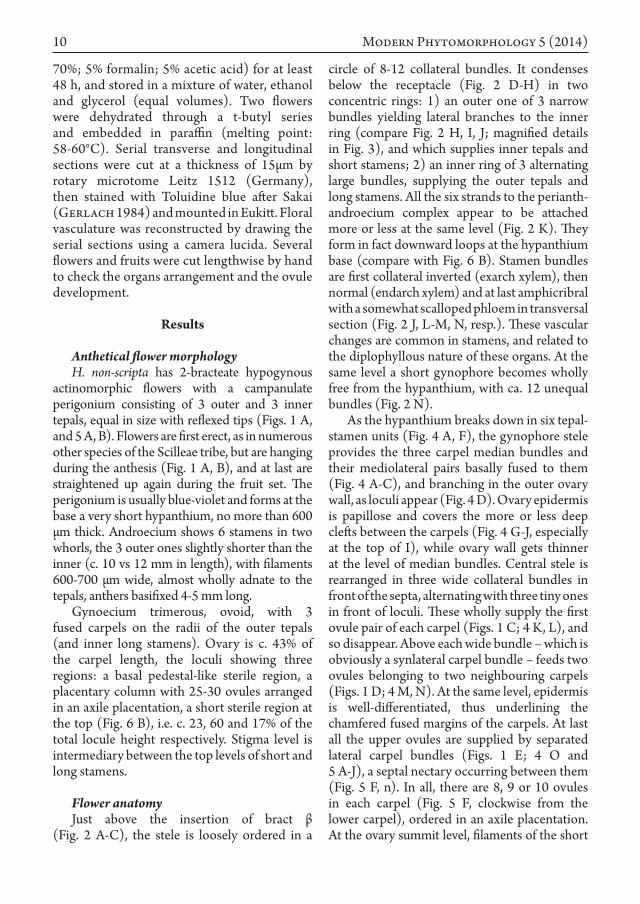
Flowers and fruits of H. non-scripta were collected at different stages during April 2012 and May 2013 in the undergrowth of the Sénart forest (“Forêt domaniale de Sénart”, parcel 111), at Draveil (France, Île-de-France region, Essonne, ca. 20 km SSE of Paris). All samples were fixed on the field by FAA (90% ethanol
VASCULAR ANATOMY OF THE FLOWER OF HYACINTHOIDES NON‑SCRIPTA (L.) CHOUARD EX ROTHM. A NEW INSIGHT
ABOUT A COMPLEX PLACENTATION PATTERN IN ASPARAGACEAE
Thierry Deroin
Abstract. Floral vasculature and gynoecium architecture were revised in Hyacinthoides non-scripta (L.) Chouard ex Rothm. Receptacle exhibits the usual regular vascular groundplan for monocots, while carpels are united at the ovary bases and by their styles, but very loosely in their middle region, so that a basket-like capsule is ripened. The septal nectary is well developed with two apertures at the ovary base and below the style insertion. Vasculature demonstrated basal ovules pairs sheltered by each carpel are in fact cauline. Thus floral axis appears to be involved in the congenital intercarpellary fusion, an unrecognized feature until now in the core-Asparagales.
Key words: Hyacinthoides non-scripta, Hyacinthaceae, anatomy, cauline placentation, floral vasculature, gynoecium, stachyospory
Muséum national d’Histoire naturelle, Département Systématique et Évolution, UMR 7205, ISYEB, CNRS MNHN UPMC EPHE, CP 39, 57 rue Cuvier, F-75231 Paris cedex 05, France; [email protected]
10 Modern Phytomorphology 5 (2014)
70%; 5% formalin; 5% acetic acid) for at least 48 h, and stored in a mixture of water, ethanol and glycerol (equal volumes). Two flowers were dehydrated through a t-butyl series and embedded in paraffin (melting point: 58-60°C). Serial transverse and longitudinal sections were cut at a thickness of 15µm by rotary microtome Leitz 1512 (Germany), then stained with Toluidine blue after Sakai (Gerlach 1984) and mounted in Eukitt. Floral vasculature was reconstructed by drawing the serial sections using a camera lucida. Several flowers and fruits were cut lengthwise by hand to check the organs arrangement and the ovule development.
Results
Anthetical flower morphologyH. non-scripta has 2-bracteate hypogynous
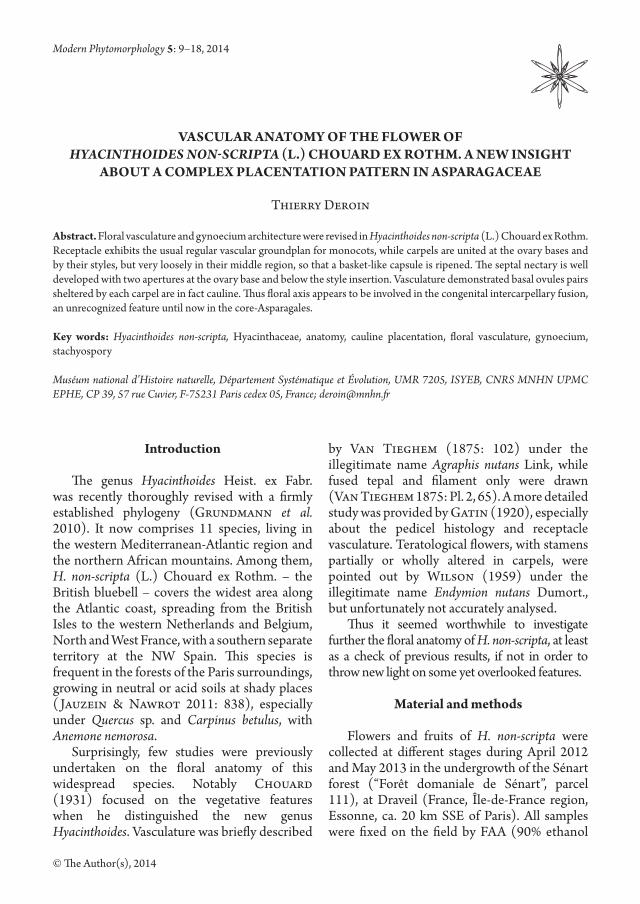
actinomorphic flowers with a campanulate perigonium consisting of 3 outer and 3 inner tepals, equal in size with reflexed tips (Figs. 1 A, and 5 A, B). Flowers are first erect, as in numerous other species of the Scilleae tribe, but are hanging during the anthesis (Fig. 1 A, B), and at last are straightened up again during the fruit set. The perigonium is usually blue-violet and forms at the base a very short hypanthium, no more than 600 µm thick. Androecium shows 6 stamens in two whorls, the 3 outer ones slightly shorter than the inner (c. 10 vs 12 mm in length), with filaments 600-700 µm wide, almost wholly adnate to the tepals, anthers basifixed 4-5 mm long.
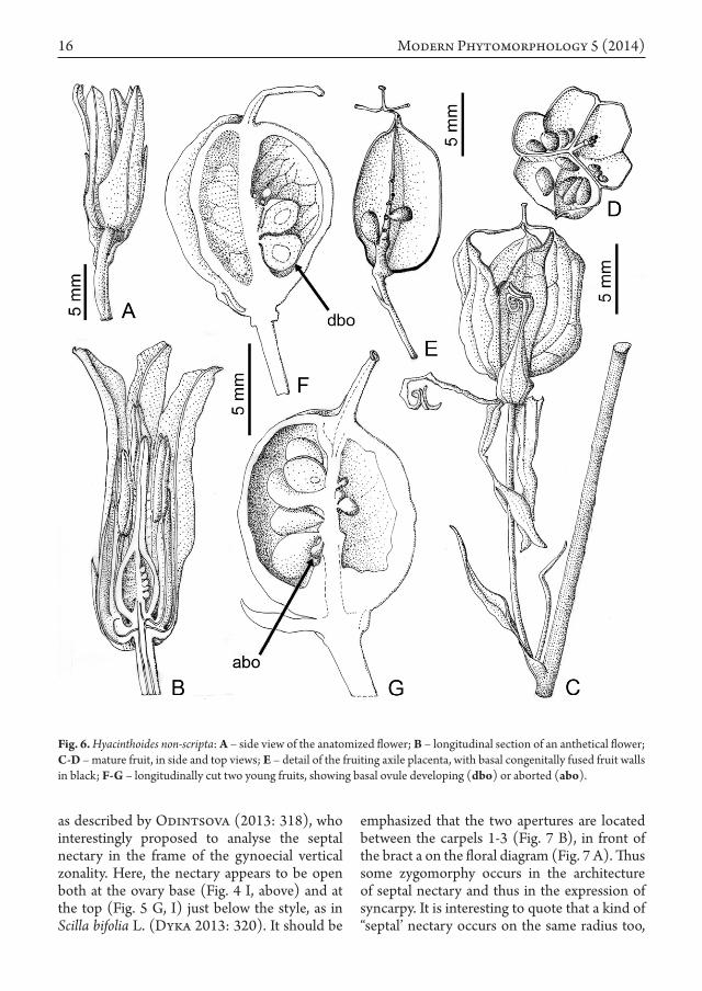
Gynoecium trimerous, ovoid, with 3 fused carpels on the radii of the outer tepals (and inner long stamens). Ovary is c. 43% of the carpel length, the loculi showing three regions: a basal pedestal-like sterile region, a placentary column with 25-30 ovules arranged in an axile placentation, a short sterile region at the top (Fig. 6 B), i.e. c. 23, 60 and 17% of the total locule height respectively. Stigma level is intermediary between the top levels of short and long stamens.
Flower anatomyJust above the insertion of bract β
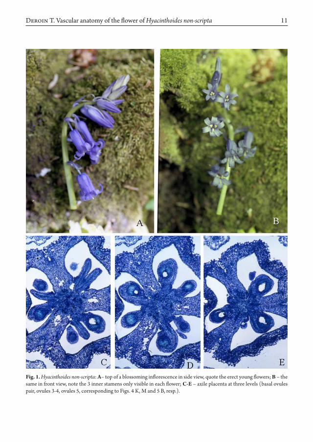
(Fig. 2 A-C), the stele is loosely ordered in a
circle of 8-12 collateral bundles. It condenses below the receptacle (Fig. 2 D-H) in two concentric rings: 1) an outer one of 3 narrow bundles yielding lateral branches to the inner ring (compare Fig. 2 H, I, J; magnified details in Fig. 3), and which supplies inner tepals and short stamens; 2) an inner ring of 3 alternating large bundles, supplying the outer tepals and long stamens. All the six strands to the perianth-androecium complex appear to be attached more or less at the same level (Fig. 2 K). They form in fact downward loops at the hypanthium base (compare with Fig. 6 B). Stamen bundles are first collateral inverted (exarch xylem), then normal (endarch xylem) and at last amphicribral with a somewhat scalloped phloem in transversal section (Fig. 2 J, L-M, N, resp.). These vascular changes are common in stamens, and related to the diplophyllous nature of these organs. At the same level a short gynophore becomes wholly free from the hypanthium, with ca. 12 unequal bundles (Fig. 2 N).
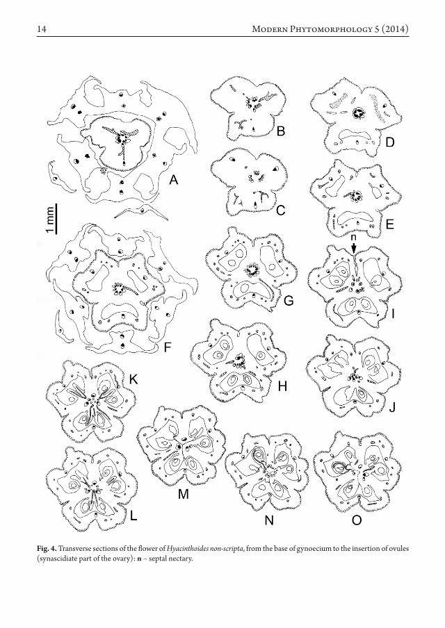
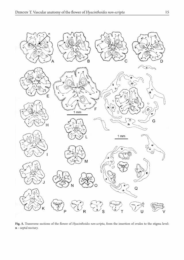
As the hypanthium breaks down in six tepal-stamen units (Fig. 4 A, F), the gynophore stele provides the three carpel median bundles and their mediolateral pairs basally fused to them (Fig. 4 A-C), and branching in the outer ovary wall, as loculi appear (Fig. 4 D). Ovary epidermis is papillose and covers the more or less deep clefts between the carpels (Fig. 4 G-J, especially at the top of I), while ovary wall gets thinner at the level of median bundles. Central stele is rearranged in three wide collateral bundles in front of the septa, alternating with three tiny ones in front of loculi. These wholly supply the first ovule pair of each carpel (Figs. 1 C; 4 K, L), and so disappear. Above each wide bundle – which is obviously a synlateral carpel bundle – feeds two ovules belonging to two neighbouring carpels (Figs. 1 D; 4 M, N). At the same level, epidermis is well-differentiated, thus underlining the chamfered fused margins of the carpels. At last all the upper ovules are supplied by separated lateral carpel bundles (Figs. 1 E; 4 O and 5 A-J), a septal nectary occurring between them (Fig. 5 F, n). In all, there are 8, 9 or 10 ovules in each carpel (Fig. 5 F, clockwise from the lower carpel), ordered in an axile placentation. At the ovary summit level, filaments of the short
11
Fig. 1. Hyacinthoides non-scripta: A– top of a blossoming inflorescence in side view, quote the erect young flowers; B – the same in front view, note the 3 inner stamens only visible in each flower; C-E – axile placenta at three levels (basal ovules pair, ovules 3-4, ovules 5, corresponding to Figs. 4 K, M and 5 B, resp.).
Deroin T. Vascular anatomy of the flower of Hyacinthoides non-scripta
12 Modern Phytomorphology 5 (2014)
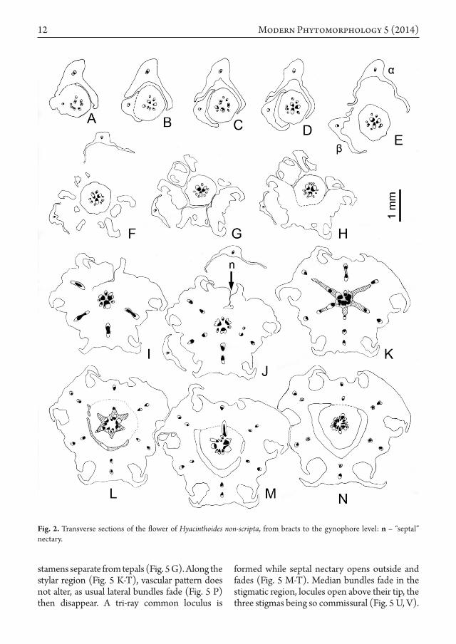
stamens separate from tepals (Fig. 5 G). Along the stylar region (Fig. 5 K-T), vascular pattern does not alter, as usual lateral bundles fade (Fig. 5 P) then disappear. A tri-ray common loculus is
formed while septal nectary opens outside and fades (Fig. 5 M-T). Median bundles fade in the stigmatic region, locules open above their tip, the three stigmas being so commissural (Fig. 5 U, V).
Fig. 2. Transverse sections of the flower of Hyacinthoides non-scripta, from bracts to the gynophore level: n – “septal” nectary.
13
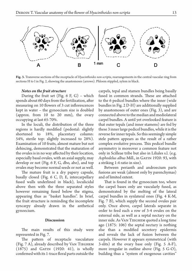
Fig. 3. Transverse sections of the receptacle of Hyacinthoides non-scripta, rearrangements in the central vascular ring from sections H to I in Fig. 2, showing the anastomoses (arrows). Phloem stippled, xylem in black.
Notes on the fruit structureDuring the fruit set (Fig. 6 F, G) – which
spends about 60 days from the fertilization, after measuring on 10 flowers of 3 cut inflorescences kept in water – the gynoecium size is doubled (approx. from 10 to 20 mm), the ovary occupying at last 65-70%.
In the loculi, the distribution of the three regions is hardly modified (pedestal: slightly shortened to 18%, placentary column: 54%, sterile top: slightly increased to 28%). Examination of 10 fruits, almost mature but not dehiscing, demonstrated that the maturation of the ovules is in no way affected by their location, especially basal ovules, with an axial supply, may develop or not (Fig. 6 F, G, dbo, abo), and top ovules may become normal seeds or not as well.
The mature fruit is a dry papery capsule, basally closed (Fig. 6 C, D, E, intercarpellary fused walls underlined in black), loculicidal above then with the three separated styles however remaining fused below the stigma, appearing thus as “basket handles”. In brief, the fruit structure is reminding the incomplete syncarpy already drawn in the anthetical gynoecium.
Discussion
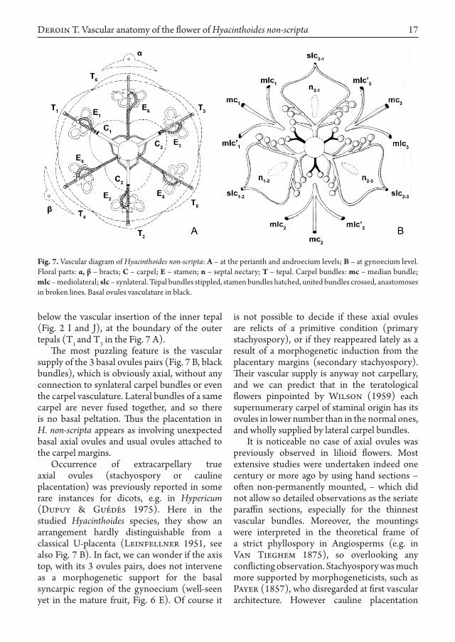
The main results of this study were represented in Fig. 7.
The pattern of receptacle vasculature (Fig. 7 A), already described by Van Tieghem (1875) and Gatin (1920: 41), is wholly confirmed with its 1-trace floral parts outside the
carpels, tepal and stamen bundles being basally fused in common strands. These are attached to the 6 pedicel bundles where the inner (wide bundles in Fig. 2 D-H) are additionally supplied by anastomoses of outer ones (Fig. 3), and are connected above to the median and mediolateral carpel bundles. A until yet overlooked feature is that outer tepals (and inner stamens) are fed by these 3 inner large pedicel bundles, while it is the reverse for inner tepals. So this seemingly simple stele pattern appears as the result of a rather complex evolutive process. This pedicel bundle asymmetry is moreover a common feature not only in Scilleae tribe but also in Liliales s.l. (e.g. Asphodelus albus Mill., in Gatin 1920: 93, with a striking 1:4 ratio in size).
Between perianth and androecium parts fusions are weak (almost only by parenchymas) and of limited extent.
That is found in the gynoecium too, where the carpel bases only are vascularly fused, as demonstrated by the melting of the lateral carpel bundles in synlateral ones (Fig. 4 K-M, Fig. 7 B), which supply the second ovules pair only. Once above, carpel laterals separate in order to feed each a row of 3-4 ovules on the external side, as well as a septal nectary on the inner side. As Van Tieghem quoted a long time ago (1875: 106) the septal nectary is nothing else than a modified secretory epidermis and reveals the lack of fusion between the carpels. However it appears symmetrical (with 3-slits) at the ovary base only (Fig. 5 A-F), more irregular in outline above (Fig. 5 G-O), building thus a “system of exogenous cavities”
Deroin T. Vascular anatomy of the flower of Hyacinthoides non-scripta
14 Modern Phytomorphology 5 (2014)
Fig. 4. Transverse sections of the flower of Hyacinthoides non-scripta, from the base of gynoecium to the insertion of ovules (synascidiate part of the ovary): n – septal nectary.
15
Fig. 5. Transverse sections of the flower of Hyacinthoides non-scripta, from the insertion of ovules to the stigma level: n – septal nectary.
Deroin T. Vascular anatomy of the flower of Hyacinthoides non-scripta
16 Modern Phytomorphology 5 (2014)
Fig. 6. Hyacinthoides non-scripta: A – side view of the anatomized flower; B – longitudinal section of an anthetical flower; C-D – mature fruit, in side and top views; E – detail of the fruiting axile placenta, with basal congenitally fused fruit walls in black; F-G – longitudinally cut two young fruits, showing basal ovule developing (dbo) or aborted (abo).
as described by Odintsova (2013: 318), who interestingly proposed to analyse the septal nectary in the frame of the gynoecial vertical zonality. Here, the nectary appears to be open both at the ovary base (Fig. 4 I, above) and at the top (Fig. 5 G, I) just below the style, as in Scilla bifolia L. (Dyka 2013: 320). It should be
emphasized that the two apertures are located between the carpels 1-3 (Fig. 7 B), in front of the bract a on the floral diagram (Fig. 7 A). Thus some zygomorphy occurs in the architecture of septal nectary and thus in the expression of syncarpy. It is interesting to quote that a kind of “septal’ nectary occurs on the same radius too,
17
Fig. 7. Vascular diagram of Hyacinthoides non-scripta: A – at the perianth and androecium levels; B – at gynoecium level. Floral parts: α, β – bracts; C – carpel; E – stamen; n – septal nectary; T – tepal. Carpel bundles: mc – median bundle; mlc – mediolateral; slc – synlateral. Tepal bundles stippled, stamen bundles hatched, united bundles crossed, anastomoses in broken lines. Basal ovules vasculature in black.
below the vascular insertion of the inner tepal (Fig. 2 I and J), at the boundary of the outer tepals (T1 and T3 in the Fig. 7 A).
The most puzzling feature is the vascular supply of the 3 basal ovules pairs (Fig. 7 B, black bundles), which is obviously axial, without any connection to synlateral carpel bundles or even the carpel vasculature. Lateral bundles of a same carpel are never fused together, and so there is no basal peltation. Thus the placentation in H. non-scripta appears as involving unexpected basal axial ovules and usual ovules attached to the carpel margins.
Occurrence of extracarpellary true axial ovules (stachyospory or cauline placentation) was previously reported in some rare instances for dicots, e.g. in Hypericum (Dupuy & Guédès 1975). Here in the studied Hyacinthoides species, they show an arrangement hardly distinguishable from a classical U-placenta (Leinfellner 1951, see also Fig. 7 B). In fact, we can wonder if the axis top, with its 3 ovules pairs, does not intervene as a morphogenetic support for the basal syncarpic region of the gynoecium (well-seen yet in the mature fruit, Fig. 6 E). Of course it
is not possible to decide if these axial ovules are relicts of a primitive condition (primary stachyospory), or if they reappeared lately as a result of a morphogenetic induction from the placentary margins (secondary stachyospory). Their vascular supply is anyway not carpellary, and we can predict that in the teratological flowers pinpointed by Wilson (1959) each supernumerary carpel of staminal origin has its ovules in lower number than in the normal ones, and wholly supplied by lateral carpel bundles.
It is noticeable no case of axial ovules was previously observed in lilioid flowers. Most extensive studies were undertaken indeed one century or more ago by using hand sections – often non-permanently mounted, – which did not allow so detailed observations as the seriate paraffin sections, especially for the thinnest vascular bundles. Moreover, the mountings were interpreted in the theoretical frame of a strict phyllospory in Angiosperms (e.g. in Van Tieghem 1875), so overlooking any conflicting observation. Stachyospory was much more supported by morphogeneticists, such as Payer (1857), who disregarded at first vascular architecture. However cauline placentation
Deroin T. Vascular anatomy of the flower of Hyacinthoides non-scripta
18 Modern Phytomorphology 5 (2014)
might be brought to the fore by it, as suggested in some cases of central placentas – especially in Caryophyllales – by Moeliono (1970).
Thus in the case of H. non-scripta, the carpels are congenitally united at the base via the floral centre, even bearing some ovules, a pathway until now unrecognized for a member of the core-Asparagales (Remizowa et al. 2010: 638). A comparable situation was met by Novikoff & Kazemirska (2012) who demonstrated in Fritillaria montana Hoppe (Liliaceae) that recurrent bundles, in parallel with the synlateral carpel bundles occur in the centre of the ovary column, a striking – and likely functionally significant – but isolated feature yet.
Conclusions
As exemplified by H. non-scripta, apocarpy and syncarpy may coexist in a same gynoecium, while stachyospory and phyllospory are no more exclusive and should be further studied in a broader scope, following a method taking in account the vertical zonality – at first described by Leinfellner (1950), – and combining different approaches such as vascular architecture, histology and development from the floral meristem to the mature fruit.
This brief study is in line with recent proposals (Novikoff & Kazemirska 2012; Dyka 2013; Odintsova 2013), that a careful revision of floral anatomy of monocots is imperative, not only for a better knowledge of character states (and thus their phylogenetical relevance), but even for an accurate understanding of the angiospermous flower. Especially carpels appear to shelter ovules but not always to bear them compulsorily. This result strengthen the previous hypotheses of Moeliono (1970), mainly based on a reappraisal of the central placenta in Caryophyllales. Any struggle about the classical opposition stachyospory vs phyllospory is to be abandoned in Angiosperms.
Acknowledgements
I thank heartily the anonymous reviewer, whose suggestions allowed to improve significantly the original draft. I am grateful to
my colleagues Emmanuel Côtez and Thibault Dumas (Service des Publications scientifiques du Muséum), and Marion Martinez (Herbarium P – GPI team) for their help in the preparation of the electronic version of plates.
References
Chouard P. 1931. Types de développement de l’appareil végétatif chez les Scillées. Ann. Sc. Nat., Bot., Paris, 10e
série 13: 132–323, 4 plates.Dupuy P., Guédès M. 1975. Placentation and possible
partial stachyospory in Hypericum sect. Eremanthe. Flora 164: 37–49.
Dyka O.O. 2013. Morphology and vascular anatomy of the flower in Scilla bifolia L. (Hyacinthaceae). Mod. Phytomorphol. 4: 319–320. (in Ukrainian)
Gatin V.C. 1920. Recherches anatomiques sur le pédoncule et la fleur des Liliacées. Imprimerie nemourienne, Henri Bouloy, Nemours.
Gerlach D. 1984. Botanische Mikrotechnik. 3d ed. Thieme, Stuttgart.
Grundmann M., Rumsey F.J., Ansell S.W., Russell S.J., Darwin S.C., Vogel J.C., Spencer M., Squirrell J., Hollingsworth P.M., Ortiz S., Schneider H. 2010. Phylogeny and taxonomy of the bluebell genus Hyacinthoides, Asparagaceae (Hyacinthaceae). Taxon 59: 68–82.
Jauzein P., Nawrot O. 2011. Flore d’Île-de-France. Quae.
Leinfellner W. 1950. Der Bauplan des synkarpen Gynözeums. Österr. Bot. Zeitschr. 97: 403–436.
Leinfellner W. 1951. Die U-förmige Plazenta als der Plazentationstypus der Angiospermen. Österr. Bot. Zeitschr. 98: 338–358.
Moeliono B.M. 1970. Cauline or carpellary placentation among dicotyledons. 2 vols. Koninklijke Van Gorcum, Assen, The Netherlands.
Novikoff A.V., Kazemirska M.A. 2012. Vascular anatomy and morphology of the flower in Fritillaria montana Hoppe (Liliaceae). Mod. Phytomorphol. 1: 27–35.
Odintsova A. 2013. Vertical zonality of septal nectaries of monocots. Mod. Phytomorphol. 4: 317–318. (in Ukrainian)
Payer J.-B. 1857. Traité d’organogénie comparée de la fleur. Texte et atlas. Masson, Paris. Reprint 1966. Cramer, Wheldon & Wesley, New York.
Remizowa M.V., Sokoloff D.D., Rudall P.J. 2010. Evolutionary history of the monocot flower. Ann. Mo. Bot. Gard. 97: 617–645.
Van Tieghem P. 1875. Recherches sur la structure du pistil et sur l’anatomie comparée de la fleur. Mém. Prés. Divers Savants Acad. Sci. Inst. Impérial France Sér. 2 21: 1–261.
Wilson J.Y. 1959. Vegetative reproduction in the bluebell, Endymion nonscriptus (L.) Garcke. New Phytol. 58: 155–163.