varus gonarthrosis predisposes to varus malalignment in tka
TRANSCRIPT
ORIGINAL ARTICLE
Varus Gonarthrosis Predisposes to Varus Malalignment in TKAThomas J. Heyse, MD &Ralf Decking, MD & Jack Davis, BS, RN, ONC & Friedrich Boettner, MD &Richard S. Laskin, MD
Received: 11 January 2009/Accepted: 30 April 2009/Published online: 20 May 2009* Hospital for Special Surgery 2009
Abstract Postoperative alignment is a predictor for long-term survival of total knee arthroplasty (TKA). The purposeof this study was to evaluate whether or not preoperativedeformities predispose to intraoperative malposition ofTKA components. A retrospective radiographic analysis of53 primary TKA cases was performed. Preoperative AP hipto ankle and lateral knee radiographs were compared withpostoperative views to evaluate component positioning. Thefollowing angles were measured: the hip–knee–ankle(HKA) angle expressing the mechanical axis of the leg,the mechanical lateral distal femur angle (mLDFA), themedial proximal tibia angle (MPTA), the posterior distalfemur angle (PDFA), and the posterior proximal tibia angle(PPTA). Postoperative measurement of the HKA revealed34.0% of the cases had a deviation of >±3° from neutralalignment. Sixteen knees (30.2%) were in varus and, withone exception, all presented with severe varus gonarthrosisprior to surgery with a mean tibiofemoral angle of 12.4°compared with 1.0° of valgus in the optimally alignedgroup. Patients (93.3%) with preoperative valgus malalign-ment showed optimal postoperative HKA. Odds ratios formalalignment of TKA for varus knees in comparison with
valgus knees were 7.1 for HKA, 2.4 for MPTA, 4.9 forPDFA, and 1.7 for PPTA. The overall number of outliers inthe presented data corresponds well with reports from otherauthors using different implants and guide systems. Thepresented data indicate that patients with preoperative varusalignment have a higher risk of postoperative implantmalposition than patients with valgus alignment. The datasupports that preoperative varus deformity predisposes tovarus malposition of TKA. The risk for intraoperativemalposition is significantly lower in valgus knees.
AbbreviationsAP AnteroposteriorHAI Hip–ankle intersectionHKA Hip–knee–ankle anglemLDFA Mechanical lateral distal femur angleMPTA Medial proximal tibia anglePDFA Posterior distal femur anglePPTA Posterior proximal tibia angleOA OsteoarthritisTKA Total knee arthroplasty
Introduction
The introduction of computer navigation in total kneearthroplasty (TKA) has focused research on implantpositioning. The debate continues as to what extentcomponent positioning affects the longevity of implants.Jeffery et al. reported on a series of 115 primary TKA. Theincidence of subsequent component loosening was 3%when Maquet’s line passed through the middle third of theprosthesis. When Maquet’s line was aberrant more than ±3°,the incidence of loosening was 24% after 8 years [1]. Randand Coventry showed a 10-year survival of 90% for kneesbetween 0° and 4° of valgus. However, they described only71% of survival for knees with 5° to 9° of valgus and 73%for knees in varus alignment [2]. On the contrary, Pagnanoet al. recently showed no association between componentposition and early failure in 399 primary cemented TKA at
HSSJ (2009) 5: 143–148DOI 10.1007/s11420-009-9118-7
Level of Evidence: Level IV: Retrospective Case SeriesRichard Laskin, MD worked for Smith & Nephew as a consultant.Thomas Heyse, MD and Ralf Decking, MD received scientific fundingfrom Smith & Nephew.Each author certifies that his or her institution has approved thereporting of these cases and that all investigations were conducted inconformity with ethical principles of research.
T. J. Heyse, MD (*)Department of Orthopedics and Rheumatology,University Hospital Marburg,Baldingerstrasse, 35043, Marburg, Germanye-mail: [email protected]
R. Decking, MDDepartment of Orthopedics,University Hospital Ulm, Ulm, Germany
J. Davis, BS, RN, ONC &F. Boettner, MD &R. S. Laskin, MDAdult Reconstruction & Joint Replacement Division,Hospital for Special Surgery, New York, NY, USA
14 years follow-up. They reported an 84.6% survival in thegroup aligned within 3° from the mechanical axis (n=293).One hundred and six knees were regarded as outliersdeviating more than 3° at a survival of 87%. It wassuggested that restoring a straight axis in TKA might aim atthe wrong target [3].
Varus alignment of a total knee replacement has beenassociated with early tibial component loosening and thepresence of radiolucent lines [4, 5]. In the normal knee, thearticular surface of the proximal tibia angle is an average of 3°of varus to the mechanical axis. To improve load distributionand optimize implant survival, however, it is recommended toposition the tibial component perpendicular to the anatomicalaxis of the tibia to provide a uniform distribution of load acrossthe interface between implant and bone [6–8].
The restoration of the physiologic anatomical valgus ofapproximately 4–8° has been shown to improve long-termimplant survival after TKA [6, 8, 9]. Hvid and Nielsendefined the optimal anatomical tibiofemoral angle as 7±5°and stated that radiolucencies were correlated with varusmalposition [10]. Even in a normal leg with a physiologicaxis at a tibiofemoral angle of 7°, loads in the medialcompartment have been found to be greater than in thelateral compartment. Hsu reported 75% of the knee jointload pass through the medial tibial plateau during simula-tion of a one-legged weight-bearing stance [11]. Varusmalalignment is thus much more likely to lead to excessivestress peaks for both natural cartilage surface and thecomponents of a total knee replacement.
Moderate malalignment following TKA is a globalfinding described by different authors using all types ofprostheses affecting many patients. Bathis et al. reported aneutral mechanical axis of the leg within a range of ±3° in78% of patients after primary TKA. In their study, 86% hada femoral alignment within ±3° of the optimal position and94% of the patients had a tibial alignment within ±3° [12].These numbers are consistent with those reported by otherauthors after primary TKA [5, 13].
The purpose of this study was to investigate thefollowing questions: (1) Does pre-op varus malalignmentpredispose to varus malalignment in the TKA? (2) Howdoes preoperative deformity influence the position of thefemoral and tibial components in the coronal and sagittalplanes? (3) Does preoperative valgus malalignment carrythe same risk as varus? (4) Are there correlating factorswhich predispose to postoperative malalignment?
Materials and methods
This retrospective study included 53 patients in a consecutiveseries undergoing primary TKA for primary or secondaryosteoarthritis of the knee between September 2006 and May2007. All surgeries were performed by the senior author. Therewere no defined exclusion criteria. All patients receivedcemented posterior stabilized TKAs with patellar resurfacing(Genesis II, Smith & Nephew, Memphis, TN, USA). Surgerywas performed following the manufacturer’s instructionsusing a conventional instrument tray through a parapatellar
approach. For both femoral and tibial referencing, intra-medullary alignment rods were used. The distal femoral cutwas made at 6° to the intramedullary rod.
Radiographic analysis was performed on all patientsincluding preoperative and 3 months postoperative weight-bearing anteroposterior (AP) and non-weight-bearing lateralradiographs. In addition, pre- and postoperative weight-bearing hip to ankle standing radiographs were analyzed.The amount of preoperative gonarthrosis was classifiedusing Ahlback’s criteria [14]. Radiographs for all 53 (32female and 21 male patients; 27 left and 26 right knees)patients were available for analysis. All X-rays wereevaluated by two independent investigators (T.H. and R.D.)who had not been involved with the surgical procedure.
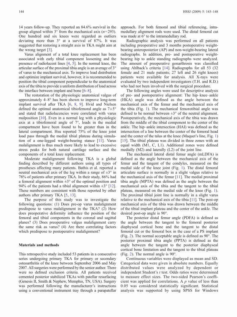
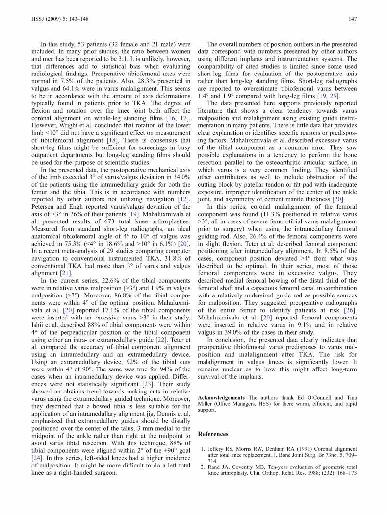
The following angles were used for descriptive analysisof pre- and postoperative alignment: The hip–knee–ankle(HKA) angle was defined as the angle between themechanical axis of the femur and the mechanical axis ofthe tibia (Fig. 1). The mechanical femorotibial angle wasdefined to be normal between ±3° of the neutral alignment.Postoperatively, the mechanical axis of the tibia was drawnfrom the middle of the tibial component to the center of theankle. The hip–ankle intersection (HAI) was defined as theintersection of a line between the center of the femoral headand the center of the talus at the knee (Maquet’s line, Fig. 1)[15]. The tibial plateau was divided in three zones with anequal width (M1, C, L1). Additional zones were definedmedially (M2) and laterally (L2) of the joint line.
The mechanical lateral distal femur angle (mLDFA) isdefined as the angle between the mechanical axis of thefemur and the tangent of the condyles, measured on themedial side of the knee joint (Fig. 1). The distal femoralarticulate surface is normally in a slight valgus relative tothe mechanical axis of the femur [11]. The medial proximaltibia angle (MPTA) was defined as the angle between themechanical axis of the tibia and the tangent to the tibialplateau, measured on the medial side of the knee (Fig. 1).The proximal tibial joint line is normally in a slight varusrelative to the mechanical axis of the tibia [11]. The post-opmechanical axis of the tibia was drawn between the middleof the tibial implant plateau and the center of the ankle. Thedesired post-op angle is 90°.
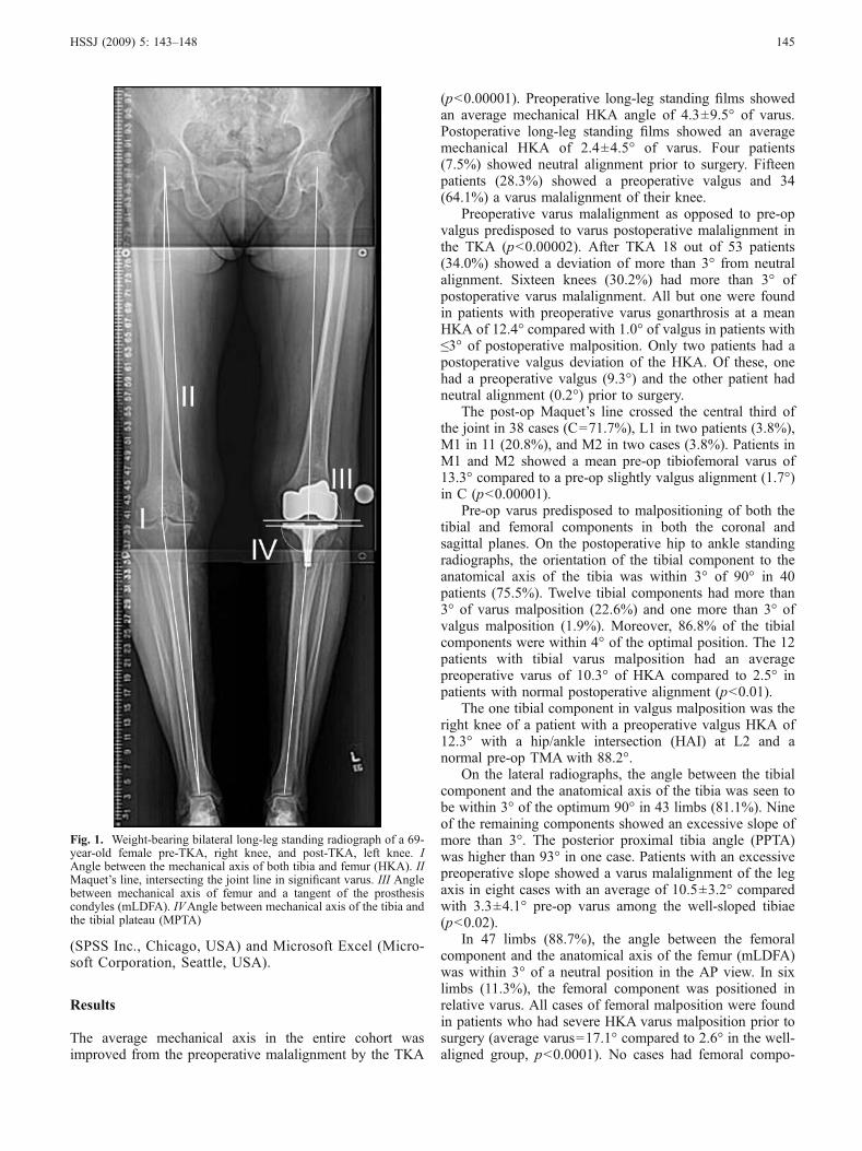
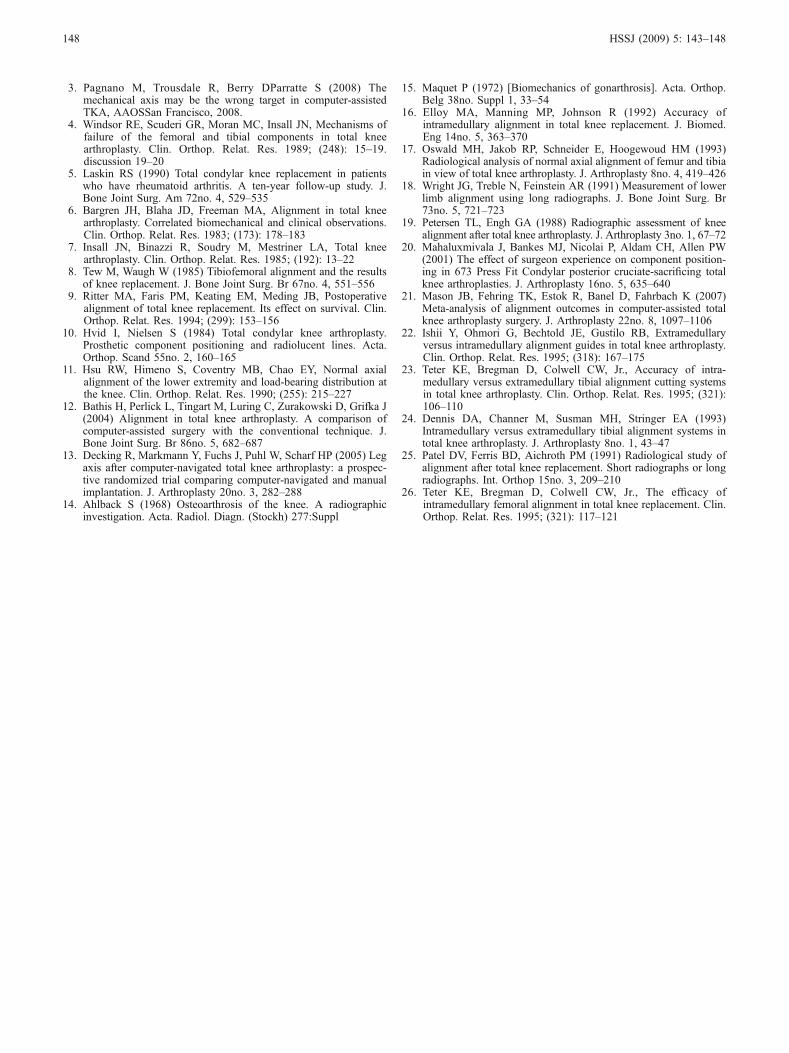
The posterior distal femur angle (PDFA) is defined asthe angle between the tangent to the femoral posteriordiaphyseal cortical bone and the tangent to the distalfemoral cut or the femoral box in the case of a PS implant(Fig. 2). The normal acceptable angle is defined as 90°. Theposterior proximal tibia angle (PPTA) is defined as theangle between the tangent to the posterior diaphysealcortical bone limitation and the tangent to the tibial plateau(Fig. 2). The normal angle is 90°.
Continuous variables were displayed as mean and SD.Categorical data were given in absolute numbers. Equallydistributed values were analyzed by dependent orindependent Student’s t test. Odds ratios were determinedto measure effect sizes. The two-sided Pearson’s coeffi-cient was applied for correlations. A p value of less than0.05 was considered statistically significant. Statisticalanalysis was supported by using SPSS for Windows
144 HSSJ (2009) 5: 143–148
(SPSS Inc., Chicago, USA) and Microsoft Excel (Micro-soft Corporation, Seattle, USA).
Results
The average mechanical axis in the entire cohort wasimproved from the preoperative malalignment by the TKA
(p<0.00001). Preoperative long-leg standing films showedan average mechanical HKA angle of 4.3±9.5° of varus.Postoperative long-leg standing films showed an averagemechanical HKA of 2.4±4.5° of varus. Four patients(7.5%) showed neutral alignment prior to surgery. Fifteenpatients (28.3%) showed a preoperative valgus and 34(64.1%) a varus malalignment of their knee.
Preoperative varus malalignment as opposed to pre-opvalgus predisposed to varus postoperative malalignment inthe TKA (p<0.00002). After TKA 18 out of 53 patients(34.0%) showed a deviation of more than 3° from neutralalignment. Sixteen knees (30.2%) had more than 3° ofpostoperative varus malalignment. All but one were foundin patients with preoperative varus gonarthrosis at a meanHKA of 12.4° compared with 1.0° of valgus in patients with≤3° of postoperative malposition. Only two patients had apostoperative valgus deviation of the HKA. Of these, onehad a preoperative valgus (9.3°) and the other patient hadneutral alignment (0.2°) prior to surgery.
The post-op Maquet’s line crossed the central third ofthe joint in 38 cases (C=71.7%), L1 in two patients (3.8%),M1 in 11 (20.8%), and M2 in two cases (3.8%). Patients inM1 and M2 showed a mean pre-op tibiofemoral varus of13.3° compared to a pre-op slightly valgus alignment (1.7°)in C (p<0.00001).
Pre-op varus predisposed to malpositioning of both thetibial and femoral components in both the coronal andsagittal planes. On the postoperative hip to ankle standingradiographs, the orientation of the tibial component to theanatomical axis of the tibia was within 3° of 90° in 40patients (75.5%). Twelve tibial components had more than3° of varus malposition (22.6%) and one more than 3° ofvalgus malposition (1.9%). Moreover, 86.8% of the tibialcomponents were within 4° of the optimal position. The 12patients with tibial varus malposition had an averagepreoperative varus of 10.3° of HKA compared to 2.5° inpatients with normal postoperative alignment (p<0.01).
The one tibial component in valgus malposition was theright knee of a patient with a preoperative valgus HKA of12.3° with a hip/ankle intersection (HAI) at L2 and anormal pre-op TMA with 88.2°.
On the lateral radiographs, the angle between the tibialcomponent and the anatomical axis of the tibia was seen tobe within 3° of the optimum 90° in 43 limbs (81.1%). Nineof the remaining components showed an excessive slope ofmore than 3°. The posterior proximal tibia angle (PPTA)was higher than 93° in one case. Patients with an excessivepreoperative slope showed a varus malalignment of the legaxis in eight cases with an average of 10.5±3.2° comparedwith 3.3±4.1° pre-op varus among the well-sloped tibiae(p<0.02).
In 47 limbs (88.7%), the angle between the femoralcomponent and the anatomical axis of the femur (mLDFA)was within 3° of a neutral position in the AP view. In sixlimbs (11.3%), the femoral component was positioned inrelative varus. All cases of femoral malposition were foundin patients who had severe HKA varus malposition prior tosurgery (average varus=17.1° compared to 2.6° in the well-aligned group, p<0.0001). No cases had femoral compo-
Fig. 1. Weight-bearing bilateral long-leg standing radiograph of a 69-year-old female pre-TKA, right knee, and post-TKA, left knee. IAngle between the mechanical axis of both tibia and femur (HKA). IIMaquet’s line, intersecting the joint line in significant varus. III Anglebetween mechanical axis of femur and a tangent of the prosthesiscondyles (mLDFA). IVAngle between mechanical axis of the tibia andthe tibial plateau (MPTA)
HSSJ (2009) 5: 143–148 145
nents placed in relative valgus. The angle between thefemoral component and the anatomical axis of the femurwas between the optimum 87° and 93° on the lateralradiographs in 39 patients (73.6%). All of the remainingcomponents were in slight flexion. Preoperative varus wascommon in these cases (ø 7.2° of varus compared to 3.2° inoptimal alignment (p=0.09)).
Fifteen patients showed a valgus malalignment of theirknee prior to TKA with a mean mechanical tibiofemoralangle of 8.3°. Fourteen showed optimal postoperativemechanical alignment of the whole leg (93.3%). In thisgroup, the coronal alignment of all femoral components wasoptimal. One femoral component was in excessive flexion(6.7%). One tibial component was positioned in varus andone in valgus. One tibial component showed excessive
slope (6.7%). Odds ratios for malalignment of TKA forvarus knees were calculated in comparison with valgusknees. In varus knees, the risk for malalignment was 7.1 forHKA, 2.4 for MPTA, 4.9 for PDFA, and 1.7 for PPTA.
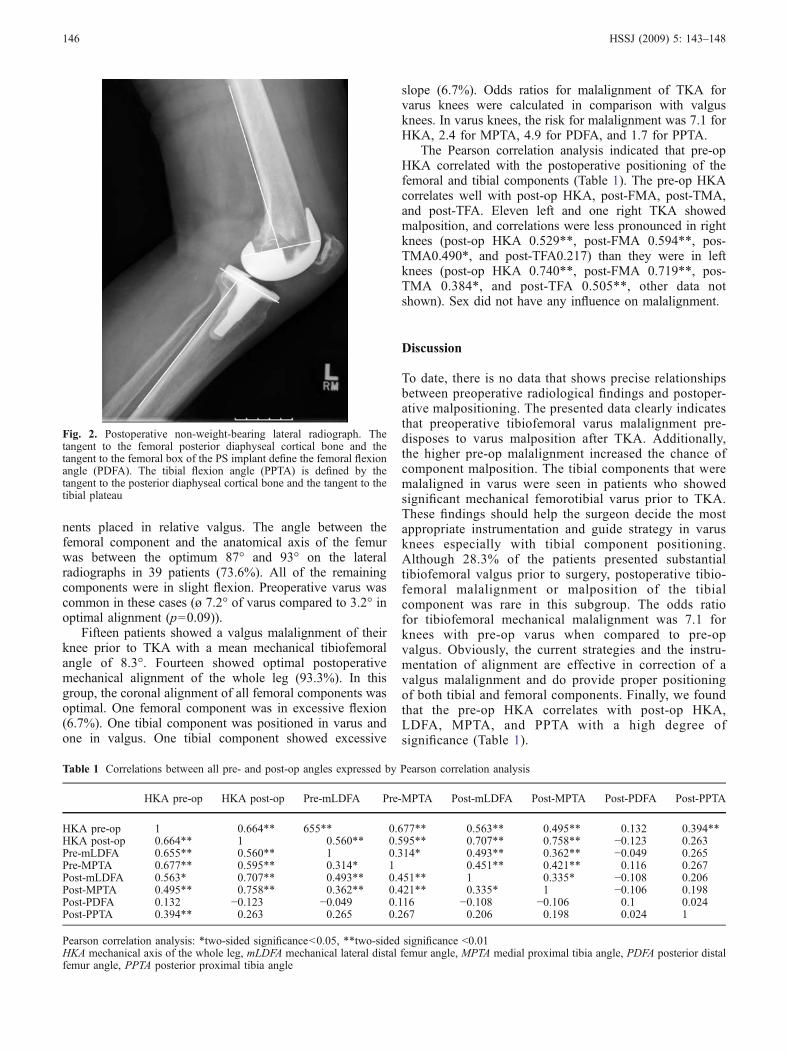
The Pearson correlation analysis indicated that pre-opHKA correlated with the postoperative positioning of thefemoral and tibial components (Table 1). The pre-op HKAcorrelates well with post-op HKA, post-FMA, post-TMA,and post-TFA. Eleven left and one right TKA showedmalposition, and correlations were less pronounced in rightknees (post-op HKA 0.529**, post-FMA 0.594**, pos-TMA0.490*, and post-TFA0.217) than they were in leftknees (post-op HKA 0.740**, post-FMA 0.719**, pos-TMA 0.384*, and post-TFA 0.505**, other data notshown). Sex did not have any influence on malalignment.
Discussion
To date, there is no data that shows precise relationshipsbetween preoperative radiological findings and postoper-ative malpositioning. The presented data clearly indicatesthat preoperative tibiofemoral varus malalignment pre-disposes to varus malposition after TKA. Additionally,the higher pre-op malalignment increased the chance ofcomponent malposition. The tibial components that weremalaligned in varus were seen in patients who showedsignificant mechanical femorotibial varus prior to TKA.These findings should help the surgeon decide the mostappropriate instrumentation and guide strategy in varusknees especially with tibial component positioning.Although 28.3% of the patients presented substantialtibiofemoral valgus prior to surgery, postoperative tibio-femoral malalignment or malposition of the tibialcomponent was rare in this subgroup. The odds ratiofor tibiofemoral mechanical malalignment was 7.1 forknees with pre-op varus when compared to pre-opvalgus. Obviously, the current strategies and the instru-mentation of alignment are effective in correction of avalgus malalignment and do provide proper positioningof both tibial and femoral components. Finally, we foundthat the pre-op HKA correlates with post-op HKA,LDFA, MPTA, and PPTA with a high degree ofsignificance (Table 1).
Fig. 2. Postoperative non-weight-bearing lateral radiograph. Thetangent to the femoral posterior diaphyseal cortical bone and thetangent to the femoral box of the PS implant define the femoral flexionangle (PDFA). The tibial flexion angle (PPTA) is defined by thetangent to the posterior diaphyseal cortical bone and the tangent to thetibial plateau
Table 1 Correlations between all pre- and post-op angles expressed by Pearson correlation analysis
HKA pre-op HKA post-op Pre-mLDFA Pre-MPTA Post-mLDFA Post-MPTA Post-PDFA Post-PPTA
HKA pre-op 1 0.664** 655** 0.677** 0.563** 0.495** 0.132 0.394**HKA post-op 0.664** 1 0.560** 0.595** 0.707** 0.758** −0.123 0.263Pre-mLDFA 0.655** 0.560** 1 0.314* 0.493** 0.362** −0.049 0.265Pre-MPTA 0.677** 0.595** 0.314* 1 0.451** 0.421** 0.116 0.267Post-mLDFA 0.563* 0.707** 0.493** 0.451** 1 0.335* −0.108 0.206Post-MPTA 0.495** 0.758** 0.362** 0.421** 0.335* 1 −0.106 0.198Post-PDFA 0.132 −0.123 −0.049 0.116 −0.108 −0.106 0.1 0.024Post-PPTA 0.394** 0.263 0.265 0.267 0.206 0.198 0.024 1
Pearson correlation analysis: *two-sided significance<0.05, **two-sided significance <0.01HKA mechanical axis of the whole leg, mLDFA mechanical lateral distal femur angle, MPTA medial proximal tibia angle, PDFA posterior distalfemur angle, PPTA posterior proximal tibia angle
146 HSSJ (2009) 5: 143–148
In this study, 53 patients (32 female and 21 male) wereincluded. In many prior studies, the ratio between womenand men has been reported to be 3:1. It is unlikely, however,that differences add to statistical bias when evaluatingradiological findings. Preoperative tibiofemoral axes werenormal in 7.5% of the patients. Also, 28.3% presented invalgus and 64.1% were in varus malalignment. This seemsto be in accordance with the amount of axis deformationstypically found in patients prior to TKA. The degree offlexion and rotation over the knee joint both affect thecoronal alignment on whole-leg standing films [16, 17].However, Wright et al. concluded that rotation of the lowerlimb <10° did not have a significant effect on measurementof tibiofemoral alignment [18]. There is consensus thatshort-leg films might be sufficient for screenings in busyoutpatient departments but long-leg standing films shouldbe used for the purpose of scientific studies.
In the presented data, the postoperative mechanical axisof the limb exceeded 3° of varus/valgus deviation in 34.0%of the patients using the intramedullary guide for both thefemur and the tibia. This is in accordance with numbersreported by other authors not utilizing navigation [12].Petersen and Engh reported varus/valgus deviation of theaxis of >3° in 26% of their patients [19]. Mahaluxmivala etal. presented results of 673 total knee arthroplasties.Measured from standard short-leg radiographs, an idealanatomical tibiofemoral angle of 4° to 10° of valgus wasachieved in 75.3% (<4° in 18.6% and >10° in 6.1%) [20].In a recent meta-analysis of 29 studies comparing computernavigation to conventional instrumented TKA, 31.8% ofconventional TKA had more than 3° of varus and valgusalignment [21].
In the current series, 22.6% of the tibial componentswere in relative varus malposition (>3°) and 1.9% in valgusmalposition (>3°). Moreover, 86.8% of the tibial compo-nents were within 4° of the optimal position. Mahaluxmi-vala et al. [20] reported 17.1% of the tibial componentswere inserted with an excessive varus >3° in their study.Ishii et al. described 88% of tibial components were within4° of the perpendicular position of the tibial componentusing either an intra- or extramedullary guide [22]. Teter etal. compared the accuracy of tibial component alignmentusing an intramedullary and an extramedullary device.Using an extramedullary device, 92% of the tibial cutswere within 4° of 90°. The same was true for 94% of thecases when an intramedullary device was applied. Differ-ences were not statistically significant [23]. Their studyshowed an obvious trend towards making cuts in relativevarus using the extramedullary guided technique. Moreover,they described that a bowed tibia is less suitable for theapplication of an intramedullary alignment jig. Dennis et al.emphasized that extramedullary guides should be distallypositioned over the center of the talus, 3 mm medial to themidpoint of the ankle rather than right at the midpoint toavoid varus tibial resection. With this technique, 88% oftibial components were aligned within 2° of the ±90° goal[24]. In this series, left-sided knees had a higher incidenceof malposition. It might be more difficult to do a left totalknee as a right-handed surgeon.
The overall numbers of position outliers in the presenteddata correspond with numbers presented by other authorsusing different implants and instrumentation systems. Thecomparability of cited studies is limited since some usedshort-leg films for evaluation of the postoperative axisrather than long-leg standing films. Short-leg radiographsare reported to overestimate tibiofemoral varus between1.4° and 1.9° compared with long-leg films [19, 25].
The data presented here supports previously reportedliterature that shows a clear tendency towards varusmalposition and malalignment using existing guide instru-mentation in many patients. There is little data that providesclear explanation or identifies specific reasons or predispos-ing factors. Mahaluxmivala et al. described excessive varusof the tibial component as a common error. They sawpossible explanations in a tendency to perform the boneresection parallel to the osteoarthritic articular surface, inwhich varus is a very common finding. They identifiedother contributors as well to include obstruction of thecutting block by patellar tendon or fat pad with inadequateexposure, improper identification of the center of the anklejoint, and asymmetry of cement mantle thickness [20].
In this series, coronal malalignment of the femoralcomponent was found (11.3% positioned in relative varus>3°, all in cases of severe femorotibial varus malalignmentprior to surgery) when using the intramedullary femoralguiding rod. Also, 26.4% of the femoral components werein slight flexion. Teter et al. described femoral componentpositioning after intramedullary alignment. In 8.5% of thecases, component position deviated ≥4° from what wasdescribed to be optimal. In their series, most of thosefemoral components were in excessive valgus. Theydescribed medial femoral bowing of the distal third of thefemoral shaft and a capacious femoral canal in combinationwith a relatively undersized guide rod as possible sourcesfor malposition. They suggested preoperative radiographsof the entire femur to identify patients at risk [26].Mahaluxmivala et al. [20] reported femoral componentswere inserted in relative varus in 9.1% and in relativevalgus in 39.0% of the cases in their study.
In conclusion, the presented data clearly indicates thatpreoperative tibiofemoral varus predisposes to varus mal-position and malalignment after TKA. The risk formalalignment in valgus knees is significantly lower. Itremains unclear as to how this might affect long-termsurvival of the implants.
Acknowledgements The authors thank Ed O’Connell and TinaMiller (Office Managers, HSS) for there warm, efficient, and rapidsupport.
References
1. Jeffery RS, Morris RW, Denham RA (1991) Coronal alignmentafter total knee replacement. J. Bone Joint Surg. Br 73no. 5, 709–714
2. Rand JA, Coventry MB, Ten-year evaluation of geometric totalknee arthroplasty. Clin. Orthop. Relat. Res. 1988; (232): 168–173
HSSJ (2009) 5: 143–148 147
3. Pagnano M, Trousdale R, Berry DParratte S (2008) Themechanical axis may be the wrong target in computer-assistedTKA, AAOSSan Francisco, 2008.
4. Windsor RE, Scuderi GR, Moran MC, Insall JN, Mechanisms offailure of the femoral and tibial components in total kneearthroplasty. Clin. Orthop. Relat. Res. 1989; (248): 15–19.discussion 19–20
5. Laskin RS (1990) Total condylar knee replacement in patientswho have rheumatoid arthritis. A ten-year follow-up study. J.Bone Joint Surg. Am 72no. 4, 529–535
6. Bargren JH, Blaha JD, Freeman MA, Alignment in total kneearthroplasty. Correlated biomechanical and clinical observations.Clin. Orthop. Relat. Res. 1983; (173): 178–183
7. Insall JN, Binazzi R, Soudry M, Mestriner LA, Total kneearthroplasty. Clin. Orthop. Relat. Res. 1985; (192): 13–22
8. Tew M, Waugh W (1985) Tibiofemoral alignment and the resultsof knee replacement. J. Bone Joint Surg. Br 67no. 4, 551–556
9. Ritter MA, Faris PM, Keating EM, Meding JB, Postoperativealignment of total knee replacement. Its effect on survival. Clin.Orthop. Relat. Res. 1994; (299): 153–156
10. Hvid I, Nielsen S (1984) Total condylar knee arthroplasty.Prosthetic component positioning and radiolucent lines. Acta.Orthop. Scand 55no. 2, 160–165
11. Hsu RW, Himeno S, Coventry MB, Chao EY, Normal axialalignment of the lower extremity and load-bearing distribution atthe knee. Clin. Orthop. Relat. Res. 1990; (255): 215–227
12. Bathis H, Perlick L, Tingart M, Luring C, Zurakowski D, Grifka J(2004) Alignment in total knee arthroplasty. A comparison ofcomputer-assisted surgery with the conventional technique. J.Bone Joint Surg. Br 86no. 5, 682–687
13. Decking R, Markmann Y, Fuchs J, Puhl W, Scharf HP (2005) Legaxis after computer-navigated total knee arthroplasty: a prospec-tive randomized trial comparing computer-navigated and manualimplantation. J. Arthroplasty 20no. 3, 282–288
14. Ahlback S (1968) Osteoarthrosis of the knee. A radiographicinvestigation. Acta. Radiol. Diagn. (Stockh) 277:Suppl
15. Maquet P (1972) [Biomechanics of gonarthrosis]. Acta. Orthop.Belg 38no. Suppl 1, 33–54
16. Elloy MA, Manning MP, Johnson R (1992) Accuracy ofintramedullary alignment in total knee replacement. J. Biomed.Eng 14no. 5, 363–370
17. Oswald MH, Jakob RP, Schneider E, Hoogewoud HM (1993)Radiological analysis of normal axial alignment of femur and tibiain view of total knee arthroplasty. J. Arthroplasty 8no. 4, 419–426
18. Wright JG, Treble N, Feinstein AR (1991) Measurement of lowerlimb alignment using long radiographs. J. Bone Joint Surg. Br73no. 5, 721–723
19. Petersen TL, Engh GA (1988) Radiographic assessment of kneealignment after total knee arthroplasty. J. Arthroplasty 3no. 1, 67–72
20. Mahaluxmivala J, Bankes MJ, Nicolai P, Aldam CH, Allen PW(2001) The effect of surgeon experience on component position-ing in 673 Press Fit Condylar posterior cruciate-sacrificing totalknee arthroplasties. J. Arthroplasty 16no. 5, 635–640
21. Mason JB, Fehring TK, Estok R, Banel D, Fahrbach K (2007)Meta-analysis of alignment outcomes in computer-assisted totalknee arthroplasty surgery. J. Arthroplasty 22no. 8, 1097–1106
22. Ishii Y, Ohmori G, Bechtold JE, Gustilo RB, Extramedullaryversus intramedullary alignment guides in total knee arthroplasty.Clin. Orthop. Relat. Res. 1995; (318): 167–175
23. Teter KE, Bregman D, Colwell CW, Jr., Accuracy of intra-medullary versus extramedullary tibial alignment cutting systemsin total knee arthroplasty. Clin. Orthop. Relat. Res. 1995; (321):106–110
24. Dennis DA, Channer M, Susman MH, Stringer EA (1993)Intramedullary versus extramedullary tibial alignment systems intotal knee arthroplasty. J. Arthroplasty 8no. 1, 43–47
25. Patel DV, Ferris BD, Aichroth PM (1991) Radiological study ofalignment after total knee replacement. Short radiographs or longradiographs. Int. Orthop 15no. 3, 209–210
26. Teter KE, Bregman D, Colwell CW, Jr., The efficacy ofintramedullary femoral alignment in total knee replacement. Clin.Orthop. Relat. Res. 1995; (321): 117–121
148 HSSJ (2009) 5: 143–148