varicose vein treatments - cigna · this coverage policy addresses varicose vein treatment....
TRANSCRIPT
Page 1 of 40 Medical Coverage Policy: 0234
Medical Coverage Policy
Effective Date ............................................11/15/2019 Next Review Date ......................................11/15/2020 Coverage Policy Number .................................. 0234
Varicose Vein Treatments
Table of Contents Overview .............................................................. 1 Coverage Policy ................................................... 1 General Background ............................................ 3 Coding/Billing Information .................................. 21 References ........................................................ 24
Related Coverage Resources
INSTRUCTIONS FOR USE The following Coverage Policy applies to health benefit plans administered by Cigna Companies. Certain Cigna Companies and/or lines of business only provide utilization review services to clients and do not make coverage determinations. References to standard benefit plan language and coverage determinations do not apply to those clients. Coverage Policies are intended to provide guidance in interpreting certain standard benefit plans administered by Cigna Companies. Please note, the terms of a customer’s particular benefit plan document [Group Service Agreement, Evidence of Coverage, Certificate of Coverage, Summary Plan Description (SPD) or similar plan document] may differ significantly from the standard benefit plans upon which these Coverage Policies are based. For example, a customer’s benefit plan document may contain a specific exclusion related to a topic addressed in a Coverage Policy. In the event of a conflict, a customer’s benefit plan document always supersedes the information in the Coverage Policies. In the absence of a controlling federal or state coverage mandate, benefits are ultimately determined by the terms of the applicable benefit plan document. Coverage determinations in each specific instance require consideration of 1) the terms of the applicable benefit plan document in effect on the date of service; 2) any applicable laws/regulations; 3) any relevant collateral source materials including Coverage Policies and; 4) the specific facts of the particular situation. Coverage Policies relate exclusively to the administration of health benefit plans. Coverage Policies are not recommendations for treatment and should never be used as treatment guidelines. In certain markets, delegated vendor guidelines may be used to support medical necessity and other coverage determinations.
Overview This Coverage Policy addresses varicose vein treatment. Varicose veins result from weakening or incompetence of a one-way valve, leading to reflux (i.e., reverse flow) of blood in the vessel. Methods of treatment that been investigated and proven effective for the treatment of varicose veins include ambulatory phlebectomy, ligation and excision, radiofrequency ablation (RFA), endovenous laser therapy (EVLT), sclerotherapy, and subfascial endoscopic perforator surgery.
Coverage Policy Coverage for treatment of varicose veins varies across plans. Refer to the customer’s benefit plan document for coverage details. If coverage is available for the treatment of varicose veins, the following conditions of coverage apply. MEDICAL NECESSITY CRITERIA FOR HIGH RISK INDICATIONS Ambulatory phlebectomy, ligation and excision, RFA (radiofrequency ablation), EVLT (endovenous laser therapy), and/or sclerotherapy* (i.e., liquid, foam, ultrasound-guided, endovenous chemical ablation,
Page 2 of 40 Medical Coverage Policy: 0234
endovenous microfoam) is considered medically necessary for ANY of the following HIGH RISK varicose vein indications:
• leg ulceration(s) due to saphenous vein insufficiency refractory to conservative management • recurrent bleeding from the saphenous vein or other varicosity • history of a significant episode of bleeding from a varicosity
* Note: Sclerotherapy using a sclerosant approved by the U.S. Food and Drug Administration for the intended use. MEDICAL NECESSITY CRITERIA FOR LOWER RISK INDICATIONS Ligation and excision, RFA and/or EVLT for the treatment of symptomatic saphenous varicose veins is considered medically necessary for ANY of the following indications:
• pain resulting in a clinically significant functional impairment (e.g., inability to perform household chores or prolonged standing, interference with essential job functions)
• recurrent phlebitis or thrombophlebitis • refractory dependent edema • persistent stasis dermatitis • chronic cellulitis
when ALL of the following criteria are met:
• a Doppler and/or Duplex ultrasonography evaluation and report, performed no more than 12 months prior to the requested procedure, confirms incompetence/reflux and documents vein size ≥ 3 mm
• documentation of BOTH of the following: previous invasive treatment(s) of varicose veins (if any) failure or intolerance of medically supervised conservative management, including but not
limited to compression stocking therapy for three consecutive months • a clearly defined treatment plan including the procedure (CPT®) codes for the planned interventions
and whether the proposed treatment is to the left leg, the right leg, or both legs Ambulatory phlebectomy or primary (i.e., initial) sclerotherapy* (liquid, foam, ultrasound-guided, or endovenous chemical ablation, endovenous microfoam) is considered medically necessary treatment of symptomatic saphenous varicose veins or tributaries greater than or equal to 3 mm when reflux proximal to the incompetence (i.e., at the saphenofemoral or saphenopopliteal junction) is concurrently being or has previously been treated (i.e., ligation and excision, RFA, and/or EVLT). *Note: Primary (i.e., initial) sclerotherapy for these indications, using a sclerosant approved by the U.S. Food and Drug Administration for the intended use, is limited to a maximum of three (3) sclerotherapy treatment sessions per leg. MEDICAL NECESSITY CRITERIA FOR SECONDARY SCLEROTHERAPY TREATMENT SESSIONS (i.e., RETREATMENT, SUBSEQUENT TREATMENTS) One or more series of three (3) secondary sclerotherapy treatment sessions (i.e., retreatment, subsequent treatments) is considered medically necessary when ALL of the following criteria are met, for each series being requested:
• symptomatic varicosities ≥ 3mm persist or have recurred following a previously completed series of primary or secondary sclerotherapy
• inadequate clinical response to a recent trial of medical management including leg elevation and compression
• absence of reflux proximal to the incompetence (i.e., at the saphenofemoral or saphenopopliteal junction)
Page 3 of 40 Medical Coverage Policy: 0234
• submission of a clearly defined treatment plan including the procedure codes requested as well as the number of treatment /procedures clinically indicated
MEDICAL NECESSITY CRITERIA FOR SUBFASCIAL ENDOSCOPIC PERFORATOR SURGERY (SEPS) Subfascial endoscopic perforator surgery (SEPS) is considered medically necessary when ALL of the following medical necessity criteria are met:
• a Doppler and/or Duplex ultrasonography evaluation and report, performed no more than 12 months prior to the requested procedure, confirms reflux of the incompetent perforator vein and location on the medial aspect of the calf being treated
• failure or intolerance of medically supervised conservative management, including but not limited to compression stocking therapy, for at least three consecutive months
• documentation of at least ONE of the following conditions: venous stasis dermatitis/ulceration chronic venous insufficiency
NOT MEDICALLY NECESSARY/EXPERIMENTAL/INVESTIGATIONAL/UNPROVEN The following varicose vein treatments are each considered cosmetic in nature and not medically necessary:
• treatment of telangiectasis or varicose veins that are less than 3 mm in diameter by any method • sclerotherapy with glycerin/glycerol • intense pulsed-light source (photothermal sclerosis) treatment of a varicose vein
The following varicose vein treatments are each is considered experimental, investigational or unproven (this list may not be all-inclusive):
• non-compressive sclerotherapy • transdermal laser therapy • transilluminated powered phlebectomy (TIPP, TriVex™) • SEPS for the treatment of venous insufficiency as a result of post-thrombotic syndrome • sclerotherapy (i.e., liquid, foam, ultra-sound guided, endovenous chemical ablation, endovenous
microfoam) when performed for ANY of the following indications: sole treatment of accessory, reticular or varicose tributaries without associated occlusion of
the saphenofemoral or saphenopopliteal junction incompetence that is isolated to the perforator veins of the Great Saphenous Vein (GSV), with or without associated ligation of the
saphenofemoral junction as a sole (i.e., standalone) treatment for reflux occurring at the saphenofemoral or
saphenopopliteal junction • endomechanical ablative approaches using rotating catheter (e.g., ClariVein™ Catheter) (e.g.,
mechanical occlusion chemically assisted ablation [MOCA], mechanic-chemical endovenous ablation [MCEA], mechanically enhanced endovenous chemical ablation [MEECA)
• endovascular embolization with cyanoacrylate adhesive (e.g., VenaSeal™ Closure System) • endovenous catheter directed chemical ablation with balloon isolation • coil embolization • cryostripping (including cryoablation, cryofreezing, transilluminated cryosurgery) of any vein
General Background The venous system of the lower extremities is separated into two main systems: the deep venous and the superficial venous system. The two systems are connected by perforator veins. The deep venous system comprises the popliteal and femoral veins; the superficial venous system comprises the great saphenous and
Page 4 of 40 Medical Coverage Policy: 0234
small saphenous veins (formerly called the short or lesser saphenous vein). The GSV generally measures 3–4 mm in diameter in the upper thigh; the GSV meets the femoral vein at the saphenofemoral junction (SFJ). Approximately 60% of patients who have varicosities have reflux in the GSV (Hamper, et al., 2007). The small saphenous vein is not usually larger than 3 mm in diameter, and connects with the deep veins at the saphenopopliteal junction (SPJ) in the knee area. Incompetence of the superficial venous system typically results from failure of valves at the SFJ and the SPJ with resulting pressure that is worse at the more distal area of the vein. Incompetence of the perforating veins also leads to increased pressure in the superficial venous system due to the pump mechanism of the calf. Varicose tributaries are veins that empty into a larger vein. Varicose veins vary in size from 3–10 mm, on average. Symptoms that have been associated with varicose veins of the lower extremities result from inadequate emptying of the vein (i.e., venous insufficiency) and include pain, cramping, aching, burning, throbbing, swelling and the feeling of heaviness or fatigue in the leg. Typically, symptoms are exacerbated by standing and warm weather (Hamper, et al., 2007). Saphenous varicose veins can ultimately result in intractable ulcerations and recurrent bleeding. Patients with larger varicosities (e.g., varicose veins greater than 3 mm in diameter) are more prone to thrombophlebitis and other complications than those with smaller varicosities. Chronic cellulitis may also be associated with varicosities. Telangiectases are permanently dilated blood vessels, also called spider veins that create fine red or blue lines on the skin. They are similar to varicose veins, but are limited to the dermis and are not usually more than 3 mm in diameter. They are not typically associated with symptoms, and treatment is generally considered cosmetic in nature and not medically necessary. Varicose veins may develop during pregnancy. Treatment is not medically necessary as most varicosities will spontaneously resolve within 4–6 months after delivery. Varicose veins of the upper extremity are rare and there are few reports in the published, peer-reviewed medical literature dealing with the management of upper extremity varicosities (Welch and Villavicencio, 1994; Duffy, et al., 1999; Lee, 2002; Bowes and Goldman, 2002). However, authors have reported successful outcomes utilizing methods of treatment similar to lower extremity varicosities (e.g., sclerotherapy, ligation and stripping, phlebectomy). DIAGNOSIS AND CLASSIFICATION Various ultrasound technologies are used in conjunction with other noninvasive testing to determine the physiological characteristics of the varicosities, as physical exam alone may not be reliable. Duplex ultrasound, Doppler ultrasound and plethysmography may all be used to diagnose varicose veins. In most cases, once the initial vein mapping is performed, it is not essential that follow-up scanning be done for subsequent sclerotherapy sessions. It has not been demonstrated in the published medical literature that repeat Duplex or Doppler studies are essential for the successful outcome of the procedure when performed as part of a series of sclerotherapy sessions. Also, routine use of any of these tools in the absence of venous symptoms or clinical evidence of venous insufficiency or reflux is not considered a medical necessity. Photographs or diagrams are helpful in assessing the size and extent of the varicosities. The CEAP classification is a method commonly used to document the severity of chronic venous disease and is based on clinical presentation (C), etiology (E), anatomy (A), and pathophysiology (P) (See Table 1). Each classification can be further defined as follows (Eklof, et al., 2004; Glovicki, et al., 2011) (See Table 1): Table 1: CEAP Classification
Class Definition C - Clinical Classification C0: No visible or palpable signs of venous disease
C1: Telangiectases or reticular veins C2: Varicose veins C3: Edema
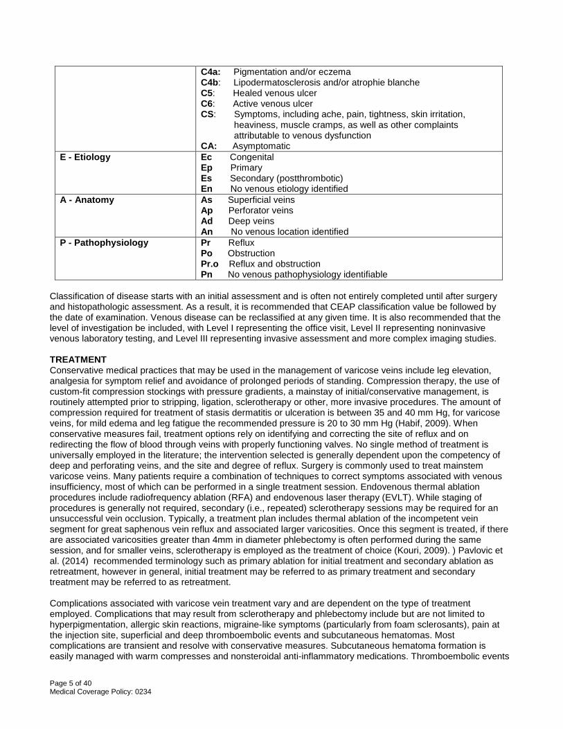
Page 5 of 40 Medical Coverage Policy: 0234
C4a: Pigmentation and/or eczema C4b: Lipodermatosclerosis and/or atrophie blanche C5: Healed venous ulcer C6: Active venous ulcer CS: Symptoms, including ache, pain, tightness, skin irritation, heaviness, muscle cramps, as well as other complaints attributable to venous dysfunction CA: Asymptomatic
E - Etiology Ec Congenital Ep Primary Es Secondary (postthrombotic) En No venous etiology identified
A - Anatomy As Superficial veins Ap Perforator veins Ad Deep veins An No venous location identified
P - Pathophysiology Pr Reflux Po Obstruction Pr.o Reflux and obstruction Pn No venous pathophysiology identifiable
Classification of disease starts with an initial assessment and is often not entirely completed until after surgery and histopathologic assessment. As a result, it is recommended that CEAP classification value be followed by the date of examination. Venous disease can be reclassified at any given time. It is also recommended that the level of investigation be included, with Level I representing the office visit, Level II representing noninvasive venous laboratory testing, and Level III representing invasive assessment and more complex imaging studies. TREATMENT Conservative medical practices that may be used in the management of varicose veins include leg elevation, analgesia for symptom relief and avoidance of prolonged periods of standing. Compression therapy, the use of custom-fit compression stockings with pressure gradients, a mainstay of initial/conservative management, is routinely attempted prior to stripping, ligation, sclerotherapy or other, more invasive procedures. The amount of compression required for treatment of stasis dermatitis or ulceration is between 35 and 40 mm Hg, for varicose veins, for mild edema and leg fatigue the recommended pressure is 20 to 30 mm Hg (Habif, 2009). When conservative measures fail, treatment options rely on identifying and correcting the site of reflux and on redirecting the flow of blood through veins with properly functioning valves. No single method of treatment is universally employed in the literature; the intervention selected is generally dependent upon the competency of deep and perforating veins, and the site and degree of reflux. Surgery is commonly used to treat mainstem varicose veins. Many patients require a combination of techniques to correct symptoms associated with venous insufficiency, most of which can be performed in a single treatment session. Endovenous thermal ablation procedures include radiofrequency ablation (RFA) and endovenous laser therapy (EVLT). While staging of procedures is generally not required, secondary (i.e., repeated) sclerotherapy sessions may be required for an unsuccessful vein occlusion. Typically, a treatment plan includes thermal ablation of the incompetent vein segment for great saphenous vein reflux and associated larger varicosities. Once this segment is treated, if there are associated varicosities greater than 4mm in diameter phlebectomy is often performed during the same session, and for smaller veins, sclerotherapy is employed as the treatment of choice (Kouri, 2009). ) Pavlovic et al. (2014) recommended terminology such as primary ablation for initial treatment and secondary ablation as retreatment, however in general, initial treatment may be referred to as primary treatment and secondary treatment may be referred to as retreatment. Complications associated with varicose vein treatment vary and are dependent on the type of treatment employed. Complications that may result from sclerotherapy and phlebectomy include but are not limited to hyperpigmentation, allergic skin reactions, migraine-like symptoms (particularly from foam sclerosants), pain at the injection site, superficial and deep thromboembolic events and subcutaneous hematomas. Most complications are transient and resolve with conservative measures. Subcutaneous hematoma formation is easily managed with warm compresses and nonsteroidal anti-inflammatory medications. Thromboembolic events
Page 6 of 40 Medical Coverage Policy: 0234
although rare can be life-threatening and may require anticoagulation (Lew, Weaver, 2015; Alaiti, 2017). Complications associated with thermal ablation techniques are usually minor and self-limiting; serious events are rare. Invasive Approaches Sclerotherapy: Sclerotherapy is an invasive procedure used to eradicate small to medium sized varicose veins of the superficial venous system (great and small saphenous veins). When reflux is present at the junction, sclerotherapy should be performed in addition to surgical ligation and division of the junction, promoting control of the point of reflux. Injection of the vein at its junction and of the incompetent perforating veins has been proposed as an alternative to ligation; however, the scientific literature does not support the efficacy of this procedure. Sclerotherapy has not been shown to be effective as a sole treatment of larger incompetent veins and is often used with other approaches to treat significant varicosities. Vahaaho and colleagues completed five year follow-up for subjects with GSV reflux (5-10 mm in diameter) who were randomized to undergo ultrasound guided foam sclerotherapy (n=76), EVLA (n=73) or open stripping with phlebectomy (n=65). At five years post treatment 77.6% of subjects were availale for follow-up. The authors reported UGFS had inferior occlusion rates in comparison to EVLA and open surgery (51%, 89% and 96% respectively; the difference between UGFS and the EVLA or surgery group was statistically significant (p< 0.001). In addition, EVLA subjects had a slightly lower rate of subsequent treatments following initial treatment, whereas the UGFS subjects required more repeat treatments. In the author’s opinion primary GSV reflux is best treated with open surgery or EVLA (Vahaaho, et al., 2018). Corabian et al. (2004) reported the role of sclerotherapy in the management of GSV and perforator incompetence has not been clearly defined, although sclerotherapy may be indicated for treatment of large saphenous veins without reflux. While a majority of varicosities are related to valvular incompetence (reflux) of the great or small saphenous veins some individuals may develop symptoms despite the absence of underlying reflux. Sclerotherapy as a sole therapy has been proposed for these individuals although the evidence base supporting this use is not robust. The authors of one randomized controlled trial (n=25) (Kahle, et al., 2004) compared injection sclerotherapy with compression using polidocanol to a placebo of saline. Only subjects with primary superficial varicose veins, 3-6mm in size, with competent saphenofemoral or saphenopopliteal junctions were included in the trial. Subjects who underwent compression sclerotherepy with polidocanol had a higher obliteration rate compared with those receiving normal saline at 12 weeks follow-up. Limitations noted by the authors of this study included small sample population and short term follow-up. Overall, the evidence to support the clinical effectiveness of sclerotherapy in the absence of incompetence at the saphenofemoral or saphenopopliteal junction is limited; effectiveness is dependent on the size, location, number of varicosities involved, and patency of the deeper veins below (Perrin, et al., 2011; Alguire, Scovell, 2018). During sclerotherapy, the abnormal vein is injected with a sclerosing agent that irritates the lining of the vein, causing it to thrombose and stenose, ultimately leading to resorption into the surrounding tissue. Echosclerotherapy using liquid or foam sclerosant, also referred to as ultrasound-guided sclerotherapy and endovenous chemical ablation (ECA), employs real-time ultrasound during the sclerotherapy procedure to help locate deep or inaccessible sites. Echosclerotherapy is indicated for treatment of veins below the surface, such as deep veins and other varices that are difficult to visualize (Corabian, et al., 2004). According to the ACP, (2008) the use of image guided techniques such as ultrasound is essential for the safe and effective performance of endovenous chemical ablation and reflects the current standard of care. Foam sclerotherapy, which involves the use of a sclerosing solution that has been forcibly mixed with air or gas (e.g., carbon dioxide) to create a foam agent, is often used in large-diameter vessels and with the use of ultrasound. Ultrasound is used to monitor the foam distribution. Foam sclerosant forces blood out of the vein and allows for less dilution of the sclerosant and more contact with the endothelium (Lew, Weaver, 2015). Overall, authors generally agree foam sclerotherapy is a safe and effective method of treating varicose veins (Rabe, et al., 2004; Wright, et al., 2006; Kendler, et al., 2007; Uurto, et al., 2007; Subramonia and Lees, 2007; Jia, et al., 2007; Darvall, et al., 2009). In addition, this method is supported by several professional societies and organizations as being safe and at least equally if not more effective than liquid sclerotherapy (Institute for Clinical Excellence [NICE], 2007; American College of Phlebology, 2008; German Society of Phlebology [Rabe, Pannier, 2010], Society for Vascular Surgery/ American Venous Forum [Glovicki, et al., 2011]; European Guideline Conference 2012 [Rabe, et al., 2013]).
Page 7 of 40 Medical Coverage Policy: 0234
As with sclerotherapy in general, the need for repeat treatment sessions when utilizing any of these methods of treatment has been reported in the literature (Barrett, et al., 2004; Darke, and Baker, 2006). Although echosclerotherapy has been investigated as an alternative to traditional saphenous vein ligation and stripping (Min, Navarro, 2000; Bountouroglou, et al., 2006), there is insufficient evidence in the medical literature to support safety, efficacy and improvement in long-term clinical outcomes when used for this indication. Evidence consists mainly of case series with few comparative trials and mixed reported clinical outcomes. There is no consensus in the published scientific literature regarding the optimal number of sclerotherapy treatments required to reduce the symptoms associated with varicose veins and the number of treatments needed to resolve symptoms varies among patients. Sclerotherapy is the treatment of choice for varicose veins that are 2–4 mm in diameter; large areas of veins can usually be eradicated using two to three treatment sessions. Vessels 4–6 mm in diameter may be treated by sclerotherapy or ambulatory phlebectomy. The primary aims of sclerotherapy are to prevent complications of varicose disease and relieve symptoms; cosmetic improvement in the leg's appearance is an added benefit. Treatment provided solely for cosmetic purposes is not considered a medical necessity. Sclerotherapy is a palliative solution and cannot prevent the formation of new varicosities. New varicosities may form, either because of an underlying illness or condition, or, in some cases, because of a genetic predisposition. In compressive sclerotherapy, the most commonly performed method of sclerotherapy, compressive dressings are applied after injection of the sclerosing agent, while the limb is elevated and the vein is drained. External compression and internal decompression (e.g., walking) stimulates fibrosis, which contributes to obliteration of the entire vein wall. Non-compressive sclerotherapy involves injecting a sclerosant into the non-elevated (blood-filled) vein without applying a compressive dressing. This method of therapy has not been shown to be effective in producing long-term obliteration of the incompetent veins. Various sclerosing agents have been approved by the U.S. Food and Drug Administration (FDA) to treat varicose veins of the lower extremities. Two most commonly used include sodium tetradecyl sulfate (Sotradecol®) and polidocanol (Asclera®); polidocanol was approved by the FDA March 2010 for the treatment of small spider veins and reticular veins. According to the manufacturer Asclera has not been studied in varicose veins larger than 3mm. Other agents such as morrhuate sodium (Scleromate™ morrhuate sodium) although FDA approved are not used as commonly. Glycerin/ glycerol is an osmotic dehydrating agent which is primarily used for the treatment of residual telangiectasias (Duffy, 2010). Nonetheless, there is no evidence-based consensus on the optimal type, dosage or concentration of the sclerosing agent. Transilluminated sclerotherapy is a procedure that employs the use of a hand-held vein light (e.g., fiberoptic illuminator) to assist with identification of varicose veins. When placed on the skin the illumination devices theoretically allow visualization of deeper veins, that often serve as feeder veins, for which sclerotherapy can then be performed. Nevertheless, the use of illumination and other similar devices is considered integral to the sclerotherapy procedure. Endovenous Microfoam (e.g., Varithena™): Varithena™ (polidocanol injectable foam) (Biocompatibles UK, Ltd.; Provensis, UK) is a type of foam sclerosant referred to as “endovenous microfoam sclerosant”, which is dispersed from a proprietary canister device. It is intended for intravenous injection under ultrasound guidance, and is administered by way of a single cannula into the lumen of the incompetent trunk veins or by direct injection into the varicosities. According to the manufacturer, in contrast to physician compounded foams, dispensing from the proprietary canister device allows for lower nitrogen content, a controlled density, and more consistent bubble size minimizing the risk of gas embolic adverse events. Varithena is recommended as an alternative to sclerotherapy (liquid or foam), surgery, and other endovenous ablative methods for treating varicose veins, as either primary or adjunctive therapy. Varithena does not require tumescent anesthesia and is intended for treating incompetent greater saphenous veins, accessory saphenous veins and visible varicosities of the great saphenous veins system above and below the knee (C2-C6). Similar to foam sclerotherapy, it is purported that ablation is achieved by foam displacement of venous blood and polidocanol-induced damage to the venous
Page 8 of 40 Medical Coverage Policy: 0234
endothelium after intravenous injection into the target vein or varicosity. A thrombus forms, occludes the vein, and is eventually replaced by fibrous tissue. Evidence in the published peer-reviewed scientific literature evaluating Varithena (polidocanol endovenous microfoam [PEM]) consists of few randomized controlled trials. Much of the evidence compares different doses of the Varithena sclerosant with placebo (Todd, et al., 2014 [VANISH-2], Todd, et al., 2015 [VANISH-2]; King, et al., 2015 [VANISH-1]). In an ongoing five-year study, Todd et al. published the preliminary eight week results of VANISH-2 (n=232), a pivotal trial evaluating subjects randomized to receive treatment of varicose veins with PEM 0.125% (control), PEM .5%, or PEM 1% compared to placebo injection. Outcomes were measured eight weeks following treatment using a primary endpoint measured by VVSymQ scores and secondary/tertiary outcomes, which included but were not limited to improvement in appearance, a clinically meaningful change, and response to treatment using Duplex ultrasound. The average GSV diameter was 8.7 mm; the range was a minimum of 3.1 mm and a maximum of 19.4 mm. At 8 weeks follow-up there were statistically significant improvements in VVSymQ scores and appearance for both treatment groups. Improvement in the treatment groups was also clinically meaningful, with a 64% reduction in symptoms in the pooled group compared with 22% for placebo. Duplex response (occlusion of the GSV and/or accessory veins) was achieved in 83% and 86% of subjects receiving either PEM 0.5% or 1.0% respectively. In the author’s opinion GSV diameter had little effect on duplex response rates (90%, 100%, 84%, 77%, and 79% in relation to vein size <5mm, 5 to <7mm, 7 to <10 mm, 10 to <12 mm, and >12 mm, respectively). No pulmonary emboli were detected and clinically important neurologic or visual adverse events were reported. In 2015 Todd and Wright reported the one year safety and efficacy data of subjects in the initial study who received PEM 1% to assess durability of response to treatment. Primary, secondary and tertiary outcomes were the same as the initial study. At one year post-treatment primary and secondary measures of efficacy, using VVSymQ score, IPR-V3 and PA-V3 scores, demonstrated sustained improvements of outcomes. Additionally, both Duplex responders (occlusion) and non responders (non-occlusion) demonstrated substantial improvements of clinical symptoms at one year (Todd, et al., 2015). In a second pivotal trial, VANISH-1, King and colleagues (2015) published their results of a phase III, multicenter, parallel study (n=279) designed to evaluate the clinical efficacy of a single administration of ≤ 15 ml of Varithena, for reducing symptoms and improving appearance of varicose veins. The study evaluated safety and efficacy of PEM using 0.5%, 1%, and 2% compared with 0.125% (control), and placebo injection. GSV diameter at baseline averaged 7.63 mm, (range 1.5 mm to 25.9 mm), inclusion criteria did not include a restriction related to vein diameter, tortuosity or prior treatments. The primary end point was efficacy measured using a 7-day average VVSymQ Instrument score at week 8, however the study was designed to follow subjects for one and five years. Other endpoints included but were not limited to appearance, PA-V3 clinically meaningful change, Duplex ultrasound of occlusion of incompetent veins or elimination of reflux at the SF junction, and change in venous clinical severity score. At eight weeks follow-up reported VVSymQ scores for pooled treatment groups and individual dose concentrations were significantly superior to placebo. Additionally mean changes from baseline to week 8 in IPR-V3 and PA-V3 scores were significantly greater for the pooled PEM than for placebo. Duplex ultrasound response rates for the pooled and individual PEM group ranged from 59% to 83% and were superior compared to those in patients treated with 0.125% PEM. No pulmonary emboli were reported and the authors noted most adverse events were mild or moderate and resolved without sequelae. Gibson and colleagues (2016) evaluated Varithena in a multicenter study involving 77 subjects with symptomatic visible varicose veins. Subjects were randomized to receive either Varithena 1% (n=39) or a placebo injection (n=38). Using patient reported assessments of symptoms including heaviness, achiness, swelling, throbbing, and itching, the results of the study support Varithena provided significantly greater symptom and appearance improvement when compared to placebo at eight weeks post treatment. In 2015, Vasquez and Gasparis published results of a multicenter RCT evaluating efficacy of endovenous ablation (i.e., laser, RFA) and Varithena (0.5%, 1.0) or placebo injection (n=117). Endovenous laser ablation was performed on the proximal incompetent GSV with either Varithena or placebo employed as treatment of the distal varicosities. Patients were assessed using Quality of Life/Symptoms questionnaire, Patient Self Assessment of Visible Varicose Veins (PA-V), and Independent Photography Review-Visible Varicose Veins (IPR-V) instruments. At eight weeks post treatment, baseline scores improved greater for the pooled Varithena group compared to placebo using both IPR-V and PA-V scores, however only IPR-V (physician-rated) reached statistical significance. At six months follow-up the percentage of subjects who achieved a clinically meaningful
Page 9 of 40 Medical Coverage Policy: 0234
benefit in appearance, evaluated by both physician and patient, was significantly higher for the subject group who underwent endovenous ablation and PEM compared to endovenous ablation/placebo. In addition, there were significantly fewer PEM subjects who required additional treatment for varices between week eight and month six (14% PEM vs 24% placebo). Elimination of reflux was achieved through the SFJ in 78.9% of endovenous ablation/placebo subjects compared with 87.3% endovenous ablation/PEM subjects. The authors concluded endovenous laser ablation combined with PEM improved efficacy for treatment of varicose veins. Davis et al. (2018) evaluated the effectiveness of PEM employed as treatment for chronic venous insufficiency for individuals unable to undergo endovenous thermal ablations due to increased risk of bleeding, severe lipodermatosclerosis, hidradenitis suppurative, fibrosis of the vein, risk of nerve injury, and/or failed prior ETA (n=10). Treated veins included GSV and AASV with follow-up at four days, six weeks, six months and one year post procedure. Preprocedure pain, symptoms, and VCSS were recorded. Clinical outcomes reported at one year follow-up included successful improvement in VCSS, pain score, HASTI symptoms (heaviness, aching, swelling, throbbing, and/or itching) , ulceration (n=3) and wound healing. The study is limited however by retrospective design and small sample size including only ten subjects. Kugler and Brown (2017) reported the results of a systematic review evaluating nonthermal ablative techniques (VenaSeal, ClariVein, and Verithena). The authors reviewed 60 publications (graded Level III and Level II) and concluded nonthermal ablative techniques for the primary management of superficial venous insufficiency have acceptable success rates compared with thermal techniques. In the authors opinion, nonthermal techniques may be preferred in certain cases in which thermal techniques may have drawbacks, such as the below knee saphenous vein or tortuous, superficial saphenous veins. In a technology assessment published by Hayes (2019) evaluating polidocanol endovenous microfam 1% for treatment of varicose veins, Hayes reported six studies met their inclusion criteria and were reviewed to evaluate safety and efficacy of PEM 1% (n=77-399 patients). The evidence consisted of two RCTs (fair quality) comparing PEM 1% with PEM 1.25% or placebo, three RCTs (poor quality) comparing PEM 1% with surgery, sclerotherapy or placebo, and one retrospective case series (very poor quality). Based on the evidence, Hayes concluded PEM 1% may improve symptoms and result in occlusion and elimination of reflux as well as improvement in appearance, clinical severity, and quality of life (QOL). However, there is uncertainty regarding the comparative effectiveness of PEM 1% in relation to other sclerosants as well as other surgical approaches. Well-designed, independent (non-manufacturer–funded) RCTs are needed to further establish the comparative safety and effectiveness of PEM 1%, determine the durability of its beneficial effects, and identify optimal patient selection criteria for its use. While the VANISH-1 and VANISH -2 trials are planned for five-year follow-up, results have not yet been published. In addition, randomized controlled trials comparing PEM as a primary therapy with other well-established primary treatments, such as endovenous ablation or stripping of the GSV are currently lacking. Evidence in the peer-reviewed published scientific literature is sufficient to support however that PEM is safe and effective when compared to placebo for the treatment of varicose veins in the short-to mid term. Transdermal Light/Laser Therapy: Photothermal sclerosis, such as PhotoDerm® Vasculite™, is also referred to as intense pulsed-light source. Used as an alternative to or to complement sclerotherapy in treating small varicose veins and telangiectases (spider veins), this type of light therapy utilizes small pulses of light energy which travel through the skin, are absorbed by the blood, are then changed to heat and ultimately destroy the vein. Successful treatment requires adequate heating of the veins, and several treatments are usually required for optimal results. Transcutaneous laser ablation, also known as transdermal laser treatment, is a type of laser therapy similar to light therapy that involves the use of a laser to treat small varicose and spider veins. Small laser pulses are delivered to the vein, causing heat, which will ultimately lead to destruction of the vein. This modality is not generally useful as a primary treatment of spider veins of the lower extremity; instead, it is employed to treat superficial vessels on the face. The treatment may result in superficial skin burns and permanent pigmentation changes. Laser or light therapy has been indicated for the treatment of telangiectasis and cutaneous vascular lesions. However, evidence in the published scientific literature indicates that transdermal light/laser therapy has not
Page 10 of 40 Medical Coverage Policy: 0234
been shown to be as effective for the lower extremities as for facial telangiectasis and smaller varicosities. The vessels in the lower extremities are located deeper and have thicker surrounding tissue. Deeper vessels require a longer wavelength and longer pulse duration to damage the vessel effectively. Additionally, because spider veins and varicosities smaller than 3 mm do not usually cause symptoms, they are considered cosmetic; hence, treatment for them is not medically necessary. Ligation, Division and/or Excision: The traditional surgical treatment of saphenous-vein varicosities consists of surgical ligation and stripping. When the GSV and SSV have reflux or incompetence, junction ligation with or without vein stripping is often recommended; in most cases, ligation is followed by GSV stripping. During the procedure, the saphenous vein and other smaller veins are exposed through an incision in the groin, where the veins are then ligated (i.e., tied off) with sutures. A second incision is made just below the knee or at the ankle to allow access for stripping the vein. When both ends of the vein are free, a wire-like instrument is threaded through the vein, extending up to the second incision in the groin area. The vein is then pulled (i.e., stripped) and removed from the leg. Removal of the superficial symptomatic vein restores venous circulation and provides relief of symptoms. Operative excision of the vein is most often reserved for large varicosities and for those located in the medial or anterior thigh. Cryostripping: Cryoablation uses extreme cold to cause injury to the vessel. Cryostripping of the GSV has been suggested as an alternative approach to traditional ligation and stripping. During this procedure, a cryoprobe is passed through the GSV, the probe freeze attaches to the GSV and stripping is performed by pulling back the probe. Theoretically cryosurgery requires less time, has fewer complications and results in less hospital day. Evidence evaluating cryosurgery techniques are limited in quantity and quality with mixed results (Kim, Kim, 2017; Lee, et al., 2015; Klem, et al., 2009; Menyhei, et al., 2008; Disselhoff, et al., 2008). Lee, et al. (2015) reported the results of a nonrandomized comparative trial evaluating cryostripping (n=32) and EVLT (n=36). Their results demonstrated similar outcomes with respect to recurrence and complication rates; three recurrences (9.4%) occurred in the cryostripping group compared with two in the EVLT group (5.6%). In one randomized clinical trial (n=494) comparing cryostripping with conventional stripping of the GSV (Klem, et al., 2009) the authors reported that cryostripping accounted for higher failures and residual GSV and offered no benefits over conventional stripping. Menyhei et al. (2008) compared conventional stripping and cryostripping and assessed quality of life outcomes and complications (n=160) in a randomized trial. The authors reported significantly improved quality of life scores for both groups, with no difference between the two groups at six months. There was less bruising in the cryo group but no difference in post-operative pain scores between the two groups. The results of another randomized trial (n=120) indicated that EVLT and cryostripping were similarly effective at two years follow-up (recurrent incompetence 77% and 66%, for EVLT and cryostripping, respectively), however EVLT was superior with regard to duration of operation, postprocedural pain, induration and resumption of normal activity (Disselhoff, et al., 2008). Results of cryotherapy procedures for treatment of varicose veins in the published scientific literature are mixed and do not lend strong support to improved clinical outcomes when compared to more conventional methods of varicose vein treatment. Further studies are needed to demonstrate safety, efficacy and the clinical utility of cryostripping. Ambulatory Phlebectomy/Stab Phlebectomy: Ambulatory phlebectomy is widely accepted as an alternative to sclerotherapy, performed for the treatment of secondary branch varicose veins. It is also referred to as miniphlebectomy, hook phlebectomy or stab avulsion. In ambulatory phlebectomy, multiple small incisions are made, and the varicose veins are grasped with a small hook or hemostat. They are then clamped, divided and finally extracted. The entire varicosity can be extracted with multiple small incisions. Compression therapy has been shown to reduce bleeding and improve resorption following this method of treatment and is thus widely used for that purpose. The procedure is often performed in combination with endovenous laser ablation. Effectiveness is dependent on the type of vein treated; the results of a one systematic review (Leopardi, et al., (2010) indicated that phlebectomy appears to be a treatment of choice for smaller veins such as the lateral accessory veins, and that for larger veins such as the saphenous veins, phlebectomy may not provide the same level of success as sclerotherapy. Transilluminated Powered Phlebectomy (TIPP): TIPP, which is similar to ambulatory phlebectomy, is another minimally invasive alternative to standard surgery for the treatment of symptomatic varicosities. Also known as the TriVex™ (Smith & Nephew Inc., Andover, MA) procedure, TIPP involves endoscopic resection and ablation of the superficial varicosity.
Page 11 of 40 Medical Coverage Policy: 0234
Subcutaneous transillumination and tumescent anesthesia help visualize and locate the varicosity, while subcutaneous vein ablation is performed using a powered resector to obliterate the vein. Tumescent anesthesia involves the infusion of large amounts of saline and lidocaine to reduce hemorrhage and of epinephrine to delay absorption of the lidocaine. During this procedure, the veins are marked with a marker, and a bright light is introduced into the leg through a small incision (2–3 cm) to enhance visualization of the veins. The power vein resector is then inserted to cut and remove the vein through suction. Proponents of this method assert the illuminating light allows quicker and more accurate removal of the vein, leading to a more effective yet less traumatic procedure. TIPP is intended for patients who are suitable candidates for conventional ambulatory phlebectomy, and may also be used as an adjunctive method to other varicose vein treatments (e.g., ligation and stripping). Eidt et al. (2016) reported the advantage of TIPP is the need for fewer incisions and that cosmetic outcomes do not appear to be superior to conventional ligation/excision techniques. The individual components of the TriVex system were approved for use by the FDA in 1999, however since that time, several other illumination and powered-resection devices have been approved and are available for use. Evidence evaluating TIPP for the treatment of varicose veins is primarily in the form of published reviews, few comparative trials (few involving randomized groups) and both retrospective and prospective case series involving small populations and evaluating short-term outcomes (Obi, et al, 2016, Lin, et al, 2016; Kim, et al., 2012; Franz and Knapp, 2008; Passman, et al., 2007; Scavee, 2006; Chetter, et al., 2006; Aremu, et al., 2004; Shamiyeh, et al., 2003; Scavee, et al., 2003; Chesire, et al., 2002; Spitz, et al., 2000). Two controlled studies specifically compared TIPP to phlebectomy (Aremu, et al., 2004; Scavee, et al., 2003), although neither of these studies were blinded. In addition, the outcomes measured in most studies include operative time, number of incisions, complications, and cosmetic satisfaction with few patient-oriented outcomes being reported. Generally, the results of these studies demonstrate that TIPP is associated with fewer incisions (Luebke, et al., 2008; Chetter, et al., 2006; Aremu, et al., 2004; Shamiyeh, et al., 2003; Scavee, et al., 2003; Spitz, et al., 2000), comparative trials support reduction of pain following TIPP procedures (Scavee, et al., 2003; Spitz, et al., 2000) and reduced complications compared to hook phlebectomy (Spitz, et al., 2000). Operative time varies among authors and with experience. Despite reports in the published literature of a reduced number of incisions, an increase in bruising, postoperative pain and decreased quality of life during the early postoperative period has been reported in some studies. Moreover, it has been reported in the literature that technical complications may be associated with inexperience. Overall evidence in the published, peer-reviewed, scientific literature does not lead to strong conclusions that TIPP results in clinical outcomes (e.g., improved pain, less varicose vein recurrence) that are as good as treatment with standard conventional methods (i.e., hook phlebectomy). Furthermore, long-term safety and efficacy of the procedure has not been adequately demonstrated. Hayes Inc. published a Medical Technology Directory report (2008) evaluating TIPP for treatment of varicose veins. Hayes concluded that the quality of the evidence was limited by small patient population, short follow-up, comparison with retrospective data for control group, no randomization, limited reporting of patient characteristics and no data regarding efficacy. According to the report the available evidence is inadequate to draw firm conclusions regarding potential benefits of transilluminated powered phlebectomy. In 2004 NICE issued an Interventional Procedure Guidance for TIPP. The advisory committee indicated that, although the evidence suggested that the procedure is effective, the data are too limited to be conclusive and there are no long-term follow-up data (NICE, 2004a). Endoluminal Radiofrequency Ablation (RFA): Radiofrequency ablation, also known as endovascular occlusion, is a treatment for symptomatic varicose veins that involves delivery of controlled radiofrequency (RF) energy through a catheter inserted into the affected vein. The heat generated by the RF energy causes the vein to contract and become occluded. The treatment is intended as a minimally-invasive alternative to standard surgery for symptomatic varicosities located mainly below the saphenofemoral or saphenopopliteal junction. RFA has also been investigated as a treatment of incompetent perforator veins (Singh and Sura, 2008; Uchino, 2007; Roth, et al. 2007; Peden and Lumsden, 2007; Gibson, et al, 2007a), however data demonstrating safety and efficacy is limited and further clinical studies are needed to support widespread use for this indication. It has
Page 12 of 40 Medical Coverage Policy: 0234
been reported that recanalization rates following RFA of the perforator veins at one year follow-up was near five times the recurrence rate compared with RFA of the GSV and SSV (Aurshina, et al., 2018). Evidence in the peer-reviewed published scientific literature supports the safety and efficacy of RFA for the treatment of symptomatic varicose veins. Most early studies were small case series with short-term follow-up (Ogawa, et al., 2005; Goldman, 2002; Weiss, 2002; Goldman, 2000), and only two included direct comparisons with standard treatments (Lurie, 2003; Rautio, 2002). RFA has been shown in a prospective nonrandomized trial to be more effective than foam sclerotherapy for closure of the GSV at one year follow-up (Gonzalez-Zeh, et al., 2008). More recently, RFA has been compared to procedures such as EVLT (Almeida, et al., 2009) and has been evaluated with and without ligation of the saphenofemoral junction (Disselhoff, et al, 2008) in randomized controlled trials. Compared to EVLT, at one month following treatment, RFA was significantly superior for measures evaluating post procedure recovery and quality of life parameters. When performed with and without ligation, at two years post procedure, there was no difference in outcomes (recurrence, degree of ablation and venous clinical severity scores) from adding the ligation procedure. The short-term results of several other studies have demonstrated that the procedure effectively occludes incompetent veins following RFA treatment (Broe, et al., 2014; Proebstle, et al., 2011; Helmy, et al., 2011; Merchant and Pichot, 2006; Hinchliffe et al., 2006; Welch, 2006; Lurie, et al., 2005). Long-term occlusion rates were reported by Merchant and Pichot (2005). This group of authors collected data to evaluate the long-term treatment outcomes of endovascular RFA and to determine risk factors that affect treatment efficacy. In their study, the authors reported on five-year follow-up results of 1006 patients (1222 limbs) treated with radiofrequency obliteration (RFO). Immediate vein occlusion was achieved in 96.8% of limbs confirmed by Duplex ultrasound examination one week or less after the procedure. The vein occlusion rate at six months, one, two, three, four and five years was 89.2%, 87.1%, 88.2%, 83.5%, 84.9% and 87.2%, respectively. The absence of reflux rate was 91.3%, 88.2%, 88.2%, 88.0%, 86.6% and 83.8%, respectively. Over a five-year follow-up period, anatomical failure was identified in 185 limbs, 19 of which received reintervention. RFA also resulted in improved pain and less bruising compared to ligation and stripping in some studies (Hinchliffe, et al., 2006). Evidence in the peer reviewed published scientific literature supports the safety and efficacy of RFA for the treatment of symptomatic saphenous varicosities, RFA is considered an appropriate alternative to conventional procedures. Hayes Inc. published a Medical Directory Technology report evaluating endovenous radio-frequency ablation (VNUS) for the treatment of varicose veins (Hayes 2010). After reviewing the available evidence Hayes concluded that the evidence evaluating RFA was moderate; RFA was successful in producing acute occlusion of the affected vessels and eliminating reflux, as demonstrated by initial response rates of 93% to 100%, and improvements in CEAP clinical class and patient-reported symptoms. Most complications were considered relatively minor, although some serious complications, including severe burns, have been reported. Results of the randomized trials directly comparing RFA with surgery and data from studies that provided up to 5-year follow-up indicate that, at least for the short- and medium-term, results of RFA are comparable to those of surgery, with earlier return to normal activities and work. In 2003 NICE issued an Interventional Procedure Guidance for RFA and reported that safety and efficacy appeared adequate to support use of the procedure as an alternative to sapheno-femoral ligation and stripping. Endovenous Laser Therapy (EVLT): EVLT, also commonly referred to as endovenous laser ablation of the saphenous vein (ELAS), is a treatment alternative to surgical stripping of the greater saphenous vein. EVLT is also considered an effective treatment for the SSV (Bhayani, Lippitz, 2009) however it is not typically used for smaller veins. EVLT is performed by threading a catheter through the greater saphenous vein and inserting an optical fiber through the catheter. The optical fiber is then connected to a surgical laser, allowing high-intensity laser light to induce photocoagulation of blood and occlusion of the vein. As the catheter is withdrawn, light pulses can be repeated at regular intervals to prevent any further blood flow through the vein. The procedure is typically used to treat larger varicose veins since catheters cannot be easily passed through a tortuous vein or a vein with several turns or bends. Small dilated branches that persist after EVLT may require additional treatments with sclerotherapy or phlebectomy (Radiological Society of North America, 2009). The FDA has granted several approvals for ablative technologies, including: Diomed 810nm laser (Diomed, Inc.); Dornier diode laser systems (Dornier MedTech, Kennesaw, GA); Biolitec, Inc. (East Longmeadow, MA); Angiodynamics, Inc. and Vascular Solutions Inc. (Minneapolis, MN).
Page 13 of 40 Medical Coverage Policy: 0234
Evidence in the medical literature evaluating EVLT for the treatment of saphenous vein reflux consists of both retrospective and prospective case series, published reviews, and randomized controlled clinical trials (El-Sheikha, et al., 2014; Rass, et al, 2012; Disselhoff, et al., 2011; Huisman, et al., 2009; Nijsten, et al., 2009; Kalteis, et al., 2008; Darwood, et al., 2008; Desmyttrere, et al., 2007; Sharif, et al., 2007; Gibson, et al., 2007; Rasmussen, et al., 2007; Ravi, et al., 2006; Puggioni, et al., 2006; Min, et al., 2003; Ho, 2003; Chang and Chua, 2002; Proebstle, et al., 2002; Navarro and Min, 2001). There is a large body of evidence to suggest that more minimally invasive techniques, which include both RFA and EVLT, are beneficial in the treatment of varicose veins when used alone (van den Bos, et al, 2009; Ravi et al., 2006; Sadick, 2005; Beale, et al., 2004; Teruya and Ballard, 2004; Elias and Frasier, 2004). Sample size and follow-up periods vary widely across studies; follow-up periods typically range at least one to four years on average. In some of the studies, duplex ultrasound demonstrated successful vein occlusion after initial treatment and throughout the various follow-up periods (Kalteis, et al., 2008; Gibson, et al., 2007; Desmyttrere, et al., 2007; Ravi, et al., 2006; Puggioni, et al., 2006; Min, et al., 2003). Some of the measured outcomes, such as complication rates, return to work, patient satisfaction and quality of life scores, are mixed—some authors report improvement compared to traditional surgical methods while others have not. Success rates and recurrence rates have been promising with several studies supporting clinical efficacy. Van den Bos, et al. (2009) published the results of meta-analysis demonstrating success rates of 78%, 84%, and 95% for ultrasound guided sclerotherapy, RFA and EVLT respectively, after three years. Min and associates (2003) reported a recurrence rate of less than 7% at a two-year follow-up, although the study had a significant number of patients lost to follow-up. Nonetheless, the authors noted their results were comparable or superior to those reported for other treatment options, including surgery, ultrasound-guided sclerotherapy, and radiofrequency ablation. Puggioni et al. (2006) concluded from a retrospective review that the overall success rate of endovenous ablation techniques for occluding the incompetent greater saphenous vein was 94% at one month, although the EVLT group developed more frequent postoperative complications compared to an RFA group. Ravi et al., (2006) reported that no GSV recanalization was found at three years post EVLT and that no saphenous vein could be identified in 82.5% of limbs in their study group. Closure rates at one month, one year, two year, three year and four years follow-up were reported by Desmyttrere, et al. (2007) as follows: 98.4%, 96.8%, 97.8%, 99.3% and 97.1%, respectively. Overall, much of the evidence available suggests that endovenous closure techniques are as good as or superior to conventional ligation and stripping of the greater saphenous vein. Hayes Inc. published two Medical Technology Directory reports (Hayes 2013a, Hayes 2013b) evaluating laser ablation of the greater saphenous vein and small saphenous vein. Based on their review of the published evidence Hayes concluded evidence from a number of randomized comparative trials and prospective studies suggests that endovenous laser therapy (EVLT) effectively provides venous occlusion to treat symptomatic varicose veins. NICE issued an Interventional Procedure Guidance for EVLT of the long saphenous vein. The guidance committee accepts the evidence on safety and efficacy as adequate to support the use of this procedure (NICE, 2004b). The evidence for efficacy was based on five case series with a mean follow-up of one to 17 months. Saphenous vein closure rates were between 90% and 100%. The authors noted that although procedure seems effective in occluding the vein, few studies have reported on patient-oriented outcomes such as improvement in symptoms. A position statement issued by the Society of Interventional Radiology in December 2003 calls the use of endovenous ablation therapy, performed with either laser or radiofrequency devices under imaging guidance and monitoring, an effective treatment of extremity venous reflux and varicose veins. The statement reports that the success rate for vein ablation ranges from 90–95% and that long-term results demonstrate recurrence rates of less than 7% at two-year follow-up. Lower rates of recurrence may be the result of the fact that imaging guidance enhances the ability to target and treat only the abnormal, incompetent venous segments. The society recommends using Duplex ultrasound prior to the procedure to map the necessary anatomy of the venous system, during the procedure for correct catheter placement and anesthetic delivery, and as necessary for follow-up. Currently, the 2003 position statement remains unchanged. Endomechanical Ablative Approach: Minimally invasive methods for treatment of varicose veins continue to evolve. One method under current investigation is the endomechanical ablative approach to varicose vein
Page 14 of 40 Medical Coverage Policy: 0234
treatment utilizing a percutaneous infusion catheter. The procedure is also referred to as mechanical occlusion chemically assisted ablation (MOCA), mechanic-chemical endovenous ablation (MCEA), and mechanically enhanced endovenous chemical ablation (MEECA).The approach involves the use of a special catheter (ClariVein™ [Vascular Insights, LLC, Madison, CT]) which combines two modalities of treatment for varicose veins: endovenous mechanical vein destruction with a rotating wire and the simultaneous infusion of an FDA approved liquid sclerosant, sodium tetradecyl sulfate to enhance venous occlusion. This mechanical-chemical ablative modality (endomechanical ablative approach) is described as minimally invasive and purported to accomplish great saphenous vein occlusion without the use of tumescent anesthesia. Information available from the manufacturer of ClariVein indicates the catheter has received 510(k) clearance from the United States Food and Drug Administration (FDA) for infusion of physician-specified agents in the peripheral vasculature. Evidence in the published peer-reviewed scientific literature evaluating endomechanical ablation is in the form few randomized controlled trials (Holewijn, et al., 2019; Vahaaho, et al., 2019; Lane, et al., 2016; Bootun, et al., 2014), one multicenter prospective observational report (Bishawi, et al., 2013) and retrospective or prospective observational cohorts involving small sample populations and evaluating short to mid-term outcomes (Kim, et al., 2019; Mohamed, et al., 2019; Khor, et al., 2018; Tang, et al., 2016; Witte, et al., 2016; Kim, et al, 2016; Boersma, et al, 2013; Elias, Raines, 2012). Sample populations in early clinical trials ranged from 25 to 155 subjects with follow-up of six weeks to one year. More recently, authors have published results of observational clinical trials involving larger sample populations although despite occlusion rates of >90%, follow-up assessment remains limited to 6-12 weeks post-procedure in these studies (Tang, et al., 2016; Deijen, et al., 2016). Clinical outcomes measured generally include occlusion rates, recanalization rates, perioperative pain, overall quality of life and general satisfaction. Compared to RFA, MOCA has been associated with a greater reduction of peri- and post-operative pain (Lane, et al., 2016; Bootun, et al., 2014). The authors of one early clinical trial (Elias and Raines, 2012) reported a 97% total occlusion rate of the treated vein segment at 6 months post procedure (N=30). A total of 22 subjects available for follow-up at one year had total occlusion of the vein treated, and at two years 96% had total occlusion. Van Eekeren and colleagues (2013) reported the results of prospective observational study comparing RFA (N=34) and MOCA (N=34) of the greater saphenous vein. Outcome measures included RAND-36 short-form health survey, the Aberdeen Varicose Vein Questionnaire, and a 100 point VAS measured at two weeks and six weeks following surgery. Treatment time was significantly shorter in the MOCA group (P=.02). At two weeks subjects who were treated with MOCA reported significantly less postoperative pain than subjects who underwent RFA. This group also required significantly less time to resume normal activities and return to work. At six weeks there were no major complications in either group and improvement in disease specific quality of life and health status was reported for both groups. Limitations of the study included small sample size and short-term follow-up with lack of randomization. In 2014 Bootun et al. reported the preliminary results of a RCT comparing the degree of pain subjects experienced while receiving mechanochemical ablation (n=60 legs) compared to RFA (n=59 legs). The primary outcome was perioperative pain measured by a 100-millimeter visual analog scale, with a score ranging 0-10 and return to normal activities one month post-surgery with secondary outcomes of change in quality of life and clinical scores, time to return to normal activities and work, and occlusion rates. At one month post procedures 66 % of subjects were available for follow-up. The mean maximum pain score was significantly lower in the mechanical ablation group compared to the RFA group (19.3 ± 19 mm, 34.5 ± 23 mm, respectively; [p<0.0001]). The mean time to normal activities was 3.5 ± 3.1 days in the ablated group versus 4.8 ± 4.3 days in the RFA group (P=0.235), and the corresponding time to return to work was a mean of 5.3 ± 8.7 days for the ablated group and 4.9 ± 3.6 days for the RFA group (P=0.887).The authors noted long term data, including occlusion rates at six months and quality of life scores, are yet to be determined. Vun et al. (2014) reported the results of a prospective, non randomized comparative study evaluating the efficacy of ClariVein (n=55) compared to EVLT (n=40) and RFA (n=50). Measured outcomes included procedure times and pain scores using a VAS. Technical success rate was 91% for ClariVein in comparison to 93% for RFA and EVLT. Procedure time and median pain scores were significantly lower for ClariVein compared to EVLT or RFA (p<0.01). The study is limited by lack of randomization, small sample size for comparison and lack of long-term outcomes.
Page 15 of 40 Medical Coverage Policy: 0234
Kim et al. (2016) published two year clinical results of a prospective observational trial (n=126) evaluating MOCA in patients with symptomatic GSV reflux. At two years post-procedure 65 subjects were available for follow-up; 14% had adjunctive treatments consisting of either phlebectomy (9%) or sclerotherapy (5%). The authors reported 92% closure rate at 24 months post procedure, with one complete and four partial recanalizations. When compared to pre-operative values VCSS and CEAP scores improved, 63% had no residual varicose veins, and 83% were asymptomatic. Limitations of the study included small sample, lack of control group comparison, and high loss of follow-up. Lane and associates (2016) published the outcomes of a randomized controlled trial evaluating the difference in pain during truncal ablation using MOCA (n=87) and RFA (n=83) with six months follow-up. The proportion of subjects completing follow-up at six months were 71% (n=121). The primary outcome was measured as maximum pain and average pain experienced during truncal ablation, secondary outcomes included disease specific quality of life, general quality of life, and clinical severity scoring, return to work/normal activities, occlusion rates, and complications. Using the VAS scale maximum pain and average pain experienced during the procedure was significantly less in the MOCA group compared to the RFA group. Between groups there was no significant differences for disease specific quality of life, general quality of life, clinical severity scores, return to work/normal activities, or occlusion rates at six month follow-up. MOCA occlusion at six months was 87% versus RFA 93%. The authors acknowledged additional studies are needed involving larger populations to assess long-term outcomes. Witte and colleagues (2016) published their results of a prospective case series evaluating MOCA for treatment of GSV insufficiency (n=85). Subjects were evaluated at baseline, four weeks, and one, two and three years post procedure using Duplex ultrasound, CEAP classification, Venous Clinical Severity Score (VCSS). Primary endpoints were clinical success (i.e., improvement of ≥ 1 VCSS) and anatomic success (i.e., occlusion of the treated vein); secondary outcomes were general and disease specific quality of life and re-intervention. During follow-up there were four RFA reinterventions for recanalized GSVs. Anatomic success after MOCA was 91.8% at 12 months, 89.5% at 24 months and 86.5% at 36 months. Clinical success was achieved in 83.1% of treatments. Quality of life measures showed an improvement in all time intervals compared with baseline. During the 12 to 36 month interval there was significant drop on VCSS scores, which was accompanied by a deterioration of disease specific and general quality of life. It was noted worsening of VCSS might be the result of a progression in comorbidities and may not reflect the recurrent nature of varicose veins. In the authors opinion MOCA is an effective treatment modality however clinical results decrease over time. In 2017 Witte and associates published a systematic review evaluating MOCA as treatment of insufficient great and/or small saphenous veins. Ten unique cohort studies (1521 veins) were included in the review. The primary outcome measure was anatomical success, defined as closure of the treated vein on follow-up Duplex imaging. Secondary outcomes included technical success (i.e., ability to complete the procedure as planned), clinical success (i.e., VCSS, Aberdeen Varicose Vein Questionnaire, and quality of life), and major complications which were defined as deep venous thrombosis, pulmonary embolism or paresthesia. Outcome data was pooled, follow-up periods were clustered, and occlusion rates were evaluated. The authors noted in six of the studies there was a financial connection with the manufacturer of ClariVein. The pooled anatomical success rate after short term follow-up was 92% and at six and 12 months it was reported as 92% and 91%, respectively. Long term anatomical success rate at two and three years were 91% and 87%, respectively. Six cohorts included in the review that described VCSS showed that MOCA led to a significant improvement in quality of life scores which remained evident at 36 months, although after two years the authors noted there was a deterioration in clinical scores. In the authors opinion this may have been related to the recurrent nature of varicose veins. Major complications and nerve injury were rare (≤ 2%). (Witte, et al., 2017). Vahaaho et al (2019) published the results of a RCT comparing MOCA (n=65) with thermal ablation, using either EVLT (n=34) or RFA (n=33), of the GSV with the primary outcome measure of occlusion rate at one year follow-up. The groups were similar in terms of age, BMI, initial GSV size, C classification, and clinical disability score. At one year 117/125 subjects were available for follow-up. At one year follow-up the treated section of the GSV was occluded in all subjects who underwent RFA or EVLT, in the MOCA group ten subjects did not have full occlusion (45/55). Larger GSV diameter in the proximal part of the GSV (8-6 mm) was associated higher recanalization rate. At one year post procedure quality of life scores were similar in all three groups. Limitations of the study included small sample population, lack of blinding and short term follow-up.
Page 16 of 40 Medical Coverage Policy: 0234
Holewijn et al. (2019) published the results of a RCT comparing MOCA (n=109) with RFA (n=104) for GSV incompetence. The primary measured outcomes included postprocedural pain two weeks post-treatment and anatomic success (i.e., occlusion) at one year. Several secondary outcomes were measured which included anatomic success, clinical success using the Venous Clinical Severity Score (VCSS), 18 to 30-day morbidity, procedural time, procedural pain, disease specific quality of life (using the Aberdeen Varicose Vein Questionnaire [AVVQ]) and general HRQoL (36-Item Short Form Health Survey [SF-36]), time to return to daily activities or work, reintervention rate, and any additional varicose vein treatment performed during 2 years of follow-up. One and two year complication rates were 86.8 and 72.4% for the MOCA group and 82.7 and 78.6% for the RFA group. The one and two year anatomic success rate was lower after MOCA (83.5% and 80.0%) compared with RFA (94.2% and 83.3%)(p=.025 and .066). There were nine complete recanalizations in the MOCA group and four in the RFA group until one year follow-up, and nine and seven complete failures, (respectively) until two years of follow-up. Absolute VCSSs were similar in both groups at one and two years follow-up, clinical success results were similar for the same timeframe (respectively: MOCA 88.75, RFA 93.2%; MOCA 93%, RFA 90.4%) and no differences in HRQoL scores were noted. In the authors opinion both techniques were associated with similar clinical outcomes at one and two years. NICE issued an update to the Interventional Procedure Guidance for Endovenous Mechanochemical Ablation for varicose veins (NICE, 2016). Within this document NICE acknowledges endovenous mechanochemical ablation is considered safe and effective for treatment of varicose veins (NICE, 2016). Hayes published a Health Technology Brief evaluating endovenous mechanical ablation (MOCA) (ClariVein Occlusion Catheter, Nonthermal Vein Ablation System [Vascular Insights LLC]) for treatment of varicose veins (Hayes, 2016). The evidence available for review included seven prospective studies published between January 2005 and February 2015. Study outcomes included immediate occlusion of the treated vein and long-term occlusion determined by duplex US, rates of recanalization, perioperative pain, satisfaction and disease-specific quality of life (QOL), procedure times, and complications. Venous incompetence was confirmed at study entry by ultrasound in the patients in all studies. Hayes reported the overall quality of evidence was low, lacked patient selection criteria, involved small samples, lacked randomization/control groups, and had insufficient follow-up time to determine long term outcomes; therefore no definitive conclusions can be made. Hayes updated the report in 2017 and noted that the body of consistent evidence, considered low quality, suggests MOCA for treatment of symptomatic varicose veins appears to be safe and efficacious in the short term. However, substantial uncertainty remains regarding the appropriate patient population, treatment parameters, and long-term durability of the procedure. Hayes updated the report in 2018 and 2019 with no change to position. Evidence in the peer-reviewed published scientific literature supporting long-term safety and efficacy of endomechanical ablative approaches to treatment of varicose veins is currently lacking. Randomized controlled trials comparing MOCA anatomical success to endothermal ablation are few. Evidence lends support to improved quality of life scores and clinical success in the short to mid-term, however anatomic success (occlusion rate) has been shown to deterioriate at one to three year followup; additional well-designed studies are needed to support the long term efficacy of this approach. Subfascial Endoscopic Perforator Surgery (SEPS): SEPS is a minimally invasive procedure for treating chronic venous insufficiency, in which incompetent perforating veins located in the calf are believed to be a contributing factor. Incompetent perforator veins result in pooling of blood in the lower extremity area, leading to vein enlargement, pain, swelling, skin discoloration and ulcers, and typically lead to chronic venous insufficiency. An alternative to open subfascial perforator vein surgery (i.e., the Linton procedure), SEPS is recommended for patients in whom conservative measures have failed to treat chronic venous insufficiency and ulceration. The Linton procedure has been associated with a high incidence of postoperative wound healing complications (Townsend, 2004). Direct visualization through endoscopy has been suggested as a more desirable approach than the Linton technique. During SEPS, an endoscope is inserted in an incision located away from the ulcer site, and a balloon dissection is performed. The veins are ligated with clips and subsequently dissected, reducing pressure. Authors claim that stasis ulcer healing rates and maintenance of healing at five years after SEPS are 90% for patients with normally functioning deep venous systems and 75–80% for patients with deep venous insufficiencies (Elias, Frazier, 2004; Gloviczki, et al., 1999). The overall goal of SEPS in treating chronic venous
Page 17 of 40 Medical Coverage Policy: 0234
ulcers is to interrupt the incompetent perforating veins in order to decrease reflux and pressure in areas above the ankle. Evidence in the form of randomized clinical trials and both retrospective and prospective case series support the safety and efficacy of SEPS as an alternative to open procedures when performed for the treatment of incompetent medial calf perforator veins (Di Battista, et al., 2012; Nelzen, Fransson, 2007; Kianifard, et al., 2007; de Rijcke, et al., 2003; Lee, et al., 2003; Kalra and Gloviczki, 2003; Sybrandy, et al., 2001; Pierek, et al., 1997). In contrast, SEPS performed for the treatment of post-thrombotic syndrome is controversial. Studies indicate that SEPS produces poorer outcomes, specifically, less ulcer healing and higher recurrence rates when used to treat limbs with post-thrombotic syndrome than when used to treat limbs with peripheral vascular insufficiency (Gloviczki, et al., 1999). NICE issued an Interventional Procedure Guidance for subfascial endoscopic perforator vein surgery. One randomized controlled trial, two non-randomized comparative studies and two case series were reviewed. The NICE specialty advisors noted that based on the evidence reviewed, efficacy of the procedure is unproven and the indications are not well established. Reported complications include nerve injury and deep vein thrombosis. There was evidence to support lower wound infection rates compared to the open procedure. Length of stay was shorter for SEPS. The rate of primary ulcer healing and cumulative ulcer recurrence rates was comparable for both open and SEPS procedures. Although SEPS has been used for individuals with post-thrombotic valvular incompetence, there is evidence when used for this indication individuals may have poorer outcomes compared with individuals with primary valvular incompetence. In summary, the advisors noted careful patient selection is particularly important and there are uncertainties regarding safety of the procedure (NICE, 2004c). Other Treatments Other proposed treatments for varicose veins, some only under investigation in countries outside the U.S., include but are not limited to endovenous steam ablation (EVSA) (Steam Vein Sclerosis System [SVS™, VenoSteam™], CermaVEIN, France); endovenous microwave ablation (Microwave Intracavitary Coagulation System, Fuzhong Medical High-Tech Co. Ltd., China), coil embolization, VariClose Vein Sealing System (Biolas, FG Group,Turkey), VenaSeal™ Closure System (Sapheon, Inc., Morrisville, NC) (also referred to as cyanoacrylate vein ablation [CAVA]) and endovenous catheter directed chemical ablation with balloon isolation. Evidence confirming steam ablation and microwave ablation technologies have been approved or cleared for marketing by the FDA is lacking. In addition, much of the evidence evaluating these emerging modalities of treatment are in preliminary stages (Yang, et al., 2013; Milleret, et al., 2013; van den Bos, et al., 2014) and strong conclusions regarding safety and efficacy compared to well-established endovenous ablation treatments cannot be made at this time. Evidence evaluating endovenous catheter directed chemical ablation with balloon isolation as a treatment of varicose veins was not found in the peer-reviewed published scientific literature. In theory adding occlusive balloon isolation to the vein wall may enhance the interaction of the sclerosant. Coil embolization, also known as coil occlusion, involves the use of a coil combined with a sclerosant to occlude the vein and is under investigation for treatment of lower extremity varicose veins. It is a technique generally reserved for larger diameter veins such as perforating veins; the coil is curled up into the vein and may involve the use of more than one coil. Evidence in the peer-reviewed published literature evaluating this method of treatment for lower extremity varicosities is very limited (Viani, 2014), additional clinical trials are necessary to develop strong conclusions regarding safety and efficacy. Evidence confirming the VariClose Vein Sealing System (Biolas, FG Group, Turkey), a cyanoacrylate-type adhesive, has been approved or cleared for marketing by the FDA is lacking. VenaSeal received FDA pre-market approval in 2015 and employs the use of cyanoacrylate embolization (CAE) and does not require tumescent anesthesia; a cyanoacrylate adhesive is injected into the vein via a catheter inserted through the skin under ultrasound, the vein is compressed, and the adhesive material changes into a solid to seal the varicose vein. In 2015 Morrison et al. published the early results of a randomized controlled trial evaluating CAE, using the VenaSeal Closure System (n=108) versus radiofrequency ablation (n=114) for the treatment of varicose veins. The study’s primary endpoint was closure of the target vein at month 3 as assessed by duplex ultrasound.
Page 18 of 40 Medical Coverage Policy: 0234
Statistical testing focused on showing non-inferiority with a 10% delta conditionally followed by superiority testing. By use of the predictive method for imputing missing data, 3-month closure rates were 99% for CAE and 96% for RFA. All primary end point analyses, which used various methods to account for the missing data rate (14%), showed evidence to support the study’s non-inferiority hypothesis (all P < .01). Some of the analyses supported a trend toward superiority (P = .07) in the predictive model. Pain experienced during the procedure was mild and similar between treatment groups (2.2 and 2.4 for CAE and RFA, respectively, on a 10-point scale; P = .11) and there was less ecchymosis in the treated region on day 3 following CAE compared with RFA (P < .01). In the author’s opinion, CAE was non-inferior to RFA for the treatment of incompetent GSVs at three month follow-up. The authors noted the results will be monitored for 3 years to determine long-term success rates, rates of recanalization, and chronic venous disorder (CVD) progression (Morrison, et al, 2015). In 2017 this same group of authors published 12 month results of the study; 192 subjects were available for followup. The complete occlusion rate was similar in both groups: 97.2% in the CAE group compared with 97% in the RFA group (Morrison, et al., 2017). In 2018 the authors published 24 month results of the VeClose trial of which 171 subjects completed; 87 from the CAE group and 84 from the RFA group. The 24 month complete closure rate was 95.3% in the CAE group compared with 94.0% in the RFA group, supporting continued non-inferiority of CAE. Symptoms and quality of life improved in both groups and there was no significant device or procedure related adverse events (Gibson, et al., 2018). In 2019 Morrison and colleagues published 36 month follow-up of the VeClose trial with follow-up on 146 (66%) patients; 72/108 from the CAC group and 74/114 subjects from RFA group. The remaining subjects either dropped out of the study or the data could not be collected in the time period dictated by the study period. Improvement in quality of life scores were sustained in both groups, closure rates of CAE were similar to RFA (94.4%, 91.9%, respectively), although not statistically significant, and continued to support noninferiority. CAE was superior to other methods in terms of patient comfort. No significant device- or procedure-related adverse events were reported for either group. The trial is limited by high loss to follow-up. In addition, both groups used compression stocking for seven days post treatment, use of compression stocking varied in other studies making comparisons difficult. As noted by the authors, followup will continue for up to 60 months. Evidence in the form of case series tends to support safety and efficacy of endovenous cyanoacrylate embolization at 12 month follow-up (Proebstle, et al., 2015; Chan, et al., 2016). Proebstle and colleagues (2015) published 12 month outcomes of a European multicenter cohort study (n=70). Subjects underwent endovenous cyanoacrylate embolization for treatment of symptomatic GSV incompetence using VenaSeal. Immediately after the procedure all but one subject had complete closure of the target GSV. The authors noted this subject also received foam sclerotherapy resulting in full occlusion. At 12 months post-procedure, 68 subjects were available for follow-up. Eight subjects developed partial recanalization during the 12 month time-frame; using life-table methods the authors reported a 12-month complete occlusion rate of 92.9%. As reported by the authors, although results are encouraging the study is limited by lack of a control group and randomized controlled trials comparing VenaSeal with endothermal ablation are still needed to establish clinical utility.Chan et al (2016) reported the results of a single center cohort of subjects who underwent endovenous cyanoacrylate embolization using VenaSeal (n=29 subjects, 59 legs). The primary outcome measure was GSV obliteration, follow-up occurred at one week, and one, six, and 12 months post procedure. The authors reported GSV closure rates were 98.2%, 94.3%, 89.7% and 78.5% respectively. At a medium follow-up of nine months no clinical recurrence of varicosity was recorded. In addition, all subjects had improvement of SF-36 physical and mental scores, venous clinical severity scores, and Aberdeen varicose vein questionnaire scores. Since 2016 additional evidence has been published evaluating cyanoacrylate adhesive (Bademci, et al., 2017; Premnath, et al., 2017; Lam, et al., 2017; Park, 2017), however this evidence lacks control groups, involves short to intermediate term outcomes (3-12 months), employed the use of different glues, and some populations had adjunctive varicose vein treatments using either sclerotherapy or phlebectomy, therefore strong evidence based conclusions cannot be made regarding safety and efficacy. Almeida and colleagues (2017) published results of prospective trial (n=38) evaluating cyanoacrylate adhesive as treatment for saphenous vein incompetence with 36 month follow-up (Almeida, et al., 2017). Thirty eight individuals were treated with cyanoacrylate adhesive by injection of small bolus using ultrasound guidance and
Page 19 of 40 Medical Coverage Policy: 0234
no anesthesia or compression stockings post procedure. Twenty nine subjects were available for follow-up at 36 months. All but two subjects had complete occlusion confirmed by duplex ultrasound (occlusion rate of 94.7%). Pain, edema, and VCSS scores improved from baseline to month 36. In the authors opinion long term occlusion rates were similar to those of other thermal and nonthermal methods. Limitations of the study include lack of a control group, high drop out rate (9/38), and small sample population. A systematic review and meta-analysis published by Shortell and colleagues (2017) evaluated MOCA and cyanoacrylate vein ablation for treatment of GSV incompetence. A total of 15 articles met the inclusion criteria and were reviewed and included seven MOCA studies (Bishwai, et al., 2014; Elias, et al., 2012; Kim, et al, 2017, Lane, et al., 2017; Ozen, et al., 2014; van Eekeren, et al., 2014; and Witte, et al., 2017) and eight CAVA studies (Almeida, et al., 2013; Almeida, et al., 2015; Bozkurt, et al., 2016; Calk, et al., 2016; Chan, et al., 2017; Kolluri, et al., 2016; Proebstle, et al., 2015; and Tekin, et al., 2016). A total of 1645 subjects were included in the studies, 691 underwent MOCA and 954 underwent CAVA. Rates were pooled using a random effects model. The pooled anatomic success for MOCA and cyanoacrylate ablation was 94.7% and 94.8% at six months and 94.1% and 89.0% at one year, respectively. Venous Clinical Severity score and Aberdeen Varicose Vein Questionnaire score significantly improved after treatment with MOCA and cyanoacrylate ablation. The authors noted that among the studies the definition of anatomic success varied, the use of adjunctive treatments varied, most of the studies had short term follow-up of six months to one year, and methodologic quality was only moderate. Although the results were promising the authors acknowledged high quality randomized controlled trials comparing these techniques with other well-established modalities are required. Within an UpToDate review for sclerotherapy techniques for the treatment of lower extremity veins (Scovell, et al., 2019) the authors note that short term outcomes reported at three months were similar to RFA, however longer term followup is needed to determine the durablility of the results of cayanoacrylate glue. Hayes, Inc. published a search and summary report to evaluate the VenaSeal Closure System for treatment of varicose veins (Hayes, 2019). The authors reported the evidence base was small, consisting of few randomized controlled trials, prospective uncontrolled trials, a retrospective comparative study and one systematic review with meta-analysis. Hayes concluded the evidence reviewed is conflicting (Hayes, 2019). At present there is insufficient evidence in the peer reviewed scientific literature to firmly establish long-term safety and efficacy of cyanoacrylate adhesive embolization for treatment of varicose veins. Additional well-designed RCTs consisting of large sample populations, with long term outocmes that consistently demonstrate anatomic and clinical success are need to firmly establish safety and efficacy. Professional Societies/Organizations Although an update to the guideline was not found, within clinical practice guidelines of the European Society of Vascular Surgeons (Witten, et al., 2015) for the management of chronic venous disease the authors noted the scientific evidence was insufficient to evaluate cyanoacrylate glue injection, and that steam ablation seems to give results comparable to other thermal ablation techniques at one year follow-up, although additional studies are needed to confirm clinical results in the medium to long- term. Treatment of great saphenous vein reflux in patients with symptoms and signs of chronic venous disease with endovascular techniques, endovenous thermal ablation is recommended in preference to surgery or foam sclerotherapy. Although the occlusion rate of MOCA at six months is 96.7% and side effects seem to be minimal, the authors note that only two cohort studies including a small number of subjects treated with MOCA have been published to date. In 2014 guidelines for Endovenous Thermal Ablation (ETA) of the treatment of varicose veins were published by the First International Consensus Conference (Pavlovic, et al., 2014). Using a systematic literature review and expert opinion recommendations were made for endovenous thermal ablation procedures, which were defined as catheter-directed ultrasound-guided thermal methods (i.e., EVLA, RFA). The authors noted that there is little evidence evaluating other ablation procedures, such as steam ablation or bipolar RFA. Patient selection criteria varies depending on venous condition and general health, however veins applicable to endovenous thermal ablation include the GSV, SSV, accessory saphenous vein, Giacomini vein and cranial extension of SSV, superficial veins located in subcutaneous tissue, insufficient perforating veins, residual intrafascial veins following treatment, and venous malformations. Specific requirements for RFA include vein segment at least 10cm long using a standard catheter (7cm) or 5cm when a shorter heating element is used. No restrictions were noted for
Page 20 of 40 Medical Coverage Policy: 0234
EVLA. Other recommendations in the guidelines addressed areas such as qualifications of ETA providers, pre and post treatment recommendations, indications, contraindications, generalized treatment plans and complications. The American College of Phlebology (ACP) published revised guidelines for the treatment of superficial venous disease of the lower leg (ACP, 2014, rev 02/03/16). The updated guidelines are based on the 2011 review by Gloviczki et al., consensus of ACP experts, and current evidence. CEAP classification and VCSS (venous clinical severity score) were included in the revised guidelines to better define “medically significant venous insufficiency” versus what may be considered cosmetic and/or not medically necessary treatments. According to the guidelines indications for treatment include pain, edema, hemorrhage, recurrent superficial phlebitis, stasis dermatitis, or ulceration. CEAP classification and VCSS evaluation should be performed, with “medically necessary” being defined as a CEAP of C2 or higher. Duplex ultrasound is recommended prior to treatment with reflux time of > 500 msec regardless of vein diameter for the GSV, SSV, Anterior Accessory of the GSV, Posterior Accessory of the GSV, and Intersaphenous Vein (Vein of Giacomini). The ACP recommends (i.e., strong recommendation) treatment such as laser and radiofrequency ablation, open ligation and stripping for veins not amenable to endovenous procedures, stab phlebectomy, and liquid and foam chemical sclerotherapy with/without ultrasound guidance, as needed. Mechanical/chemical ablation (e.g., Clarivein) used to treat truncal reflux is a suggested treatment (i.e., weak recommendation [2B]). Treatment of incompetent perforator veins below a healed or open venous ulcer when reflux has an outward flow of 500 ms and a diameter is 3.5 cm is also a suggested treatment. In 2017 the American College of Phlebology Guidelines “Treatment of Refluxing Accessory Saphenous Veins” were published. Within these guidelines the authors acknowledged the FDA approvals for MOCA and cyanoacrylate glue does not exclude their use in accessory saphenous veins and that the Committee supports MOCA and cyanoacrylate techniques are likely effective for accessory saphenous veins although there are no case series published to date specifically evaluating efficacy in accessory veins. Treatment of accessory veins using endovenous thermal ablation of ultrasound guided foam sclerotherapy is as safe and effective as treatment of the GSV and SSV using the same modalities. Vein closure rates and improvement in quality of life scores are similar to results achieved with treatment of GSV and SSVs (Strength of Recommendation Grade 1, level of evidence C) (Gibson, et al., 2017). In 2011 the Society for Vascular Surgery and the American Venous Forum (Gloviczki. et al., 2011) developed clinical practice guidelines for care of patients with varicose veins of the lower limbs and pelvis. Although not all-inclusive, the main recommendations of the committee may be summarized as follows:
• in patients with varicose veins or more severe chronic venous disease (CVD), a complete history and detailed physical examination are complemented by duplex ultrasound scanning of the deep and superficial veins
• the use of CEAP classification for patients with CVD and the revised Venous Clinical Severity Score to assess treatment outcome
• regarding Duplex scanning results: a cutoff value of 1 second for abnormally reversed flow (reflux) in the femoral and popliteal veins a cutoff value of 500 ms for abnormally reversed flow (reflux)for the great saphenous vein, the small
saphenous vein, the tibial, deep femoral, and the perforating veins in patients with chronic venous insufficiency, duplex scanning of the perforating veins is performed
selectively; the definition of “pathologic” perforating veins includes those with an outward flow of duration of ≤ 500 ms, with a diameter of ≤ 3.5 mm and a location beneath healed or open venous ulcers (CEAP class C5-C6)
• compression therapy (pressure 20-30 mm Hg): is suggested for patients with symptomatic varicose veins as the primary treatment to aid healing of venous ulceration in addition to ablation of incompetent superficial veins in order to decrease the recurrence of
venous ulcers is not recommended as the primary treatment if the patient is a candidate for saphenous
vein ablation • ligation and stripping for the treatment of incompetent great, small saphenous and superficial veins • recommend the following:
Page 21 of 40 Medical Coverage Policy: 0234
endovenous thermal ablation (radiofrequency or laser) for treatment of incompetent saphenous vein rather than high ligation and inversion stripping of the saphenous vein to the level of the knee
phlebectomy or sclerotherapy to treat varicose tributaries foam sclerotherapy as an option for the treatment of the incompetent saphenous vein
(endovenous thermal ablation is recommended over foam sclerotherapy) treatment of pathologic perforating veins (outward flow duration >500 ms, vein diameter >3.5
mm) located underneath healed or active ulcers (CEAP class C5-C6) • recommend against selective treatment of perforating vein incompetence in patients with simple varicose
veins (CEAP class C2). The American Board of Internal Medicine’s (ABIM) Foundation Choosing Wisely® Initiative: The Society for Vascular Surgery Choosing Wisely statements (2015) does not support routine venous ultrasound tests for individuals with asymptomatic telangiectasia. Routine testing could result in unnecessary saphenous vein ablation procedures. Centers for Medicare & Medicaid Services (CMS)
• National Coverage Determinations (NCDs): NCD not found. • Local Coverage Determinations (LCDs): Multiple LCDs found. Refer to the LCD table of contents link in
the reference section. Use Outside of the US: ClariVein Occlusion Catheter has received CE Marking. Varithena is not a device and is not subject to CE Marking. The National Institute of Clinical Excellence (NICE, 2013) issued guidance for the treatment of varicose veins and recommends the following interventional treatments for individuals with confirmed varicose veins and truncal reflux:
• endothermal ablation using radiofrequency or laser ablation • if endothermal ablation is unsuitable, offer foam sclerotherapy with ultrasound guidance • if foam sclerotherapy is unsuitable, offer truncal vein stripping • if incompetent varicose tributaries are present treatment should be considered at the same time
In a separate guidance document, NICE notes the current evidence for cyanoacrylate glue is limited in quantity and quality, and the procedure should only be used with special arrangements for clinical governance, consent and audit or research (NICE, 2015). In May 2016 NICE published guidance supporting endovenous mechanochemical ablation. Within this document NICE acknowledges endovenous mechanochemical ablation is considered safe and effective for treatment of varicose veins (NICE, 2016). Coding/Billing Information Note: 1) This list of codes may not be all-inclusive. 2) Deleted codes and codes which are not effective at the time the service is rendered may not be eligible for reimbursement. The treatment of varicose veins is covered only when coverage is available under the plan for varicose vein treatment. Benefit exclusions and limitations may apply. Invasive treatment of varicose veins is excluded under many plans and therefore the services listed below may not be covered. Considered Medically Necessary when criteria in the applicable policy statements listed above are met: Ambulatory Phlebectomy
CPT®* Codes Description 37765 Stab phlebectomy of varicose veins, 1 extremity; 10-20 stab incisions 37766 Stab phlebectomy of varicose veins, 1 extremity; more than 20 incisions
Page 22 of 40 Medical Coverage Policy: 0234
37799† Unlisted procedure, vascular surgery
†Note: Considered Medically Necessary when used to report stab phlebectomy of varicose veins, one extremity; less than 10 incisions. Ligation and Excision
CPT®* Codes Description 37700 Ligation and division of long saphenous vein at saphenofemoral junction, or distal
interruptions 37718 Ligation, division, and stripping, short saphenous vein 37722 Ligation, division, and stripping, long (greater) saphenous veins from saphenofemoral
junction to knee or below 37735 Ligation and division and complete stripping of long or short saphenous veins with radical
excision of ulcer and skin graft and/or interruption of communicating veins of lower leg, with excision of deep fascia
37780 Ligation and division of short saphenous vein at saphenopopliteal junction (separate procedure)
37785 Ligation, division, and/or excision of varicose vein cluster(s), 1 leg Radiofrequency Ablation
CPT®* Codes Description 36475 Endovenous ablation therapy of incompetent vein, extremity, inclusive of all imaging
guidance and monitoring, percutaneous, radiofrequency; first vein treated 36476 Endovenous ablation therapy of incompetent vein, extremity, inclusive of all imaging
guidance and monitoring, percutaneous, radiofrequency; subsequent veins treated in a single extremity, each through separate access sites (List separately in addition to code for primary procedure)
Endovenous Laser Ablation
CPT®* Codes Description 36478 Endovenous ablation therapy of incompetent vein, extremity, inclusive of all imaging
guidance and monitoring, percutaneous, laser,; first vein treated 36479 Endovenous ablation therapy of incompetent vein, extremity, inclusive of all imaging
guidance and monitoring, percutaneous, laser; subsequent veins treated in a single extremity, each through separate access sites (List separately in addition to code for primary procedure)
Sclerotherapy
CPT®* Codes Description 36470 Injection(s) of sclerosant; single incompetent vein (other than telangiectasia) 36471 Injection(s) of sclerosant; multiple incompetent veins (other than telangiectasia), same leg
HCPCS Codes Description S2202 Echosclerotherapy
Sclerotherapy using Ultrasound Guidance and a Microfoam Sclerosant (e.g., Varithena™):
CPT®* Codes Description
Page 23 of 40 Medical Coverage Policy: 0234
36465 Injection of non-compounded foam sclerosant with ultrasound compression maneuvers to guide dispersion of the injectate, inclusive of all imaging guidance and monitoring; single incompetent extremity truncal vein (eg, great saphenous vein, accessory saphenous vein)
36466 Injection of non-compounded foam sclerosant with ultrasound compression maneuvers to guide dispersion of the injectate, inclusive of all imaging guidance and monitoring; multiple incompetent truncal veins (eg, great saphenous vein, accessory saphenous vein), same leg
Subfascial Endoscopic Perforator Surgery (SEPS) (Including Open Ligation Procedures)
CPT®* Codes Description 37500 Vascular endoscopy, surgical, with ligation of perforator veins, subfascial (SEPS) 37760 Ligation of perforator veins, subfascial, radical (Linton type), including skin graft, when
performed, open, 1 leg 37761 Ligation of perforator vein(s), subfascial, open, including ultrasound guidance, when
performed, 1 leg Considered Cosmetic/ Not Medically Necessary: Sclerotherapy for Treatment of Telangiectasia
CPT®* Codes Description
36468 Injection(s) of sclerosant for spider veins (telangiectasia), limb or trunk Intense Pulsed Light Source
CPT®* Codes Description 17106 Destruction of cutaneous vascular proliferative lesions (eg, laser technique); less than 10
sq cm 17107 Destruction of cutaneous vascular proliferative lesions (eg, laser technique); 10.0 to 50.0
sq cm 17108 Destruction of cutaneous vascular proliferative lesions (eg, laser technique); over 50.0 sq
cm Considered Experimental/Investigational/Unproven: Transilluminated Powered phlebectomy (TIPP, TriVex™)
CPT®* Codes Description 37799 Unlisted procedure, vascular surgery
Endomechanical Ablative Approach
CPT®* Codes Description 36473 Endovenous ablation therapy of incompetent vein, extremity inclusive of all imaging
guidance and monitoring, percutaneous, mechanochemical; first vein treated 36474 Endovenous ablation therapy of incompetent vein, extremity inclusive of all imaging
guidance and monitoring, percutaneous, mechanochemical; subsequent vein(s) treated in a single extremity, each through separate access sites (List separately in addition to code for primary procedure)
37241† Vascular embolization or occlusion, inclusive of all radiological supervision and interpretation, intraprocedural roadmapping, and imaging guidance necessary to complete the intervention; venous, other than hemorrhage (eg, congenital or acquired venous malformations, venous and capillary hemangiomas, varices, varicoceles)
Page 24 of 40 Medical Coverage Policy: 0234
37244 Vascular embolization or occlusion, inclusive of all radiological supervision and interpretation, intraprocedural roadmapping, and imaging guidance necessary to complete the intervention; for arterial or venous hemorrhage or lymphatic extravasation
75894 Transcatheter therapy, embolization, any method, radiological supervision and interpretation
† Note: In scope of this policy 37241 refers to varicose veins of the extremities. Endovascular Ablation Cyanoacrylate Adhesive (e.g., VenaSeal Closure System)
CPT®* Codes Description 36482 Endovenous ablation therapy of incompetent vein, extremity, by transcatheter delivery of a
chemical adhesive (eg, cyanoacrylate) remote from the access site, inclusive of all imaging guidance and monitoring, percutaneous; first vein treated
36483 Endovenous ablation therapy of incompetent vein, extremity, by transcatheter delivery of a chemical adhesive (eg, cyanoacrylate) remote from the access site, inclusive of all imaging guidance and monitoring, percutaneous; subsequent vein(s) treated in a single extremity, each through separate access sites (List separately in addition to code for primary procedure)
Endovenous Catheter Directed Chemical Ablation with Balloon Isolation
CPT®* Codes Description 0524T Endovenous catheter directed chemical ablation with balloon isolation of incompetent
extremity vein, open or percutaneous, including all vascular access, catheter manipulation, diagnostic imaging, imaging guidance and monitoring
Coil Embolization
CPT®* Codes Description 37241† Vascular embolization or occlusion, inclusive of all radiological supervision and
interpretation, intraprocedural roadmapping, and imaging guidance necessary to complete the intervention; venous, other than hemorrhage (eg, congenital or acquired venous malformations, venous and capillary hemangiomas, varices, varicoceles)
37799 Unlisted procedure, vascular surgery † Note: In scope of this policy 37241 refers to extremity varicose veins of the extremities. Cryostripping (including cryoablation, cryofreezing, and transilluminated cryosurgery)
CPT®* Codes Description 37799 Unlisted procedure, vascular surgery
*Current Procedural Terminology (CPT®) ©2018 American Medical Association: Chicago, IL. References
1. Alaiti S. Sclerotherapy: Treatment. eMedicine Specialties. Updated May 19, 201, April 3, 2019.
Accessed September 20, 2019. Available at URL address: http://emedicine.medscape.com/article/1271091-treatment
2. Alder LS, Rahi MA. Single-visit endovenous laser treatment and tributary procedures for symptomatic
great saphenous varicose veins. Ann R Coll Surg Engl. 2014 May;96(4):279-83.
Page 25 of 40 Medical Coverage Policy: 0234
3. Almeida JI, Javier JJ, Mackay EG, Bautista C4 Cher DJ, Proebstle TM. Thirty-sixth-month follow-up of first-in-human use of cyanoacrylate adhesive for treatment of saphenous vein incompetence. J Vasc Surg Venous Lymphat Disord. 2017 Sep;5(5):658-666.
4. Alguire PC, Scovell S. Overview and management of lower extremity chronic venous disease. In:
UpToDate, Post TW (Ed), UpToDate, Waltham, MA. Updated July 27, 2016, May 29, 2018. Accessed September 20, 2019.
5. Almeida JI, Kaufman J, Göckeritz O, Chopra P, Evans MT, Hoheim DF, et al. Radiofrequency
endovenous ClosureFAST versus laser ablation for the treatment of great saphenous reflux: a multicenter, single-blinded, randomized study (RECOVERY study). J Vasc Interv Radiol. 2009 Jun;20(6):752-9.
6. American College of Phlebology. Guidelines for varicose vein surgery. 2008. Accessed October 6, 2016.
Available at URL address: http://vejledning.eu/filer%20til%20fag-ret/Varicose-Vein-Rx-Guidelines.pdf 7. American College of Phlebology. Treatment of Superficial Venous Disease of the Lower leg. Practice
Guidelines. Rev 102314, 020316. Accessed September 20, 2019. Available at URL address: http://www.phlebology.org/acp-publishes-treatment-superficial-venous-disease-lower-leg-guidelines
8. Aremu MA, Mahendran B, Butcher W, Khan Z, Colgan MP, Moore DJ, et al. Prospective randomized
controlled trial: conventional versus powered phlebectomy. J Vasc Surg. 2004 Jan;39(1):88-94. 9. Aurshina A, Alsheekh A, Kibrik P, et al. Recanalization After Endovenous Thermal Ablation. Ann Vasc
Surg. 2018 Oct;52:158-162. 10. Bellmunt-Montoya S, Escribano JM, Dilme J, Martinez-Zapata MJ. CHIVA method for the treatment of
chronic venous insufficiency. Cochrane Database Syst Rev. 2015 Jun 29;(6):CD009648. 11. Barrett JM, Allen B, Ockelford A, Goldman MP. Microfoam ultrasound-guided sclerotherapy of varicose
veins in 100 legs. Dermatol Surg. 2004 Jan 1;30(1):6-12. 12. Beale RJ, Mavor AI, Gough MJ. Minimally invasive treatment for varicose veins: a review of endovenous
laser treatment and radiofrequency ablation. Int J Low Extrem Wounds. 2004 Dec;3(4):188-97. 13. Bellam Premnath KP, Joy B, Raghavendra VA, Toms A, Sleeba T. Cyanoacrylate adhesive
embolization and sclerotherapy for primary varicose veins. Phlebology. 2017 Jan 1:268355517733339. 14. Bhayani R, Lippitz J. Varicose veins. Dis Man. 2009 April; 55:212-22. 15. Bishawi M, Bernstein R, Boter M, Draughn D, Gould C, Hamilton C, Koziarski J. Mechanochemical
ablation in patients with chronic venous disease: A prospective multicenter report. Phlebology. 2013 Jul 2.
16. Boersma D, Kornmann VN, van Eekeren RR, Tromp E, Ünlü Ç, Reijnen MM, de Vries JP. Treatment
Modalities for Small Saphenous Vein Insufficiency: Systematic Review and Meta-analysis. J Endovasc Ther. 2016 Feb;23(1):199-211.
17. Boersma D, van Eekeren RR, Werson DA, van der Waal RI, Reijnen MM, de Vries JP.
Mechanochemical endovenous ablation of small saphenous vein insufficiency using the ClariVein® device: one-year results of a prospective series. Eur J Vasc Endovasc Surg. 2013 Mar;45(3):299-303.
18. Bootun R, Lane T, Dharmarajah B, Lim C, Najem M, Renton S, Sritharan K, Davies A. Intra-procedural
pain score in a randomised controlled trial comparing mechanochemical ablation to radiofrequency ablation: The Multicentre Venefit™ versus ClariVein® for varicose veins trial. Phlebology. 2014 Sep 5.
Page 26 of 40 Medical Coverage Policy: 0234
19. Bootun R, Lane TR, Dharmarajah B, Lim CS, Najem M, Renton S, Sritharan K, Davies AH. Intra-procedural pain score in a randomised controlled trial comparing mechanochemical ablation to radiofrequency ablation: The Multicentre Venefit™ versus ClariVein® for varicose veins trial. Phlebology. 2016 Feb;31(1):61-5.
20. Bountouroglou DG, Azzam M, Kakkos SK, Pathmarajah M, Young P, Geroulakos G. Ultrasound-guided
foam sclerotherapy combined with sapheno-femoral ligation compared to surgical treatment of varicose veins: early results of a randomized controlled trial. Eur J Vasc Endovasc Surg. 2006 Jan;31(1):93-100.
21. Bowes LE, Goldman MP. Sclerotherapy of reticular and telangiectatic veins of the face, hands, and
chest. Dermatol Surg. 2002 Jan;28(1):46-51. 22. Brittenden J, Cotton SC, Elders A, Ramsay CR, Norrie J, Burr J, Campbell B, Bachoo P, Chetter I,
Gough M, Earnshaw J, Lees T, Scott J, Baker SA, Francis J, Tassie E, Scotland G, Wileman S, Campbell MK. A randomized trial comparing treatments for varicose veins. N Engl J Med. 2014 Sep 25;371(13):1218-27.
23. Broe M, Shaikh FM, Leahy A. Endovenous radiofrequency ablation: no value in short-term duplex
ultrasound follow-up. Ir J Med Sci. 2014 Sep 14. 24. Brar R, Nordon IM, Hinchliffe RJ, Loftus IM, Thompson MM. Surgical management of varicose veins:
meta-analysis. Vascular. 2010 Jul-Aug;18(4):205-20. 25. Bush RG, Bush P, Flanagan J, Fritz R, Gueldner T, Koziarski J, McMullen K, Zumbro G. Factors
associated with recurrence of varicose veins after thermal ablation: results of the recurrent veins after thermal ablation study. Scientific World Journal. 2014 Jan 27;2014:505843.
26. Çalık ES, Arslan Ü, Erkut B. Ablation therapy with cyanoacrylate glue and laser for refluxing great
saphenous veins - a prospective randomised study. (Abstract only) Vasa. 2019 Aug;48(5):405-412. 27. Carradice D, Mekako AI, Mazari FA, Samuel N, Hatfield J, Chetter IC. Clinical and technical outcomes
from a randomized clinical trial of endovenous laser ablation compared with conventional surgery for great saphenous varicose veins. Br J Surg. 2011 Aug;98(8):1117-23.
28. Carugo D, Ankrett DN, Zhao X, et al. Benefits of polidocanol endovenous microfoam (Varithena®)
compared with physician-compounded foams. Phlebology. 2016 May;31(4):283-95. 29. Centers for Medicaid and Medicare Services (CMS). Local coverage determinations. Varicose Vein
treatments. Available at URL address: https://www.cms.gov/medicare-coverage-database/indexes/lcd-alphabetical-index.aspx?Cntrctr=373&ContrVer=1&CntrctrSelected=373*1&DocType=Active%7cFuture&s=All&bc=AggAAAQAAAAA&
30. Chan YC, Law Y, Cheung GC, Ting AC, Cheng SW. Cyanoacrylate glue used to treat great saphenous
reflux: Measures of outcome. Phlebology. 2016 Apr 6. 31. Chang CJ, Chua JJ. Endovenous laser photocoagulation (EVLP) for varicose veins. Lasers Surg Med.
2002;31(4):257-62. 32. Chen CH, Chiu CS, Yang CH. Ultrasound-guided foam sclerotherapy for treating incompetent great
saphenous veins--results of 5 years of analysis and morphologic evolvement study. Dermatol Surg. 2012 Jun;38(6):851-7.
33. Cheshire N, Elias SM, Keagy B, Kolvenbach R, Leahy AL, Marston W, Pannier-Fischer F, Rabe E, Spitz
GA. Powered phlebectomy (TriVex) in treatment of varicose veins. Ann Vasc Surg. 2002 Jul;16(4):488-94.
Page 27 of 40 Medical Coverage Policy: 0234
34. Chetter IC, Mylankal KJ, Hughes H, Fitridge R. Randomized clinical trial comparing multiple stab incision
phlebectomy and transilluminated powered phlebectomy for varicose veins. Br J Surg. 2006 Feb;93(2):169-74.
35. Choi JH, Park HC, Joh JH. The occlusion rate and patterns of saphenous vein after radiofrequency
ablation. J Korean Surg Soc. 2013 Feb;84(2):107-13. 36. Chwała M, Szczeklik W, Szczeklik M, Aleksiejew-Kleszczyński T, Jagielska-Chwała M. Varicose veins of
lower extremities, hemodynamics and treatment methods. Adv Clin Exp Med. 2015 Jan-Feb;24(1):5-14. 37. Corabian P, Harstall C. Sclerotherapy for leg varicose veins. Alberta Heritage Foundation for Medical
Research. Technology Assessment. Ip-19 Information paper. May 2004. ©Copyright Alberta Heritage Foundation for Medical Research, 2004.
38. Darke SG, Baker SJ. Ultrasound-guided foam sclerotherapy for the treatment of varicose veins. Br J
Surg. 2006 Aug;93(8):969-74. 39. Darvall KA, Bate GR, Silverman SH, Adam DJ, Bradbury AW. Medium-term results of ultrasound-guided
foam sclerotherapy for small saphenous varicose veins. Br J Surg. 2009 Nov;96(11):1268-73. 40. Darwood RJ, Theivacumar N, Dellagrammaticas D, Mavor AI, Gough MJ. Randomized clinical trial
comparing endovenous laser ablation with surgery for the treatment of primary great saphenous varicose veins. Br J Surg. 2008 Mar;95(3):294-301.
41. Davies HO, Popplewell M, Darvall K, Bate G, Bradbury AW. A review of randomised controlled trials
comparing ultrasound-guided foam sclerotherapy with endothermal ablation for the treatment of great saphenous varicose veins. Phlebology. 2016 May;31(4):234-40.
42. Davis PE, Phillips J, Kolluri R. Use of Polidocanol Endovenous Microfoam to Improve Hemodynamics
and Symptomology in Patients with Challenging Clinical Presentations: A Case Series. Ann Vasc Surg. 2018 Oct;52:176-182.
43. Deijen CL, Schreve MA, Bosma J, et al. Clarivein mechanochemical ablation of the great and small
saphenous vein: Early treatment outcomes of two hospitals. Phlebology 2016. Apr;31(3):192-7. 44. Di Battista L, D'Andrea V, Galani A, De Cristofaro F, Guarino S, Pulcini A, Nardi M, Maturo A, Palermo
S, De Antoni E, Stio F. Subfascial endoscopic perforator surgery (SEPS) in chronic venous insufficiency. A 14 years’ experience. G Chir. 2012 Mar;33(3):89-94.
45. Disselhoff BC, der Kinderen DJ, Kelder JC, Moll FL. Randomized clinical trial comparing endovenous
laser ablation of the great Saphenous vein with and without ligation of the sapheno-femoral junction: 2-year results. Eur J Vasc Endovasc Surg. 2008 Dec;36(6):713-8.
46. Disselhoff BC, der Kinderen DJ, Kelder JC, Moll FL. Randomized clinical trial comparing endovenous
laser with cryostripping for great saphenous varicose veins. Br J Surg. 2008 Oct;95(10):1232-8. 47. Disselhoff BC, der Kinderen DJ, Kelder JC, Moll FL. Five-year results of a randomized clinical trial
comparing endovenous laser ablation with cryostripping for great saphenous varicose veins. Br J Surg. 2011 Aug;98(8):1107-11.
48. Duffy DM. Sclerosants: a comparative review. Dermatol Surg. 2010 Jun;36 Suppl 2:1010-25. 49. Duffy DM, Garcia C, Clarke RE. The role of sclerotherapy in abnormal varicose hand veins. Plast
Reconstr Surg. 1999 Oct;104(5):1474-9; discussion 1480-1.
Page 28 of 40 Medical Coverage Policy: 0234
50. Eidt JF. Open surgical techniques for lower extremity vain ablation. In: UpToDate®. Mills JL, Collins KA UpToDate, Waltham, MA. Updated March 09, 2016. Topic 8182. Version 17.0. Accessed October 24, 2017.
51. Eklöf B, Rutherford RB, Bergan JJ, Carpentier PH, Gloviczki P, Kistner RL, et al., American Venous
Forum International Ad Hoc Committee for Revision of the CEAP Classification. Revision of the CEAP classification for chronic venous disorders: consensus statement. J Vasc Surg. 2004 Dec;40(6):1248-52.
52. Elias S, Raines JK. Mechanochemical tumescentless endovenous ablation: final results of the initial
clinical trial. Phlebology. 2012 Mar;27(2):67-72. 53. El-Sheikha J, Nandhra S, Carradice D, Wallace T, Samuel N, Smith GE, Chetter IC. Clinical outcomes
and quality of life 5 years after a randomized trial of concomitant or sequential phlebectomy following endovenous laser ablation for varicose veins. Br J Surg. 2014 Aug;101(9):1093-7.
54. Eroglu E, Yasim A. A Randomised Clinical Trial Comparing N-Butyl Cyanoacrylate, Radiofrequency
Ablation and Endovenous Laser Ablation for the Treatment of Superficial Venous Incompetence: Two Year Follow up Results. Eur J Vasc Endovasc Surg. 2018 Oct;56(4):553-560.
55. Freischlag JA, Heller JA. Venous disease. In: Townsend CM Jr., Beauchamp RD, Evers BM, Mattox KL,
editors. Sabiston textbook of surgery. 19th ed. Philadelphia, PA: W.B. Saunders Co.; ch 65. Copyright © 2012 Elsevier.
56. Franz RW, Knapp ED. Transilluminated Powered Phlebectomy Surgery for Varicose Veins: A Review of
339 Consecutive Patients. Ann Vasc Surg. 2008 Sep 5. 57. Gibson K, Ferris B. Cyanoacrylate closure of incompetent great, small and accessory saphenous veins
without the use of post-procedure compression: Initial outcomes of a post-market evaluation of the VenaSeal System (the WAVES Study). Vascular. 2016 May 20.
58. Gibson KD, Ferris BL, Pepper D. Endovenous laser treatment of varicose veins. Surg Clin North Am.
2007a Oct;87(5):1253-65, xii. 59. Gibson KD, Ferris BL, Pepper D. Foam sclerotherapy for the treatment of superficial venous
insufficiency. Surg Clin North Am. 2007b Oct;87(5):1285-95, xii-xiii. 60. Gibson KD, Ferris BL, Polissar N, Neradilek B, Pepper D. Endovenous laser treatment of the small
[corrected] saphenous vein: efficacy and complications. J Vasc Surg. 2007 Apr;45(4):795-801; discussion 801-3.
61. Gibson K, Kabnick L; Varithena® 013 Investigator Group. A multicenter, randomized, placebo-controlled
study to evaluate the efficacy and safety of Varithena® (polidocanol endovenous microfoam 1%) for symptomatic, visible varicose veins with saphenofemoral junction incompetence. Phlebology. 2016 Mar 24.
62. Gibson K, Khilnani N, Schul M, Meissner M; American College of Phlebology Guidelines Committee.
American College of Phlebology Guidelines - Treatment of refluxing accessory saphenous veins. Phlebology. 2017 Aug;32(7):448-452.
63. Gibson K, Minjarez R, Gunderson K, Ferris B. Need for adjunctive procedures following cyanoacrylate
closure of incompetent great, small and accessory saphenous veins without the use of postprocedure compression: Three-month data from a postmarket evaluation of the VenaSeal System (the WAVES Study). Phlebology. 2019 May;34(4):231-237.
Page 29 of 40 Medical Coverage Policy: 0234
64. Gibson K, Morrison N, Kolluri R, et al. Twenty-four month results from a randomized trial of cyanoacrylate closure versus radiofrequency ablation for the treatment of incompetent great saphenous veins. J Vasc Surg Venous Lymphat Disord. 2018 Sep;6(5):606-613.
65. Gloviczki P, Bergan JJ, Rhodes JM, Canton LG, Harmsen S, Ilstrup DM. Mid-term results of endoscopic
perforator vein interruption for chronic venous insufficiency: lessons learned from the North American subfascial endoscopic perforator surgery registry. The North American Study Group. J Vasc Surg. 1999;29(3):489-502.
66. Gloviczki P, Comerota AJ, Dalsing MC, Eklof BG, Gillespie DL, Gloviczki ML, Lohr JM, McLafferty RB,
Meissner MH, Murad MH, Padberg FT, Pappas PJ, Passman MA, Raffetto JD, Vasquez MA, Wakefield TW; Society for Vascular Surgery; American Venous Forum. The care of patients with varicose veins and associated chronic venous diseases: clinical practice guidelines of the Society for Vascular Surgery and the American Venous Forum. J Vasc Surg. 2011 May;53(5 Suppl):2S-48S.
67. Gloviczki P, Gloviczki ML. Guidelines for the management of varicose veins. Phlebology. 2012 Mar;27
Suppl 1:2-9. 68. Goldman MP. Closure of the greater saphenous vein with endoluminal radiofrequency thermal heating of
the vein wall in combination with ambulatory phlebectomy: preliminary 6-month follow-up. Dermatol Surg. 2000;26:452-56.
69. Gonzalez-Zeh R, Armisen R, Barahona S. Endovenous laser and echo-guided foam ablation in great
saphenous vein reflux: one-year follow-up results. J Vasc Surg. 2008 Oct;48(4):940-6. 70. Goode SD, Chowdhury A, Crockett M, Beech A, Simpson R, Richards T, Braithwaite BD. Laser and
radiofrequency ablation study (LARA study): a randomised study comparing radiofrequency ablation and endovenous laser ablation (810 nm). Eur J Vasc Endovasc Surg. 2010 Aug;40(2):246-53.
71. Guo, LL, Huang, RR, Zhao, DD, Xu, GG, Liu, HH, Yang, JJ, Guo, TT. Long-term efficacy of different
procedures for treatment of varicose veins: A network meta-analysis. Medicine (Baltimore), 2019 Feb 15;98(7).
72. Habif: Clinical Dermatology, 5th ed, Ch 3. Stasis dermatitis and venous ulceration: postphlebitic
syndromes. Copyright © 2009 Mosby. 73. Hamann SAS, Giang J, De Maeseneer MGR, Nijsten TEC, van den Bos RR. Five Year Results of Great
Saphenous Vein Treatment: A Meta-analysis. Eur J Vasc Endovasc Surg. 2017 Oct 12. pii: S1078-5884(17)30538-5.
74. Hamper UM, DeJong MR, Scoutt LM. Ultrasound Evaluation of the Lower Extremity Veins. Radiol Clinic
North Am. 2007 May;45(3):525-47. 75. Hayes, Inc. Medical Technology Directory. Endoluminal Radiofrequency Ablation for Varicose Veins of
the Leg. Lansdale, PA: Hayes, Inc. May 2006, Annual review June 2010, Archived 2011. 76. Hayes, Inc. Medical Technology Directory. Comparative effectiveness. Endovenous Radiofrequency
Ablation Versus Conventional Surgery for Symptomatic Varicose Veins: A Review of Reviews. Lansdale, PA: Hayes, Inc. October 17, 2017.
77. Hayes, Inc. Medical Technology Directory. Comparative effectiveness. Endovenous Laser Therapy
Versus Conventional Surgery for Symptomatic Varicose Veins: A Review of Reviews. Lansdale, PA: Hayes, Inc. September 7, 2017.
Page 30 of 40 Medical Coverage Policy: 0234
78. Hayes, Inc. Medical Technology Directory. Endovenous Laser Therapy for Varicose Veins Due to Small Saphenous Vein Reflux. Lansdale, PA: Hayes, Inc. March 2009; Annual review Feb 2013a, Archived April 2014.
79. Hayes, Inc. Medical Technology Directory. Endovenous Laser Therapy for Varicose Veins Due to Great
Saphenous Vein Reflux. Lansdale, PA: Hayes, Inc. March 2009; Annual review Feb 2013b, Archived April 2014.
80. Hayes, Inc. Medical Technology Directory. Polidocanol Endovenous Microfoam (Varithena) 1% for
Treatment of Varicose Veins. Lansdale, PA.: Hayes, Inc. Sep 2019. 81. Hayes, Inc. Medical Technology Directory. Transilluminated Powered Phlebectomy for Symptomatic
Varicose Veins. Lansdale, PA: Hayes, Inc.: May 2002, Annual review 2007, Archived 2008. 82. Hayes, Inc. Health Technology Brief. Endovenous Mechanochemical Ablation (MOCA) (ClariVein
Occlusion Catheter, Nonthermal Vein Ablation System [Vascular Insights LLC]) for Treatment of Varicose Veins. Lansdale, PA: Hayes, Inc.: March 12, 2015, Annual Review March 14, 2016, June 30, 2017, January 2018, August 2019.
83. He G, Zheng C, Yu MA, Zhang H. Comparison of ultrasound-guided endovenous laser ablation and
radiofrequency for the varicose veins treatment: An updated meta-analysis. Int J Surg. 2017 Mar;39:267-275.
84. Helmy ElKaffas K, ElKashef O, ElBaz W. Great saphenous vein radiofrequency ablation versus standard
stripping in the management of primary varicose veins - a randomized clinical trial. Angiology. 2011;62(1):49-54.
85. Hinchliffe RJ, Ubhi J, Beech A, Ellison J, Braithwaite BD. A prospective randomized controlled trial of
VNUS closure versus surgery for the treatment of recurrent long saphenous varicose veins. Eur J Vasc Endovasc Surg. 2006 Feb;31(2):212-8.
86. Ho P, Poon JT, Cho SY, Cheung G, Tam YF, Yuen WK, Cheng SW. Day surgery varicose vein
treatment using endovenous laser. Hong Kong Med J. 2009 Feb;15(1):39-43. 87. Holewijn S, van Eekeren RRJP, Vahl A, et al. Two-year results of a multicenter randomized controlled
trial comparing Mechanochemical endovenous Ablation to RADiOfrequeNcy Ablation in the treatment of primary great saphenous vein incompetence (MARADONA trial). J Vasc Surg Venous Lymphat Disord. 2019 May;7(3):364-374.
88. Huisman LC, Bruins RM, van den Berg M, Hissink RJ. Endovenous laser ablation of the small
saphenous vein: prospective analysis of 150 patients, a cohort study. Eur J Vasc Endovasc Surg. 2009 Aug;38(2):199-202.
89. Jia X, Mowatt G, Burr JM, Cassar K, Cook J, Fraser C. Systematic review of foam sclerotherapy for
varicose veins. Br J Surg. 2007 Aug;94(8):925-36. 90. Kahle B, Leng K. Efficacy of sclerotherapy in varicose veins—prospective, blinded, placebo-controlled
study. Dermatol Surg. 2004 May;30(5):723-8; discussion 728. 91. Kakkos SK, Bountouroglou DG, Azzam M, Kalodiki E, Daskalopoulos M, Geroulakos G. Effectiveness
and safety of ultrasound-guided foam sclerotherapy for recurrent varicose veins: immediate results. J Endovasc Ther. 2006 Jun;13(3):357-64.
92. Kalra M, Gloviczki P. Surgical treatment of venous ulcers: role of subfascial endoscopic perforator vein
ligation. Surg Clin North Am. Jun 2003;83(3):671-705.
Page 31 of 40 Medical Coverage Policy: 0234
93. Kalteis M, Berger I, Messie-Werndl S, Pistrich R, Schimetta W, Pölz W, Hieller F. High ligation combined with stripping and endovenous laser ablation of the great saphenous vein: early results of a randomized controlled study. J Vasc Surg. 2008 Apr;47(4):822-9; discussion 829.
94. Kayssi A, Oreopoulos G, Tan KT, Jaskolka J. Combined Coil Embolization and Foam Sclerotherapy for
the Management of Varicose Veins. Ann Vasc Surg. 2016 Aug 12. pii: S0890-5096(16)30624-0. 95. Kendler M, Wetzig T, Simon JC. Foam sclerotherapy--a possible option in therapy of varicose veins. J
Dtsch Dermatol Ges. 2007 Aug;5(8):648-54. 96. Kheirelseid EAH, Crowe G, Sehgal R, et al. Systematic review and meta-analysis of randomized
controlled trials evaluating long-term outcomes of endovenous management of lower extremity varicose veins. J Vasc Surg Venous Lymphat Disord. 2018 Mar;6(2):256-270.
97. Khilani NM, de la Torre JI, Winokur RS, Varicose vein treatment with endovenous laser. eMedicine
Specialties. Medscape. Updated Jan 15, 2016, May 23, 2018. Accessed September 20, 2019. Available at URL address: http://emedicine.medscape.com/article/1815850-overview#a2
98. Khor SN1, Lei J1, Kam JW2, et al., ClariVein™ - One year results of mechano-chemical ablation for
varicose veins in a multi-ethnic Asian population from Singapore. Phlebology. 2018 Dec;33(10):687-694. 99. Kianifard B, Holdstock J, Allen C, Smith C, Price B, Whiteley MS. Randomized clinical trial of the effect
of adding subfascial endoscopic perforator surgery to standard great saphenous vein stripping. Br J Surg. 2007 Sep;94(9):1075-80.
100. Kim PS, Bishawi M, Draughn D, et al. Mechanochemical ablation for symptomatic great saphenous vein
reflux: A two-year follow-up. Phlebology. 2016 Jan 24. 101. Kim JW, Han JW, Jung SY, Lim MS, Jung JP, Cho JW. Outcome of transilluminated powered
phlebectomy for varicose vein: review of 299 patients (447 limbs). Surg Today. 2012 Mar 6. 102. Kim KY, Kim JW. Early experience of transilluminated cryosurgery for varicose vein with saphenofemoral
reflux: review of 84 patients (131 limbs). Ann Surg Treat Res. 2017 Aug;93(2):98-102. 103. King JT, O'Byrne M, Vasquez M, Wright D; VANISH-1 Investigator Group. Treatment of Truncal
Incompetence and Varicose Veins with a Single Administration of a New Polidocanol Endovenous Microfoam Preparation Improves Symptoms and Appearance. Eur J Vasc Endovasc Surg. 2015 Dec;50(6):784-93.
104. Klem TM, Schnater JM, Schütte PR, Hop W, van der Ham AC, Wittens CH.. A randomized trial of cryo
stripping versus conventional stripping of the great saphenous vein. J Vasc Surg. 2009 Feb;49(2):403-9. 105. Kolluri R, Gibson K, Cher D, Madsen M, Weiss R, Morrison N. Roll-in phase analysis of clinical study of
cyanoacrylate closure for incompetent great saphenous veins. J Vasc Surg Venous Lymphat Disord. 2016 Oct;4(4):407-15.
106. Koramaz İ, El Kılıç H, Gökalp F, et al. Ablation of the great saphenous vein with nontumescent n-butyl
cyanoacrylate versus endovenous laser therapy. J Vasc Surg Venous Lymphat Disord. 2017 Mar;5(2):210-215.
107. Kouri B. Current evaluation and treatment of lower extremity varicose veins. Am J Med. 2009
Jun;122(6):513-5. 108. Kugler NW, Brown KR. An update on the currently available nonthermal ablative options in the
management of superficial venous disease. J Vasc Surg Venous Lymphat Disord. 2017 May;5(3):422-429.
Page 32 of 40 Medical Coverage Policy: 0234
109. Labas P, Ohradka B, Cambal M, Reis R, Fillo J. Long term results of compression sclerotherapy. Bratisl
Lek Listy. 2003;104(2):78-81. 110. Lam YL, Toonder IM, Wittens CH. Clarivein® mechano-chemical ablation an interim analysis of a
randomized controlled trial dose-finding study. Phlebology. 2016 Apr;31(3):170-6. 111. Lane T, Bootun R, Dharmarajah B, et al. A multi-centre randomised controlled trial comparing
radiofrequency and mechanical occlusion chemically assisted ablation of varicose veins - Final results of the Venefit versus Clarivein for varicose veins trial. Phlebology. 2016 May 24.
112. Lane TR, Kelleher D, Shepherd AC, Franklin IJ, Davies AH. Ambulatory Varicosity Avulsion Later or
Synchronized (AVULS): A Randomized Clinical Trial. Ann Surg. 2014 Jun 19. 113. Lawson J, Gauw S, van Vlijmen C, Pronk P, Gaastra M, Mooij M, Wittens CH. Sapheon: the solution?
Phlebology. 2013 Mar;28 Suppl 1:2-9. 114. Lee BJ. The role of sclerotherapy in abnormal varicose hand veins. Plast Reconstr Surg. 2000
Jul;106(1):227-9. 115. Lee KH, Chung JH, Kim KT, Lee SH, Son HS, Jung JS, Kim HJ, Lee SH. Comparative Study of
Cryostripping and Endovenous Laser Therapy for Varicose Veins: Mid-Term Results. Korean J Thorac Cardiovasc Surg. 2015 Oct;48(5):345-50.
116. Lee DW, Lam YH, Chan AC, Chung SC. Subfascial endoscopic perforator surgery for venous ulcers.
Hong Kong Med J. 2003 Aug;9(4):279-82. 117. Leopardi D, Hoggan BL, Fitridge RA, Woodruff PW, Maddern GJ. Systematic review of treatments for
varicose veins. Ann Vasc Surg. 2009 Mar;23(2):264-76. 118. Lew WK, Weaver FA. Varicose Veins: Treatment. eMedicine Specialties. Updated Nov 18, 2015.
Accessed October 12, 2017. Available at URL address: http://emedicine.medscape.com/article/462579-treatment
119. Lin PH, Matos JM, Chen A, Kim W, Poi MJ, Jiang JS, Bechara CF. Treatment Outcomes and Lessons
Learned From Transilluminated Powered Phlebectomy for Varicose Veins in 1034 Patients. Vasc Endovascular Surg. 2016 May;50(4):277-82.
120. Luebke T, Brunkwall J. Meta-analysis of transilluminated powered phlebectomy for superficial
varicosities. J Cardiovasc Surg (Torino). 2008 Dec;49(6):757-64. 121. Lurie F, Cretan D, Ekloff B, Kabnick LS, Kistner RI, Pichot O, et al. Prospective randomized study of
endovenous radiofrequency obliteration (closure procedure) versus ligation and stripping in selected patient population (EVOLVeS Study). J Vasc Surg. 2003;Aug 38(2):207-14.
122. Lurie F, Creton D, Eklof B, Kabnick LS, Kistner RL, Pichot O, et al. Prospective randomised study of
endovenous radiofrequency obliteration (Closure) versus ligation and vein stripping (EVOLVeS): Two-year follow-up. Eur J Vasc Endovasc Surg. 2005 Jan;29:67-73.
123. Mariani F, Marone EM, Gasbarro V, Bucalossi M, Spelta S, Amsler F, Agnati M, Chiesa R. Multicenter
randomized trial comparing compression with elastic stocking versus bandage after surgery for varicose veins. J Vasc Surg. 2011 Jan;53(1):115-22.
124. Menyhei G, Gyevnár Z, Arató E, Kelemen O, Kollár L . Conventional stripping versus cryostripping: a
prospective randomised trial to compare improvement in quality of life and complications. Eur J Vasc Endovasc Surg. 2008 Feb;35(2):218-23.
Page 33 of 40 Medical Coverage Policy: 0234
125. Merchant RF, Pichot O, for the Closure Study Group. Long-term outcomes of endovenous
radiofrequency obliteration of saphenous reflux as a treatment for superficial venous insufficiency. J Vasc Surg. 2005 Sep;42(3):502-9,discussion 509.
126. Michaels JA, Campbell WB, Brazier JE, Macintyre JB, Ratcliffe J, Rigby K. Randomised clinical trial,
observational study and assessment of cost-effectiveness of the treatment of varicose veins (REACTIV trial). Health Technol Assess. 2006 Apr;10(13):1-196, iii-iv.
127. Milleret R, Huot L, Nicolini P, Creton D, Roux AS, Decullier E, Chapuis FR, Camelot G. Great saphenous
vein ablation with steam injection: results of a multicentre study. Eur J Vasc Endovasc Surg. 2013 Apr;45(4):391-6.
128. Min RJ, Khilnani N, Zimmet SE. Endovenous laser treatment of saphenous vein reflux: long term results.
J Vasc Interv Radiol. 2003 Aug;14(8):991-6. 129. Min RJ, Navarro L. Transcatheter duplex ultrasound-guided sclerotherapy for treatment of greater
saphenous vein reflux: preliminary report. Dermatol Surg. 2000 May;26(5):410-4; discussion 413-4. 130. Mohamed AH, Leung C, Wallace T, et al. Mechanochemical ablation for the treatment of superficial
venous incompetence: A cohort study of a single centre's early experience. Phlebology. 2019 Aug;34(7):466-473.
131. Morrison N, Gibson K, McEnroe S, et al. Randomized trial comparing cyanoacrylate embolization and
radiofrequency ablation for incompetent great saphenous veins (VeClose). J Vasc Surg 2015 Apr;61(4):985-94.
132. Morrison N, Gibson K, Vasquez M, Weiss R, Cher D, Madsen M, Jones A. VeClose trial 12-month
outcomes of cyanoacrylate closure versus radiofrequency ablation for incompetent great saphenous veins. J Vasc Surg Venous Lymphat Disord. 2017 May;5(3):321-330.
133. Morrison N, Kolluri R, Vasquez M, Madsen M, Jones A, Gibson K. Comparison of cyanoacrylate closure
and radiofrequency ablation for the treatment of incompetent great saphenous veins: 36-Month outcomes of the VeClose randomized controlled trial. Phlebology. 2019 Jul;34(6):380-390.
134. Mozafar M, Atqiaee K, Haghighatkhah H, Taheri MS, Tabatabaey A, Lotfollahzadeh S. Endovenous
laser ablation of the great saphenous vein versus high ligation: long-term results. Lasers Med Sci. 2013 Aug 14.
135. Mueller RL, Raines JK. ClariVein mechanochemical ablation: background and procedural details. Vasc
Endovascular Surg. 2013 Apr;47(3):195-206. 136. Murad MH, Coto-Yglesias F, Zumaeta-Garcia M, Elamin MB, Duggirala MK, Erwin PJ, Montori VM,
Gloviczki P. A systematic review and meta-analysis of the treatments of varicose veins. J Vasc Surg. 2011 May;53(5 Suppl):49S-65S.
137. Nandhra S, el-sheikha J, Carradice D, et al. A randomized clinical trial of endovenous laser ablation
versus conventional surgery for small saphenous varicose veins. J Vasc Surg 2015 Mar;61(3):741-6. 138. National Institute for Health and Care Excellence (NICE). Cyanoacrylate glue for occlusion for varicose
veins. Interventional procedural guidance. Issued June 2015. Accessed September 20, 2019. Available at URL address: https://www.nice.org.uk/search?q=varicose+vein+
139. National Institute for Health and Care Excellence (NICE). Endovenous laser treatment of the long
saphenous vein. Guidance. Issued 2004b Mar 4. Accessed September 20, 2019. Available at URL address: https://www.nice.org.uk/search?q=varicose+vein+
Page 34 of 40 Medical Coverage Policy: 0234
140. National Institute for Health and Care Excellence. Endovenous mechanochemical ablation for varicose
veins. Interventional procedural guidance. May, 2016. Accessed September 20, 2019. Available at URL address: https://www.nice.org.uk/search?q=varicose+vein+
141. National Institute for Health and Care Excellence (NICE). Radiofrequency ablation of varicose veins.
Guidance. Issued September 2003. Accessed September 20, 2019. Available at URL address: https://www.nice.org.uk/search?q=varicose+vein+
142. National Institute for Health and Care Excellence (NICE). Subfascial endoscopic perforator surgery,
Guidance. Issued 2004c June. Accessed October 11, 2013. Available at URL address: https://www.nice.org.uk/search?q=varicose+vein+
143. National Institute for Health and Care Excellence (NICE). Transilluminated powered phlebectomy for
varicose veins, Guidance. Issued 2004a Jan. Accessed September 20, 2019. Available at URL address: http:// https://www.nice.org.uk/search?q=varicose+vein+
144. National Institute for Health and Care Excellence (NICE). Ultrasound guided foam sclerotherapy for
varicose veins. Guidance. Issued June 2006. Re-issued May 2007, published 2013. Accessed September 20, 2019. Available at URL address: https://www.nice.org.uk/search?q=varicose+vein+
145. Navarro L, Min RJ, Bon E. Endovenous laser: a new minimally invasive method of treatment for varicose
veins -- preliminary observations using an 810 nm Diode Laser. Dermatol Surg. 2001; 27(2):117-22. 146. Nesbitt C, Bedenis R, Bhattacharya V, Stansby G. Endovenous ablation (radiofrequency and laser) and
foam sclerotherapy versus open surgery for great saphenous vein varices. Cochrane Database Syst Rev. 2014 Jul 30;7.
147. Nelzén O, Fransson I. True long-term healing and recurrence of venous leg ulcers following SEPS
combined with superficial venous surgery: a prospective study. Eur J Vasc Endovasc Surg. 2007 Nov;34(5):605-12.
148. Nijsten T, van den Bos RR, Goldman MP, Kockaert MA, Proebstle TM, Rabe E, et al. Minimally invasive
techniques in the treatment of saphenous varicose veins. J Am Acad Dermatol. 2009 Jan;60(1):110-9. 149. Nordon IM, Hinchliffe RJ, Brar R, Moxey P, Black SA, Thompson MM, Loftus IM. A prospective double-
blind randomized controlled trial of radiofrequency versus laser treatment of the great saphenous vein in patients with varicose veins. Ann Surg. 2011 Dec;254(6):876-81.
150. Obi AT, Reames BN, Rook TJ, Mouch SO, Zarinsefat A, Stabler C, Rectenwald JE, Coleman DM,
Wakefield TW; Michigan Vein Health Program. Outcomes associated with ablation compared to combined ablation and transilluminated powered phlebectomy in the treatment of venous varicosities. Phlebology. 2016 Oct;31(9):618-24.
151. O'Donnell TF Jr, Passman MA, Marston WA, et al; Society for Vascular Surgery; American Venous
Forum. Management of venous leg ulcers: clinical practice guidelines of the Society for Vascular Surgery ® and the American Venous Forum. J Vasc Surg 2014 Aug;60(2 Suppl):3S-59S.
152. Ogawa T, Hoshino S, Midorikawa H, Sato K. Clinical results of radiofrequency endovenous obliteration
for varicose veins. Surg Today. 2005;35(1):47-51. 153. Ozkan U. Endovenous laser ablation of incompetent perforator veins: a new technique in treatment of
chronic venous disease. Cardiovasc Intervent Radiol. 2009 Sep;32(5):1067-70. 154. Pan Y, Zhao J, Mei J, Shao M, Zhang J. Comparison of endovenous laser ablation and high ligation and
stripping for varicose vein treatment: a meta-analysis. Phlebology 2014 Mar;29(2):109-19.
Page 35 of 40 Medical Coverage Policy: 0234
155. Parés JO, Juan J, Tellez R, Mata A, Moreno C, Quer FX, Suarez D, Codony I, Roca J. Varicose vein
surgery: stripping versus the CHIVA method: a randomized controlled trial. Ann Surg. 2010 Apr;251(4):624-31.
156. Park HS, Kwon Y, Eom BW, Lee T. Prospective nonrandomized comparison of quality of life and
recurrence between high ligation and stripping and radiofrequency ablation for varicose veins. J Korean Surg Soc. 2013 Jan;84(1):48-56.
157. Passman MA, Dattilo JB, Guzman RJ, Naslund TC. Combined endovenous ablation and transilluminated
powered phlebectomy: is less invasive better? Vasc Endovascular Surg. 2007 Feb-Mar;41(1):41-7. 158. Pavlovic MD, Schuller-Petrovic S, Pichot O, Rabe E, Maurins U, Morrison N, Pannier F. Guidelines of
the First International Consensus Conference on Endovenous Thermal Ablation for Varicose Vein Disease - ETAV Consensus Meeting 2012. Phlebology. 2014 Feb 17.
159. Peden E, Lumsden A. Radiofrequency ablation of incompetent perforator veins. Perspect Vasc Surg
Endovasc Ther. 2007 Mar;19(1):73-7. 160. Perrin M. Role of Surgery in the Treatment of Varicose Veins. Ch 10. IN: Sclerotherapy: Treatment of
Varicose and Telangietactic Leg Veins, Fifth edition. Goldman MP, Guex JJ, Weiss RA. © 2011, Elsevier Inc. All rights reserved.
161. Proebstle TM, Alm J, Dimitri S, Rasmussen L, Whiteley M, Lawson J, Davies A. Twelve-Month Follow-up
of the European Multicenter Study on Cyanoacrylate Embolization of Incompetent Great Saphenous Veins. J Vasc Surg Venous Lymphat Disord. 2014 Jan;2(1):105-6.
162. Proebstle TM, Alm J, Dimitri S, et al. The European multicenter cohort study on cyanoacrylate
embolization of refluxing great saphenous veins. J Vasc Surg Venous Lymphat Disord. 2015 Jan;3(1):2-7.
163. Proebstle TM, Alm J, Göckeritz O, Wenzel C, Noppeney T, Lebard C, Pichot O, Sessa C, Creton D;
European Closure Fast Clinical Study Group. Three-year European follow-up of endovenous radiofrequency-powered segmental thermal ablation of the great saphenous vein with or without treatment of calf varicosities. J Vasc Surg. 2011 Jul;54(1):146-52.
164. Proebstle TM, Herdemann S. Early results and feasibility of incompetent perforator vein ablation by
endovenous laser treatment. Dermatol Surg. 2007 Feb;33(2):162-8. 165. Proebstle TM, Lehr HA, Kargl A, Espinola-Klein C, Rother W, Bethge S, Knop J. Endovenous treatment
of the greater saphenous vein with 940-nm diode laser: thrombic occlusion after endoluminal thermal damage by laser-generated steam bubbles. J Vasc Surg. 2002;35(4):729-36.
166. Puggioni A, Kalra M, Carmo M, Mozes G, Gloviczki P. Endovenous laser therapy and radiofrequency
ablation of the great saphenous vein: analysis of early efficacy and complications. J Vasc Surg. 2005 Sep;42(3):488-93.
167. Rabe E, Breu F, Cavezzi A, Smith PC, Frullini A, Gillet J, Guex J, Hamel-Desnos C, Kern P, Partsch B,
Ramelet A, Tessari L, Pannier F; for the Guideline Group. European guidelines for sclerotherapy in chronic venous disorders. Phlebology. 2013 May 3;29(6):338-354.
168. Rabe E, Pannier F. Sclerotherapy of varicose veins with polidocanol based on the guidelines of the
German Society of Phlebology. Dermatol Surg. 2010 Jun;36 Suppl 2:968-75.
Page 36 of 40 Medical Coverage Policy: 0234
169. Rabe E, Pannier-Fischer F, Gerlach H, Breu FX, Guggenbichler S, Zabel M; German Society of Phlebology. Guidelines for sclerotherapy of varicose veins (ICD 10: I83.0, I83.1, I83.2, and I83.9). Dermatol Surg. 2004 May;30(5):687-93; discussion 693.
170. Rabe E, Otto J, Schliephake D, Pannier F. Efficacy and safety of great saphenous vein sclerotherapy
using standardised polidocanol foam (ESAF): a randomised controlled multicentre clinical trial. Eur J Vasc Endovasc Surg. 2008 Feb;35(2):238-45.
171. Radiological Society of North America. Varicose vein treatment (Endovenous ablation of varicose vein).
June 29, 2009. Reviewed March 21, 2016. Copyright © 2016 Radiological Society of North America, Inc. (RSNA). Accessed October 12, 2017. Available at URL address: http://www.radiologyinfo.org/en/info.cfm?PG=varicoseabl
172. Rasmussen LH, Bjoern L, Lawaetz M, Blemings A, Lawaetz B, Eklof B. Randomized trial comparing
endovenous laser ablation of the great saphenous vein with high ligation and stripping in patients with varicose veins: short-term results. J Vasc Surg. 2007 Aug;46(2):308-15.
173. Rasmussen LH, Lawaetz M, Bjoern L, Vennits B, Blemings A, Eklof B. Randomized clinical trial
comparing endovenous laser ablation, radiofrequency ablation, foam sclerotherapy and surgical stripping for great saphenous varicose veins. Br J Surg. 2011 Aug;98(8):1079-87.
174. Rass K, Frings N, Glowacki P, et al. Same site recurrence is more frequent after endovenous laser
ablation compared with high ligation and stripping of the great saphenous vein: 5 year results of a randomized clinical trial (RELACS Study). Eur J Vasc Endovasc Surg 2015 Nov;50(5):648-56.
175. Rass K, Frings N, Glowacki P, Hamsch C, Gräber S, Vogt T, Tilgen W. Comparable effectiveness of
endovenous laser ablation and high ligation with stripping of the great saphenous vein: two-year results of a randomized clinical trial (RELACS study). Arch Dermatol. 2012 Jan;148(1):49-58.
176. Rathbun S, Norris A, Stoner J. Efficacy and safety of endovenous foam sclerotherapy: meta-analysis for
treatment of venous disorders. Phlebology. 2012 Apr;27(3):105-17. 177. Rautio R, Saarinen J, Laranne J, Salenius JP, Keski-Nisula L. Endovascular treatment of venous
malformations in extremities: results of sclerotherapy and the quality of life after treatment. Acta Radiol. 2004 Jul;45(4):397-403.
178. Ravi R, Rodriguez-Lopez JA, Trayler EA, Barrett DA, Ramaiah V, Diethrich EB. Endovenous ablation of
incompetent saphenous veins: a large single-center experience. J Endovasc Ther. 2006 Apr;13(2):244-8.
179. Rigby KA, Palfreyman SJ, Beverley C, Michaels JA. Surgery for varicose veins: use of tourniquet.
Cochrane Database of Systematic Reviews 2002. In: The Cochrane Library, 2007, Issue 4. Copyright © 2007 The Cochrane Collaboration.
180. Rigby KA, Palfreyman SJ, Beverley C, Michaels JA. Surgery versus sclerotherapy for the treatment of
varicose veins. Cochrane Database of Systematic Reviews 2004. In: The Cochrane Library, 2007 Issue 4. Copyright © 2007 The Cochrane Collaboration.
181. Roth SM. Endovenous radiofrequency ablation of superficial and perforator veins. Surg Clin North Am.
2007 Oct;87-5: 1267-84, xii. 182. Sadick NS. Advances in the treatment of varicose veins: Ambulatory phlebectomy, foam sclerotherapy,
endovascular laser, and radiofrequency closure. Dermatol Clin. 2005 Jul;23(3):443-55,vi. 183. Sarac A. Two-year follow-up of a n-butyl-2-cyanoacrylate glue ablation for the treatment of saphenous
vein insufficiency with a novel application catheter with guiding light. Vascular. 2019 Aug;27(4):352-358.
Page 37 of 40 Medical Coverage Policy: 0234
184. Scavee V. Transilluminated powered phlebectomy: not enough advantages? Review of the literature.
Eur J Vasc Endovasc Surge. Eur J Vasc Endovasc Surg. 2006 Mar;31(3):316-9. Epub 2005 Dec 15. 185. Scavee V, Lemaire E, Haxhe JP. Transilluminated powered phlebectomy. Mid-term clinical experience.
Int Angiol. 2005 Mar;24(1):75-9. 186. Scavee V, Lesceu O, Theys S, Jamart J, Louagie Y, Schoevaerdts JC. Hook phlebectomy versus
transilluminated powered phlebectomy for varicose vein surgery: early results. Eur J Vasc Endovasc Surg. 2003 May;25(5):473-5.
187. Scovell S. Liquid, foam, and glue sclerotherpay techniques for the treatment of lower extremity veins.
Updated June 1, 2016. , Updated Jan 2019. In: UpToDate, Post TW (Ed), UpToDate, Waltham, MA. Accessed September 29, 2016. ©2019 UpToDate®.
188. Shadid N, Nelemans P, Lawson J, sommer A. Predictors of recurrence of great saphenous vein reflux
following treatment with ultrasound-guided foam sclerotherapy. Phlebology 2015 Apr;30(3):194-9. 189. Shamiyeh A, Schrenk P, Huber E, Danis J, Wayand WU. Transilluminated powered phlebectomy:
advantages and disadvantages of a new technique. Dermatol Surg. 2003 Jun;29(6):616-9. 190. Sharif MA, Lau LL, Lee B, Hannon RJ, Soong CV. Role of endovenous laser treatment in the
management of chronic venous insufficiency. Ann Vasc Surg. 2007 Sep;21(5):551-5. 191. Shepherd AC, Gohel MS, Brown LC, Metcalfe MJ, Hamish M, Davies AH. Randomized clinical trial of
VNUS ClosureFAST radiofrequency ablation versus laser for varicose veins. Br J Surg. 2010 Jun;97(6):810-8.
192. Singh MJ, Sura C. Endovenous saphenous and perforator vein ablation. Operative techniques in general
Surgery. 2008 Sep; 10(3):131-135. 193. Siribumrungwong B, Noorit P, Wilasrusmee C, Attia J, Thakkinstian A. A Systematic Review and Meta-
analysis of Randomised Controlled Trials Comparing Endovenous Ablation and Surgical Intervention in Patients with Varicose Vein. Eur J Vasc Endovasc Surg. 2012 Aug;44(2):214-23.
194. Society for Interventional Radiology. Position statement. Endovenous ablation. 2003 Dec. Accessed
September 27, 2016. Available at URL address: http://www.sirweb.org/clinical/cpg/SIR_venous_ablation_statement_final_Dec03.pdf
195. Society of Vaascular Surgery. Choosing Wisely. Released January 29, 2015, #4 and #5 updated July 1,
2016. Accessed September 20, 2019. Available at URL address: http://www.choosingwisely.org/societies/society-for-vascular-surgery/
196. Spitz GA, Braxton JM, Bergan JJ. Outpatient varicose vein surgery with transilluminated powered
phlebectomy. Vasc Surg. 2000;34(6);547-55. 197. Stanisic MG, Subramonia S, Lees TA. The treatment of varicose veins. Ann R Coll Surg Engl. 2007
Mar;89(2):96-100. 198. Sybrandy JE, van Gent WB, Pierik EG, Wittens CH. Endoscopic versus open subfascial division of
incompetent perforating veins in the treatment of venous leg ulceration: long-term follow-up. J Vasc Surg. 2001 May;33(5):1028-32.
199. Tang TY, Kam JW, Gaunt ME. ClariVein® - Early results from a large single-centre series of
mechanochemical endovenous ablation for varicose veins. Phlebology. 2016 Feb 22.
Page 38 of 40 Medical Coverage Policy: 0234
200. Tang TY, Rathnaweera HP, Kam JW, Chong TT, Choke EC, Tan YK. Endovenous cyanoacrylate glue to treat varicose veins and chronic venous insufficiency-Experience gained from our first 100+ truncal venous ablations in a multi-ethnic Asian population using the Medtronic VenaSeal™ Closure System. Phlebology. 2019 Sep;34(8):543-551.
201. Teruya TH, Ballard JL. New approaches for the treatment of varicose veins. Surg Clin N Am. 2004
Oct;84(5):1397-417viii-ix. 202. Tisi PV, Beverley CA. Injection sclerotherapy for varicose veins. Cochrane database of systematic
reviews. In: The Cochrane Library, Issue 2, 2005. ©2005 The Cochrane Collaboration. 203. Todd KL 3rd, Wright DI; VANISH-2 Investigator Group. Durability of treatment effect with polidocanol
endovenous microfoam on varicose vein symptoms and appearance (VANISH-2). J Vasc Surg Venous Lymphat Disord. 2015 Jul;3(3):258-264.e1.
204. Todd KL 3rd, Wright DI; VANISH-2 Investigator Group. The VANISH-2 study: a randomized, blinded,
multicenter study to evaluate the efficacy and safety of polidocanol endovenous microfoam 0.5% and 1.0% compared with placebo for the treatment of saphenofemoral junction incompetence. Phlebology. 2014 Oct;29(9):608-18.
205. Tolva VS, Cireni LV, Bianchi PG, Lombardo A, Keller GC, Casana RM. Radiofrequency ablation of the
great saphenous vein with the ClosureFAST™ procedure: mid-term experience on 400 patients from a single centre. Surg Today. 2012 Aug 30.
206. Trelles MA, Allones I, Alvarez J, Velez M, Martin-Vazquez M, Trellas OR, et al. The 800-nm diode laser
in the treatment of leg veins: Assessment at 6 months. J Am Acad Dermatol. 2006 Feb;54(2):282-9. 207. Uchino IJ. Endovenous laser closure of the perforating vein of the leg. Phlebology. 2007;22(2):80-2. 208. Uurto I, Hannukainen J, Aarnio P. Single-center experience with foam sclerotherapy without ultrasound
guidance for treatment of varicose veins. Dermatol Surg. 2007 Nov;33(11):1334-9; discussion 1339. 209. Vähäaho S, Halmesmäki K, Albäck A, Saarinen E, Venermo M. Five-year follow-up of a randomized
clinical trial comparing open surgery, foam sclerotherapy and endovenous laser ablation for great saphenous varicose veins. Br J Surg. 2018 May;105(6):686-691.
210. Vähäaho S, Mahmoud O, Halmesmäki K, et al. Randomized clinical trial of mechanochemical and
endovenous thermal ablation of great saphenous varicose veins. Br J Surg. 2019 Apr;106(5):548-554. 211. van den Bos R, Arends L, Kockaert M, Neumann M, Nijsten T. Endovenous therapies of lower extremity
varicosities: a meta-analysis. J Vasc Surg. 2009 Jan;49(1):230-9. 212. van den Bos RR, Malskat WS, De Maeseneer MG, de Roos KP, Groeneweg DA, Kockaert MA,
Neumann HA, Nijsten T. Randomized clinical trial of endovenous laser ablation versus steam ablation (LAST trial) for great saphenous varicose veins. Br J Surg. 2014 Aug;101(9):1077-83.
213. van der Velden SK, Biemans AA, De Maeseneer MG, et al. Five-year results of a randomized clinical
trial of conventional surgery, endovenous laser ablation and ultrasound-guided foam sclerotherapy in patients with great saphenous varicose veins. Br J Surg. 2015 Sep;102(10):1184-94.
214. van Eekeren RR, Boersma D, Konijn V, de Vries JP, Reijnen MM. Postoperative pain and early quality of
life after radiofrequency ablation and mechanochemical endovenous ablation of incompetent great saphenous veins. J Vasc Surg. 2013 Feb;57(2):445-50.
215. Vasquez M, Gasparis AP; Varithena® 017 Investigator Group. A multicenter, randomized, placebo-
controlled trial of endovenous thermal ablation with or without polidocanol endovenous microfoam
Page 39 of 40 Medical Coverage Policy: 0234
treatment in patients with great saphenous vein incompetence and visible varicosities. Phlebology. 2016 Mar 7.
216. Vemulapalli S, Parikh K, Coeytaux R, et al. Systematic review and meta-analysis of endovascular and
surgical revascularization for patients with chronic lower extremity venous insufficiency and varicose veins. Am Heart J. 2018 Feb;196:131-143.
217. Vos CG, Ünlü Ç, Bosma J, van Vlijmen CJ, de Nie AJ, Schreve MA. A systematic review and meta-
analysis of two novel techniques of nonthermal endovenous ablation of the great saphenous vein. J Vasc Surg Venous Lymphat Disord. 2017 Nov;5(6):880-896.
218. Vun S, Rashid S, Blest N, Spark J. Lower pain and faster treatment with mechanico-chemical
endovenous ablation using ClariVein®. Phlebology. 2014 Oct 8. 219. Weiss, Hsu JT, Neuhaus I, Sadick NS, Duffy DM. Consensus for sclerotherapy. Dermatol Surg. 2014
Dec;40(12):1309-18. 220. Weiss RA, Dover JS. Laser surgery of leg veins. Dermatol Clin. 2002 Jan;20(1):19-36. 221. Weiss RA. Evaluation of the venous system by Doppler ultrasound and photoplethysmography or light
reflection rheography before sclerotherapy. Semin Dermatol. 1993;12:78-87. 222. Weiss RA, Goldman MP. Transillumination mapping prior to ambulatory phlebectomy. Dermatol Surg.
1998 Apr;24(4):447-50. 223. Weiss RA, Heagle CR, Raymond-Martimbeau P. The Bulletin of the North American Society of
Phlebology. Insurance Advisory Committee Report. J Dermatol Surg Oncol. 1992 Jul;18(7):609-16. 224. Weiss RA, Weiss MA. Controlled radiofrequency endovenous occlusion using a unique radiofrequency
catheter under duplex guidance to eliminate saphenous varicose vein reflux: a 2-year follow-up. Dermatol Surg. 2002 Jan;28(1):38-42.
225. Welch HJ. Endovenous ablation of the great saphenous vein may avert phlebectomy for branch
varicose veins. J Vasc Surg. 2006 Sep;44(3):601-5. 226. Welch HJ, Villavicencio JL. Primary varicose veins of the upper extremity: a report of three cases. J
Vasc Surg. 1994 Nov;20(5):839-43. 227. Witte ME, Holewijn S, van Eekeren RR, de Vries JP, Zeebregts CJ, Reijnen MM. Midterm Outcome of
Mechanochemical Endovenous Ablation for the Treatment of Great Saphenous Vein Insufficiency. J Endovasc Ther. 2016 Oct 14.
228. Witte ME, Zeebregts CJ, de Borst GJ, Reijnen MMPJ, Boersma D. Mechanochemical endovenous
ablation of saphenous veins using the ClariVein: A systematic review. Phlebology. 2017 Jan 1:268355517702068.
229. Wittens C, et al; European Society for Vascular Surgery. Editor's choice - management of chronic
venous disease: clinical practice guidelines of the European Society for Vascular Surgery (ESVS). Eur J Vasc Endovasc Surg 2015 Jun;49(6):678-737.
230. Woo H, Kim SM, Kim D, et al. Outcome of ClosureFAST radiofrequency ablation for large-diameter
incompetent great saphenous vein. Ann Surg Treat Res. 2019 Jun;96(6):313-318. 231. Woźniak W, Mlosek RK, Ciostek P. Complications and Failure of Endovenous Laser Ablation and
Radiofrequency Ablation Procedures in Patients With Lower Extremity Varicose Veins in a 5-Year Follow-Up. Vasc Endovascular Surg. 2016 Sep 28.
Page 40 of 40 Medical Coverage Policy: 0234
232. Wright D, Gobin JP, Bradbury AW, Coleridge-Smith P, Spoelstra H, Berridge D, et al. Varisolve®
polidocanol microfoam compared with surgery or sclerotherapy in the management of varicose veins in the presence of trunk vein incompetence: European randomized controlled trial. Phlebology 2006;21(4):180-190.
233. Yang GK, Parapini M, Gagnon J, Chen JC. Comparison of cyanoacrylate embolization and
radiofrequency ablation for the treatment of varicose veins. Phlebology. 2018 Aug 16:268355518794105.
234. Yang L, Wang XP, Su WJ, Zhang Y, Wang Y. Randomized clinical trial of endovenous microwave
ablation combined with high ligation versus conventional surgery for varicose veins. Eur J Vasc Endovasc Surg. 2013 Oct;46(4):473-9.
235. Yi EJ, Lee SH, Cho JH, Kim KT. Early results of cryosurgery in varicose veins in Korea: safety and
feasibility. Korean J Thorac Cardiovasc Surg. 2012 Jun;45(3):155-60. 236. Yin H, He H, Wang M, et al. Prospective Randomized Study of Ultrasound-Guided Foam Sclerotherapy
Combined with GSV High Ligation in the Treatment of Severe Lower Extremity Varicosis. Ann Vasc Surg. 2016 Sep 23.
237. Zhao ZY, Zhang XJ, Li JH, Huang M. Comparison of high ligation and stripping of the great saphenous
vein combined with foam sclerotherapy versus conventional surgery for the treatment of superficial venous varicosities of the lower extremity. Int J Clin Exp Med. 2015 May 15;8(5):7843-8.
“Cigna Companies” refers to operating subsidiaries of Cigna Corporation. All products and services are provided exclusively by or through such operating subsidiaries, including Cigna Health and Life Insurance Company, Connecticut General Life Insurance Company, Cigna Behavioral Health, Inc., Cigna Health Management, Inc., QualCare, Inc., and HMO or service company subsidiaries of Cigna Health Corporation. The Cigna name, logo, and other Cigna marks are owned by Cigna Intellectual Property, Inc. © 2019 Cigna.