variability of subjective classifi cations of corneal...
TRANSCRIPT
1Journal of Refractive Surgery • Vol. xx, No. x, 201X
O R I G I N A L A R T I C L E
omputerized videokeratography (corneal topogra-phy) emerged as an important complementary ex-amination in ophthalmology. With the popularity of
refractive surgery using excimer laser ablation in the 1990s, corneal topography became an essential tool to identify pre-operative patterns that would indicate a less favorable prog-nosis and risk of postoperative corneal ectasia.1-6 Specifi c indices to detect corneal abnormalities were developed by Maeda et al.7,8 and Rabinowitz.9 In these cases, subjective analysis of the topography by the investigator to determine topographic pattern combined with patient-specifi c clinical information defi ned the diagnoses.
In 2008, the Ectasia Risk Scoring System (ERSS) was pub-lished by Randleman et al.10 based on a retrospective case-con-trol study that evaluated Placido-based corneal topography, central corneal thickness, the degree of preoperative myopia, residual stromal bed thickness, and patient age in a weighted fashion. As part of the ERSS, a topographic pattern scoring sys-tem was also defi ned. This study and a follow-up validation
CABSTRACT
PURPOSE: To evaluate the variability of subjective cor-neal topography map classifi cation between different experienced examiners and the impact of changing from an absolute to a normative scale on the classifi cations.
METHODS: Preoperative axial curvature maps using Scheimpfl ug imaging obtained with the Pentacam HR (Oculus Optikgeräte, Wetzlar, Germany) and clinical pa-rameters were sent to 11 corneal topography special-ists for subjective classifi cation according to the Ectasia Risk Scoring System. The study population included two groups: 11 eyes that developed ectasia after LASIK and 14 eyes that had successful and stable LASIK out-comes. Each case was fi rst reviewed using the absolute scale masked to the patient group. After 3 months, the same cases were represented using a normative scale and reviewed again by the same examiners for new clas-sifi cations masked to the patient group.
RESULTS: Using the absolute scale, 17 of 25 (68%) cases had variations on the classifi cations from 0 to 4 for the same eye across examiners, and the over-all agreement with the mode was 60%. Using the nor-mative scale, the classifi cations from 11 of 25 (44%) cases varied from 0 to 4 for the same eye across ex-aminers, and the overall agreement with the mode was 61%. Eight examiners (73%) reported statistically higher scores (P < .05) when using the normative scale. Con-sidering all 550 topographic analyses (25 cases, 11 ex-aminers, and two scales), the same classifi cation from the two scales was reported for 121 case pairs (44%).
CONCLUSION: There was signifi cant inter-observer vari-ability in the subjective classifi cations using the same scale, and signifi cant intra-observer variability between scales. Changing from an absolute to a normative scale increased the scores on the classifi cations by the same examiner, but signifi cant inter-observer variability in the subjective interpretation of the maps still persisted.
[J Refract Surg. 20XX;XX:XX-XX.]
From Rio de Janeiro Corneal Tomography and Biomechanics Study Group, Rio de Janeiro, Brazil (ICR, RC, FPG, BMF, RA); Instituto de Olhos Renato Ambrósio, Rio de Janeiro, Brazil (ICR, RC, FPG, RA); Hospital de Olhos Santa Luzia, Maceió, Brazil (ICR); Center for Excellence on Eye Care, Miami, Florida (RC, WT); Southern Arizona VA Healthcare System, University of Arizona, Tucson, Arizona (MWB); the Department of Ophthalmology, Mount Sinai School of Medicine, New York, New York (SDK); the Department of Ophthalmology, Federal University of São Paulo, São Paulo, Brazil (PS, RA); CLEVER Eye Institute, Pearl River, Louisiana (MKS); the Department of Ophthalmology, University of Florida, Gainesville, Florida (DGD); the Department of Ophthalmology, University of Brasília, Brasília, Brazil (MRC); Universidad Autónoma de Barcelona, Barcelona, Spain (JOC); the Department of Ophthalmology, University of São Paulo, São Paulo, Brazil (MR); and the Department of Ophthalmology, Emory University, Atlanta, Georgia (JBR).
Submitted: February 14, 2013; Accepted: June 12, 2013; Posted online: August 30, 2013
Drs. Ambrósio and Belin are consultants for Oculus Optikgeräte, Wetzlar, Germany. The remaining authors have no financial or proprietary interest in the materials presented herein.
Dr. Randleman did not participate in the editorial review of this manuscript.
Correspondence: Renato Ambrósio, Jr., MD, PhD, Conde do Bonfim, 211/702, Rio de Janeiro-RJ 20520-050, Brazil. E-mail: [email protected]
doi:10.3928/1081597X-20130823-01
Variability of Subjective Classifi cations of Corneal Topography Maps From LASIK CandidatesIsaac C. Ramos, MD; Rosane Correa, MD; Frederico P. Guerra, MD; William Trattler, MD; Michael W. Belin, MD; Stephen D. Klyce, PhD; Bruno M. Fontes, MD, PhD; Paulo Schor, MD; Michael K. Smolek, PhD; Daniel G. Dawson, MD; Maria Regina Chalita, MD, PhD; Jorge O. Cazal, MD; Milton Ruiz, MD; J. Bradley Randleman, MD; Renato Ambrósio, Jr., MD, PhD
2 Copyright © SLACK Incorporated
Subjective Classifications of Corneal Topography Maps/Ramos et al
study by Randleman et al.11 confi rmed abnormal pre-operative topography as the most signifi cant predictive variable. However, the ERSS had a false-negative rate of 4% to 8% and a false-positive rate of 6%.10,11 Other stud-ies have reported relatively high false-negative rates,12 but a higher number of false-positive results has been reported. In addition, lower true positive rates were also reported,13,14 although none of these publications included patient topographies for review or analysis.
The purpose of this study was to evaluate variability of corneal topographic pattern scores generated from the Scheimpfl ug images obtained with the Pentacam HR (Oculus Optikgeräte, Wetzlar, Germany) based on ERSS criteria among a group of experienced examin-ers using two separate color-coded scales (0.5 and 1.5 diopters [D]), using two groups of patients who did and did not develop postoperative corneal ectasia after LASIK with examiners blind to patient outcomes.
PATIENTS AND METHODSEleven eyes that developed ectasia after LASIK and
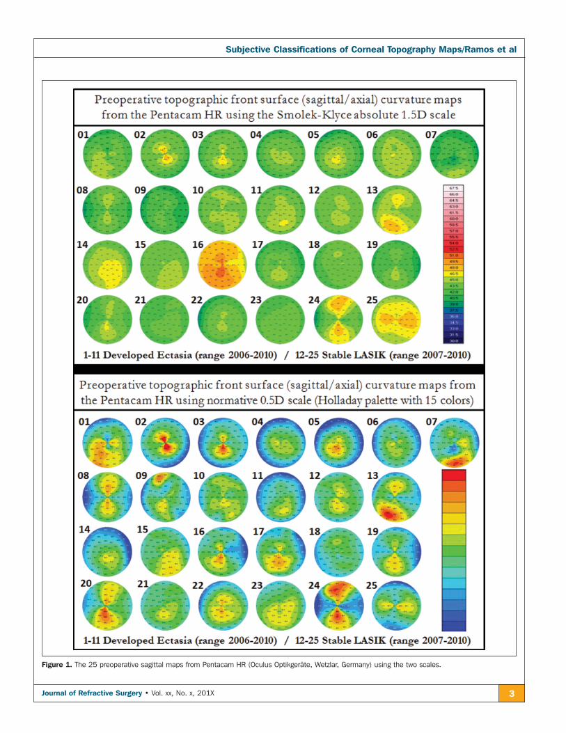
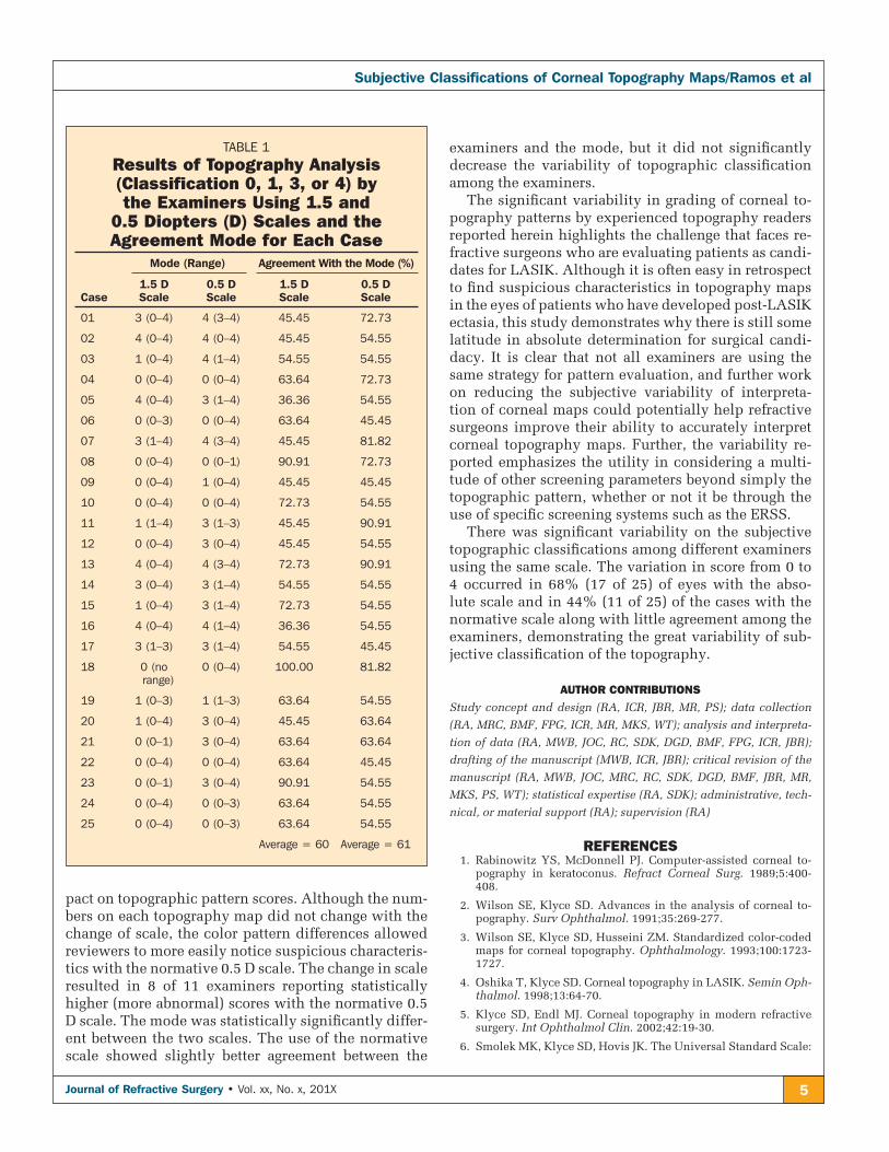
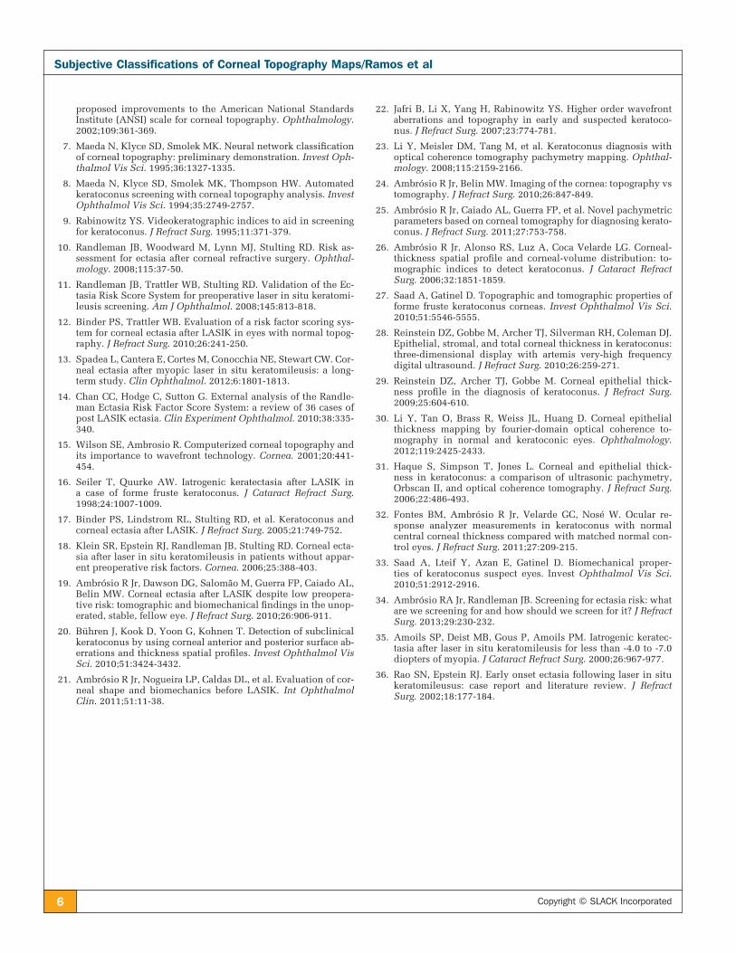
14 eyes with stable LASIK outcomes (follow-up > 18 months; mean: 19.8 months, range: 18 to 28 months) were selected for this study. The preoperative axial (or sagittal) curvature map of each case was obtained from corneal tomography (Pentacam HR). All examina-tions showed high rates of quality, including quality approval by the instrument software (quality specifi -cation). These maps and the clinical parameters (age, manifest refraction, visual acuity, central corneal thickness, and predicted residual stromal bed) were sent to 11 corneal topography specialists for masked subjective classifi cation accordingly to the ERSS (0 = normal or symmetric bowtie; 1 = asymmetric bowtie; 3 = inferior steepening pattern or skewed radial axis; 4 = abnormal topography). Each case was fi rst sent us-ing the absolute Smolek–Klyce 1.5 D scale and then resent to the same examiners after a period of 3 months for a new classifi cation using a 0.5 D normative scale (Figures 1-2) in a masked fashion.
Statistical analyses were performed using the Wil-coxon signed rank and Kruskal–Wallis tests to com-pare the differences between the examiners using the same scale and to compare the differences between the two scales by the same examiner.
RESULTSIn the fi rst evaluation with the absolute 1.5 D scale,
the variation of classifi cations from 0 to 4 across the examiners was observed in 17 to 25 cases (68%). The average agreement with the mode (number of examin-ers who choose the same classifi cation as the mode [the value that appears most often in a set of data] in each
case) was 60% among all cases (ranging from 36% to 100%). Only one case had 100% of agreement among all examiners (Table 1).
In the second evaluation with the normative 0.5 D scale, 11 of 25 cases (44%) had a classifi cation varia-tion from 0 to 4 across examiners. The average agree-ment with the mode was 61% among all cases (ranging from 45% to 91%). None of the cases had 100% agree-ment among all examiners (Table 1). The Appendix (available in the online version of this article) includes all results of the topography analysis by the examiners using 1.5 and 0.5 D scales, along with the agreement with the mode in circumstance.
The mode of classifi cations with the absolute Smolek–Klyce 1.5 D scale was statistically lower (Wil-coxon test, P = .0033) compared to the mode obtained with the normative 0.5 D scale. Considering all 275 classifi cations (11 examiners multiplied by 25 cases), statistical differences were found between the two scales (Wilcoxon test, P < .0001), with higher values on the normative 0.5 D scale (2.13 ± 1.56) compared with the absolute Smolek–Klyce 1.5 D scale (1.47 ± 1.55). Forty-four percent of the 275 analyses were exactly equal on the two scales, 42% had higher values on the normative 0.5 D scale, and 14% had higher values on the absolute Smolek–Klyce 1.5 D scale. Eight of 11 ex-aminers (73%) had statistically higher classifi cations using the 0.5 D normative scale (Wilcoxon, P < .05).
DISCUSSIONThis study assessed the variability of subjective as-
sessment of corneal topographic pattern in 11 eyes that developed ectasia after LASIK and 14 eyes that under-went LASIK and were stable, as judged by experienced reviewers not informed of the patient outcomes during the review. The results demonstrate great variability of these subjective topographic classifi cations among and between examiners for both the normative 0.5 D and the American National Standards Institute standard absolute 1.5 D color scales.
The development of computerized corneal topog-raphy from the 1990s is closely linked to advances in corneal refractive surgery.15 This topic has gained signifi cant importance since the fi rst reports of post-operative corneal ectasia,16,17 and subsequent reported cases considered to be without preoperative risk fac-tors for ectasia.18,19 Thereafter, subjective evaluations of corneal topography performed preoperatively in these patients started to be questioned.17 However, despite the emergence of new tests for the character-ization of the cornea,20-22 including anterior segment tomography,23-27 epithelial thickness profi les,28-31 and direct biomechanical measurements,19,32,33 the correct
3Journal of Refractive Surgery • Vol. xx, No. x, 201X
Subjective Classifications of Corneal Topography Maps/Ramos et al
Figure 1. The 25 preoperative sagittal maps from Pentacam HR (Oculus Optikgeräte, Wetzlar, Germany) using the two scales.
4 Copyright © SLACK Incorporated
Subjective Classifications of Corneal Topography Maps/Ramos et al
subjective evaluation of topographic patterns is still essential in the preoperative assessment of refractive surgery candidates.34
Retrospective studies involving careful evaluation of the preoperative corneal topography of patients who developed ectasia after LASIK have been per-formed.18,35,36 Clinical parameters have been analyzed
together to quantify the risk for developing ectasia in these eyes. However, it is important to emphasize that the subjective nature of the retrospective analysis be-comes even more clouded when the reviewer knows the patient’s diagnosis and outcome.
As hypothesized, changing the color scale from a 1.5 D step to a 0.5 D step scale had a signifi cant im-
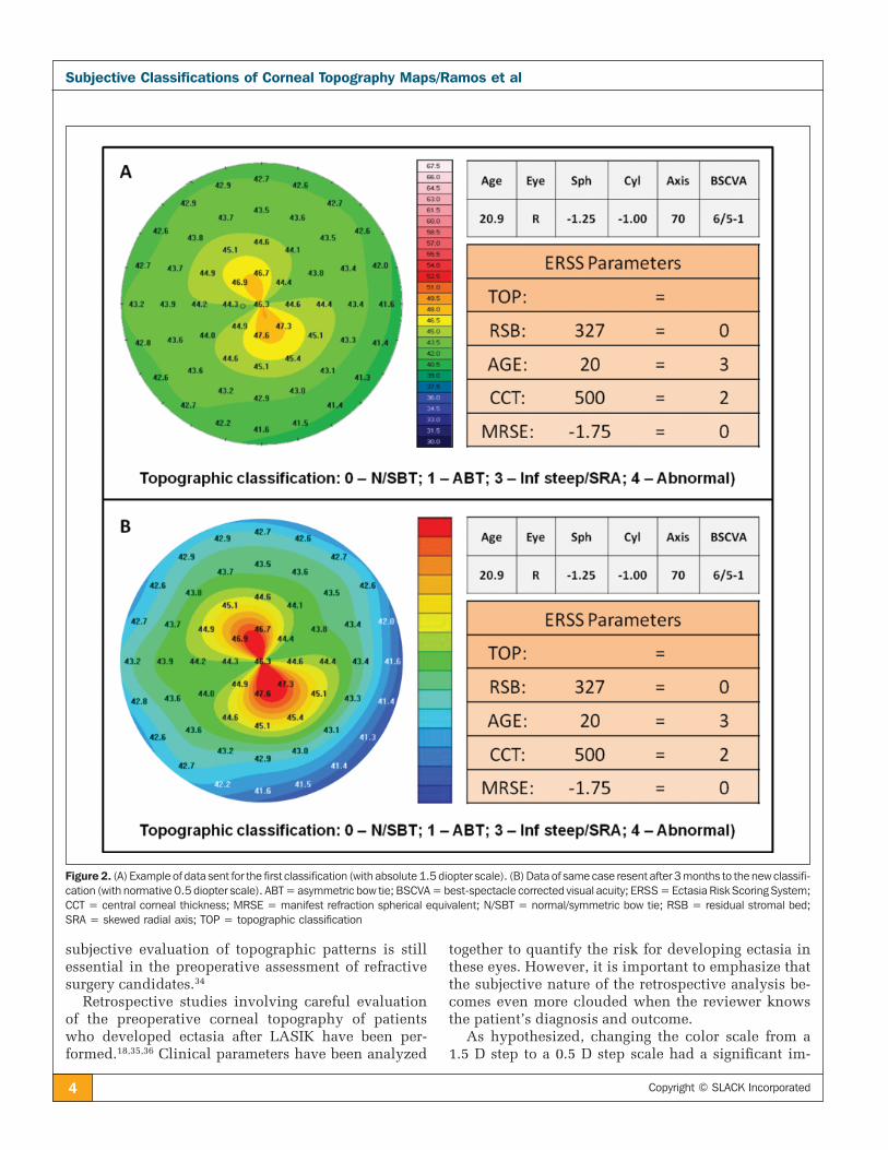
Figure 2. (A) Example of data sent for the first classification (with absolute 1.5 diopter scale). (B) Data of same case resent after 3 months to the new classifi-cation (with normative 0.5 diopter scale). ABT = asymmetric bow tie; BSCVA = best-spectacle corrected visual acuity; ERSS = Ectasia Risk Scoring System; CCT = central corneal thickness; MRSE = manifest refraction spherical equivalent; N/SBT = normal/symmetric bow tie; RSB = residual stromal bed; SRA = skewed radial axis; TOP = topographic classification
5Journal of Refractive Surgery • Vol. xx, No. x, 201X
Subjective Classifications of Corneal Topography Maps/Ramos et al
pact on topographic pattern scores. Although the num-bers on each topography map did not change with the change of scale, the color pattern differences allowed reviewers to more easily notice suspicious characteris-tics with the normative 0.5 D scale. The change in scale resulted in 8 of 11 examiners reporting statistically higher (more abnormal) scores with the normative 0.5 D scale. The mode was statistically signifi cantly differ-ent between the two scales. The use of the normative scale showed slightly better agreement between the
examiners and the mode, but it did not signifi cantly decrease the variability of topographic classifi cation among the examiners.
The signifi cant variability in grading of corneal to-pography patterns by experienced topography readers reported herein highlights the challenge that faces re-fractive surgeons who are evaluating patients as candi-dates for LASIK. Although it is often easy in retrospect to fi nd suspicious characteristics in topography maps in the eyes of patients who have developed post-LASIK ectasia, this study demonstrates why there is still some latitude in absolute determination for surgical candi-dacy. It is clear that not all examiners are using the same strategy for pattern evaluation, and further work on reducing the subjective variability of interpreta-tion of corneal maps could potentially help refractive surgeons improve their ability to accurately interpret corneal topography maps. Further, the variability re-ported emphasizes the utility in considering a multi-tude of other screening parameters beyond simply the topographic pattern, whether or not it be through the use of specifi c screening systems such as the ERSS.
There was signifi cant variability on the subjective topographic classifi cations among different examiners using the same scale. The variation in score from 0 to 4 occurred in 68% (17 of 25) of eyes with the abso-lute scale and in 44% (11 of 25) of the cases with the normative scale along with little agreement among the examiners, demonstrating the great variability of sub-jective classifi cation of the topography.
AUTHOR CONTRIBUTIONSStudy concept and design (RA, ICR, JBR, MR, PS); data collection
(RA, MRC, BMF, FPG, ICR, MR, MKS, WT); analysis and interpreta-
tion of data (RA, MWB, JOC, RC, SDK, DGD, BMF, FPG, ICR, JBR);
drafting of the manuscript (MWB, ICR, JBR); critical revision of the
manuscript (RA, MWB, JOC, MRC, RC, SDK, DGD, BMF, JBR, MR,
MKS, PS, WT); statistical expertise (RA, SDK); administrative, tech-
nical, or material support (RA); supervision (RA)
REFERENCES 1. Rabinowitz YS, McDonnell PJ. Computer-assisted corneal to-
pography in keratoconus. Refract Corneal Surg. 1989;5:400-408.
2. Wilson SE, Klyce SD. Advances in the analysis of corneal to-pography. Surv Ophthalmol. 1991;35:269-277.
3. Wilson SE, Klyce SD, Husseini ZM. Standardized color-coded maps for corneal topography. Ophthalmology. 1993;100:1723-1727.
4. Oshika T, Klyce SD. Corneal topography in LASIK. Semin Oph-thalmol. 1998;13:64-70.
5. Klyce SD, Endl MJ. Corneal topography in modern refractive surgery. Int Ophthalmol Clin. 2002;42:19-30.
6. Smolek MK, Klyce SD, Hovis JK. The Universal Standard Scale:
TABLE 1Results of Topography Analysis (Classification 0, 1, 3, or 4) by the Examiners Using 1.5 and
0.5 Diopters (D) Scales and the Agreement Mode for Each Case
Mode (Range) Agreement With the Mode (%)
Case1.5 D Scale
0.5 D Scale
1.5 D Scale
0.5 D Scale
01 3 (0–4) 4 (3–4) 45.45 72.73
02 4 (0–4) 4 (0–4) 45.45 54.55
03 1 (0–4) 4 (1–4) 54.55 54.55
04 0 (0–4) 0 (0–4) 63.64 72.73
05 4 (0–4) 3 (1–4) 36.36 54.55
06 0 (0–3) 0 (0–4) 63.64 45.45
07 3 (1–4) 4 (3–4) 45.45 81.82
08 0 (0–4) 0 (0–1) 90.91 72.73
09 0 (0–4) 1 (0–4) 45.45 45.45
10 0 (0–4) 0 (0–4) 72.73 54.55
11 1 (1–4) 3 (1–3) 45.45 90.91
12 0 (0–4) 3 (0–4) 45.45 54.55
13 4 (0–4) 4 (3–4) 72.73 90.91
14 3 (0–4) 3 (1–4) 54.55 54.55
15 1 (0–4) 3 (1–4) 72.73 54.55
16 4 (0–4) 4 (1–4) 36.36 54.55
17 3 (1–3) 3 (1–4) 54.55 45.45
18 0 (no range)
0 (0–4) 100.00 81.82
19 1 (0–3) 1 (1–3) 63.64 54.55
20 1 (0–4) 3 (0–4) 45.45 63.64
21 0 (0–1) 3 (0–4) 63.64 63.64
22 0 (0–4) 0 (0–4) 63.64 45.45
23 0 (0–1) 3 (0–4) 90.91 54.55
24 0 (0–4) 0 (0–3) 63.64 54.55
25 0 (0–4) 0 (0–3) 63.64 54.55
Average = 60 Average = 61
6 Copyright © SLACK Incorporated
Subjective Classifications of Corneal Topography Maps/Ramos et al
proposed improvements to the American National Standards Institute (ANSI) scale for corneal topography. Ophthalmology. 2002;109:361-369.
7. Maeda N, Klyce SD, Smolek MK. Neural network classifi cation of corneal topography: preliminary demonstration. Invest Oph-thalmol Vis Sci. 1995;36:1327-1335.
8. Maeda N, Klyce SD, Smolek MK, Thompson HW. Automated keratoconus screening with corneal topography analysis. Invest Ophthalmol Vis Sci. 1994;35:2749-2757.
9. Rabinowitz YS. Videokeratographic indices to aid in screening for keratoconus. J Refract Surg. 1995;11:371-379.
10. Randleman JB, Woodward M, Lynn MJ, Stulting RD. Risk as-sessment for ectasia after corneal refractive surgery. Ophthal-mology. 2008;115:37-50.
11. Randleman JB, Trattler WB, Stulting RD. Validation of the Ec-tasia Risk Score System for preoperative laser in situ keratomi-leusis screening. Am J Ophthalmol. 2008;145:813-818.
12. Binder PS, Trattler WB. Evaluation of a risk factor scoring sys-tem for corneal ectasia after LASIK in eyes with normal topog-raphy. J Refract Surg. 2010;26:241-250.
13. Spadea L, Cantera E, Cortes M, Conocchia NE, Stewart CW. Cor-neal ectasia after myopic laser in situ keratomileusis: a long-term study. Clin Ophthalmol. 2012;6:1801-1813.
14. Chan CC, Hodge C, Sutton G. External analysis of the Randle-man Ectasia Risk Factor Score System: a review of 36 cases of post LASIK ectasia. Clin Experiment Ophthalmol. 2010;38:335-340.
15. Wilson SE, Ambrosio R. Computerized corneal topography and its importance to wavefront technology. Cornea. 2001;20:441-454.
16. Seiler T, Quurke AW. Iatrogenic keratectasia after LASIK in a case of forme fruste keratoconus. J Cataract Refract Surg. 1998;24:1007-1009.
17. Binder PS, Lindstrom RL, Stulting RD, et al. Keratoconus and corneal ectasia after LASIK. J Refract Surg. 2005;21:749-752.
18. Klein SR, Epstein RJ, Randleman JB, Stulting RD. Corneal ecta-sia after laser in situ keratomileusis in patients without appar-ent preoperative risk factors. Cornea. 2006;25:388-403.
19. Ambrósio R Jr, Dawson DG, Salomão M, Guerra FP, Caiado AL, Belin MW. Corneal ectasia after LASIK despite low preopera-tive risk: tomographic and biomechanical fi ndings in the unop-erated, stable, fellow eye. J Refract Surg. 2010;26:906-911.
20. Bühren J, Kook D, Yoon G, Kohnen T. Detection of subclinical keratoconus by using corneal anterior and posterior surface ab-errations and thickness spatial profi les. Invest Ophthalmol Vis Sci. 2010;51:3424-3432.
21. Ambrósio R Jr, Nogueira LP, Caldas DL, et al. Evaluation of cor-neal shape and biomechanics before LASIK. Int Ophthalmol Clin. 2011;51:11-38.
22. Jafri B, Li X, Yang H, Rabinowitz YS. Higher order wavefront aberrations and topography in early and suspected keratoco-nus. J Refract Surg. 2007;23:774-781.
23. Li Y, Meisler DM, Tang M, et al. Keratoconus diagnosis with optical coherence tomography pachymetry mapping. Ophthal-mology. 2008;115:2159-2166.
24. Ambrósio R Jr, Belin MW. Imaging of the cornea: topography vs tomography. J Refract Surg. 2010;26:847-849.
25. Ambrósio R Jr, Caiado AL, Guerra FP, et al. Novel pachymetric parameters based on corneal tomography for diagnosing kerato-conus. J Refract Surg. 2011;27:753-758.
26. Ambrósio R Jr, Alonso RS, Luz A, Coca Velarde LG. Corneal-thickness spatial profi le and corneal-volume distribution: to-mographic indices to detect keratoconus. J Cataract Refract Surg. 2006;32:1851-1859.
27. Saad A, Gatinel D. Topographic and tomographic properties of forme fruste keratoconus corneas. Invest Ophthalmol Vis Sci. 2010;51:5546-5555.
28. Reinstein DZ, Gobbe M, Archer TJ, Silverman RH, Coleman DJ. Epithelial, stromal, and total corneal thickness in keratoconus: three-dimensional display with artemis very-high frequency digital ultrasound. J Refract Surg. 2010;26:259-271.
29. Reinstein DZ, Archer TJ, Gobbe M. Corneal epithelial thick-ness profi le in the diagnosis of keratoconus. J Refract Surg. 2009;25:604-610.
30. Li Y, Tan O, Brass R, Weiss JL, Huang D. Corneal epithelial thickness mapping by fourier-domain optical coherence to-mography in normal and keratoconic eyes. Ophthalmology. 2012;119:2425-2433.
31. Haque S, Simpson T, Jones L. Corneal and epithelial thick-ness in keratoconus: a comparison of ultrasonic pachymetry, Orbscan II, and optical coherence tomography. J Refract Surg. 2006;22:486-493.
32. Fontes BM, Ambrósio R Jr, Velarde GC, Nosé W. Ocular re-sponse analyzer measurements in keratoconus with normal central corneal thickness compared with matched normal con-trol eyes. J Refract Surg. 2011;27:209-215.
33. Saad A, Lteif Y, Azan E, Gatinel D. Biomechanical proper-ties of keratoconus suspect eyes. Invest Ophthalmol Vis Sci. 2010;51:2912-2916.
34. Ambrósio RA Jr, Randleman JB. Screening for ectasia risk: what are we screening for and how should we screen for it? J Refract Surg. 2013;29:230-232.
35. Amoils SP, Deist MB, Gous P, Amoils PM. Iatrogenic keratec-tasia after laser in situ keratomileusis for less than -4.0 to -7.0 diopters of myopia. J Cataract Refract Surg. 2000;26:967-977.
36. Rao SN, Epstein RJ. Early onset ectasia following laser in situ keratomileusus: case report and literature review. J Refract Surg. 2002;18:177-184.

APPE
ND
IX
All
Resu
lts
of T
opog
raph
y A
naly
sis
(Cla
ssif
icat
ion
0,
1,
3,
or 4
) by
the
Exa
min
ers
Usi
ng 1
.5 a
nd 0
.5 D
Sca
les
and
the
Agr
ee
me
nt W
ith
Mod
e i
n E
ach
Cas
e
Exa
min
er 1
Exa
min
er 2
Ex
amin
er 3
Exa
min
er 4
Exa
min
er 5
Exa
min
er 6
Exa
min
er 7
Exa
min
er 8
Exa
min
er 9
Exam
iner
10
Exam
iner
11
Mod
eAg
reem
ent
Wit
h M
ode
(%)
Cas
e1.
5 D
0.5 D
1.5
D0.
5 D
1.5
D0.
5 D
1.5
D0.
5 D
1.5
D0.
5 D
1.5
D0.
5 D
1.5
D0.
5 D
1.5
D0.
5 D
1.5
D0.
5 D
1.
5 D
0.5
D
1.5
D0.
5 D
1.
5 D
0.5
D
1.5
D0.
5 D
011
40
43
34
43
44
43
41
33
34
43
43
445
.45
72.7
3
024
44
00
01
40
04
44
03
40
40
04
44
445
.45
54.5
5
034
44
10
11
41
14
44
41
41
11
41
11
454
.55
54.5
5
040
01
00
01
10
04
40
00
00
01
30
00
063
.64
72.7
3
053
34
31
33
33
14
40
31
34
14
41
14
336
.36
54.5
5
060
31
10
01
30
00
43
01
00
00
10
10
063
.64
45.4
5
073
41
43
44
31
44
41
43
33
44
43
43
445
.45
81.8
2
080
04
10
00
00
00
10
00
10
00
00
00
090
.91
72.7
3
090
13
30
10
11
14
40
40
13
03
33
30
145
.45
45.4
5
100
11
00
10
10
04
40
00
03
40
00
00
072
.73
54.5
5
111
14
33
31
31
33
33
31
33
34
31
31
345
.45
90.9
1
120
31
33
30
10
04
41
30
33
03
30
10
345
.45
54.5
5
131
44
40
44
44
43
44
44
44
34
44
44
472
.73
90.9
1
140
34
30
13
33
13
33
43
33
34
40
13
354
.55
54.5
5
151
31
41
11
31
34
31
41
30
34
41
11
372
.73
54.5
5
164
31
40
43
43
14
31
44
43
34
43
34
436
.36
54.5
5
171
31
41
11
33
13
31
33
33
13
43
13
354
.55
45.4
5
180
00
40
10
00
00
00
00
00
00
00
00
010
0.00
81.8
2
191
11
10
11
31
13
31
33
30
11
31
11
163
.64
54.5
5
201
31
30
11
33
13
31
33
33
04
41
31
345
.45
63.6
4
210
11
40
31
31
30
30
40
30
01
30
30
363
.64
63.6
4
220
10
30
00
30
04
40
01
13
01
00
10
063
.64
45.4
5
230
10
40
30
30
30
40
40
30
01
30
30
390
.91
54.5
5
240
01
10
11
31
00
10
00
14
00
00
00
063
.64
54.5
5
250
00
10
11
31
00
10
00
14
00
01
00
063
.64
54.5
5
Aver
age
12.
041.
722.
520.
601.
641.
322.
641.
241.
282.
643.
161.
242.
321.
322.
282.
001.
362.
042.
481.
201.
721.
322.
1260
.00
61.0
9
Wilc
oxon
sig
ned
rank
(P)
.000
7.0
502
.000
3.0
002
.389
9.0
332
.016
6.0
010
.070
1.0
191
.020
7.0
033
.354
4
D =
dio
pter