uva-dare (digital academic repository) motor nerve ... · c h ap t e r |l somatosensory evoked...
TRANSCRIPT
UvA-DARE is a service provided by the library of the University of Amsterdam (http://dare.uva.nl)
UvA-DARE (Digital Academic Repository)
Motor nerve conduction velocity and somatosensory evoked potentials in the newbornand young child in relation to thyroid functionSmit, B.J.
Link to publication
Citation for published version (APA):Smit, B. J. (1999). Motor nerve conduction velocity and somatosensory evoked potentials in the newborn andyoung child in relation to thyroid function
General rightsIt is not permitted to download or to forward/distribute the text or part of it without the consent of the author(s) and/or copyright holder(s),other than for strictly personal, individual use, unless the work is under an open content license (like Creative Commons).
Disclaimer/Complaints regulationsIf you believe that digital publication of certain material infringes any of your rights or (privacy) interests, please let the Library know, statingyour reasons. In case of a legitimate complaint, the Library will make the material inaccessible and/or remove it from the website. Please Askthe Library: http://uba.uva.nl/en/contact, or a letter to: Library of the University of Amsterdam, Secretariat, Singel 425, 1012 WP Amsterdam,The Netherlands. You will be contacted as soon as possible.
Download date: 07 Jul 2018
C h a p t e r | l
Somatosensory evoked potentials in very preterm infants
Bert J. Smit1, MD, Bram W. Ongerboer de Visser2, MD, PhD, Linda S. de Vries3, MD, PhD, Friedo W. Dekker4, MD, PhD, Joke H. Kok1, MD, PhD.
Department of Neonatology1, Emma Children's Hospital AMC, University of Amsterdam, Clinical Neurophysiology Unit of the Department of Neurology2, Academic Medical Center, University of Amsterdam, Department of Neonatology3, Wilhelmina Children's Hospital, University of Utrecht, Department of Epidemiology and Biostatistics4, Academic Medical Center, University of Amsterdam, the Netherlands.
Submitted
54 Chapter 5
Abstract
Sufficient reference values of cortical Nj peak latency of the median nerve SEP in
very preterm infants are not yet available. In the placebo treated infants within an
L-thyroxine supplementation trial, born at less than 30 weeks' gestation, cortical
Nj peak latency was measured at two weeks, at term, and at six months corrected
age. Cross-sectional cortical Nj peak latency values obtained in 50 infants and complete
series of longitudinal values obtained in 15 infants were analyzed in relation to
postmenstrual age (PMA). Mean cortical N : peak latency decreased from 66 to 20
ms. Longitudinal cortical Nj peak latency values decreased as well and were consistent
with cross-sectional cortical Nj peak latency values. Possible confounding factors did
not have any significant effect on cortical N : peak latency at two weeks or at term age
except cranial ultrasound abnormalities at two weeks of age. The observed cortical
Nj peak latencies at term and at six months corrected age suggest that extrauterine
maturation of the somatosensory pathway in infants born at less than 30 weeks'
gestation, is delayed by extrauterine life.
Key words electrophysiology, evoked potentials, preterm infant, somatosensory.
SEPs in very preterm infants 55
Introduction
In recent years, median nerve somatosensory evoked potentials (SEPs) in newborns
have received increased attention.1"4 Sufficient reference values of peak latencies of
the first cortical potential (N ) in infants born at less than 30 weeks' gestation are not
yet available.56 Moreover, the validity of median nerve SEPs as a prognostic test to
predict neurodevelopmental outcome especially in preterm infants is not yet fully
established.14
To determine whether L-thyroxine supplementation improved neurodevelopmental
outcome at the age of two years, we recently carried out a randomized, double-
blind, placebo controlled trial in 200 very preterm infants, irrespective of their
clinical condition and thyroid hormone levels, who were born at less than 30 weeks'
gestation.78 SEP recordings were obtained at two weeks, at term and at six months
corrected age.
The present paper describes cross-sectional and longitudinal reference values of the
first cortical peak latency (N ) after stimulation of the median nerve based on data
obtained in the placebo group. Because cortical peak N latency reflects the functional
integrity of the somatosensory pathway in the nervous system, it can be abnormal due
to intracerebral hemorrhages, periventricular leucomalacia and ventriculomegaly All
of these conditions are not infrequently occurring in very preterm infants. Therefore,
we examined all cranial ultrasound examinations of the infants during admission. Other
possible confounding factors were also analyzed. Moereover, cortical peak N latency
values were studied in relation to neurologic development at two years of age.
Patients and Methods
Between January 1991 and July 1993, 200 very preterm infants were enrolled in the
thyroxine supplementation trial. The study was approved by the Research and Ethics
Committee of the Academic Medical Center in Amsterdam. Criteria for inclusion were
birth at a gestational age of 25 to 30 weeks and admission to our neonatal intensive care
unit within the first 24 hours after birth. Exclusion criteria were severe congenital
malformations, maternal endocrine disease and maternal illicit drug dependency. After
informed consent by at least one parent, each infant was randomly and blindly assigned
to receive either L-thyroxine or a placebo.7 For the purpose of the study presented here
56 Chapter 5
cortical Nx peak latency values of median nerve SEPs obtained in the placebo group
were analyzed.
SEP measurement
SEPs were recorded after stimulating the median nerve with a mobile SEP recording
machine (Neuropack Four Mini, model MEB-5304K - Nihon Kohden, Tokyo, Japan).
A hand-held device was placed at the wrist overlying the median nerve and 0.5 Hz
electrical pulse stimuli of 0.1 ms duration were given. The intensity of the stimulus
used was set at a level that was necessary to produce a minimal thumb twitch. The
skin temperature was guaranteed to be above 36° C. Recordings were obtained from
the left somatosensory cortex, following stimulation of the right median nerve. Only
in case of an infusion line in the right arm or left-sided brain lesions, recordings were
obtained from the right somatosensory cortex, following stimulation of the left median
nerve. Recordings were done using 10 mm diameter silver-silverchloride disk
electrodes. The negative electrode was placed over the primary somatosensory area
for the upper extremities, i.e. two cm posterior to C3/C4 (C3'/C4'), according to the
international 10 - 20 electrode system. The reference electrode was placed in
midfrontal position (Fz) and one electrode was placed on the lower arm as ground.
The skin-electrode resistance was usually kept under 5 kQ. The analysis time was
200 ms. Thirty to fifty cortical potentials were averaged through a bandpass of 2-100
Hz. For reproducibility, each recording was duplicated. After stimulating the median
nerve, the latency to the peak of the first negative wave (N ), i.e. the first cortical
potential was measured according to the criteria of Desmedt et al.9 When a bilobed
Nj-wave was present the latency to the first prominent peak was determined. All
infants were tested without sedation. At two weeks of age, the infant's behavioral
state during recording was noted according to Prechtl.10 At term and at the corrected
age of six months, the behavioral state of the infant was classified as awake or asleep.
Furthermore, actual medication was recorded. Each SEP recording took about 45
minutes to complete. All potentials were stored on a disk. Later on they were reassessed
and classified for quality according to predefined criteria with respect to configuration
and reproducibility. If both configuration and reproducibility were good, the quality
was considered good. If the configuration was good, but lacked good reproducibility,
i.e. two cortical Nj peak latencies with a difference more than 10 % of the largest
value, the quality was considered modera te . If ne i ther configuration nor
reproducibility were good, the quality of the potentials was considered poor. In cases
of good or moderate quality the mean value of the two cortical N peak latencies was
SEPs in very preterm infants 57
used for the analyses. Potentials of poor quality were excluded from statistical analyses.
In each patient SEPs were obtained at two weeks (14-17 days), at term (37-43 weeks
postmenstrual age [PMA]) and at six months corrected age (63-69 weeks PMA).
PMA is defined as gestational age at birth plus postnatal age. Corrected age is defined
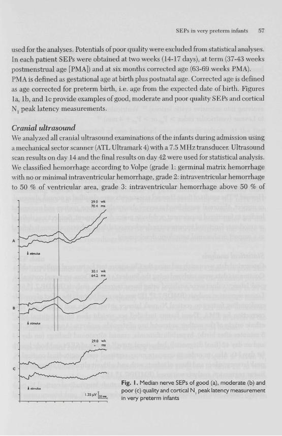
as age corrected for preterm birth, i.e. age from the expected date of birth. Figures
la, lb , and lc provide examples of good, moderate and poor quality SEPs and cortical
Nj peak latency measurements.
Cranial ultrasound
We analyzed all cranial ultrasound examinations of the infants during admission using
a mechanical sector scanner (ATL Ultramark 4) with a 7.5 MHz transducer. Ultrasound
scan results on day 14 and the final results on day 42 were used for statistical analysis.
We classified hemorrhage according to Volpe (grade 1: germinal matrix hemorrhage
with no or minimal intraventricular hemorrhage, grade 2: intraventricular hemorrhage
to 50 % of ventricular area, grade 3: intraventricular hemorrhage above 50 % of
Fig. I . Median nerve SEPs of good (a), moderate (b) and poor (c) quality and cortical N : peak latency measurement in very preterm infants
58 Chapter 5
ventricular area, usually with distention of lateral ventricle and grade 4: hemorrhagic
intracerebral involvement or other parenchymal lesion),11 and periventricular
leucomalacia according to De Vries et al. (grade 1: transient periventricular densities,
persisting for > 7 days, grade 2: transient periventricular densities, evolving in small
localized fronto-parietal cysts, grade 3: periventricular densities, evolving into
extensive periventricular cysts and grade 4: densities extending into deep white matter
evolving into extensive cystic lesions).12 Ventriculomegaly was classified according
to Levene (ventricular index > Pg7 or > Pg7 + 4 mm).13
Follow up with respect to neurologic development at two years
In this study, in contrast to the few earlier studies in low-risk preterm infants,5 6 a group
of high-risk preterm infants for abnormal neurologic development was investigated.
Therefore, we studied whether latency values of median nerve SEPs (good / moderate
quality SEPs) and poor quality SEPs (without a N peak latency value) were related to
neurologic development at two years of age, assessed according to the method of
Hempel.14 The results of these Hempel assessments were classified as normal, abnormal,
or suspect. Abnormal was defined as severe abnormality in tone, posture and movement
leading to functional impairment or delay in motor development. Suspect was defined
as a moderate functional impairment or developmental delay. Adverse outcome is defined
as a suspect or abnormal neurologic development.
Statistical analysis
Categorical data were analyzed using the Chi-square test for two and multiway tables.
Continuous data were analyzed using the Student's t-test. Cross-sectional cortical Nj
peak latency values were analyzed using linear regression analysis (BMDP-7.1® 6D).
Linear regression analysis (BMDP-7.1® 1R) was also used to study the effect of possible
confounding factors on cortical N peak latency at two weeks and at term age after
correction for PMA. These factors included sex, weight class for gestational age,10
ethnic origin of the mother, antenatal use of steroids, asphyxia (Apgar score < 7 at
5 minutes after birth), hyperbilirubinemia, cranial ultrasound findings (on day 14
and on day 42 [final diagnosis]), behavioral state,10 quality of SEPs and body length
(at day 14). Also, in order to compare cross-sectional and longitudinal cortical N t
peak latency values and those of infants with and without ultrasound abnormalities,
linear regression analysis was used (BMDP-7.1® 1R).
We used analysis of variance (BMDP-7.1® 5V) to study longitudinal cortical N peak
latency values, which provided a reference formula for cortical N peak latency corrected
SEPs in very preterm infants 59
for PMA. Linear regression analysis (BMDP-7.1® 1R) was used to analyze cross-sectional
and longitudinal cortical N peak latency values in relation to neurologic development
at two years of age. A p value of less than 0.05 was considered statistically significant.
Results
Patient population
The placebo group consisted of one hundred very preterm infants. At the first
measurement at two weeks 59 infants were studied. At the second and third
measurements, performed at term and six months corrected age, 65 and 54 infants
were studied, respectively. The reasons for the reduction in numbers at first
measurement, were death and poor clinical condition. At the second and third
measurements, other reasons were found such as parental discomfort with the time-
consuming aspects of measuring cortical Nj peak latency in addition to other
investigations on the same day. The clinical characteristics at the first SEP
measurement at two weeks of age are summarized in Table 1. All infants, in whom
SEPs were recorded, were treated with caffeine at two weeks. Later on this
medication was discontinued at about 34 weeks PMA. Throughout the study, in
none of the infants SEP recordings were performed during anticonvulsive treatment.
SEP data Only SEPs classified as good or moderate quality were analyzed. Causes for poor SEP
quality included an unstable clinical condition especially during the neonatal period.
At two weeks of age, we were able to record SEPs of good or moderate quality in 24 of
the 59 infants, while SEPs of poor quality were recorded in the remaining 35 infants.
The latter were more preterm, had a lower birth weight and had more ultrasound
abnormalities (Table 1). At term age, poor quality SEPs were recorded in seven of the
65 infants. At 6 months corrected age this number was four of the 54 infants.
Cross-sectional cortical N1 peak latency values
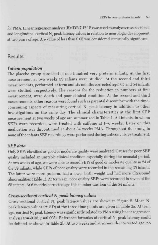
Cross-sectional cortical NL peak latency values are shown in Figure 2. Mean N\
peak latency values (± SD) at the three time points are given in Table 2a. At term
age, cortical N t peak latency was significantly related to PMA using linear regression
analysis (r=-0.38; p = 0.003). Reference formulas of cortical Nj peak latency could
be defined as shown in Table 2b. At two weeks and at six months corrected age, no
60 Chapter 5
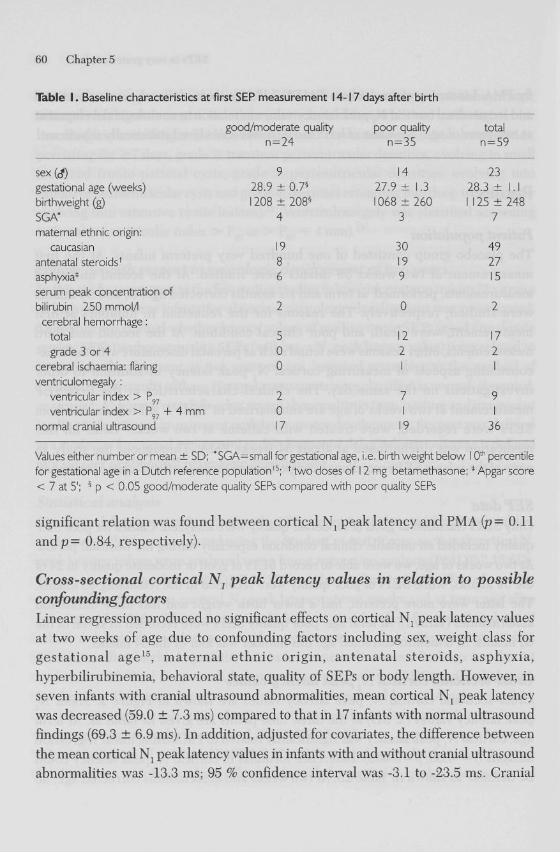
Table I. Baseline characteristics at first SEP measurement 14-17 days after birth
good/moderate quality n=24
poor quality n=35
total n = 59
sex (<?) gestational age (weeks) birthweight (g)
9 28.9 ± 0.7« 1208 ± 208s
I4 27.9 ± 1.3 I 0 6 8 ± 2 6 0
23 28.3 ± I. I 1125 ± 248
SGA- 4 3 7 maternal ethnic origin:
Caucasian I9 30 49 antenatal steroids1" 8 I9 27 asphyxia* serum peak concentration of bilirubin 250 mmol/l
cerebral hemorrhage : total
6
2
5
9
0
I2
I5
2
I7
grade 3 or 4 cerebral ischaemia: flaring ventriculomegaly :
ventricular index > P97
ventricular index > P97 + 4 mm
0 0
2 0
2 I
7 I
2 I
9 I
normal cranial ultrasound I7 I9 36
Values either number or mean ± SD; 'SGA=small for gestational age, i.e. birth weight below 10th percentile for gestational age in a Dutch reference population 5; + two doses of 11 mg betamethasone; * Apgar score < 7 at 5'; § p < 0.05 good/moderate quality SEPs compared with poor quality SEPs
significant relation was found between cortical N peak latency and PMA (p= 0.11
a n d p = 0.84, respectively).
Cross-sectional cortical 2V7 peak latency values in relation to possible confounding factors Linear regression produced no significant effects on cortical Nj peak latency values
at two weeks of age due to confounding factors including sex, weight class for
ges ta t ional age15, materna l e thnic origin, an tenata l s te ro ids , asphyxia,
hyperbilirubinemia, behavioral state, quality of SEPs or body length. However, in
seven infants with cranial ultrasound abnormalities, mean cortical N peak latency
was decreased (59.0 ± 7.3 ms) compared to that in 17 infants with normal ultrasound
findings (69.3 ± 6.9 ms). In addition, adjusted for covariates, the difference between
the mean cortical N peak latency values in infants with and without cranial ultrasound
abnormalities was -13.3 ms; 95 % confidence interval was -3.1 to -23.5 ms. Cranial
SEPs in very preterm infants 61
Z 40
28 29 30 31 32 PMA (weeks)
14-17 days after birth
33 37 38 39 40 41 PMA (weeks)
term age
63 64 65 66 67 68
PMA (weeks) 6 months (corrected) age
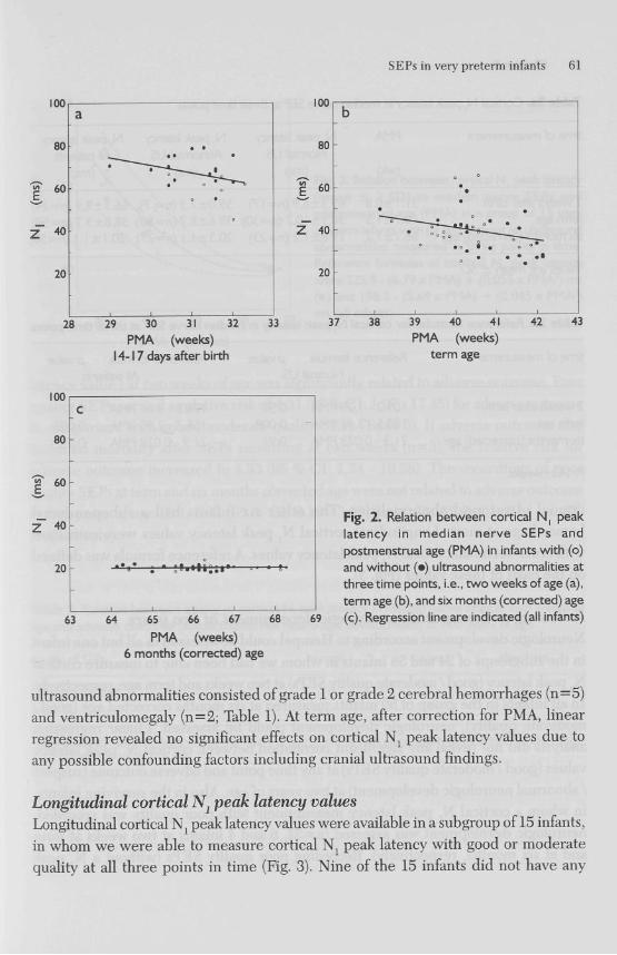
Fig. 2. Relation between cortical N : peak latency in median nerve SEPs and postmenstrual age (PMA) in infants with (o) and without (•) ultrasound abnormalities at three time points, i.e., two weeks of age (a), term age (b), and six months (corrected) age (c). Regression line are indicated (all infants)
ultrasound abnormalities consisted of grade 1 or grade 2 cerebral hemorrhages (n=5) and ventriculomegaly (n=2; Table 1). At term age, after correction for PMA, linear regression revealed no significant effects on cortical N( peak latency values due to any possible confounding factors including cranial ultrasound findings.
Longitudinal cortical Nj peak latency values Longitudinal cortical N peak latency values were available in a subgroup of 15 infants, in whom we were able to measure cortical Nj peak latency with good or moderate quality at all three points in time (Fig. 3). Nine of the 15 infants did not have any
62 Chapter 5
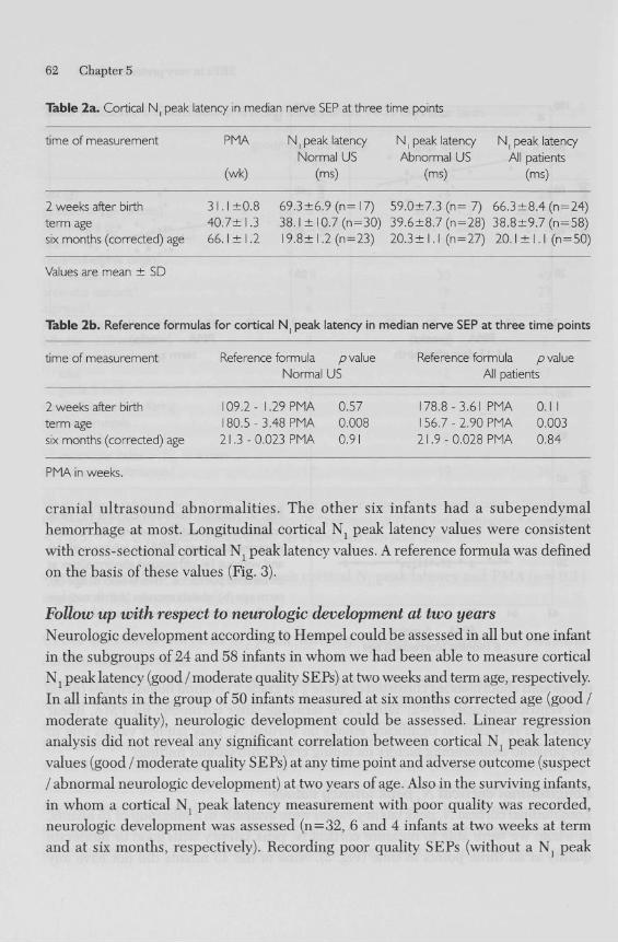
Table 2a. Cortical N : peak latency in median nerve SEP at three time points
time of measurement PMA N, peak latency N ( peak latency N peak latency Normal US Abnormal US All patients
(wk) (ms) (ms) (ms)
2 weeks after birth 31.1+0.8 69.3 ±6.9 (n= 17) 59.0±7.3 (n= 7) 66.3 ±8.4 (n = 24) term age 40.7±l .3 38.1 ± 10.7 (n=30) 39.6±8.7 (n = 28) 38.8±9.7 (n=58) six months (corrected) age 66.1 ± 1.2 19.8± 1.2 (n = 23) 20.3 ± 1.1 (n = 27) 20.1 ± I. I (n = 50)
Values are mean ± SD
Table 2b. Reference formulas for cortical N ( peak latency in median nerve SEP at three time points
time of measurement Reference formula p value Normal US
Reference formula p value All patients
2 weeks after birth term age six months (corrected) age
109.2- 1.29 PMA 0.57 180.5-3.48 PMA 0.008 21.3-0.023 PMA 0.91
178.8-3.61 PMA 0.1 1 156.7-2.90 PMA 0.003 21.9-0.028 PMA 0.84
PMA in weeks.
cranial ultrasound abnormalities. The other six infants had a subependymal
hemorrhage at most. Longitudinal cortical N peak latency values were consistent
with cross-sectional cortical N peak latency values. A reference formula was defined
on the basis of these values (Fig. 3).
Follow up with respect to neurologic development at two years
Neurologic development according to Hempel could be assessed in all but one infant
in the subgroups of 24 and 58 infants in whom we had been able to measure cortical
N j peak latency (good / moderate quality SEPs) at two weeks and term age, respectively.
In all infants in the group of 50 infants measured at six months corrected age (good /
moderate quality), neurologic development could be assessed. Linear regression
analysis did not reveal any significant correlation between cortical N peak latency
values (good / moderate quality SEPs) at any time point and adverse outcome (suspect
/abnormal neurologic development) at two years of age. Also in the surviving infants,
in whom a cortical N peak latency measurement with poor quality was recorded,
neurologic development was assessed (n = 32, 6 and 4 infants at two weeks at term
and at six months, respectively). Recording poor quality SEPs (without a N peak
SEPs in very preterm infants 63
30 40 50 60
PMA (weeks)
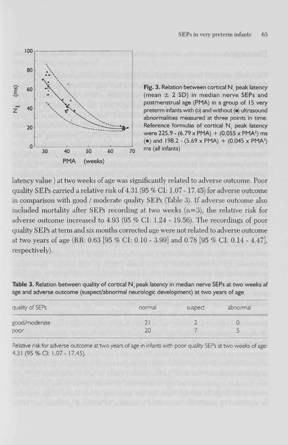
Fig. 3. Relation between cortical N ( peak latency (mean ± 2 SD) in median nerve SEPs and postmenstrual age (PMA) in a group of 15 very preterm infants with (o) and without (•) ultrasound abnormalities measured at three points in time. Reference formulas of cortical N : peak latency were 225.9 - (6.79 x PMA) + (0.055 x PMA2) ms (•) and 198.2 - (5.69 x PMA) + (0.045 x PMA2) ms (all infants)
latency value ) at two weeks of age was significantly related to adverse outcome. Poor
quality SEPs carried a relative risk of 4.31 (95 % CI: 1.07 -17.45) for adverse outcome
in comparison with good / moderate quality SEPs (Table 3). If adverse outcome also
included mortality after SEPs recording at two weeks (n=3), the relative risk for
adverse outcome increased to 4.93 (95 % CI: 1.24 - 19.56). The recordings of poor
quality SEPs at term and six months corrected age were not related to adverse outcome
at two years of age (RR: 0.63 [95 % CI: 0.10 - 3.99] and 0.78 [95 % CI: 0.14 - 4.47],
respectively).
Table 3. Relation between quality of cortical N ( peak latency In median nerve SEPs at two weeks af age and adverse outcome (suspect/abnormal neurologic development) at two years of age
quality of SEPs normal suspect abnormal
good/moderate poor
21 20
0 5
Relative risk for adverse outcome at two years of age in infants with poor quality SEPs at two weeks of age: 4.31 ( 9 5 % CI: 1.07- 17.45).
64 Chapter 5
Discussion
This study presents cross-sectional and longitudinal reference values of cortical
Nj peak latency in very preterm infants born at less than 30 weeks' gestation. Mean
cortical N t peak latency decreased from a mean value of 66 ms at two weeks to 38 ms
at term and 20 ms at six months corrected age. With the increment of PMA the
decrease of cortical Nl peak latency diminished. Longitudinal cortical N peak
latency values in a subgroup of 15 infants were consistent with cross-sectional cortical
Nj peak latency values in a larger group of about 50 infants, suggesting that these
values and the derived reference formula can be used as reliable reference values.
Until now, only two other reports regarding reference values of latencies in median nerve
SEPs in preterm infants included infants born at less than 30 weeks' gestation.5 6 Klimach
and Cooke described median nerve SEP findings in 102 neurologically normal preterm
infants with a median gestational age of 32.0 weeks (range 26.7 - 40.4 weeks) in stable
clinical condition without ventilator or oxygen requirement. At the first SEP recording,
their median postmenstrual age is 33.9 weeks (range 27.7 - 41.4 weeks). The success
rate of an acceptable SEP recording is 90.2 %. In fact, only 3 infants born at less than
30 weeks' gestation contribute to the data presented. Taylor et al. report on
maturational patterns of median nerve SEPs in a group of 22 preterm infants selected
on the basis of normal cranial ultrasounds during admission, no signs of asphyxia and
normal neurological examination at a mean corrected age of 14.8 months. The mean
gestational age of these infants is 28.4 weeks (range 27 - 32 weeks). Their mean
postmenstrual age when first tested is 29.6 weeks. The success rate is 100 %. Repeated
SEPs were done in 20 infants. Sixteen infants were tested twice and four infants
were tested three times. With a total of 46 SEPs, of which 22 completed at a
postmenstrual age of less than 30 weeks, latency values are presented, showing a
rapid maturational change in the SEPs over the early preterm period.
In contrast to these two studies, we studied a large group of infants born at less than
30 weeks' gestation who had not been selected on the basis of clinical condition,
ultrasound findings and follow-up. Such a group of high-risk preterm infants
represents an important part of a level III intensive care unit population. Presumably
as a result of this, at the age of two weeks the succes rate of a recordable SEP classified
as good or moderate was 41 %. In the present study, in fact, a recordable SEP was
only found at a postmenstrual age of more than 30 weeks except in one infant (at 29
weeks PMA [Figs, la and 2a]. Thus, the low succs rate at two weeks of age may also
be explained by insufficient maturation in addition to technical problems and brain
SEPs in very preterm infants 65
damage. At term and six months corrected age, succes rate was much higher with
89% and 93%, respectively.
The mean cortical N peak latencies found at two weeks and at term age (66.3 ± 8.4
ms and 38.8 ± 9.7 ms, respectively) were in agreement with those reported by Klimach
and Cooke, and Taylor et al.5 6 However, compared to values reported by others in
infants born at full-term (for term age: 24.6 - 31.0 ms and for six months of age: about
17.5 ms )16"23, in the presented study cortical Nl peak latencies found at term and six
months corrected age were longer (38.8 ± 9.7 ms and 20.1 ± 1.1 ms, respectively).
This could suggest that extrauterine maturation of the somatosensory pathway in
high-risk preterm infants is delayed compared to maturation in fullterm infants,
probably due to a delay in central myelination in preterm infants at term age compared
with fullterm infants.24 Former suggestions that maturation of the sensory pathway is
unaffected by the extrauterine environment is possibly true in a selected group of
low risk preterm infants.
In studies regarding cortical N peak latency in the preterm infant, several possible
confounding factors should be taken into account. In this study cortical N peak
latency values were not affected significantly by any possible confounding factor except
cranial ultrasound abnormalities at two weeks of age. In contrast to what we expected,
we found a decreased cortical N peak latency at 2 weeks of age in a small subgroup
of seven infants with cranial ultrasound abnormalities. This difference was even greater
after adjustment for covariates (-13.3 ms). However, the clinical relevance of this
finding may be rather small in view of the large confidence interval (95 % CI: -3.1 to
-23.5 ms). Our finding could be explained by the assumption that the first negative
peak which was measured was not, in fact, the cortical N ; peak but a peak derived
from subcortical structures, such as the thalamus rather than the somatosensory cortex
itself due to injury to the thalamocortical projections. Confounding by other factors
such as body length could be excluded.
De Vries et al. described an increase in cortical N peak latency in nine infants with
rapidly progressive ventricular dilatation.25 Others report conflicting information about
the effect of intracerebral hemorrhage and periventricular leucomalacia on cortical
Nj peak latency.2628
At term age, we found no difference in mean cortical N peak latency in infants with
and without cranial ultrasound abnormalities. This suggests that maturational state
(PMA) may be of predominant importance at that time.
Observed cortical N peak latency values were not related to neurologic development,
assessed according to Hempel at the age of two years. However, our analysis of
66 Chapter 5
comparing the poor quality with good/moderate quality SEPs at two weeks of age in
relation to neurologic development at two years (Table 3), supported the hypothesis
that poor quality SEPs predict adverse outcome. A correlation with outcome is in
agreement with Ekert et al. who found that cortical Nj peak latency in median nerve
SEPs in the first weeks of life is associated with later cerebral palsy29 At term and at
six months corrected age, we could not demonstrate a relation between poor quality
SEPs and adverse outcome.
We may conclude that this study presents cross-sectional and longitudinal values of
cortical Nj peak latency that can be used as reference values in very preterm infants,
born at less than 30 weeks' gestation. However, measuring N, peak latency in median
nerve SEPs at two weeks of age can be impeded in such a group of high-risk preterm
infants by unstable clinical condition, insufficient maturation and brain damage. All
these factors could be interrelated. They could also be an epiphenomenon of a common
factor as prematurity.
Acknowledgments
We would like to thank the participating children, their parents and the medical
and nursing staffs of our neonatal department for their cooperation. We are grateful
to T Brantsma-van Wulfften Palthe, EH.C. de Jongh, EE. Posthumus Meyjes and
J.J.M. de Vijlder for their enthusiastic support. This work was supported by the
Praeventiefonds (grant 28-2051), The Hague, the Netherlands.
References
1 De Vries LS, Pierrat V, Eken E The use of evoked potentials in the neonatal intensive care unit. J Perinat Med 1994;22:547-555.
2 Mercuri E, Von Siebenthal K, Daniels H, Guzetta E Casaer E Multimodality evoked responses in the neurological assessment of the newborn. Eur J Pediatr 1994;153:622-631.
3 Majnemer A, Rosenblatt B. Evoked potentials as predictors of outcome in neonatal intensive care unit survivors: Review of the literature. Pediatr Neurol 1996;14:189-195.
4 Taylor M J, Saliba E, Laugier. Use of evoked potentials in preterm neonates. Arch Dis Child 1996;74:F70-76.
5 Klimach VJ, Cooke RWI. Maturation of the neonatal somatosensory evoked response in
SEPs in very preterm infants 67
preterm infants. Dev Med Child Neurol 1988;30:208-214. 6 Taylor M J, Boor R, Ekert PG. Preterm maturation of the somatosensory evoked potential.
Electroenceph Clin Neurophysiol 1996;100:448-452. 7 Van Wassenaer AG, Kok JH, de Vijlder JJM, Briët JM, Smit BJ, Tamminga P, et al.
Effects of thyroxine supplementation on neurologic development in infants born at less than 30 weeks' gestation. N Engl J Med 1997;336:21-26.
8 Smit BJ, Kok JH, de Vries LS, van Wassenaer AG, Dekker FW, Ongerboer de Visser BW. Somatosensory evoked potentials in very preterm infants in relation to L-thyroxine supplementation. Pediatrics 1998;101:865-869.
9 Desmedt JE, Brunko E, Debecker J. Maturation of the somatosensory evoked potentials in normal infants and children with special reference to the early Nj component. Elecroencephalogr Clin Neurophysiol 1976;40:43-58.
10 Prechtl HFR. The behavioural states of the newborn infant (a review). Brain Res 1974;76:185-212.
11 Volpe JJ. Neurology of the newborn. 2nd ed. W.B. Saunders Company, Philadelphia; 1987:331. 12 De Vries LS, Eken P Dubowitz LMS. The spectrum ofleucomalacia using cranial ultrasound.
Behav Brain Res 1992a;49:l-6. 13 Levene MI. Measurement of the growth of the lateral ventricles in preterm infants with real
time ultrasound. Arch Dis Child 1981;56:900-904. 14 Hempel MS. The neurological examination for todler-age. Ph.D. thesis. University of
Groningen, the Netherlands: Groningen, 1993. 15 Kloosterman GJ. On intrauterine growth. Int J Gynaecol Obstet 1970;8:895-912. 16 Laureau E, Majnemer A, Rosenblatt B, Riley R A longitudinal study of short latency
somatosensory evoked responses in healthy newborns and infants. Electroenceph Clin Neurophysiol 1988;71:100-108.
17 Taylor MJ, Fagan ER. SEPs to median nerve stimulation: normative data for pediartrics. Electroenceph Clin Neurophysiol 1988;71:323-330.
18 Bongers-Schokking CT, Colon E J, Hoogland RA, Van Den Brande JL, De Groot CJ. The somatosensory evoked potentials of normal infants: influence of filter bandpass, arousal state and number of stimuli. Brain Dev 1989;11:33-39.
19 Bongers-Schokking CT, Colon EJ, Hoogland RA, Van Den Brande JL, De Groot CJ. Somatosensory evoked potentials in term and preterm infants in relation to postconceptional age and birth weight. Neuropediatrics 1990;21:32-36.
20 Laureau E, Marlot D. Somatosensory evoked responses after median and tibial nerve stimulation in healthy newborns. Electroenceph Clin Neurophysiol 1990;76:453-458.
21 Majnemer A, Rosenblatt B, Willis D, Lavallee. The effect of gestational age at birth on somatosensory-evoked potentials performed at term. J Child Neurol 1990;5:329-335.
22 George SR, Taylor MJ. Somatosensory evoked potential in neonates and infants: developmental and normative data. Electroenceph Clin Neurophysiol 1991;80:94-102.
23 Gibson NA, Brezinova V, Levene MI. Somatosensory evoked potentials in the term newborn. Electroenceph Clin Neurophysiol 1992;84:26-31.
24 Hüppi PS, Schuknecht B, Boesch C, Bossi E, Felblinger J, Fusch C, Herschkowitz N. Structural and neurobehavioral delay in postnatal brain development of preterm infants. Pediatr Res 1996;39:895-901.

68 Chapter 5
25 De Vries LS, Pierrat V, Minami T, Smet M, Casaer E The role of short latency somatosensory evoked responses in infants with rapidly progressive ventricular dilatation. Neuropediatrics 1990;21:136-139.
26 Klimach VJ, Cooke RWI. Short latency cortical somatosensory evoked responses of preterm infants with ultrasound abnormality of the brain. Dev Med Child Neurol 1988;30:215-221.
27 Willis J, Duncan MC, Bell R, Pappas E Moniz M. Somatosensory evoked potentials predict neuromotor outcome after periventricular hemorrhage. Dev Med Child Neurol 1989;31:435-439.
28 Pierrat V, Eken P, Duquennoy C, Rousseau S, De Vries LS. Prognostic value of early somatosensory evoked potentials in neonates with cystic leukomalacia. Dev Med Child Neurol 993;35:683-690.
29 Ekert PG, Taylor MJ, Keenan NK, Boulton JE, Whyte HE. Early somatosensory evoked potentials in preterm infants: their prognostic utility. Biol Neonate 1997;71:83-91.