uterine anomaly, fibroid uterus, ovarian tumor, uterine prolapse
DESCRIPTION
UTERINE ANOMALY, FIBROID UTERUS, OVARIAN TUMOR, UTERINE PROLAPSETRANSCRIPT
UTERINE ANOMALIES
Uterine anomalies are quite often asymptomatic and so are hard to recognize. Women with a history of miscarriage, pre-term deliveries, etc., have a higher incidence of anomalies There is no universal classification of defects, however the American Fertility Society sorted m üllerian defects into 7 categories .Anomalies are
Class I: Mullerian Agenesis/Hypoplasia-segmental,
Class II: Unicornuate uterus Class III: Didelphys uterus Class IV: Bicornuate uterus Class V : Septate uterus Class VI: Arcuate uterus Class VII: Diethyl stilboestrol realted abnormality
Failure of development of one or both Mullerian ductsThe absence of both ducts leads too absence of uterus including oviducts. There is absence of vaginal as well. Primary amenorrhoea is the chief complaint.The absence of one duct leads to a unicornuate uterus with a single oviduct.
Failure of recanalisation of the Mullerian ductsAgenesis of the upper vagina or of the ccervix – This may lead to haematometra s the uterus is functioning.
Failure of fusion of Mullerian ductsIn majority, the presence of deformity escapes attention. In some, the detection is made accidentally during investigation of infertility or repeated pregnancy wastage. In others, the diagnosis is made during D+E operation, manual removal of placenta or during caesarean section.
Types of fusion anomalies Arcuate (18%)– the corneal parts of the uterus
remain separated. The uterine fundus looks concave with heart shaped cavity outline.
Uterine didelphys (8%) – There is complete lack of fusion of the Mullerian ducts with a double uterus cervix and double vaginal
Uterus bicornis (26%) – There is varying degrees of fusion of the muscle walls of the two ducts
Uterus bicornis bicollis – there are two uterine cavities with double cervix with or without vaginal septum
Uterus bicornis unicollis – there are two uterine cavities with one cervix. The horns may be equal or one horn may be rudimentary and have no communication with the developed horn.
Septate uterus (35%) – The two Mullerian ducts fused together but there is persistence of septum in between the two either partially (subsequent) or completely.
Uterus unicornis (10%) – Failure of development of one Mullerian duct.
DES related abnormality – It is due to DES exposure during intrauterine life. Varieties of malformations are included, eg. Vagina: Adenosis, Adenocarcinoma. Cervix: Cockscomb cervix, Cervical collar. Uterus: Hypoplasia, T shaped cavity, Uterine synechae. Fallpian tube: corneal budding, abnormal fimbriae.
CLINICAL FEATURESGynaecological Infrertility and
dypareunia are often related in association with vaginal septum
Dysmenorrhoea in bicornuate uterus or due to crypotmenorrhoea (pent up menstrual flow in rudimentary horn)
Menorrhagia – due to increased surface area in bicornuate uterus
Obstetrical Mid trimester abortion which may be
recurrent. Cornual pregnancy – with inevitable
rupture around 16th week – if pregnancy occurs in the rudimentary horn
Increased incidence of malpresentation – transverse lie in arcuate or subseptate, breech in bicornuate, unicornuate or complete septate uterus.
Preterm labour, IUGR, IUD Prolonged labour – due to inordinate
uterine action Obstructed labour – obstruction by
the non gravid horn of the uterus or rudimentary horn
Retained placenta and postpartum haemorrhage where the placenta is implanted over the uterine septum

Diagnosis Internal examination reveals septate vagina and
two cervices. Passage of sound can diagnose two separate cavities. In fact in significant number of cases, the clinical diagnosis is made during uterine curettage, manual removal of placenta or caesarean section. For exact diagnosis is made during uterine curettage, manual removal of placenta or caesarean section. For exact diagnosis of the malforamation, internal as well as external architecture of the uterus must be visualised. For this reason several investigations in different combinations are done, such as hysterography, hysteroscopy, laparoscopy, ultrasonography (vaginal probe) and MRI. Urological tract is also evaluated at the same time. The reanal tract abnormality in association with Mullerian abnormality is about 40%. Skeletal system anomaly is also associated.
Treatment Mere presence of any uterine malformation
per se is not an indication of surgical intervention. Better obstetric outcome in septate uterus (86%), bicornuate uterus (50%) and unicornuate uterus (40%) has been mentioned. Didelphic uterus has high successful pregnancy outcome (64%) without metroplasty. Other causes of infertility or recurrent fetal loss must be excluded. Rudimentary horn should be excised. Unification operation ( bicornuate/septate uterus) is therefore indicated in an otherwise unexplained case. Abdominal metroplasty could be done either by excising the septum. Success rate of abdominal metroplasty in terms of live birth is high(5-75%).
Hysteroscopic metroplasty is becoming popular these days. Resection of the septum can be done either by resectoscope or by laser Advantages are: (a) High success rate (80-89%) (b)short hospital stay (c) reduced post operative morbidity (infection and adhesions); subsequent chances of vaginal delivery is high compared to abdominal metroplasty where caesarean section is mandatory.
FIBROID UTERUS
INCIDENCE: the incidence of fibroid with pregnancy is about 1 in 1000.
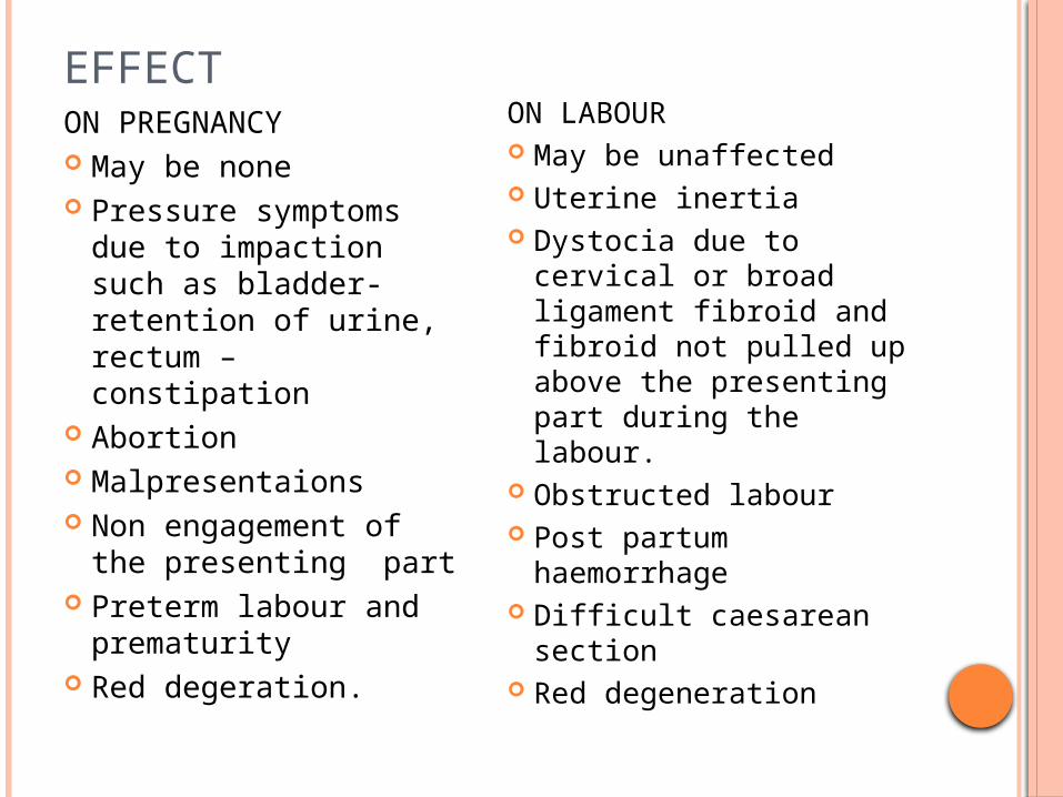
EFFECTON PREGNANCY May be none Pressure symptoms
due to impaction such as bladder- retention of urine, rectum – constipation
Abortion Malpresentaions Non engagement of
the presenting part Preterm labour and
prematurity Red degeration.
ON LABOUR May be unaffected Uterine inertia Dystocia due to
cervical or broad ligament fibroid and fibroid not pulled up above the presenting part during the labour.
Obstructed labour Post partum
haemorrhage Difficult caesarean
section Red degeneration
EFFECT
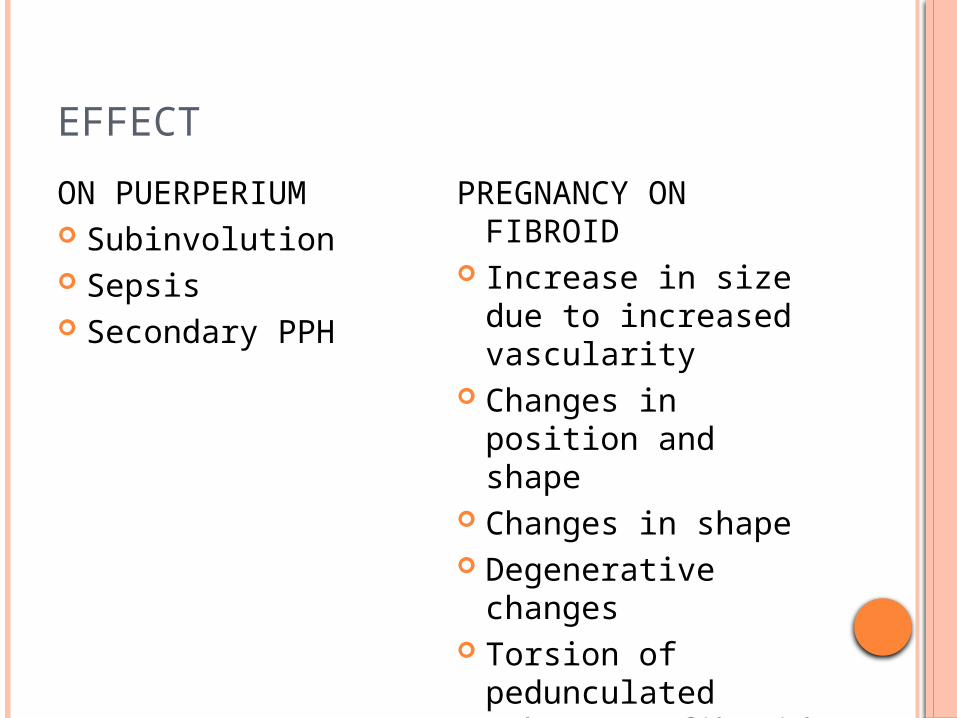
ON PUERPERIUM Subinvolution Sepsis Secondary PPH
PREGNANCY ON FIBROID
Increase in size due to increased vascularity
Changes in position and shape
Changes in shape Degenerative
changes Torsion of
pedunculated subserous fibroid
DIAGNOSIS Marked softening and alteration (flattening)
make it difficult to differentiate from the pregnant uterus. In uncomplicated tumour, it is confused with ovarian tumour, retroverted gravid uterus, non- gravid half of a double uterus. In early months, fibroid is diagnosed but pregnancy is missed where as in later moths, pregnancy is diagnosed but the fibroid is missed. Sonography confirms the diagnosis with certainty
TREATMENT. The basic principle in the management of pregnancy
complicated by a fibroid is not to do anything to the fibroid whenever possible.
During pregnancy Uncomplicated- usual antenatal care followed. All cases
are to be assessed at 38 weeks to formulate the method of delivery.
Impaction in early moths followed by retention of urine- if manual correction fails, laparotomy and myomectomy, is rarely indicated leaving behind the undisturbed pregnancy.
Red degeneration- conservative management should be followed patient should be put on the bed. Ampicillin 500mg capsule trice daily for seven days is given. Analgesics and sedatives are frequently needed. The symptoms usually clears off within 10 days.
Place of elective caesarean section- 1) cervical or broad ligament fibroid. 2) associated complicating factors such as elderly primigravida or malpresentations
During labour Fibroid situated above the presenting part
usually result in uneventfull vaginal delivery Fibroid situated below the presenting part –
spontaneous vaginal delivery may occur. If it fails, caesarean section is to be done. Myomectomy should be avoided duringing caesarean section.
One should be alert for postpartum haemorrhage and retained placenta. The fibroid usually reverts to a smaller size during puerperium.
OVARIAN TUMOR IN PREGNANCY
INCIDENCE: the incidence of ovarian tumour with pregnancy is about 2000.
EFFECTS OF TUMOUROn pregnancy: There is increased chance of Impaction leading to retention of urine Mechanical distress in presence of large tumour Mal presentation Non-engagement of head at termOn labour: There is increased chance of obstructed
labour if the tumour is impacted in the pelvisEFFECTS ON THE TUMOR: All the complications that
occur in the non-pregnant state are likely to occur with increased frequency except malignancy.
Torsion of the pedicle Intracystic haemorrhage is due to increased
vascularity Rupture Infection
DIAGNOSISPatient may remain asymptomatic or presents
with the symptoms of Retention of uterine due to impaction of the
tumour Mechanical distress due to the large cyst Acute abdomen due to complications of
tumour. Abdominal examination reveals the cystic swelling felt separated from the gravid the uterus. In later months of pregnancy confusion may arise. The patient is examined vaginally in head down Trendlenberg position to elicit the groove between the two swellings, eg. gravid uterus and the ovarian tumour (Hingorani sign). Ultrasonography is useful to have the details of pregnancy and the ovarian tumour.
TREATMENTThe principle is to remove the tumour as soon
as the diagnosis is made.During pregnancy Uncomplicated – the best time of elective
operation is between 14-18th week, as the chance of abortion is less and access to the pedicle is easy. Beyond 36 weeks the operation is better to withheld till delivery and the tumour is removed in as early as puerperium as possible.
Complicated – the tumour should be removed irrespective of the period of gestation. Adequate pain relief should be offered for 48 hours following surgery.
During labour If the tumour is well above the presenting
part, a watchful expectancy hoping for vaginal delivery is followed.
If the tumour is impacted in the pelvis causing obstruction , caesarean section should be done followed by removal of the tumour in the same sitting.
During puerperium On occasion, the diagnosis made following
delivery. The tumour should be removed as early in puerperium as possible. Following operation the specimen is sent for histological examination.

PROLAPSE OF GRAVID UTERUS
Pregnancy is not uncommon in first degree uterine prolapse with cystocele and rectocele. Pregnancy, is however, unlikely when the cervix remains outside the introitus and continuation of pregnancy in 3rd degree prolapse is an extremely rare event. The incidence of prolapse is about 1 in 250 pregnancies
EFFECT OF PROLAPSE: There is aggravation of the morbid anatomical changes in prolapse
Marked hypertrophy and oedema of the cervix.
First degree becomes second degree. Cystocele and rectocele become pronounced. There is aggravation of stress incontinence. These are marked during early pregnancy and
the effect are due to the weight of the uterus and increased vascularity.
Vaginal discharge may be copious and decubitus ulcer may develop when the cervix remains outside the introtious
There is chance of incarceration, if the uterus fails to rise above the pelvis by 16th week of pregnancy.
EFFECTS:On pregnancy: There is increased chance of Abortion Discomfort due to increased aliments Premature rupture of the membranes Chorioamnionitis.During labour: There is increased chance of Early rupture of membranes Cervical dystocia Prolonged labour due to non dilatation of cervix
and obstruction due to sagging cystocele and rectocele.
Operative interference.During puerperium: Sub involution Uterine sepsis
TREATMENT DURING PREGNANCY: The symptoms are mostly pronounced
in early pregnancy. If the cervix is outside the introitus – The cervix is to be
replaced inside the vagina and is kept in position by a ring pessary. The pessary is to be kept until 18-20th week of pregnancy when the body of the uterus will be sufficiently enlarged to sit on the brim of the pelvis.
The pelvic floor is too much lax- the patient is to lie in bed with the foot end raised by about 20 cm. to relieve oedema and congestion of the prolapsed mass, it should be covered by gauze soaked with glycerine and acriflavine. The treatment is continued until 18-20th week of pregnancy till the prolapsed mass is reduced in size and replaced inside the vagina. Thereafter, the patient is allowed to walk about. -If reposition is not possible and there is incarceration, termination of pregnancy may be indicated.-If the cervix remains outside the introitus even in the later months, it is preferable to admit the patient at 36th week
DURING LABOUR: The patient should be in bed, not only to prevent
early rupture of the membranes but also to facilitate replacement of the prolapsed cervix inside the vagina.
Intra vaginal plugging soaked with glycerine and acriflavine not only helps in reduction of cervical oedema but also facilitates its dilatation.
Prophylactic antibiotic, in cases of premature rupture of the membranes or when the cervix remains outside, should be administered.
Manual stretching of the cervix or pushing up the cystocele or rectocele pastthe presenting part during uterine contractions facilitates progressive descent of the head.
If the head is deeply engaged with the cervix remaining oedematous, thick or undilated- caesarean section is a safe procedure.
PUERPERIUM The patient should lie flat on the bed. If the mass remains outside, it should be
covered with guaze soaked in glycerine and acriflavine.
If sub involution is evident, a ring pessary may be put in until involution is completed.
Prophylactic antibiotic is administered.