use of atp bioluminescence for rapid detection and...
TRANSCRIPT
5
Use of ATP Bioluminescence for Rapid
Detection and Enumeration of Contaminants: The Milliflex Rapid Microbiology
Detection and Enumeration System
Renaud Chollet and Sébastien Ribault Merck-Millipore
France
1. Introduction
Rapid microbial detection becomes increasingly essential to many companies in
pharmaceutical, clinical and in food and beverage areas. Faster microbiological methods are
required to contribute to a better control of raw materials as well as finished products. Rapid
microbiological methods can also provide a better reactivity throughout the manufacturing
process. Implementing rapid technologies would allow companies for cost saving and
would speed up products release. Despite clear advantages, traditional methods are still
widely used. Current methods require incubation of products in liquid or solid culture
media for routinely 2 to 7 days before getting the contamination result. This necessary long
incubation time is mainly due to the fact that stressed microorganisms found in complex
matrices require several days to grow to visible colonies to be detected. Moreover, this
incubation period can be increased up to 14 days in specific application like sterility testing
for the release of pharmaceutical compounds. Although these techniques show advantages
like simplicity, the use of inexpensive materials and their acceptability to the regulatory
authorities, the major drawback is the length of time taken to get microbiological results.
Thus, face to the growing demand for rapid detection methods, various alternative
technologies have been developed. In the field of rapid microorganisms detection, ATP-
bioluminescence based on luciferine/luciferase reaction has shown great interest. Indeed,
adenosine triphosphate (ATP) is found in all living organisms and is an excellent marker for
viability and cellular contamination. Detection of ATP through ATP-luminescence
technology is therefore a method of choice to replace traditional method and significantly
shorten time to detection without loosing reliability.
This chapter will address the ATP-bioluminescence principle as a sensitive and rapid
detection technology in the Milliflex Rapid Microbiology Detection and Enumeration System (RMDS). This system combines membrane filtration principle, detection of microorganisms by ATP-bioluminescence and light capture triggered by a Charged Coupled Device camera (CCD) followed by software analysis.
www.intechopen.com
Bioluminescence – Recent Advances in Oceanic Measurements and Laboratory Applications
100
2. ATP-Bioluminescence
2.1 ATP-Bioluminescence principle
Light-producing living organisms are widespread in nature and from diverse origins. The
process of light emission from organisms is called bioluminescence and represents a
chemical conversion of energy into light. Since the work of William D McElroy showing that
ATP is a limiting and key factor of the bioluminescent reaction, research has lead to a better
understanding of how light is produced by fireflies (McElroy, 1947; McElroy, 1951; McElroy
et al., 1953). The bioluminescence mechanism involving Luciferase enzyme is a multistep
process which mainly requires Luciferin substrat, Oxygen (O2), Magnesium cation (Mg++)
and ATP (DeLuca & McElroy, 1974; McElroy et al., 1953; Seliger, 1989). ATP-
bioluminescence using luciferine/luciferase relies on luciferine oxidation by the luciferase
and the integrated light intensity is directly proportional to ATP contents. Luciferase
converts in presence of ATP and Magnesium firefly D-luciferin into the corresponding
enzyme-bound luciferil adenylate. The luciferil adenylate complex is then the substrate of
the subsequent oxidative reaction leading to oxyluciferin. The light emission is a
consequence of a rapid loss of energy of the oxyluciferine molecule from an excited state to a
stable one. This reaction induces the emission of photons with a efficient quantum yield of
about 90% (Seliger, 1989; Wilson & Hasting, 1998) (Fig1).
1/ D-luciferin + luciferase + ATPMg Luciferil adenylate complex +PPi
2/ Luciferil adenylate complex 2O Oxyluciferin + AMP+ CO2 + light
Fig. 1. Chemical reactions of the ATP-bioluminescence based on luciferin/luciferase system (PPi:inorganic pyrophosphate, CO2: Carbon Dioxide). Photons of yellow-green light (550 to 570 nm) are emitted.
2.2 Luciferase protein
Luciferase is a common term used to describe enzymes able to catalyze light emission.
Luciferase belongs to the adelynate-forming protein family and is an oxygen-4-
oxidoreductase gathering decarboxylation and ATP-hydrolysing main activities. Structural
studies have shown that Photinus pyralis Luciferase protein is folded into 2 domains: a large
N-terminal body and a small C-terminal domain linked by a flexible peptide creating a wide
cleft (Conti et al., 1996). Amino acids critical for bioluminescence phenomenon belong
mainly to the N-terminal domain (Branchini et al., 2000; Thompson et al., 1997; Zako et al.,
2003). This implies that luciferine-binding site is mediated by conformational change to
bring the 2 domains closer. This conformational change is consistent with the study of
Nakatsu et al (2006) showing that luciferase from luciola cruciata exists in an “open form”
and in a “closed form”, the later form creates an hydrophobic pocket around the active site
and is responsible of light emission. Two kinds of colored light emission are described for
luciferine/luciferase reaction. The typical high energy yellow-green light emission with a
peak at 562 nm at pH 7.5 and red light emission with a peak at 620nm when the pH
decreases to 5 (Seliger et al., 1964; Seliger & McElroy, 1964). This surprising phenomenon
where Luciferase is able to emit light of different colors is not clearly understood but the
isolation of colored luciferase variants shows that single amino acid substitution in
www.intechopen.com
Use of ATP Bioluminescence for Rapid Detection and Enumeration of Contaminants: The Milliflex Rapid Microbiology Detection and Enumeration System
101
N-terminal domain affects bioluminescence color by modulating slightly the polarity of the
active site environment (Hosseinkhani, 2011; Shapiro et al., 2005). This interesting feature
opens the way to wide applications in biotechnology (Branchini et al., 2005).
2.3 ATP-Bioluminescence applications
With the isolation, cloning and purification of various luciferases from many
bioluminescence-producing organisms (bacteria, beetles, marines organisms, etc),
bioluminescent assays have been developed and widely used in microbiology to detect
bacterial contamination by measuring presence of ATP and in molecular and cellular
biology with luciferase as reporter gene to monitor gene expression, protein-protein
interaction, etc (Francis et al., 2000; Roda et al, 2004; Thorne et al., 2010). The average
intracellular ATP content in various microorganisms has been quantified and ATP has been
shown to be a reliable biomarker of the presence of living organisms (Kodata et al., 1996;
Thore et al., 1975; Venkateswaran et al., 2003). To be able to specifically detect living
organisms by ATP-bioluminescence, the first step is to extract ATP from cells. This step is
critical and impacts directly the reliability of the detection (Selan et al., 1992). Chemical
solution or physical extraction methods were used in liquid samples (Selan et al., 1992; Siro
et al., 1982). Some false negative results were described in few studies (Conn et al., 1975;
Kolbeck et al., 1985). Additional studies investigated the cause of false negative results and
demonstrated that ATP extraction was not efficient. Indeed, extensive sonication of bacterial
samples for instance caused a significant increase of Relative Light Unit (RLU) measured
(Selan et al., 1992). Taking into account this limitation, ATP-bioluminescent assay has
already proved to provide good detection properties in many areas. Bioluminescent assay is
broadly used to monitor air and surface cleanliness and product quality mainly in food
industries and in less extent in pharmaceutical industries (Aycicek et al., 2006; Bautisda et
al., 1995; Davidson et al., 1999; Dostalek & Branyik, 2005; Girotti et al., 1997; Hawronskyj &
Holah, 1999). Studies shows that the level of contamination assessed though surface
swabbing, ATP extraction and bioluminescent assay correlate well for 80 % of the samples
tested with traditional plate method (Poulis et al., 1993). Availability of sensitive
luminometers as well as many commercial ATP-bioluminescent kits has allowed the
development of various protocols and applications in industrial microbiology. Currently,
ATP- bioluminescence is an accepted and common technology used to monitor
contamination in areas such as food and beverage, ecology, cosmetic, and clinical
(Andreotti & Berthold, 1999; Chen & Godwin, 2006; Davidson et al., 1999; Deininger & Lee,
2001; Frundzhyan & Ugarova, 2007; Miller et al., 1992; Nielsen & Van Dellen, 1989; Selan
et al., 1992; Yan et al., 2011).
3. Milliflex rapid microbiological detection and enumeration system
3.1 System description
RMDS offers a way to detect and quantify living microorganisms grown on a membrane. By combining ATP-bioluminescence and sensitive detection system, the microbial detection is obtained more rapidly than traditional method. In order to detect a colony or a micro-colony on a membrane by ATP-bioluminescence, the first step is to release ATP from cells. This critical step is achieved by nebulizing automatically an ATP-releasing solution onto the
www.intechopen.com
Bioluminescence – Recent Advances in Oceanic Measurements and Laboratory Applications
102
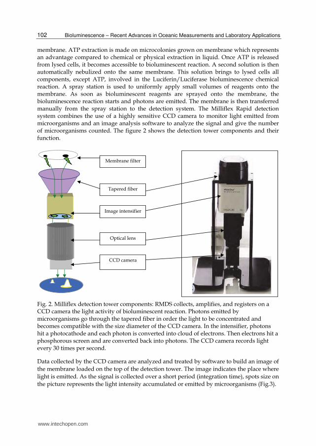
membrane. ATP extraction is made on microcolonies grown on membrane which represents an advantage compared to chemical or physical extraction in liquid. Once ATP is released from lysed cells, it becomes accessible to bioluminescent reaction. A second solution is then automatically nebulized onto the same membrane. This solution brings to lysed cells all components, except ATP, involved in the Luciferin/Luciferase bioluminescence chemical reaction. A spray station is used to uniformly apply small volumes of reagents onto the membrane. As soon as bioluminescent reagents are sprayed onto the membrane, the bioluminescence reaction starts and photons are emitted. The membrane is then transferred manually from the spray station to the detection system. The Milliflex Rapid detection system combines the use of a highly sensitive CCD camera to monitor light emitted from microorganisms and an image analysis software to analyze the signal and give the number of microorganisms counted. The figure 2 shows the detection tower components and their function.
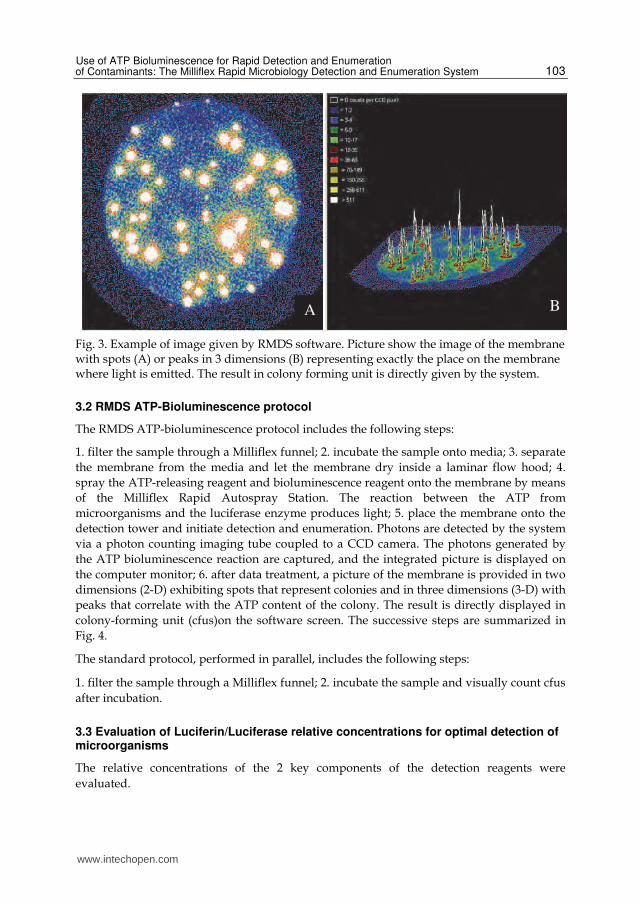
Fig. 2. Milliflex detection tower components: RMDS collects, amplifies, and registers on a CCD camera the light activity of bioluminescent reaction. Photons emitted by microorganisms go through the tapered fiber in order the light to be concentrated and becomes compatible with the size diameter of the CCD camera. In the intensifier, photons hit a photocathode and each photon is converted into cloud of electrons. Then electrons hit a phosphorous screen and are converted back into photons. The CCD camera records light every 30 times per second.
Data collected by the CCD camera are analyzed and treated by software to build an image of
the membrane loaded on the top of the detection tower. The image indicates the place where
light is emitted. As the signal is collected over a short period (integration time), spots size on
the picture represents the light intensity accumulated or emitted by microorganisms (Fig.3).
www.intechopen.com
Use of ATP Bioluminescence for Rapid Detection and Enumeration of Contaminants: The Milliflex Rapid Microbiology Detection and Enumeration System
103
Fig. 3. Example of image given by RMDS software. Picture show the image of the membrane with spots (A) or peaks in 3 dimensions (B) representing exactly the place on the membrane where light is emitted. The result in colony forming unit is directly given by the system.
3.2 RMDS ATP-Bioluminescence protocol
The RMDS ATP-bioluminescence protocol includes the following steps:
1. filter the sample through a Milliflex funnel; 2. incubate the sample onto media; 3. separate
the membrane from the media and let the membrane dry inside a laminar flow hood; 4.
spray the ATP-releasing reagent and bioluminescence reagent onto the membrane by means
of the Milliflex Rapid Autospray Station. The reaction between the ATP from
microorganisms and the luciferase enzyme produces light; 5. place the membrane onto the
detection tower and initiate detection and enumeration. Photons are detected by the system
via a photon counting imaging tube coupled to a CCD camera. The photons generated by
the ATP bioluminescence reaction are captured, and the integrated picture is displayed on
the computer monitor; 6. after data treatment, a picture of the membrane is provided in two
dimensions (2-D) exhibiting spots that represent colonies and in three dimensions (3-D) with
peaks that correlate with the ATP content of the colony. The result is directly displayed in
colony-forming unit (cfus)on the software screen. The successive steps are summarized in
Fig. 4.
The standard protocol, performed in parallel, includes the following steps:
1. filter the sample through a Milliflex funnel; 2. incubate the sample and visually count cfus
after incubation.
3.3 Evaluation of Luciferin/Luciferase relative concentrations for optimal detection of microorganisms
The relative concentrations of the 2 key components of the detection reagents were
evaluated.
A B
www.intechopen.com
Bioluminescence – Recent Advances in Oceanic Measurements and Laboratory Applications
104
Fig. 4. RMDS ATP-bioluminescence protocol
The protocol used is described in the previous paragraph “RMDS ATP bioluminescence protocol”. Only the reagent used for detection varies for the 2 components relative concentrations as described in table 1.
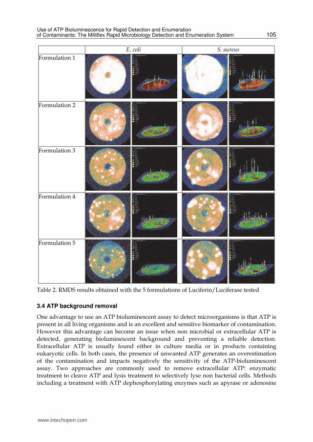
Formulation 1 Formulation 2 Formulation 3 Formulation 4 Formulation 5
Luciferase 3x 1.5x 1x 1.5x 1x
Luciferin 1x 1x 1x 0.5x 0.5x
Table 1. Formulations relative concentrations of Luciferin/Luciferase tested
The signal and background were determined using membranes incubated during 6h at
32.5°C on Tryptic Soy Agar inoculated with Escherichia coli or Staphylococcus aureus (table 2).
Formulation 1 gave a signal so strong that the detection system was almost saturated. This
saturation did not allow the accurate detection of bacteria on the membrane. The same issue
occurred to a weaker extent using formulation 2. On the other hand, while the detection of
S. aureus was accurate using formulation 5, the signal was too weak to allow all colonies of
E. coli to be counted. Formulations 3 and 4 were both able to generate a good signal
associated with low background. We can conclude from these results that the luciferin and
luciferase concentration can be increased to optimize the signal but also that the balance
between the 2 components is key. Signal will be increased while increasing concentrations
but background as well. Formulation 3 which benefits from the best signal on background
ratio has been used during the rest of the studies presented here. It is noticeable that
depending on the application, the type of sample tested and the resulting background, this
luciferase to luciferin balance can be adjusted to better match the detection criteria and
increase signal on background ratio.
Sample
Incubation on
growth medium Imaging
Results in
CFUs
90s
Spraying Membrane
Filtration
via Milliflex
www.intechopen.com
Use of ATP Bioluminescence for Rapid Detection and Enumeration of Contaminants: The Milliflex Rapid Microbiology Detection and Enumeration System
105
E. coli S. aureus
Formulation 1
Formulation 2
Formulation 3
Formulation 4
Formulation 5
Table 2. RMDS results obtained with the 5 formulations of Luciferin/Luciferase tested
3.4 ATP background removal
One advantage to use an ATP bioluminescent assay to detect microorganisms is that ATP is present in all living organisms and is an excellent and sensitive biomarker of contamination. However this advantage can become an issue when non microbial or extracellular ATP is detected, generating bioluminescent background and preventing a reliable detection. Extracellular ATP is usually found either in culture media or in products containing eukaryotic cells. In both cases, the presence of unwanted ATP generates an overestimation of the contamination and impacts negatively the sensitivity of the ATP-bioluminescent assay. Two approaches are commonly used to remove extracellular ATP: enzymatic treatment to cleave ATP and lysis treatment to selectively lyse non bacterial cells. Methods including a treatment with ATP dephosphorylating enzymes such as apyrase or adenosine
www.intechopen.com
Bioluminescence – Recent Advances in Oceanic Measurements and Laboratory Applications
106
phosphatase, have been described and used to remove efficiently ATP (Askgaard et al., 1995; Thore et al., 1975). Combination of apyrase and adenosine phosphate deaminase showed a good reduction of extracellular ATP and was applied to successfully detect E. coli and S. aureus in media broth and biological specimens (Sakakibara et al., 1997). When the objective of the assay is to detect and quantify bacterial contamination from a mixed population containing eukaryotic cells and bacteria, a differential lysis can be applied to selectively remove eukaryotic cells from the sample. This approach was used to separate bacterial ATP from biological fluids by lysing somatic cells with detergent as Triton X 100 at low concentration and combining this step with an enzymatic degradation of ATP released from lysed cells (Chapelle et al., 1978). RMDS protocol is based on sample filtration through membrane which naturally helps to eliminate extracellular ATP. If background ATP remains after filtration, rinsing the membrane with physiological serum or sterile water contributes to removal of residual ATP and allows bacterial detection. The figure 5 shows the impact of adding rinsing steps to reduce background on beverage products.
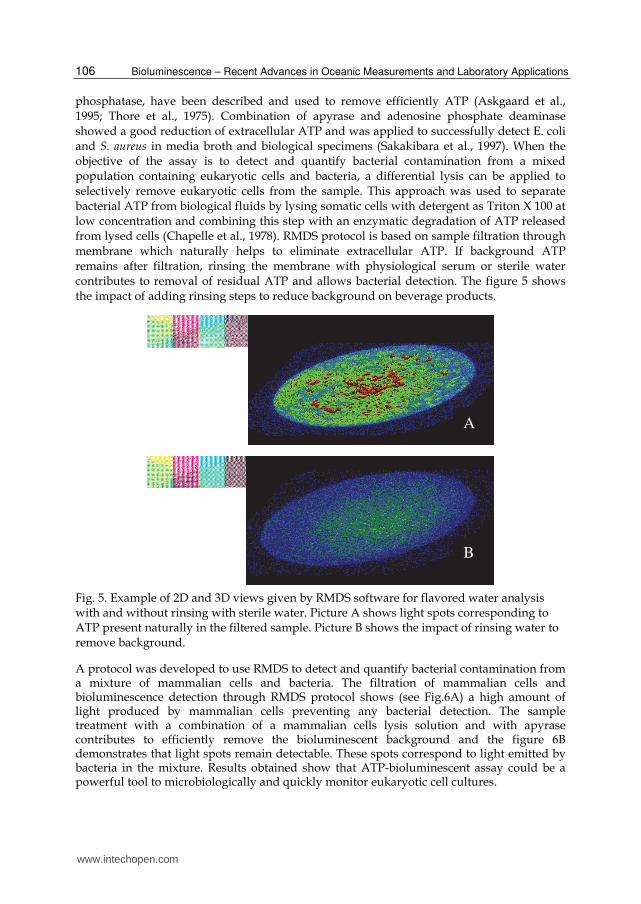
Fig. 5. Example of 2D and 3D views given by RMDS software for flavored water analysis with and without rinsing with sterile water. Picture A shows light spots corresponding to ATP present naturally in the filtered sample. Picture B shows the impact of rinsing water to remove background.
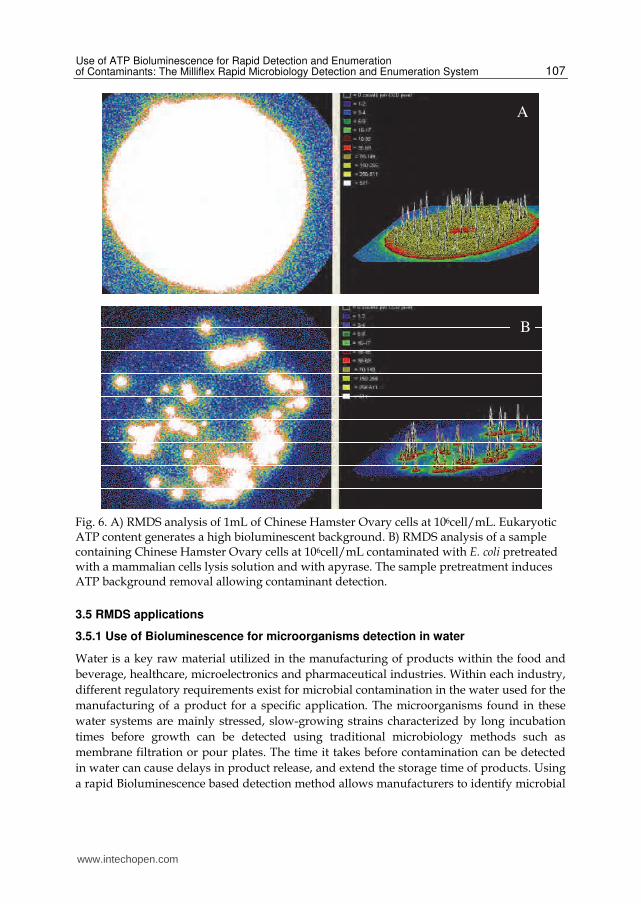
A protocol was developed to use RMDS to detect and quantify bacterial contamination from a mixture of mammalian cells and bacteria. The filtration of mammalian cells and bioluminescence detection through RMDS protocol shows (see Fig.6A) a high amount of light produced by mammalian cells preventing any bacterial detection. The sample treatment with a combination of a mammalian cells lysis solution and with apyrase contributes to efficiently remove the bioluminescent background and the figure 6B demonstrates that light spots remain detectable. These spots correspond to light emitted by bacteria in the mixture. Results obtained show that ATP-bioluminescent assay could be a powerful tool to microbiologically and quickly monitor eukaryotic cell cultures.
A
B
www.intechopen.com
Use of ATP Bioluminescence for Rapid Detection and Enumeration of Contaminants: The Milliflex Rapid Microbiology Detection and Enumeration System
107
Fig. 6. A) RMDS analysis of 1mL of Chinese Hamster Ovary cells at 106cell/mL. Eukaryotic ATP content generates a high bioluminescent background. B) RMDS analysis of a sample containing Chinese Hamster Ovary cells at 106cell/mL contaminated with E. coli pretreated with a mammalian cells lysis solution and with apyrase. The sample pretreatment induces ATP background removal allowing contaminant detection.
3.5 RMDS applications
3.5.1 Use of Bioluminescence for microorganisms detection in water
Water is a key raw material utilized in the manufacturing of products within the food and
beverage, healthcare, microelectronics and pharmaceutical industries. Within each industry,
different regulatory requirements exist for microbial contamination in the water used for the
manufacturing of a product for a specific application. The microorganisms found in these
water systems are mainly stressed, slow-growing strains characterized by long incubation
times before growth can be detected using traditional microbiology methods such as
membrane filtration or pour plates. The time it takes before contamination can be detected
in water can cause delays in product release, and extend the storage time of products. Using
a rapid Bioluminescence based detection method allows manufacturers to identify microbial
A
B
www.intechopen.com
Bioluminescence – Recent Advances in Oceanic Measurements and Laboratory Applications
108
contamination earlier, which provides them with better process control, product yield, and
shortens time to market.
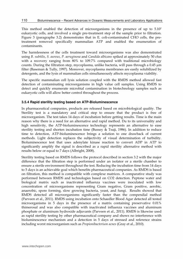
The following table 3 provides the incubation times for detectable growth, by organism, for the traditional microbiology method and RMDS. The detection time is significantly reduced using RMDS. Detection of growth is on average 4.5 times faster than traditional microbiology, and up to 6 times faster for the very slow growers tested (Methylobacterium mesophilicum ATCC 29983, stressed strain of Methylobacterium and a mix of various slow-growing strains). RMDS allows for overnight detection of the industrial-stressed microorganisms tested. The incubation temperature also has an influence on time-to-result. Incubating at 25 °C showed that longer incubation times were required (data not shown). The mean recovery between RMDS and the traditional microbiology method in these experiments was 92.7%, which shows the equivalence of the two methods.
Microorganisms Traditionnal Microbiology 30°C
Milliflex Rapid Detection System 30°C
R2A PCA TSA R2A PCA TSA
ATCC Strains
P. aeruginosa ATCC 9207 1 day 1 day 1 day 9 hrs 9 hrs 9 hrs
M. mesophilicum ATCC 29983 6 days 6 days MNA 26 hrs 26 hrs MNA
E. coli ATCC 8739 1 day 1 day 1 day 6 hrs 6 hrs 6 hrs
B. cepacia ATCC 25416 ND ND 2 days ND ND 16 hrs
S. epidermidis ATCC 12228 ND ND 1 day ND ND 9 hrs
Industrial-Stressed Microorganisms
Mix of various slow-growing strains
6 days 6 days MNA 24 hrs 24 hrs MNA
Stressed strain of Methylobacterium
6 days 6 days MNA 24 hrs 24 hrs MNA
Environmental isolate of R. pickettii
2 days 2 days ND 11 hrs 11 hrs ND
MNA : Medium Not Appropriate for growth of microorganism, ND : Not Done
Table 3. Detection time of reference strains and water isolates in traditional method and RMDS using either R2A agar, Tryptic Soy Agar (TSA) or Plate Count Agar (PCA).
3.5.2 Rapid detection of spores
Spores are major food spoilages and are also a concern in pharmaceutical samples. The classical microbiological method to enumerate spore contamination combines heat shock and on average 5 days incubation into sterile and molten specific medium Agar (Wayne et al., 1990). The amount of ATP in spores is very low and germination is necessary to increase ATP content and develop a rapid detection method based on ATP-bioluminescence (Kodata et al., 1996). ATP-bioluminescence rapid screening assay has been described showing that after germination, spore containing powder has been detected in a short time with a detection limit of 100 spores (Lee & Deininger, 2004). Fujinami et al (2004) also showed that short incubation of the sample in nutrient broth medium containing L-alanine increased RLU from spores and optimize the ATP- bioluminescent assay.
www.intechopen.com
Use of ATP Bioluminescence for Rapid Detection and Enumeration of Contaminants: The Milliflex Rapid Microbiology Detection and Enumeration System
109
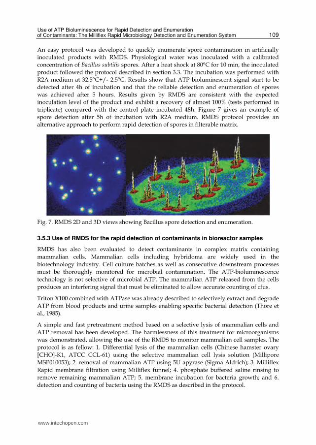
An easy protocol was developed to quickly enumerate spore contamination in artificially
inoculated products with RMDS. Physiological water was inoculated with a calibrated
concentration of Bacillus subtilis spores. After a heat shock at 80°C for 10 min, the inoculated
product followed the protocol described in section 3.3. The incubation was performed with
R2A medium at 32.5°C+/- 2.5°C. Results show that ATP bioluminescent signal start to be
detected after 4h of incubation and that the reliable detection and enumeration of spores
was achieved after 5 hours. Results given by RMDS are consistent with the expected
inoculation level of the product and exhibit a recovery of almost 100% (tests performed in
triplicate) compared with the control plate incubated 48h. Figure 7 gives an example of
spore detection after 5h of incubation with R2A medium. RMDS protocol provides an
alternative approach to perform rapid detection of spores in filterable matrix.
Fig. 7. RMDS 2D and 3D views showing Bacillus spore detection and enumeration.
3.5.3 Use of RMDS for the rapid detection of contaminants in bioreactor samples
RMDS has also been evaluated to detect contaminants in complex matrix containing
mammalian cells. Mammalian cells including hybridoma are widely used in the
biotechnology industry. Cell culture batches as well as consecutive downstream processes
must be thoroughly monitored for microbial contamination. The ATP-bioluminescence
technology is not selective of microbial ATP. The mammalian ATP released from the cells
produces an interfering signal that must be eliminated to allow accurate counting of cfus.
Triton X100 combined with ATPase was already described to selectively extract and degrade
ATP from blood products and urine samples enabling specific bacterial detection (Thore et
al., 1985).
A simple and fast pretreatment method based on a selective lysis of mammalian cells and
ATP removal has been developed. The harmlessness of this treatment for microorganisms
was demonstrated, allowing the use of the RMDS to monitor mammalian cell samples. The
protocol is as fellow: 1. Differential lysis of the mammalian cells (Chinese hamster ovary
[CHO]-K1, ATCC CCL-61) using the selective mammalian cell lysis solution (Millipore
MSP010053); 2. removal of mammalian ATP using 5U apyrase (Sigma Aldrich); 3. Milliflex
Rapid membrane filtration using Milliflex funnel; 4. phosphate buffered saline rinsing to
remove remaining mammalian ATP; 5. membrane incubation for bacteria growth; and 6.
detection and counting of bacteria using the RMDS as described in the protocol.
www.intechopen.com
Bioluminescence – Recent Advances in Oceanic Measurements and Laboratory Applications
110
This method enabled the detection of microorganisms in the presence of up to 5.107 eukaryotic cells, and involved a single pre-treatment step of the sample prior to filtration. Figure 3 (paragraphe 3.2) demonstrates that in E. coli-contaminated CHO cells, the pre-treatment removed specifically mammalian ATP and enabled the enumeration of contaminants.
The harmlessness of the cells treatment toward microorganisms was also demonstrated using B. subtilis, S. aureus, P. aeruginosa and Candida albicans spiked at approximately 50 cfus with a recovery ranging from 80% to 109.7% compared with traditional microbiology counts. During the filtration step, mycoplasma, unlike bacteria, will pass through a 0.45 µm filter (Baseman & Tully, 1997). Moreover, mycoplasma membranes are easily solubilized by detergents, and the lysis of mammalian cells simultaneously affects mycoplasma viability.
The specific mammalian cell lysis solution coupled with the RMDS method allowed fast detection of contaminating microorganisms in high value cell samples. Using RMDS to detect and quickly enumerate microbial contamination in biotechnology samples such as eukaryotic cells will allow better control throughout the process.
3.5.4 Rapid sterility testing based on ATP-Bioluminescence
In pharmaceutical companies, products are released based on microbiological quality. The Sterility test is a mandatory and critical step to ensure that the product is free of microorganism. The test takes 14 days of incubation before getting results. Time is the main reason why there is a need for an alternative and rapid method. Du to its universality and high sensitivity, the ATP-bioluminescence technology represents an alternative to ease sterility testing and shorten incubation time (Bussey & Tsuji, 1986). In addition to reduce time to detection, ATP-bioluminescence brings a solution to one drawback of current methods. Light detection replaces the subjectivity of visual determination of turbidity. Bioluminescence test that uses adenylate kinase reaction to convert ADP in ATP to significantly amplify the signal is described as a rapid sterility alternative method with results below or equal to 7 days (Albright, 2008).
Sterility testing based on RMDS follows the protocol described in section 3.2 with the major difference that the filtration step is performed under an isolator or a sterile chamber to ensure a sterile environment throughout the test. Reducing the incubation time from 14 days to 5 days is an achievable goal which benefits pharmaceutical companies. As RMDS is based on filtration, this method is compatible with complexe matrices. A comparative study was performed between RMDS and technologies based on CO2 detection. Peptone water and biological matrix such as inactivated influenza vaccines were inoculated with low concentration of microorganisms representing Gram negative, Gram positive, aerobic, anaerobic, spore forming, slow growing bacteria, yeast, and fungi. Results showed that RMDS detected all microorganisms significantly faster than the compendial method (Parveen et al., 2011). RMDS using incubation onto Schaedler Blood Agar detected all tested microorganisms in 5 days in the presence of a matrix containing preservative 0.01% thimerosal and was also compatible with inactivated influenza vaccines and aluminum phosphate or aluminum hydroxide adjuvants (Parveen et al., 2011). RMDS is likewise used as rapid sterility testing by other pharmaceutical company and shows no interference with bioluminescence mechanism and a detection in 5 days of stressed and reference strains including worst microorganism such as Propionibacterium acnes (Gray et al., 2010).
www.intechopen.com
Use of ATP Bioluminescence for Rapid Detection and Enumeration of Contaminants: The Milliflex Rapid Microbiology Detection and Enumeration System
111
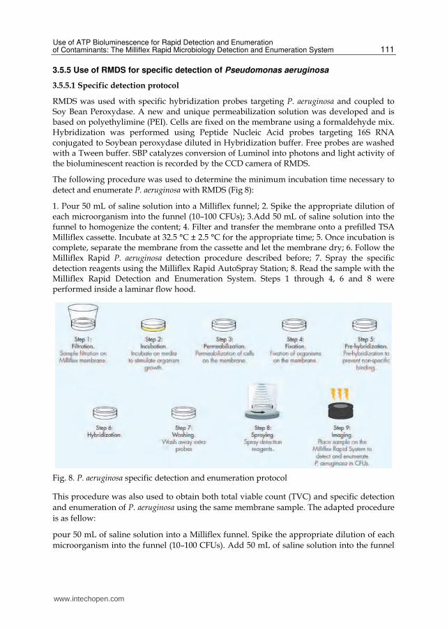
3.5.5 Use of RMDS for specific detection of Pseudomonas aeruginosa
3.5.5.1 Specific detection protocol
RMDS was used with specific hybridization probes targeting P. aeruginosa and coupled to Soy Bean Peroxydase. A new and unique permeabilization solution was developed and is based on polyethylimine (PEI). Cells are fixed on the membrane using a formaldehyde mix. Hybridization was performed using Peptide Nucleic Acid probes targeting 16S RNA conjugated to Soybean peroxydase diluted in Hybridization buffer. Free probes are washed with a Tween buffer. SBP catalyzes conversion of Luminol into photons and light activity of the bioluminescent reaction is recorded by the CCD camera of RMDS.
The following procedure was used to determine the minimum incubation time necessary to
detect and enumerate P. aeruginosa with RMDS (Fig 8):
1. Pour 50 mL of saline solution into a Milliflex funnel; 2. Spike the appropriate dilution of each microorganism into the funnel (10–100 CFUs); 3.Add 50 mL of saline solution into the funnel to homogenize the content; 4. Filter and transfer the membrane onto a prefilled TSA Milliflex cassette. Incubate at 32.5 °C ± 2.5 °C for the appropriate time; 5. Once incubation is complete, separate the membrane from the cassette and let the membrane dry; 6. Follow the Milliflex Rapid P. aeruginosa detection procedure described before; 7. Spray the specific detection reagents using the Milliflex Rapid AutoSpray Station; 8. Read the sample with the Milliflex Rapid Detection and Enumeration System. Steps 1 through 4, 6 and 8 were performed inside a laminar flow hood.
Fig. 8. P. aeruginosa specific detection and enumeration protocol
This procedure was also used to obtain both total viable count (TVC) and specific detection
and enumeration of P. aeruginosa using the same membrane sample. The adapted procedure
is as fellow:
pour 50 mL of saline solution into a Milliflex funnel. Spike the appropriate dilution of each
microorganism into the funnel (10–100 CFUs). Add 50 mL of saline solution into the funnel
www.intechopen.com
Bioluminescence – Recent Advances in Oceanic Measurements and Laboratory Applications
112
to homogenize the content. Filter and transfer the membrane onto a pre-filled TSA Milliflex
cassette. Incubate at 32.5 °C ± 2.5 °C for the appropriate time. Once incubation is complete,
separate the membrane from the cassette and let the membrane dry. Spray the ATP
releasing and bioluminescence reagents using the Milliflex Rapid AutoSpray Station. Read
the sample with the RMDS. Then, follow the Milliflex Rapid P. aeruginosa detection
procedure starting from fixation step. Spray the specific detection reagents using the
Milliflex Rapid AutoSpray Station and read the sample with the Milliflex Rapid Detection
and Enumeration System.
3.5.5.2 Specific Pseudomonas aeruginosa detection and total viable count results
The Milliflex Rapid system is a proven automated solution for the rapid detection and
enumeration of total viable count (TVC) in purified water and Water For Injection. Based on
membrane filtration and image analysis together with an adenosine triphosphate (ATP)
bioluminescence reagent, the Milliflex Rapid System delivers TVC test results faster than
traditional methods. We have developed a hybridization assay that enables the Milliflex
Rapid system to specifically detect and enumerate P. aeruginosa. The hybridization assay is
performed with a peroxidase-conjugated DNA-oligonucleotide probe targeted to a specific
RNA-sequence of P. aeruginosa. Applying luminol and peroxide substrates to the membrane
filtration sample generates light that is detected by the Milliflex Rapid system.
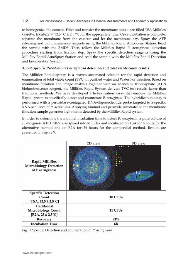
In order to determine the minimal incubation time to detect P. aeruginosa, a pure culture of
P. aeruginosa ATCC 9027 was spiked into Milliflex and incubated on TSA for 6 hours for the
alternative method and on R2A for 24 hours for the compendial method. Results are
presented in Figure 9.
Rapid Milliflex Microbiology Detection
of P.aeruginosa
2D view 3D view
Specific Detection
Count [TSA, 32.5 ± 2.5°C]
10 CFUs
Traditional Microbiology Count
[R2A, 25 ± 2.5°C] 11 CFUs
Recovery 91%
Incubation Time 6h
Fig. 9. Specific Detection and enumeration of P. aeruginosa
www.intechopen.com
Use of ATP Bioluminescence for Rapid Detection and Enumeration of Contaminants: The Milliflex Rapid Microbiology Detection and Enumeration System
113
Using the specific detection procedure described above P. aeruginosa was detected and enumerated in 8 hours in a water sample. The specificity of the method has been assessed against numerous microorganisms and only P. aeruginosa was detected in this panel of contaminants. The limit of the sensitivity is 1 CFU (data not shown).
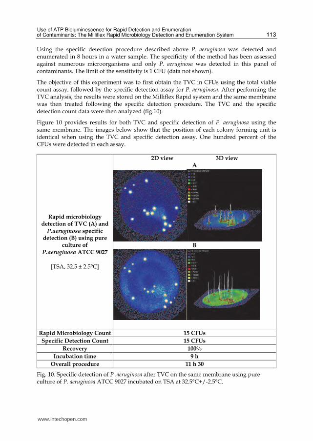
The objective of this experiment was to first obtain the TVC in CFUs using the total viable count assay, followed by the specific detection assay for P. aeruginosa. After performing the TVC analysis, the results were stored on the Milliflex Rapid system and the same membrane was then treated following the specific detection procedure. The TVC and the specific detection count data were then analyzed (fig.10).
Figure 10 provides results for both TVC and specific detection of P. aeruginosa using the same membrane. The images below show that the position of each colony forming unit is identical when using the TVC and specific detection assay. One hundred percent of the CFUs were detected in each assay.
Rapid microbiology detection of TVC (A) and
P.aeruginosa specific detection (B) using pure
culture of P.aeruginosa ATCC 9027
[TSA, 32.5 ± 2.5°C]
2D view 3D view A
B
Rapid Microbiology Count 15 CFUs
Specific Detection Count 15 CFUs
Recovery 100%
Incubation time 9 h
Overall procedure 11 h 30
Fig. 10. Specific detection of P .aeruginosa after TVC on the same membrane using pure culture of P. aeruginosa ATCC 9027 incubated on TSA at 32.5°C+/-2.5°C.
www.intechopen.com
Bioluminescence – Recent Advances in Oceanic Measurements and Laboratory Applications
114
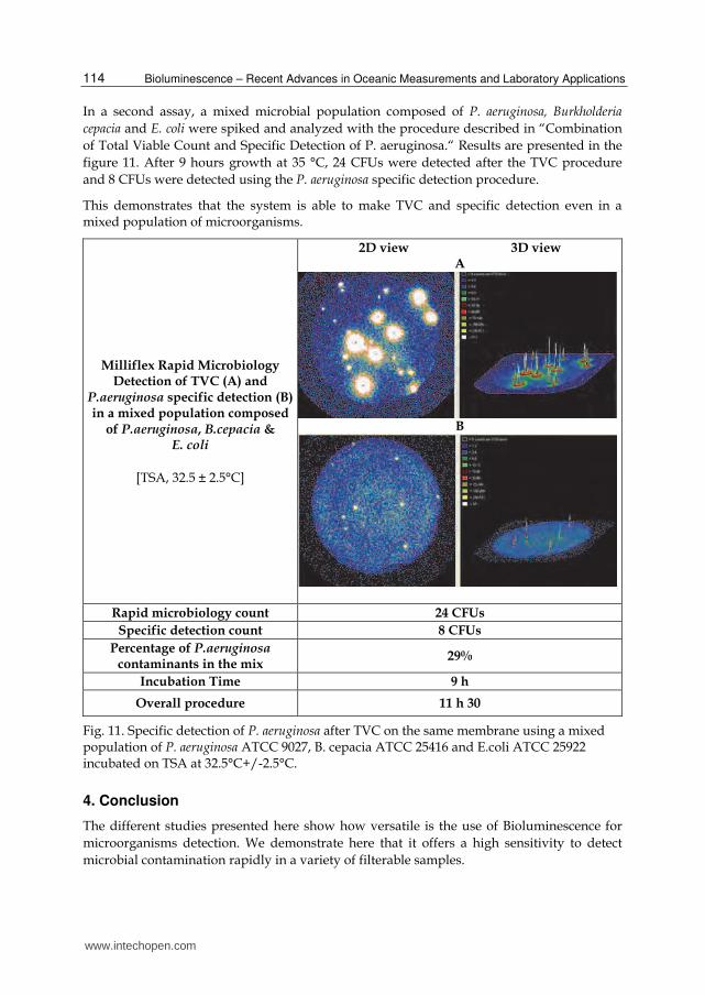
In a second assay, a mixed microbial population composed of P. aeruginosa, Burkholderia
cepacia and E. coli were spiked and analyzed with the procedure described in “Combination
of Total Viable Count and Specific Detection of P. aeruginosa.“ Results are presented in the
figure 11. After 9 hours growth at 35 °C, 24 CFUs were detected after the TVC procedure
and 8 CFUs were detected using the P. aeruginosa specific detection procedure.
This demonstrates that the system is able to make TVC and specific detection even in a mixed population of microorganisms.
Milliflex Rapid Microbiology Detection of TVC (A) and
P.aeruginosa specific detection (B) in a mixed population composed
of P.aeruginosa, B.cepacia & E. coli
[TSA, 32.5 ± 2.5°C]
2D view 3D view A
B
Rapid microbiology count 24 CFUs
Specific detection count 8 CFUs
Percentage of P.aeruginosa contaminants in the mix
29%
Incubation Time 9 h
Overall procedure 11 h 30
Fig. 11. Specific detection of P. aeruginosa after TVC on the same membrane using a mixed population of P. aeruginosa ATCC 9027, B. cepacia ATCC 25416 and E.coli ATCC 25922 incubated on TSA at 32.5°C+/-2.5°C.
4. Conclusion
The different studies presented here show how versatile is the use of Bioluminescence for
microorganisms detection. We demonstrate here that it offers a high sensitivity to detect
microbial contamination rapidly in a variety of filterable samples.
www.intechopen.com
Use of ATP Bioluminescence for Rapid Detection and Enumeration of Contaminants: The Milliflex Rapid Microbiology Detection and Enumeration System
115
The association of Bioluminescence to sensitive sensors such as RMDS provides a result in colony forming units equivalent to the standard plate count but is 4 times faster than classical microbiology. This method can be used in samples from industrial water, to food and beverage samples for the detection of any type of bacteria, yeasts and molds including spores. We also showed that it can be used to detect bacterial contamination in cell culture matrices containing high concentrations of eukaryotic cells.
Interestingly, Bioluminescence was also coupled to molecular biology through the use of 16S RNA probes for specific detection of bacteria. The example presented here allowed not only the detection of P. aeruginosa but also the total viable count using Luciferin and luciferase followed by specific detection of this very specific bacterium.
Finally, the development of the method in a pharmaceutical environment allowed sterility testing of drug products 3 times faster than the compendial method. This recent developments in the pharmaceutical field show that the method is also able to help patients taking drugs usually associated with a very short shelf life (gene therapy products, cell therapies...) as the result is delivered before the injection of the product while the traditional systems usually deliver after the treatment.
In conclusion, the use of Bioluminescence either in its “classical” or molecular format allows for a number of developments in the field of microorganisms detection. The flexibility of the method and its ease of use coupled to the considerable savings in time compared to the traditional method make it a valuable tool for life scientists as well as for other clinical applications.
5. Acknowledgment
Authors would like to thanks colleagues from Merck-Millipore Application group, Development group and Predevelopment - Technology – Collaboration for their technical collaboration. The research described in this paper was carried out at the Merck-Millipore R&D laboratory (Molsheim, France).
6. References
Albright, J. (2009). Implementing Rapid Sterility using the Celsis Enhanced ATP Bioluminescence Test.
www.celsis.com/media/pdf/rdpdfs/Poster_RapidSterilityTesting_PDA0904.pdf Andreotti, P. E. & Berthold, F. (1999). Application of a new high sensitivity luminometer for
industrial microbiology and molecular biology. Luminescence, 14(1), 19-22. Askgaard, D. S.; Gottschau, A; Knudsen, K. & Bennedsen, J. (1995). Firefly luciferase assay of
adenosine triphosphate as a tool of quantitation of the viability of BCG vaccines. Biologicals, 23(1), 55-60.
Aycicek, K., Oguz, U. & Karci, K. (2006). Comparison of results of ATP bioluminescence and traditional hygiene swabbing methods for the determination of surface cleanliness at a hospital kitchen. International Journal of Hygiene and Environmental Health, 209(2), 203-206.
Baseman, J. B. & Tully, J.G. (1997). Mycoplasmas: Sophisticated, reemerging and burdened by their notoriety. Emerging Infectious Disease, 3, 21-32.
www.intechopen.com
Bioluminescence – Recent Advances in Oceanic Measurements and Laboratory Applications
116
Bautisda, D. A.; Vaillancourt, J. P., Clarke, R. A. ; Renwick S. & Griffiths M. W. (1995). Rapid assessment of the microbiological quality of poultry carcasses using ATP-bioluminescence. Journal of Food Protection, 58, 551-554.
Branchini, B. R.; Murtiashaw, M. H.; Magyar, R. A. & Anderson, S. M. (2000). The role of lysine 529, a conserved residue of the acyl-adenylate-forming enzyme superfamily, in firefly luciferase. Biochemistry, 39, 5433–5440.
Branchini, B. R.; Southworth, T. L.; Khattak, N. F.; Michelini, E. & Roda, A. (2005). Red- and green-emitting firefly luciferase mutants for bioluminescent reporter applications. Analytical Biochemistry, 345, 140–148.
Bussey, D. M. & Tsuji, K. (1986). Bioluminescence for USP sterility testing of pharmaceutical suspension products. Applied Environmental Microbiology, 51(2), 349-355.
Chapelle, E. W.; Picciolo, G.L. & Deming, J.W. (1978). Determination of bacterial contents in fluid. Methods in Enzymology. 57, 65-72.
Chen, F. C. & Godwin, S. L. (2006). Comparison of a rapid ATP bioluminescence assay and standard plate count methods for assessing microbial contamination of consumers' refrigerators. Journal of Food Protection®, 69(10), 2534-2538.
Conn, R. B.; Charache, P. & Chapelle, E. W. (1975). Limits of applicability of the firefly luminescence ATP assay for the detection of bacteria in clinical specimens. American Journal of Clinical Pathology, 63, 493-501.
Conti, E.; Franks, N. P. & Brick, P. (1996) Crystal structure of firefly luciferase throws light on a superfamily of adenylate-forming enzymes. Structure, 4, 287–298.
Davidson, C. A.; Griffith, C. J.; Peters, A. C. & Fielding, L. M. (1999). Evaluation of two methods for monitoring surface cleanliness-ATP bioluminescence and traditional hygiene swabbing. Luminescence, 14(1), 33-38.
DeLuca, M. & McElroy, W. D. (1974). Kinetics of the firefly luciferase catalyzed reactions. Biochemistry, 13, 921–925.
Deininger, R. A. & Lee, J. Y. (2001). Rapid determination of bacteria in drinking water using an ATP assay. Field Analytical Chemistry & Technology, 5(4), 185-189.
Dostalek, P. & Branyik T. (2005). Prospects for rapid bioluminescent detection methods in the food industry - a review. Czech Journal of Food Sciences, 23(3), 85-92.
Francis, K. P.; Joh, D.; Bellinger-Kawahara, C.; Hawkinson, M. J.; Purchio, T. F. & Contag, P. R. (2000). Monitoring bioluminescent Staphylococcus aureus infections in living mice using a novel luxABCDE construct. Infection. Immunity, 68, 3594–3600.
Frundzhyan, V. & Ugarova, N. (2007). Bioluminescent assay of total bacterial contamination of drinking water. Luminescence, 22(3); 241-244.
Fujinami, Y.; Kataoka, M.; Matsushita, K.; Sekigushi, H.; Itoi, T.; Tsuge, K. & Seto, Y. (2004). Sensitive Detection of Bacteria and Spores Using a Portable Bioluminescence ATP Measurement. Assay System Distinguishing from White Powder Materials. Journal of Health science, 50, 126-132.
Girotti, S.; Ferri, E.N.; Fini, F.; Righetti, S.; Bolelli, L.; Budini, R.; Lasi, G.; Roubal, P.; Fukal, L.; Hochel, I. & Rauch, P. (1997). Determination of microbial contamination in milk by ATP assay. Czech Journal of Food Science, 15, 241-248.
Gray, J. C.; Steark, A.; Berchtold, M.; Mercier, M.; Neuhaus, G. & Wirth A. (2010). Introduction of a Rapid Microbiological Method as an Alternative to the Pharmacopoeial Method for the Sterility test. American Pharmaceutical Review. 13(6), 88-94.
www.intechopen.com
Use of ATP Bioluminescence for Rapid Detection and Enumeration of Contaminants: The Milliflex Rapid Microbiology Detection and Enumeration System
117
Hawronskyj, J. M. & Holah, J. (1997) ATP: a universal hygiene monitor. Trends in Food Science & Technology, 8, 79–84.
Hosseinkhani, S. (2011). Molecular enigma of multicolor bioluminescence of firefly luciferase. Cellular and Molecular Life Sciences, 68(7),1167-1182.
Kodaka, H.; Fukuda, K.; Mizuochi, S. & Horigome, K. (1996). Adenosine Triphosphate Content of Microorganisms Related with food Spoilage. Japanese Journal of Food Microbiology, 13, 29-34.
Kolbeck, J. C.; Padgett, R. A.; Estevez, E. G. & Harell, L. J. (1985). Bioluminescence screening for bacteriuria. Journal of clinical Microbiology, 21, 527-530.
McElroy, W. D. (1947). The energy source for bioluminescence in an isolated system. Proceedings of the National Academy of Sciences USA, 33, 342–345.
McElroy, W. D. (1951). Properties of the reaction utilizing adenosinetriphosphate for bioluminescence. Journal of Biological Chemistry, 191, 547–557.
McElroy, W., D., Hastings, J., W.; Coulombre, J. & Sonnenfeld, V. (1953). The mechanism of action of pyrophosphate in firefly luminescence. Archives of Biochemistry and Biophysics, 46, 399–416.
Miller, J. N.; Nawawi, M. B. & Burgess, C. (1992). Detection of bacterial ATP by reversed flow-injection analysis with luminescence detection. Analytica Chimica Acta, 266, 339-343.
Nakatsu, T.; Ichiyama, S.; Hiratake, J.; Saldanha, A.; Kobashi, N.; Sakata, K. & Kato, H. (2006). Structural basis for the spectral difference in luciferase bioluminescence. Nature, 440, 372–376.
Nielsen, P. & Van Dellen, E. J. (1989). Rapid bacteriological screening of cosmetic raw materials by using bioluminescence. Association of Official Analytical Chemists, 72(5), 708-711.
Parveen S.; Kaur S.; David S. A.; Kenney J. L.; McCormick W. M. & Gupta R.K. (2011). Evaluation of growth based rapid microbiological methods for sterility testing of vaccines and other biological products. Vaccine.
Poulis, J. A. ; Phper, M. & Mossel, D. A. A. (1993). Assessment of cleaning and desinfection in the food industry with the rapid ATP-bioluminescence technique combined with the tissue fluid contamination test and a conventional microbiological method. International Journal of food Microbiology, 20, 109-116.
Roda, A.; Pasini, P.; Mirasoli, M.; Michelini, E. & M. Guardigli. (2004). Biotechnological applications of bioluminescence and chemiluminescence. Trends in Biotechnology, 22, 295-303.
Sakakibara, T.; Murakami, S.; Hattori, N.; Nakajima, M. & Imai, K. (1997). Enzymatic treatment to eliminate the extracellular ATP for improving the detectability of bacterial intracellular ATP. Analytical Biochemistry, 250(2), 157-161.
Selan, L.; Berlutti, F.; Passariello, C.; Thaller, M. C. & Renzini, G. (1992). Reliability of a bioluminescence ATP assay for detection of bacteria. Journal of Clinical Microbiology, 30, 1739-1742.
Seliger, H. H. (1989). Some reflections on McElroy and bioluminescence. Journal of Bioluminescence and Chemiluminescence, 4(1), 26-28.
Seliger, H. H.; Buck, J. B.; Fastie, W. G. & McElroy, W. D. (1964). The spectral distribution of firefly light. Journal of General Physiology, 48, 95–104.
www.intechopen.com
Bioluminescence – Recent Advances in Oceanic Measurements and Laboratory Applications
118
Seliger, H. H. & McElroy, W. D. (1964). The colors of firefly bioluminescence: enzyme configuration and species specificity. Proceedings of the National Academy of Sciences USA, 52, 75–81.
Shapiro, E.; Lu, C. & Baneyx, F. (2005). A set of multicolored Photinus pyralis luciferase mutants for in vivo bioluminescence applications. PEDS 18 (12), 581-587.
Siro, M-R.; Romar, H. & Lövgren, T. (1982). Continuous flow method for extraction and bioluminescence assay of ATP in baker's yeast. Applied Microbiology and Biotechnology, 15, 258-264.
Thompson, J. F.; Geoghegan, K. F.; Lloyd, D. B.; Lanzetti, A. J.; Magyar, R. A.; Anderson, S. M., & Branchini, B. R. (1997). Mutation of a protease-sensitive region in firefly luciferase alters light emission properties. Journal of Biological Chemistry, 272, 18766–18771.
Thorne, N; Inglese, J. & Auld, D. S. (2010). Illuminating insights into firefly luciferase and other bioluminescent reporters used in chemical biology. Chemistry & Biology, 17(6), 646-657.
Thore, A.; Ansehn,S.; Lundin, A. & Bergman, S. (1975). Detection of bacteria by luciferase assay of adenosine triphosphate. Journal of Clinical Microbioliology, 1, 1-8.
Venkateswaran, K.; Hattori, N.; La Duc, M.T. & Kern, R. (2003). ATP as a biomarker of viable microorganisms in clean-room facilities. Journal of Microbiological Methods, 52(3), 367-377.
Wilson, T. & Hastings, J.W. (1998). Bioluminescence. Annual Review of Cell and Developmental biology, 14, 197-230.
Yan, S. L.; Miao, S. N.; Deng, S. Y.; Zou, M. J.; Zhong, F. S.; Huang, W. B.; Pan, S. Y. & Wang, Q. Z. (2011). ATP bioluminescence rapid detection of total viable count in soy sauce. Luminescence.
Zako, T.; Ayabe, K.; Aburatani, T.; Kamiya, N.; Kitayama, A.; Ueda, H. & Nagamune, T. (2003). Luminescent and substrate binding activities of firefly luciferase N-terminal domain. Biochimica et Biophysica Acta - Proteins & Proteomics, 1649, 183–189.
www.intechopen.com
Bioluminescence - Recent Advances in Oceanic Measurementsand Laboratory ApplicationsEdited by Dr. David Lapota
ISBN 978-953-307-940-0Hard cover, 190 pagesPublisher InTechPublished online 01, February, 2012Published in print edition February, 2012
InTech EuropeUniversity Campus STeP Ri Slavka Krautzeka 83/A 51000 Rijeka, Croatia Phone: +385 (51) 770 447 Fax: +385 (51) 686 166www.intechopen.com
InTech ChinaUnit 405, Office Block, Hotel Equatorial Shanghai No.65, Yan An Road (West), Shanghai, 200040, China
Phone: +86-21-62489820 Fax: +86-21-62489821
We now find ourselves utilizing luciferase - luciferin proteins, ATP, genes and the whole complex of theseinteractions to observe and follow the progress or inhibition of tumors in animal models by measuringbioluminescence intensity, spatially and temporally using highly sophisticated camera systems. This bookdescribes applications in preclinical oncology research by bioluminescence imaging (BLI) with a variety ofapplications. Chapters describe current methodologies for rapid detection of contaminants using the Milliflexsystem, and the use of bioluminescence resonance energy transfer (BRET) technology for monitoring physicalinteractions between proteins in living cells. Others are using bioluminescent proteins for high sensitive opticalreporters imaging in living animals, developing pH-tolerant luciferase for brighter in vivo imaging, andoscillation characteristics in bacterial bioluminescence. The book also contains descriptions of the long-termseasonal characteristics of oceanic bioluminescence and the responsible planktonic species producingbioluminescence. Such studies are few and rare.
How to referenceIn order to correctly reference this scholarly work, feel free to copy and paste the following:
Renaud Chollet and Sébastien Ribault (2012). Use of ATP Bioluminescence for Rapid Detection andEnumeration of Contaminants: The Milliflex Rapid Microbiology Detection and Enumeration System,Bioluminescence - Recent Advances in Oceanic Measurements and Laboratory Applications, Dr. David Lapota(Ed.), ISBN: 978-953-307-940-0, InTech, Available from: http://www.intechopen.com/books/bioluminescence-recent-advances-in-oceanic-measurements-and-laboratory-applications/use-of-atp-bioluminescence-for-rapid-detection-and-enumeration-of-contaminants-the-milliflex-rapid-m