universidad de murcia - digitum.um.es lópez... · a mi tíos mª sol y tomás por su cariño y...
TRANSCRIPT
UNIVERSIDAD DE MURCIA
FACULTAD DE VETERINARIA
Selección de los espermatozoides en la especie porcina: relación con su funcionalidad y capacidad
de fecundación in vitro
Spermatozoa selection in porcine species: relation with their functionality and in vitro fertilization
competence
Dña. Rebeca López Úbeda 2015

Dra. Carmen Matás Parra Facultad de Veterinaria
Departamento de Fisiología
Email: [email protected] Campus Universitario de Espinardo. 30.100 Murcia
T. 868 88 7256 – F. 868 88 4147 – www.um.es/grupo-fisiovet
Dña. CARMEN MATÁS PARRA, Profesora Titular del Departamento de Fisiología
AUTORIZA:
La presentación de la Tesis Doctoral titulada ‘‘Selección de los espermatozoides en la
especie porcina: relación con su funcionalidad y capacidad de fecundación in vitro”,
realizada por Dña. Rebeca López Úbeda, bajo su inmediata dirección y supervisión, y
que presenta para la obtención del grado de Doctor por la Universidad de Murcia.
Murcia, a 22 de Julio de 2015.
Fdo. Carmen Matás Parra

Dr. Francisco Alberto García Vázquez Facultad de Veterinaria
Departamento de Fisiología
Email: [email protected] Campus Universitario de Espinardo. 30100 Murcia
T. 868 88 8009 – F. 868 88 4147 – www.um.es/grupo-fisiovet
D. FRANCISCO ALBERTO GARCÍA VÁZQUEZ, Profesor Contratado Doctor del
Departamento de Fisiología
AUTORIZA:
La presentación de la Tesis Doctoral titulada ‘‘Selección de los espermatozoides en la
especie porcina: relación con su funcionalidad y capacidad de fecundación in vitro”,
realizada por Dña. Rebeca López Úbeda, bajo su inmediata dirección y supervisión, y
que presenta para la obtención del grado de Doctor por la Universidad de Murcia.
Murcia, a 22 de Julio de 2015.
Fdo. Francisco Alberto García Vázquez

Funding
The experimental part of this thesis has been
supported by the Spanish Ministry of Economy
and Competitiveness (MINECO) and the
European Regional Development Fund (FEDER),
Grant AGL2009‐12512‐C02‐01 and AGL2012‐
40180‐C03‐01 and by Fundación Séneca
08752/PI/08.

Acknowledgements/
Agradecimientos

Acknowledgements/Agradecimientos
Agradecimientos
La vida consiste en ir hacia delante quemando etapas, con esta memoria cierro una
etapa muy importante en mi vida durante la cual he tenido la suerte de conocer a
personas maravillosas y de la que me llevo grandes amigos.
Quiero dar las gracias a todos los componentes del departamento de Fisiología por
abrirme sus puertas y colaborar, cada uno a su manera, en esta tesis. Sin duda sois un
gran equipo científico, pero si en algo destacáis todos es en la gran valía personal.
Realmente espero que nuestro contacto no termine aquí y que podamos seguir
colaborando en el futuro.
A Carmen por aceptar iniciar conmigo esta dura e increíble etapa, por su constancia, su
apoyo, su inestimable ayuda y por creer en todo momento que esta tesis llegaría a su
fin. Gracias también por haberme dado la oportunidad de trabajar en “esto de la
reproducción”. Por último, gracias por tu paciencia en los momentos de desánimo y
por enseñarme otra forma de ver la vida.
A Fran al que estoy enormemente agradecida por haber aceptado formar parte de este
proyecto y ser un ejemplo de que con ganas las cosas se pueden hacer rápido y bien.
A Pilar por confiar en mí y darme la oportunidad de trabajar en cosas nuevas. A
Joaquín por sus consejos y soluciones, y por todos los momentos que le he robado con
mis consultas. A Raquel por su forma de ver las cosas, por tener siempre abiertas las
puertas de su despacho, por tratar de solucionarme cualquier problema en el
momento y hacerme ver que al final todo saldría bien (gracias Jefa!!). A Salvador por
hacer que muchas cosas funcionen desde “el fondo del pasillo”.
A nuestros técnicos (Juan, Sole y Darío) sin los que este trabajo no habría podido
realizarse, a Juan por prepararme numerosos litros de Percoll y por estar dispuesto a
solucionar cualquier problema, y a Sole por preocuparse por mí y porque siempre, sin
fallar un sólo día, me ha preguntado cómo iba.
A los doctorandos que estaban cuando llegué que me acogieron y me enseñaron
muchas cosas de buena gana y a los que poco a poco he visto convertirse en doctores,
Irene, Karen, Jon y, en especial, a Luis Vieira por estar dispuesto a colaborar en todo y
tener siempre palabras de ánimo (se te echa mucho de menos amigo). A los que
llegaron conmigo como Cristina, o después como Laura, Silvia y Analuce que han
sufrido conmigo y han alegrado el despacho, los congresos y las horas de laboratorio.
Summary/Resumen
Cómo no recordar a todos los alumnos y visitantes que han pasado por el
departamento y que han traído ilusión, alegría y muchas ganas de hacer cosas: Noly,
Fernando, Ernesto, Luis, Ángela, Irene, Roberto, Mercedes. En especial, quiero dar las
gracias a Virginia que llegó en un momento de caos y trabajó muchísimo para que todo
saliese adelante. A Sonia por su forma de ser, por ver la vida con optimismo y hacer
que los momentos de trabajo pasasen a ser de ocio, sin ti habría sido un año
muchísimo más duro. A Rodrigo por estar siempre de buen humor y sacarme una
sonrisa en cualquier momento, muchas gracias por ayudarme a pesar de tu tiempo
limitado.
A Manuel Avilés por su enorme entusiasmo, a Mª José Izquierdo, Carla Moros, Blanca
Algarra y Ascensión Guillen del departamento de Biología Celular e Histología, por
confiar en mí y permitirme trabajar con ellos. Gracias por vuestros consejos y por
enseñarme otra manera de hacer las cosas.
A todo el departamento de Anatomía por abrirme sus puertas y dejarme usar sus
instalaciones, especialmente a Octavio por su entusiasmo y su total disponibilidad para
prestarme su ayuda en cualquier momento.
A todos mis amigos por acompañarme en los distintos momentos de mi vida y hacerlos
mejor. A los de siempre: Carol, Alfred, Lidia, Nieves, Beatriz, Lorena, Tania y Sarai con
los que aprendí que hay que luchar por lo que se quiere sin importar el tiempo que
cueste ni lo lejos que debamos ir para lograrlo. A los que se unieron más tarde para
quedarse: Pepa, Mónica, Rosa y, en especial, a Eva por hacer del infierno un lugar
mejor y verme donde nadie más. Sin olvidar a los recién llegados, todos los
componentes de “Il pulcino pio” por enseñarme que siempre se puede celebrar algo y
que cualquier momento es bueno para ir de tapas. Y a todos los que me han
acompañado en algún momento del camino haciéndolo mejor y me han hecho ser
quien soy.
A Vicky por ser de esas personas que el destino pone en tu vida para que forme parte
importante de ella. Muchísimas gracias por creer en mí, darme fuerzas y estar a mi
lado desde la distancia. No quiero olvidarme tampoco de su familia por acogerme en
su casa y tratarme como un miembro más.
Gracias especialmente a toda mi familia por el tiempo y la paciencia que les he robado.
A mi tíos Mª Sol y Tomás por su cariño y apoyo. A mis primos Noelia y Tomás que
llegaron para alegrar y revolucionar nuestro mundo.
A mi abuela Antonia, que siempre estuvo orgullosa de mí, por su constancia y cariño,
por sus buenas palabras y estar siempre de buen humor. Muchísimas gracias por
cuidar de mí, por tus juegos, tus besos y tus manos que nunca olvidaré. Gracias yaya
por darlo todo y más.
Acknowledgements/Agradecimientos
A Isabel, mi hermana, mi amiga y mi cómplice, por ser ejemplo de constancia,
paciencia, dedicación y esfuerzo. Por ser fuente inagotable de sabiduría y enseñarme
algo cada día, muchísimas gracias por estar en los buenos y no tan buenos momentos,
reír conmigo y luchar junto a mí en la vida, sin ti todo habría sido imposible!!!
A Fran por encontrarme y quererme como soy sin intentar cambiarme. Gracias por
creer que podía lograrlo y empujarme para hacer aquello que me hiciese feliz, por
hacer buenos los días malos y los buenos mejor. Sin olvidarme de toda su familia por
aceptarme desde el primer momento.
A mis padres sin los cuales nada de esto habría sido posible, por ayudarme en todos los
sentidos a llegar aquí. Mil gracias por apoyarme incondicionalmente en todas las
decisiones, por tirar de mí hacia delante y por todo lo que me habéis dado en la vida.
Gracias a todos y cada uno por formar parte de mi vida y por colaborar en este trabajo,
haciéndolo una realidad.
“Toda mente debe aprender por sí misma la lección entera, debe recorrer todo el
terreno. Lo que no ve, lo que no vive, no lo sabrá”
Ralph Waldo Emerson


A mis padres


A Isabel


A Fran

Index

Index
Summary/Resumen
Literature review ........................................................................................... 1
1. Introduction ................................................................................................... 3
2. Sperm modifications in the female genital tract .......................................... 4
3. Functional changes and molecular pathways during sperm capacitation .... 6
4. Molecules and mechanisms involved in the process of capacitation ........... 7
4.1. Bicarbonate and sperm capacitation
4.2. Calcium and sperm capacitation
4.3. Tyrosine phosphorylation of sperm proteins
4.4. Nitric oxide (NO) and sperm capacitation
5. Summary and perspective ............................................................................. 15
References ......................................................................................................... 16
Objectives/Objetivos ...................................................................................... 27
Chapters .......................................................................................................... 33
Chapter 1: Effects of centrifugation through three different discontinuous
Percoll gradients on boar sperm function ...................................... 35
Chapter 2: Boar sperm tyrosine phosphorylation patterns in the presence of
oviductal epithelial cells: in vitro, ex vivo, and in vivo models ........ 59
Chapter 3: Sperm selection by oviductal epithelial cells is not merely a
morphological‐based phenomenon .............................................. 81
Conclusions/Conclusiones ............................................................................. 107
Appendix: Publications derived from the Doctoral Thesis .............................. 111

Summary/Resumen

Summary/Resumen
SUMMARY
Once spermatozoa are deposited in the female after natural mating or artificial
insemination they ascend through the female genital tract. During their ascent, sperm
cells acquire the ability to fertilize through a process called capacitation. The
modifications produced by this process have several consequences for the male
gamete such as changes in the pattern of movement, the generation of free radicals,
increased calcium levels, modification of lipids of the membrane or protein
phosphorylation.
Several different mechanisms along the female genital tract allow the gradual selection
of the most suitable sperm for fertilization purposes by establishing different sperm
subpopulations. These subpopulations are partially or totally deficient in one or more
phenotypic or functional aspects necessary to participate in the fertilization process.
This means that if the same sperm concentration is used, the number of spermatozoa
in the oviduct varies from one individual to another, because each male produces its
particular ratio of subpopulations. Among these phenotypic variations that determine
different sperm populations, some affect the morphology of the head, motility or
flagellar amplitude, and others involve the efficiency of transduction and the integrity
of the genetic and epigenetic information that is sent to the oocyte.
The utero‐tubal junction is one of the main mechanisms of sperm selection, since
about 96% of the sperm in the oviduct are located here. Only those spermatozoa with
a progressive motility and a specific biochemical composition membrane can cross this
area. The molecular integrity of the surface of the sperm is crucial for entry into the
oviduct, so, processes such as capacitation, which produces structural changes, reduce
the ability of sperm to reach and bind to the oviductal epithelial cells.
In the oviduct, only a small proportion of sperm is able to bind to epithelial cells of the
oviduct in the so‐called sperm reservoir (located in the caudal part of the oviductal
isthmus). The spermatozoa remain in this area until ovulation, when they are released
sequentially mainly due to endocrine changes, which involve a number of changes in
the plasma membrane of the oviduct epithelium or in the intraluminal fluid, known as
oviductal fluid. The oviduct plays an important modulatory role in the reproductive
process as it monitors and synchronizes gametes until their union.
For many years, attempts have been made to control capacitation and sperm selection
in order to simulate under in vitro conditions. The used methods eliminate both the
seminal plasma and the spermatozoa of low quality, but the results are highly variable
and are far from in vivo ones. So, a selected sperm in vitro, which is a priori
Summary/Resumen
morphologically normal and able to fertilize in vitro, could not be selected by the
oviductal epithelial cells to bind them. All this leads us to the aim of this thesis: the
study of sperm subpopulations that have been previously selected by different
methods: in vitro (Percoll gradients in Chapter 1, oviductal cells culture in Chapter 2
and by combining both in Chapter 3), ex vivo (in oviductal explants, Chapter 2) and in
vivo (in the oviduct of the sow after insemination, Chapter 2), and their relationship
with functionality, sperm capacitation status and fertilizing capacity. To achieve this
main objective, different specific objectives for each of the chapters of this thesis were
established.
One of the in vitro methods used in many laboratories for sperm selection and to start
the capacitation process is centrifugation through density gradients. In theory, the
seminal plasma and sperm of lower quality are eliminated by this method. This led us
to consider whether different subpopulations obtained from an ejaculate after
separation with different density gradients might present different sperm parameters
and fertilization competence.
In the first study (Chapter 1) centrifugation by Percoll gradients was analysed as a
sperm selection and capacitation method, and its efficiency for selecting sperm was
evaluated. Three different Percoll gradient combinations (45/60, 60/75 and 45/90%)
were used, in order to separate different sperm populations that form an ejaculate.
Firstly, in experiment 1, several parameters related to sperm function (morphology,
acrosome status, motility and motion parameters, ROS generation, chromatin
condensation, DNA fragmentation, tyrosine phosphorylation and intracellular calcium
concentration) were analysed in the different sperm populations obtained through the
three experimental techniques (Percoll 45/60, 60/75 and 45/90%). The results of this
experiment showed that as the Percoll gradient increased, the number of
morphologically abnormal sperm or sperm with DNA fragmentation is reduced,
recovering especially those sperm that have begun the process of sperm capacitation,
since 95% of spermatozoa present tyrosine phosphorylation in any of the studied areas
(acrosome, equatorial segment and/or tail). So, we can say that the separation of
sperm by Percoll gradients is primarily based on their functional differences.
Second, in experiment 2, the different sperm populations were used in an in vitro
fertilization system to evaluate the penetration rate, checking which Percoll gradient
selects the best sperm for fertilization purposes. The penetration rate and the mean
number of spermatozoa found in each oocyte were evaluated. The results showed that
spermatozoa obtained from the most restrictive Percoll gradient (45/90%) had higher
levels of penetration, since a greater amount of sperm were selected with high levels
of capacitation, which initially must be more prepared to interact favourably with the
oocytes.
Summary/Resumen
Although sperm selection by Percoll gradients provides good results in in vitro
fertilization, there are still differences with the results obtained in vivo. In pig, the main
problem is a high degree of polyspermy, because under current working conditions a
lot of spermatozoa reach, very close to the eggs and in the same time, a high state of
capacitation, without any barrier or selection mechanism, which prevents polyspermy.
This led us to focus our interest and go one step further in sperm selection through a
study of the effect of the oviductal epithelial cells, which are responsible for selecting
and binding the spermatozoa in the sperm reservoir.
That is why in the second study (Chapter 2) the capacitation status of sperm selected
by the oviductal epithelial cells was evaluated (by measuring levels of tyrosine
phosphorylation) under three conditions: 1) In vitro: by culture of epithelial cells
obtained from the oviducts of slaughtered animals. 2) Ex vivo: through the use of
oviduct explants, also collected from the slaughterhouse. 3) In vivo: in physiological
conditions in the oviduct of the sow after artificial insemination.
The main contribution of the results of this experiment is the observation that the
phosphorylation pattern changes when sperm are incubated with the epithelial cells of
the oviduct, with differences between sperm bound or unbound to the cells. Among
bound spermatozoa, the unusual presence of phosphorylation in the acrosome, which
is indicative of an advanced stage of capacitation was observed, it was concluded that
the epithelial cells of the oviduct are able to select spermatozoa with a low level of
tyrosine phosphorylation in any of the studied conditions (in vitro, ex vivo or in vivo).
Based on the results from previous experiments (Chapter 1 and 2) it is clear that it is
possible to separate different populations in vitro of an ejaculate by their morphology,
and that oviductal epithelial cells exercise a second sperm selection process and are
capable of binding sperm with certain characteristics. This led us to consider how the
oviductal epithelial cells exercise this second sperm selection, the features of the
selected sperm, and what be the fertilization competence of these doubly selected
sperm might be.
In the final study (Chapter 3), sperm selected by Percoll gradients (45/90%) were
incubated with in vitro cultured oviductal epithelial cells. This incubation resulted in
two distinct sperm populations: sperm bound to cells and sperm unbound. The study
was divided into two experiments.
In experiment 1 the ability of the two populations to fertilize was evaluated using
different parameters (penetration rate, number of sperm bound to the zona pellucida,
number of sperm per oocyte penetrated, the number of swollen spermatozoa and the
formation of the male pronucleus). The results showed that the doubly selected sperm
Summary/Resumen
(using gradients and later bound by oviductal cells) led to better results in the in vitro
fertilization systems, significantly improving penetration rates.
In experiment 2 the functional characteristics of sperm from the two populations
(tyrosine phosphorylation, translocation of phosphatidylserine in the plasma
membrane, DNA fragmentation and chromatin condensation) were analysed. The
results indicated that the oviductal epithelial cells bind higher quality sperm according
to different functional parameters and perform their own sperm selection.
In summary, the results of this thesis show that sperm selection by Percoll gradients
(45/90%) is a suitable technique to obtain sperm subpopulations with higher levels of
functionality from an ejaculate. However, this sperm selection may be more restrictive
with the use of oviductal epithelial cells, which perform a second more rigorous
selection based on numerous parameters. This improves the results of fertilization.
Finally, it is concluded that the oviductal epithelial cells are able to distinguish those
sperm with a higher fertilization capacity. In the future this could provide a solution to
obtaining the best sperm for fertilization and embryo development in vitro. Using a
non‐invasive technique that does not damage or impair the sperm for later use would
avoid the subjective selection that currently exists in conventional sperm selection
techniques.
Summary/Resumen
RESUMEN
Los espermatozoides una vez depositados en la hembra tras una monta natural o una
inseminación artificial ascienden por el tracto genital femenino. Durante este recorrido
los espermatozoides adquieren la capacidad para fecundar a través de un proceso
denominado capacitación. Las modificaciones producidas por este proceso tienen
varias consecuencias en el gameto masculino como son, entre otras, la modificación en
el patrón de movimiento, la generación de radicales libres, el incremento del nivel de
calcio intracelular, la modificación de lípidos de membrana o la fosforilación de
proteínas.
A lo largo del tracto genital femenino existen diferentes mecanismos que realizan una
selección progresiva de la población espermática más apta para fecundar, de tal
manera que se establecen diferentes subpoblaciones espermáticas. Estas
subpoblaciones son parcial o totalmente deficientes en uno o más aspectos fenotípicos
o funcionales necesarios para participar en los diferentes aspectos del proceso de
fecundación. Esto quiere decir que, si inseminamos con la misma concentración de
espermatozoides, aquellos capaces de acceder al oviducto variará de un individuo a
otro ya que, cada macho produce su particular proporción de subpoblaciones. Entre
estas variaciones fenotípicas que determinan las diferentes poblaciones espermáticas,
podemos encontrar desde aquellas que afectan a la forma de la cabeza, la motilidad o
la amplitud flagelar, a otras que involucran la eficiencia de las señales de transducción
y la integridad de la información genética y epigenética que se transmite al ovocito.
El paso por la unión útero‐tubárica es uno de los principales mecanismos de selección
espermática, ya que alrededor del 96% de los espermatozoides localizados en el
oviducto se encuentran en dicha unión. Esta zona sólo será atravesada por aquellos
que presenten motilidad progresiva y una determinada composición bioquímica a nivel
de membrana, la cual se encuentra relacionada con el estado de capacitación. La
integridad molecular de la superficie del espermatozoide es determinante para su
entrada al oviducto, por lo que procesos como la capacitación que producen
modificaciones estructurales hacen que disminuya la capacidad de los
espermatozoides para llegar al oviducto y unirse a las células epiteliales.
Una vez en el oviducto solo una pequeña proporción de espermatozoides será capaz
de unirse a las células epiteliales en el denominado reservorio espermático (localizado
en la parte caudal del istmo oviductal). Los espermatozoides permanecerán en esta
zona del oviducto hasta la ovulación, momento en el que serán liberados de manera
secuencial gracias principalmente a modificaciones endocrinas, las cuales provocan
una serie de cambios a nivel de la membrana plasmática del epitelio oviductal o en el
fluido intraluminal, conocido como fluido oviductal.
Summary/Resumen
De todas las partes que recorren los espermatozoides durante su ascenso, el oviducto
juega un importante papel modulador en el proceso reproductivo, ya que controla y
sincroniza a los gametos hasta el momento de su unión.
Durante muchos años se ha tratado de simular bajo condiciones in vitro el proceso de
capacitación y selección espermática que lleva a cabo el oviducto en condiciones
fisiológicas y para ello se han desarrollado diferentes sistemas. Aunque mediante estos
sistemas se ha logrado eliminar tanto el plasma seminal como la mayor parte de los
espermatozoides de baja calidad de las muestras espermáticas, los resultados
obtenidos son muy variables y distan mucho de la selección que ejerce el oviducto in
vivo. De modo que, un espermatozoide seleccionado in vitro, que a priori es
morfológicamente normal y capaz de fecundar en condiciones in vitro, podría no ser
seleccionado por las células epiteliales del oviducto como un espermatozoide apto
para unirse a ellas. Todo esto nos lleva a plantearnos el objetivo principal de esta tesis
doctoral: el estudio de las subpoblaciones espermáticas previamente seleccionadas a
través de métodos in vitro (por gradientes de Percoll, Capítulo 1; mediante cultivo de
células oviductales, Capítulo 2; y por la combinación de ambos sistemas, Capítulo 3), ex
vivo (en explantes oviductales, Capítulo 2) e in vivo (en el oviducto de la cerda tras una
inseminación artificial, Capítulo 2), y su relación con la funcionalidad y el estado de
capacitación espermática, así como su capacidad fecundante. Para alcanzar este
objetivo principal se establecieron diferentes objetivos específicos para cada uno de
los capítulos que conforman esta tesis doctoral.
Uno de los métodos utilizados de rutina in vitro en muchos laboratorios para llevar a
cabo la selección espermática y para iniciar la capacitación es la centrifugación a través
de gradientes de densidad. En teoría, mediante este método se elimina el plasma
seminal así como los espermatozoides de menor calidad. Esto nos llevó a plantearnos
si las diferentes subpoblaciones que se obtienen de un eyaculado tras la separación
mediante diferentes gradientes de densidad presentarían diferencias entre ciertos
parámetros espermáticos y cuál sería la fertilidad de estas subpoblaciones en un
sistema de fecundación in vitro.
Para lograr esto, en el primero de los estudios (Capítulo 1) se analizó la centrifugación
mediante gradientes de Percoll como método de selección y capacitación espermática
y se evaluó la eficacia en la selección de espermatozoides en función del gradiente
utilizado. Se emplearon tres combinaciones diferentes de Percoll (45/60, 60/75 y
45/90%), con el fin de separar diferentes poblaciones espermáticas que forman parte
del eyaculado, y con ellas se realizaron dos experimentos.
En primer lugar, en el experimento 1, se analizaron diferentes parámetros relacionados
con la funcionalidad espermática (morfología, estado del acrosoma, motilidad y
parámetros motiles, generación de ROS, condensación de la cromatina, fragmentación
del ADN, fosforilación de la tirosina y concentración de calcio intracelular) de las

Summary/Resumen
diferentes poblaciones obtenidas a través de los 3 grupos experimentales (Percoll
45/60, 60/75 y 45/90%). Los resultados alcanzados en este experimento demostraron
que a medida que aumentamos la diferencia en la densidad del gradiente de Percoll se
reduce el número de espermatozoides morfológicamente anormales o con
fragmentación del ADN, de manera que se recuperan, principalmente, aquellos
espermatozoides que han iniciado el proceso de capacitación espermática ya que un
95% de éstos presentan fosforilación de tirosina en alguna de las zonas estudiadas
(acrosoma, segmento ecuatorial y/o flagelo). De modo que podemos decir que la
separación de espermatozoides mediante gradientes de Percoll se fundamenta
principalmente en sus diferencias funcionales.
En segundo lugar, en el experimento 2, se emplearon las diferentes poblaciones
espermáticas en un sistema de fecundación in vitro para evaluar la capacidad de
penetración de cada una de ellas, y determinar qué gradiente de Percoll selecciona los
espermatozoides más aptos para fecundar y su posible relación con los parámetros
estudiados en el experimento 1. Para ello, se evaluó el porcentaje de penetración y el
número medio de espermatozoides que presentaba cada ovocito. Los resultados
mostraron que aquellos espermatozoides obtenidos a partir del gradiente de Percoll
más restrictivo (45/90%) dan lugar a mayores niveles de penetración, ya que se
selecciona una mayor cantidad de espermatozoides con mejores propiedades para
completar el proceso de capacitación que, en un principio, deben corresponder a los
más preparados para interactuar de forma favorable con los ovocitos y posteriormente
penetrarlos.
Aunque mediante la selección de espermatozoides a través de gradientes de Percoll se
logran unos buenos resultados de fecundación in vitro, aún existen diferencias con los
resultados obtenidos in vivo. En el caso de la especie porcina el principal problema es
un alto grado de polispermia, el cual puede deberse a las actuales condiciones de
trabajo. Bajo estas condiciones una gran cantidad de espermatozoides alcanzan, muy
próximos a los ovocitos y de manera simultánea, un alto estado de capacitación. Dado
que no existe ningún mecanismo barrera o de selección, la gran cantidad de
espermatozoides capacitados imposibilitan el correcto funcionamiento de los
mecanismos de bloqueo de la polispermia del ovocito entre otros. Esto nos llevó a
centrar nuestro interés en el efecto que tienen las células epiteliales del oviducto,
encargadas de seleccionar y unir los espermatozoides en el reservorio espermático,
sobre la capacitación espermática.
Es por ello que en el segundo de los estudios (Capítulo 2) se evaluó el estado de
capacitación (mediante la distribución de la fosforilación de la tirosina) de los
espermatozoides seleccionados mediante células epiteliales del oviducto bajo tres
condiciones: 1) In vitro: mediante cultivos de células epiteliales obtenidas de oviductos
de matadero. 2) Ex vivo: a través del uso de explantes de oviducto recolectados de
Summary/Resumen
animales de matadero. 3) In vivo: en condiciones lo más fisiológicas posible como es en
el propio oviducto de la cerda tras una inseminación artificial. Los espermatozoides
utilizados fueron previamente seleccionados mediante lavados por gradientes de
Percoll (en condiciones in vitro y ex vivo) o directamente por el propio tracto genital
femenino (in vivo).
La principal aportación de los resultados de este experimento es que el patrón de
fosforilación de los espermatozoides se modifica cuando se incuban con las células
epiteliales del oviducto. Entre los espermatozoides unidos o no unidos a las células
existen diferencias significativas en los patrones de fosforilación. En los
espermatozoides unidos destaca la ausencia de fosforilación en el acrosoma, lo que es
indicativo de un avanzado estado de capacitación. De estos resultados se puede
concluir que las células epiteliales del oviducto son capaces de seleccionar los
espermatozoides con un bajo grado de fosforilación en tirosina en cualquiera de las
condiciones estudiadas (in vitro, ex vivo e in vivo).
A partir de los estudios anteriores (Capítulo 1 y 2) queda claro que es posible separar in
vitro las diferentes poblaciones de un eyaculado mediante su morfología, que las
células oviductales ejercen una segunda selección y son capaces de unir
espermatozoides con determinadas características. Esto nos llevó a plantearnos cómo
ejercen esta segunda selección espermática las células epiteliales del oviducto, qué
características presentan estos espermatozoides para ser seleccionados por las células
oviductales y cuál sería la capacidad de fecundación de estos espermatozoides
doblemente seleccionados al emplearlos en un sistema de fecundación in vitro.
Para tratar de contestar a las preguntas planteadas en el párrafo anterior, en el último
de los estudios (Capítulo 3) se emplearon espermatozoides seleccionados a través de
gradientes de Percoll (45/90%) que posteriormente fueron incubados sobre cultivos in
vitro de células epiteliales del oviducto. Esta incubación dio lugar a dos poblaciones
espermáticas claramente diferenciadas: espermatozoides unidos a las células y
espermatozoides no unidos. El estudio fue dividido en dos experimentos.
En el experimento 1 se evaluó la capacidad para fecundar de las dos poblaciones de
espermatozoides obtenidas, mediante el análisis de diferentes parámetros de
fecundación (tasa de penetración, número de espermatozoides unidos a la zona
pelúcida, número de espermatozoides por ovocito penetrado, el número de
espermatozoides descondensados y la formación del pronúcleo masculino). Los
resultados del experimento 1 demostraron que los espermatozoides doblemente
seleccionados (mediante gradientes y posteriormente unidos por las células
oviductales) dan lugar a mejores resultados en los sistemas de fecundación in vitro,
con una mejora significativa de las tasas de penetración.
Summary/Resumen
En el experimento 2 se analizaron las características funcionales (fosforilación de la
tirosina, translocación de la fosfatidilserina en la membrana plasmática, fragmentación
del ADN y condensación de la cromatina) que presentan las diferentes poblaciones de
espermatozoides (espermatozoides unidos o no unidos). Los resultados del
experimento 2 mostraron que los espermatozoides unidos presentaban menor
fosforilación de tirosina así como menores niveles de translocación de fosfatidilserina
en sus membranas. Esto nos lleva a pensar que las células epiteliales del oviducto unen
los espermatozoides de mayor calidad en base a diferentes parámetros funcionales y
que llevan a cabo su propia selección incluso partiendo de espermatozoides
previamente seleccionados.
En resumen, los resultados de esta tesis doctoral muestran que la selección
espermática a través de gradientes de Percoll (45/90%) es una técnica adecuada para
obtener de un eyaculado la subpoblación espermática con una mayor funcionalidad.
Sin embargo, esta selección espermática puede ser más restrictiva con el empleo de
células epiteliales del oviducto, que ejercen una segunda selección mucho más
rigurosa basada en diversos parámetros funcionales, lo que mejora los resultados de
fecundación. Así, a partir de todos los resultados obtenidos, se concluye que las células
epiteliales del oviducto son capaces de distinguir aquellos espermatozoides con una
capacidad de fecundación superior, lo que en un futuro podría representar una
solución a la obtención de los mejores espermatozoides para la fecundación y el
desarrollo embrionario in vitro. A todo lo anterior, se añade la ventaja de ser una
técnica no invasiva, que no daña los espermatozoides lo que permite su uso posterior y
que evitaría la selección subjetiva que existe actualmente en las técnicas
convencionales de selección de espermatozoides.

Literature review

Literature review
3
LITERATURE REVIEW
An approach to the factors related with the sperm capacitation process
1. INTRODUCTION
Once sperm are in the genital tract of the female they begin a long journey to meet the
oocyte, during which they will have to overcome numerous morphological and
functional barriers, so that only those spermatozoa with certain characteristics will
reach their goal.
Most spermatozoa that come into contact with the female reproductive tract are
unable to fertilize an oocyte, and it is on their way through the genital tract that sperm
cells undergo a series of functional and molecular changes that enable them to be able
to complete the process of fertilization. The biological changes that sperm undergo in
the female genital tract are jointly known as “capacitation” (Austin, 1952), which
appears to be controlled by crosstalk between different pathways (de Lamirande et al.,
1997; Fraser, 2010). Although the process of capacitation was discovered and
described many years ago (Austin, 1951; Chang, 1951), and although sperm
capacitation has been reproduced in many species under in vitro conditions, many of
the factors and molecular pathways directly involved in the regulation of this complex
process remain unknown.
While it is true that under physiological conditions the oviduct plays an important role
in sperm capacitation, it is also able to perform a much more important role – the
selection of high quality spermatozoa from a heterogeneous sperm population
(Immler, 2008), favouring very efficient fertilization (Fitzpatrick & Lüpold, 2014). Only
selected spermatozoa will bind to form the oviductal sperm reservoir, maintaining
their viability and avoiding full premature capacitation until the time of ovulation
(Brüssow et al., 2006). Therefore, despite the large number of sperm which are
released into the reproductive tract after ejaculation (37.5 billion in the case of boar),
only few will be able to reach the site of fertilization (5000) (Avilés et al., 2015).
Despite being a very important physiological process since it leads to highly successful
fertilization, how this process is regulated and the functional status of spermatozoa
after the sperm selection, have been poorly studied. Any increase in our knowledge of
the capacitation process and sperm selection, which will allow us to simulate what
happens in vivo, should provide information on the specific characteristics of these
spermatozoa and therefore could significantly improve the different techniques of
assisted reproduction used in both livestock and human fertility processes.
Summary/Resumen
4
2. SPERM JOURNEY IN THE FEMALE GENITAL TRACT
After mating or artificial insemination, millions of sperm are deposited in the female
genital tract, of which only a small proportion is able to reach the oviduct (Rodriguez‐
Martinez et al., 2005; Holt, 2009). During the ascent of the reproductive tract, most
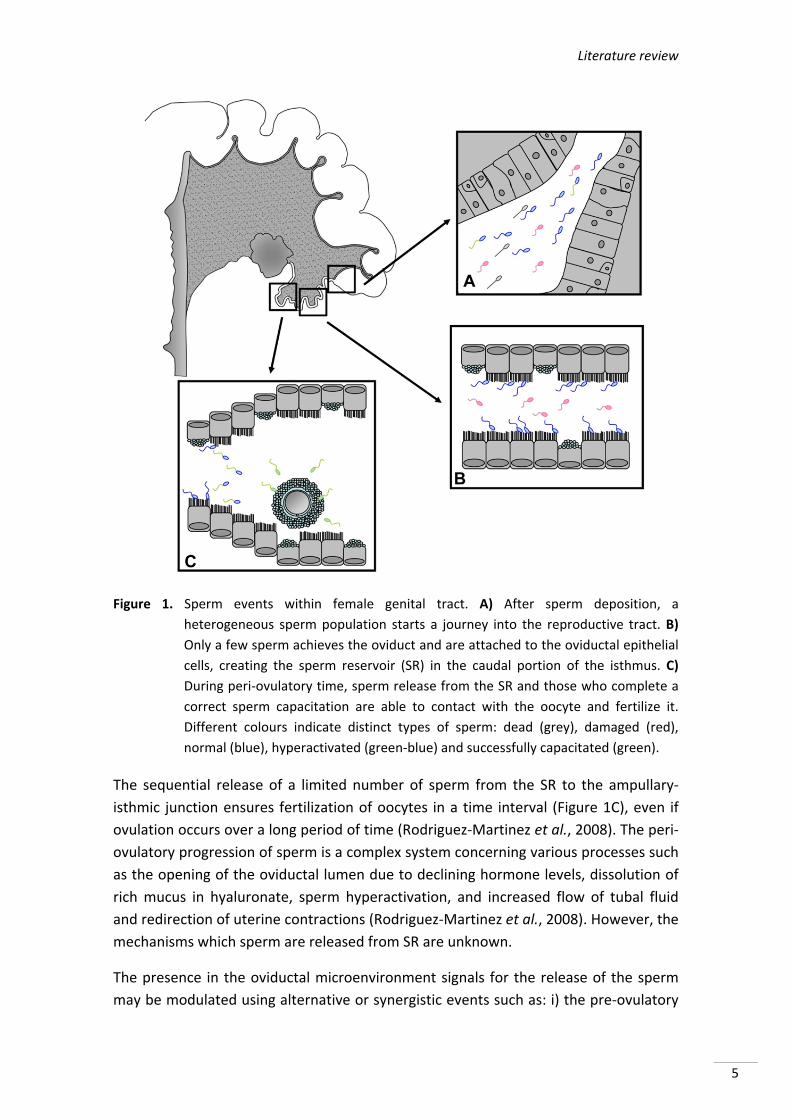
sperm (>70%) are eliminated in the uterine lumen (Figure 1A) (Einarsson & Viring,
1972), either by retrograde reflux (25‐45%) (Matthijs et al., 2003; Hernández‐Caravaca
et al., 2012) or by action of polymorphonuclear (Rozeboom et al., 1999). This removal
mainly is mediated by leukocytes that migrate from the lumen of endometrium within
30 minutes after mating (Matthijs et al., 2003). This latency period allows some sperm
to pass through the uterus which cannot be phagocytosed (Rodriguez‐Martinez et al.,
2005). During such advancement there is also a progressive modification of some
factors derived from the seminal plasma, which are attached to the surface of the
sperm plasma (Hunter & Rodriguez‐Martinez, 2004). These factors that prevent
premature capacitation are involved in binding the oviductal cells (Rodriguez‐Martinez
et al., 2009) and have also been shown as a requirement for sperm survival in the
uterus, at least in mouse (Kawano et al., 2014). Finally, only a small proportion will
pass through the utero‐tubal junction (UTJ) (revised by Soriano‐Úbeda et al., 2013).
From the UTJ sperm travel to the caudal portion of the isthmus, where the population
of motile sperm encounters a sticky secretion of glycoprotein that modifies the sperm
surface (Rodriguez‐Martinez et al., 2000). Motility decreases in this viscous medium
and facilitates the sperm adhesion to the epithelium (Figure 1B). Experimental studies
in domestic animals like goat, cow and pig have shown that in the pre‐ovulatory period
(between 25‐40 hours) sperm are unable to move and so, they stay together until near
ovulation. Between 1‐2 hours before ovulation, a small number of sperm will be
reactivated and will go progressively toward fertilization site (Hunter, 2008). As the
number of sperm is released and activated consequently, there are increased
progressively drift towards the ampullary‐isthmic junction. This brief description of
sperm activity in the caudal portion of the isthmus suggests that there is an influence
of ovaries on oviduct and on sperm bounded (Hunter, 2008).
Sperm remains in the caudal portion of the oviductal isthmus, during the pre and peri‐
ovulatory time, forming the sperm reservoir (SR) (Suarez et al., 1991; Töpfer‐Petersen
et al., 2002). Several factors may be involved in the formation of this SR. The binding
occurs through direct contact via ligand‐receptor interaction between the molecules
present in the rostral aspect of the sperm and membrane carbohydrates of oviductal
cells. This interaction is species‐specific (Lefebvre et al., 1997; Lapointe et al., 1998).
The binding is a reversible process involving, in all species studied, different sugars
(Dobrinski et al., 1996; Suarez, 1998). Other factors such as mucus, the chemical
properties of oviductal fluid and temperature gradients could contribute to the
formation of the SR (revised by Soriano‐Úbeda et al., 2013).
Literature review
5
Figure 1. Sperm events within female genital tract. A) After sperm deposition, a
heterogeneous sperm population starts a journey into the reproductive tract. B)
Only a few sperm achieves the oviduct and are attached to the oviductal epithelial
cells, creating the sperm reservoir (SR) in the caudal portion of the isthmus. C)
During peri‐ovulatory time, sperm release from the SR and those who complete a
correct sperm capacitation are able to contact with the oocyte and fertilize it.
Different colours indicate distinct types of sperm: dead (grey), damaged (red),
normal (blue), hyperactivated (green‐blue) and successfully capacitated (green).
The sequential release of a limited number of sperm from the SR to the ampullary‐
isthmic junction ensures fertilization of oocytes in a time interval (Figure 1C), even if
ovulation occurs over a long period of time (Rodriguez‐Martinez et al., 2008). The peri‐
ovulatory progression of sperm is a complex system concerning various processes such
as the opening of the oviductal lumen due to declining hormone levels, dissolution of
rich mucus in hyaluronate, sperm hyperactivation, and increased flow of tubal fluid
and redirection of uterine contractions (Rodriguez‐Martinez et al., 2008). However, the
mechanisms which sperm are released from SR are unknown.
The presence in the oviductal microenvironment signals for the release of the sperm
may be modulated using alternative or synergistic events such as: i) the pre‐ovulatory
Summary/Resumen
6
phase of the endocrine environment, ii) the presence of gametes, which can change
the secretory activity of the oviduct; and iii) the effect of cumulus‐oocyte complex. In
this sense, it has been shown that injection of progesterone or preovulatory follicular
fluid on the serosal layer or directly into the SR causes a massive release of
spermatozoa, leading to high rates of polyspermy (Hunter, 2008). This has led to the
assumption that progesterone may be a direct or an indirect signal to the release of
the spermatozoa (Talevi & Gualtieri, 2010). Furthermore unsulfated
glycosaminoglycans (components of the extracellular matrix, whose accumulation in
pig oocytes increases around ovulation), could also be involved in sperm capacitation
and the release of SR (Brüssow et al., 2008).
3. FUNCTIONAL CHANGES AND MOLECULAR PATHWAYS DURING SPERM
CAPACITATION
During the capacitation process, spermatozoa undergo a series of functional changes,
which enables them to bind to the extracellular matrix of the oocyte and consequently
require the acrosome reaction. Although the latter is under discussion as recently
shown by Jin et al. (2011) that the acrosome reaction in mouse sperm occurs before
binding to the zona pellucida. Besides, the pattern of movement of sperm flagellum
changes allowing penetration of the zona pellucida (Suarez, 2008).
Capacitation process implied several changes sequentially. Some of these changes are
rapid and occur at the moment of ejaculation. Others require a longer period of time in
the female genital tract (in vivo) or in a medium capable of supporting this process (in
vitro). All these processes (both rapid and slow), appear to be regulated by protein
kinase A (PKA) and HCO3‐, soluble adenylate cyclase (SACY or sAC), and cyclic
adenosine 3’,5’‐monophosphate (cAMP) participate in this process (revised by
Visconti, 2009).
Traditionally, reactive oxygen species (ROS) are considered to be injurious by products
of cellular metabolism but also fundamentally participants in cell signalling and
regulation mechanisms (Finkel, 2001). This apparent paradox also is true for
spermatozoa, which are particularly susceptible to ROS‐induced damage because their
plasma membranes contain relatively large amounts of polyunsaturated fatty acids
and their cytoplasm contains relatively low concentrations of scavenging enzymes
(Aitken & Fisher, 1994), but require low concentration of ROS to acquire the fertilizing
ability (Aitken, 1997; de Lamirande et al., 1997). The essential role of ROS as
modulators of capacitation is recognized in human (Herrero et al., 2003), bovine
(O'Flaherty et al., 2006), mouse (Herrero et al., 2003), and boar spermatozoa
(Funahashi, 2002; Aquila et al., 2011).
Literature review
7
Some authors consider that capacitation occurs in two steps, fast and low (Visconti,
2009):
Facts during fast sperm capacitation: An early event during capacitation is the
activation of sperm motility. Although sperm stored in the cauda epididymis being
practically immobile consume oxygen in large proportions. The flagellum movement
starts immediately after sperm are released from the epididymis and contact has been
made with seminal plasma. This is due to exposure of sperm to the HCO3‐ (Wennemuth
et al., 2003).
Facts during slow sperm capacitation: In contrast to the rapid activation of
motility, other processes associated with capacitation require a longer period of time.
During slow capacitation, sperm acquire the ability to fertilize, which is preceded by
the preparation of the sperm to undergo the acrosome reaction and change the
pattern of motility called hyperactivation. Components in oviductal fluid such as high
weight molecular proteins and high density lipoproteins promote cholesterol efflux
resulting in an increased capacitation and tyrosine phosphorylation (TP) using the
cAMP signalling pathway/PKA (Visconti et al., 1999). Additionally, these slow processes
also are achievable in vitro by incubation of spermatozoa in defined media, which
contain a protein source (usually bovine serum albumin‐BSA), and different ions,
including HCO3‐ and Ca2+.
4. MOLECULES AND MECHANISMS INVOLVED IN THE PROCESS OF CAPACITATION
As we mentioned above, HCO3‐, Ca2+ and cholesterol acceptor are essential during
capacitation process entirely (Figure 2). These substances induce modifications lipid
membrane, loss of cholesterol, activation of cAMP/PKA pathway, increase Ca2+ uptake
and intracellular pH (pHi), hyperpolarisation of membrane potential, and TP (Aitken &
Nixon, 2013). However, there are other pathways in relation to capacitation as
NO/sGC/cGMP or protein nitrosylation are being studied.
4.1. Bicarbonate and sperm capacitation
Several studies have shown that HCO3‐ plays a key role in sperm capacitation and
therefore achieve fertilization under both in vivo and in vitro (Okamura et al., 1985;
Visconti et al., 1995; Harrison et al., 1996; Harrison, 2004; Boerke et al., 2013).
Epididymal spermatozoa are exposed to low HCO3‐ concentrations (3‐4 mM). However,
when they arrive before the capacitation, takes place (oviduct). They are found in
much higher level (>20 mM) (Rodriguez‐Martinez et al., 1990). Movement of HCO3‐
through the membrane has been associated with increased pHi during capacitation
(Zeng et al., 1996). Moreover, another likely target for the action of HCO3‐ on sperm
metabolism is the regulation of cAMP (Garbers et al., 1982) by activation of sAC
Summary/Resumen
8
(Visconti, 2009). This in turn stimulates PKA to phosphorylate substrates, thereby
allowing TP (Visconti et al., 1998; Gadella & Harrison, 2002). Furthermore, activation of
the PKA results in activation of phospholipase D (PLD), which stimulates the
polymerization of F‐actin (Cohen et al., 2004), which is an event associated with the
process of acrosome reaction.
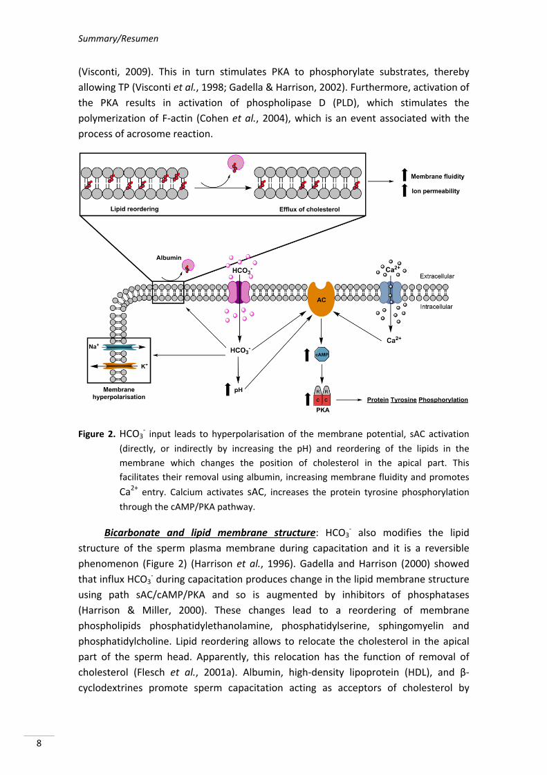
Figure 2. HCO3‐ input leads to hyperpolarisation of the membrane potential, sAC activation
(directly, or indirectly by increasing the pH) and reordering of the lipids in the
membrane which changes the position of cholesterol in the apical part. This
facilitates their removal using albumin, increasing membrane fluidity and promotes
Ca2+ entry. Calcium activates sAC, increases the protein tyrosine phosphorylation
through the cAMP/PKA pathway.
Bicarbonate and lipid membrane structure: HCO3‐ also modifies the lipid
structure of the sperm plasma membrane during capacitation and it is a reversible
phenomenon (Figure 2) (Harrison et al., 1996). Gadella and Harrison (2000) showed
that influx HCO3‐ during capacitation produces change in the lipid membrane structure
using path sAC/cAMP/PKA and so is augmented by inhibitors of phosphatases
(Harrison & Miller, 2000). These changes lead to a reordering of membrane
phospholipids phosphatidylethanolamine, phosphatidylserine, sphingomyelin and
phosphatidylcholine. Lipid reordering allows to relocate the cholesterol in the apical
part of the sperm head. Apparently, this relocation has the function of removal of
cholesterol (Flesch et al., 2001a). Albumin, high‐density lipoprotein (HDL), and β‐
cyclodextrines promote sperm capacitation acting as acceptors of cholesterol by
Literature review
9
removing it from the plasma membrane (Vadnais et al., 2007). As a result of this
process, decrease ratio of cholesterol/phospholipid consequently contributes to an
increased membrane fluidity promoting increase of ion permeability (Davis, 1976;
Cross, 1998; Visconti et al., 1999; Travis & Kopf, 2002).
Bicarbonate and sterol depletion: Albumin acts in synergy with HCO3‐ by
mediating efflux of sterols from the sperm surface (Boerke et al., 2008; Brouwers et
al., 2011). Flesch et al. (2001a) observed that the addition of albumin causes
cholesterol efflux (Figure 2), but only in HCO3‐‐responding cells that exhibited virtually
no filipin labelling in the sperm head area. In the absence of HCO3‐, albumin had no
effect on other lipid components and no affinity to cholesterol. HCO3‐ also induces
sperm surface oxysterol formation by activation of signalling pathway of the ROS,
which can be inhibited or blocked by addition of antioxidants as vitamin E or vitamin A
(Boerke et al., 2013). These sterols oxidation products (oxysterols), which are more
hydrophilic can be extracted using albumin (Brouwers et al., 2011) or can facilitate an
oxysterol dependent scavenger‐sensitive transport of free sterols to albumin (Jessup et
al., 2006).
Bicarbonate and sperm plasma membrane potential: Under normal conditions,
spermatozoa maintain intracellular ion concentration markedly different from
extracellular environment and these differences provide the resting membrane
potential (Salicioni et al., 2007). When spermatozoa are exposed to different
environments during transport through the male and female genital tracts, they find
different extracellular ion concentration. For example, the epididymal fluid contains
high K+, low Na+, and even lower concentrations of HCO3‐. After ejaculation there is a
drastic change in the concentrations of these ions in the seminal fluid and finally into
the female tract, where there concentrations of low potassium and high HCO3‐ are
present (Brooks, 1983; Setchell et al., 1994). As a result of changes in extracellular ion
concentrations, there will be changes in intracellular concentrations of these ions
leaving alterations in membrane potential (Muñoz‐Garay et al., 2001; Demarco et al.,
2003) which consequently occurs in the hyperpolarisation of sperm plasma membrane
(Zeng et al., 1995). It has been shown in mouse sperm that changes in membrane
potential do not occur in BSA or HCO3‐ absence (Demarco et al., 2003). These results
suggest that HCO3‐ present in capacitation media as well as cholesterol efflux may have
a direct or indirect function of events allowing hyperpolarisation of the sperm plasma
membrane (Salicioni et al., 2007). Arnoult et al. (1999) showed that only
hyperpolarized sperm populations are capable of undergoing the acrosome reaction in
presence of solubilised zona pellucida material.
4.2. Calcium and sperm capacitation
In 1915, Loeb was the first to demonstrate that Ca2+ is required in the extracellular
medium for fertilization to occur in invertebrates. Of all intracellular signalling
Summary/Resumen
10
mechanisms, perhaps the most studied and best characterized one is the mobilization
of Ca2+. This pathway involves transitory increase of intracellular Ca2+ concentrations
produced by multitude intercellular messengers.
One of the most important consequences of cholesterol efflux from membranes is a
massive influx of extracellular Ca2+, which is considered a prerequisite for the
acrosome reaction process (Flesch & Gadella, 2000). This Ca2+ influx may be due to
changes occurring in the membrane fluidity. The intracellular Ca2+ increase in sperm
can activate one or more enzymatic pathways (Figure 2). For example, the adenylate
cyclase (sAC) increases during capacitation in response to Ca2+, this enzyme will
catalyse the conversion of ATP to cAMP (revised by Vadnais et al., 2007).
In 1998, Visconti & Kopf suggested a cooperative effect of Ca2+ and HCO3‐ in
modulating sperm capacitation requiring the presence of both as well as increase in
cAMP levels and the subsequent phosphorylation of different proteins. In swine, both
Ca2+ and HCO3‐ appear to be required for capacitation and their roles are synergistic,
since it has been shown that the HCO3‐ will stimulate the entry of Ca2+ in this species
(Harrison et al., 1993). However, in mouse spermatozoa, Tateno et al. (2013) showed
that Ca2+ ionophore A23187 can make spermatozoa capable of fertilizing in vitro
without activation of cAMP‐dependent phosphorylation pathways in media HCO3‐ free.
Ca2+ is important to sperm hyperactivation during capacitation. CatSperm 1 and 2 are
voltage dependent Ca2+channels that are located in the tail of the sperm. Sperm from
mice deficient in these Ca2+ channels are infertile and do not exhibit hyperactivation
during capacitation despite having TP (Carlson et al., 2003).
Another aspect that influences capacitation related to Ca2+ is pHi. Sperm not
capacitated maintain an acidified intracellular pH (Parrish et al., 1989). This fact acts as
a regulator of Ca2+ influx (Florman et al., 1992) preventing capacitation and acrosome
reaction. Intracellular pH becomes more alkaline during capacitation (Vredenburgh‐
Wilberg & Parrish, 1995). Today it is believed that increasing intracellular Ca2+, HCO3‐
and the pH during sperm capacitation produce sAC activation and consequently cAMP
(Travis & Kopf, 2002; Breitbart, 2003; Harrison & Gadella, 2005; Hess et al., 2005a).
In addition, calmodulin, which is a protein binding Ca2+ considered to be an important
transducer of Ca2+ signals, appears to be diminished during capacitation. This
mechanism could be based on inhibition of Ca2+‐ATPase plasma membrane by
increasing cAMP levels through PDE1 inhibition (reviewed by Bailey, 2010).
4.3. Tyrosine phosphorylation of sperm proteins
Protein phosphorylation or de‐phosphorylation is controlled by activity of protein
kinases and protein phosphatases, which provide cells a “switch” through which they
can activate function of various proteins (Signorelli et al., 2013). Phosphorylation
Literature review
11
occurs in serine, threonine, and tyrosine. TP is related to capacitation process and
sperm hyperactivation in many mammal species (human (Baldi et al., 2002), bovine
(Galantino‐Homer et al., 1997), murine (Visconti & Kopf, 1998) or porcine (Tardif et al.,
2001)). In opposite, it has been shown that protein phosphatases decrease their
activity during capacitation (Signorelli et al., 2013).
Increasing TP during capacitation is regulated by a cAMP‐dependent pathway which
involves PKA (Kalab et al., 1998). cGMP‐PKG pathway is also involved in this process
(Cisneros‐Mejorado et al., 2014). In 2002, Visconti et al. described the possible
mechanisms, which could regulate the TP dependent signalling pathway cAMP/PKA: i)
the direct or indirect stimulation of a tyrosine kinase by PKA, ii) the direct or indirect
inhibition of a tyrosine phosphatase, and iii) direct or indirect phosphorylation of
proteins by PKA on serine or threonine residues to prepare these proteins for
subsequent phosphorylation on tyrosine residues.
TP is specific for each species. For example, in man TP during sperm capacitation
requires the presence of BSA, and HCO3‐ but no Ca2+ (Muratori et al., 2010). In the case
of stallion TP during capacitation requires HCO3‐ but neither BSA nor Ca2+ (González‐
Fernández et al., 2012). Another factor to consider in TP is time. TP in boar sperm
occurs close to 1 hour after the addition of HCO3‐ (Gadella & Van Gestel, 2004),
whereas in bull sperm it occurs 4 hours after addition of heparin (Galantino‐Homer et
al., 1997).
Although TP is an important key in capacitation, it is not yet entirely clear how the
phosphorylation of these proteins is involved in sperm‐zona recognition, gamete
interaction, or exocytosis of acrosomal content (Flesch et al., 2001b). The level of TP in
human sperm correlates strongly with the sperm‐zona‐binding capacity (Liu et al.,
2006) and alterations in TP have been found in subfertile subjects (Buffone et al.,
2009) indicating its physiological role in fertilization. In pigs ejaculated spermatozoa
selected in the oviduct adhere to the epithelial cells and suppress TP of sperm
proteins. This modulation by the oviductal epithelium on TP and, therefore
capacitation could help synchronize sperm functions to the time of ovulation.
4.4. Nitric oxide (NO) and sperm capacitation
Previous papers showed that NOS (nitric oxide synthase) is present in the oviduct
(Rosselli et al., 1996; Ekerhovd et al., 1999; Lapointe et al., 2006), oocyte, cumulus and
corona cells (Reyes et al., 2004; Tao et al., 2004) of different species (Bryant et al.,
1995; Rosselli et al., 1996; Ekerhovd et al., 1997). NOS isoforms are hormonally
regulated in the oviduct and expresses differently throughout the oestrous cycle. In
the oviduct, nitric oxide (NO) has been shown to regulate contractility (Rosselli et al.,
1994), ciliary beating of the ciliated epithelial cells, the sperm motility or even inducing
Summary/Resumen
12
chemotaxis (Miraglia et al., 2007). For this reason NO also module sperm capacitation
although the pathway is not known totally.
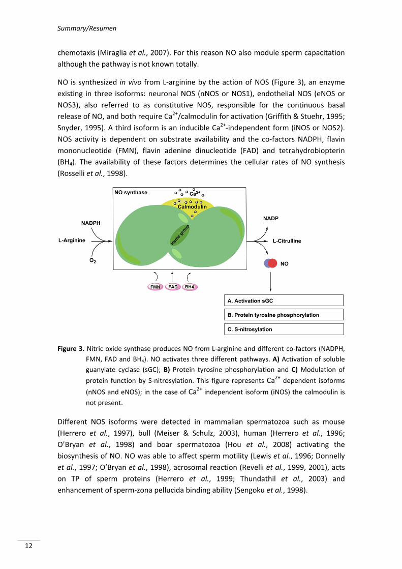
NO is synthesized in vivo from L‐arginine by the action of NOS (Figure 3), an enzyme
existing in three isoforms: neuronal NOS (nNOS or NOS1), endothelial NOS (eNOS or
NOS3), also referred to as constitutive NOS, responsible for the continuous basal
release of NO, and both require Ca2+/calmodulin for activation (Griffith & Stuehr, 1995;
Snyder, 1995). A third isoform is an inducible Ca2+‐independent form (iNOS or NOS2).
NOS activity is dependent on substrate availability and the co‐factors NADPH, flavin
mononucleotide (FMN), flavin adenine dinucleotide (FAD) and tetrahydrobiopterin
(BH4). The availability of these factors determines the cellular rates of NO synthesis
(Rosselli et al., 1998).
Figure 3. Nitric oxide synthase produces NO from L‐arginine and different co‐factors (NADPH,
FMN, FAD and BH4). NO activates three different pathways. A) Activation of soluble
guanylate cyclase (sGC); B) Protein tyrosine phosphorylation and C) Modulation of
protein function by S‐nitrosylation. This figure represents Ca2+ dependent isoforms
(nNOS and eNOS); in the case of Ca2+ independent isoform (iNOS) the calmodulin is
not present.
Different NOS isoforms were detected in mammalian spermatozoa such as mouse
(Herrero et al., 1997), bull (Meiser & Schulz, 2003), human (Herrero et al., 1996;
O’Bryan et al., 1998) and boar spermatozoa (Hou et al., 2008) activating the
biosynthesis of NO. NO was able to affect sperm motility (Lewis et al., 1996; Donnelly
et al., 1997; O’Bryan et al., 1998), acrosomal reaction (Revelli et al., 1999, 2001), acts
on TP of sperm proteins (Herrero et al., 1999; Thundathil et al., 2003) and
enhancement of sperm‐zona pellucida binding ability (Sengoku et al., 1998).
Literature review
13
NO has different functions in the spermatozoa, acting on different pathways that result
in sequential and parallel processes (Figure 3). The main actions of NO are:
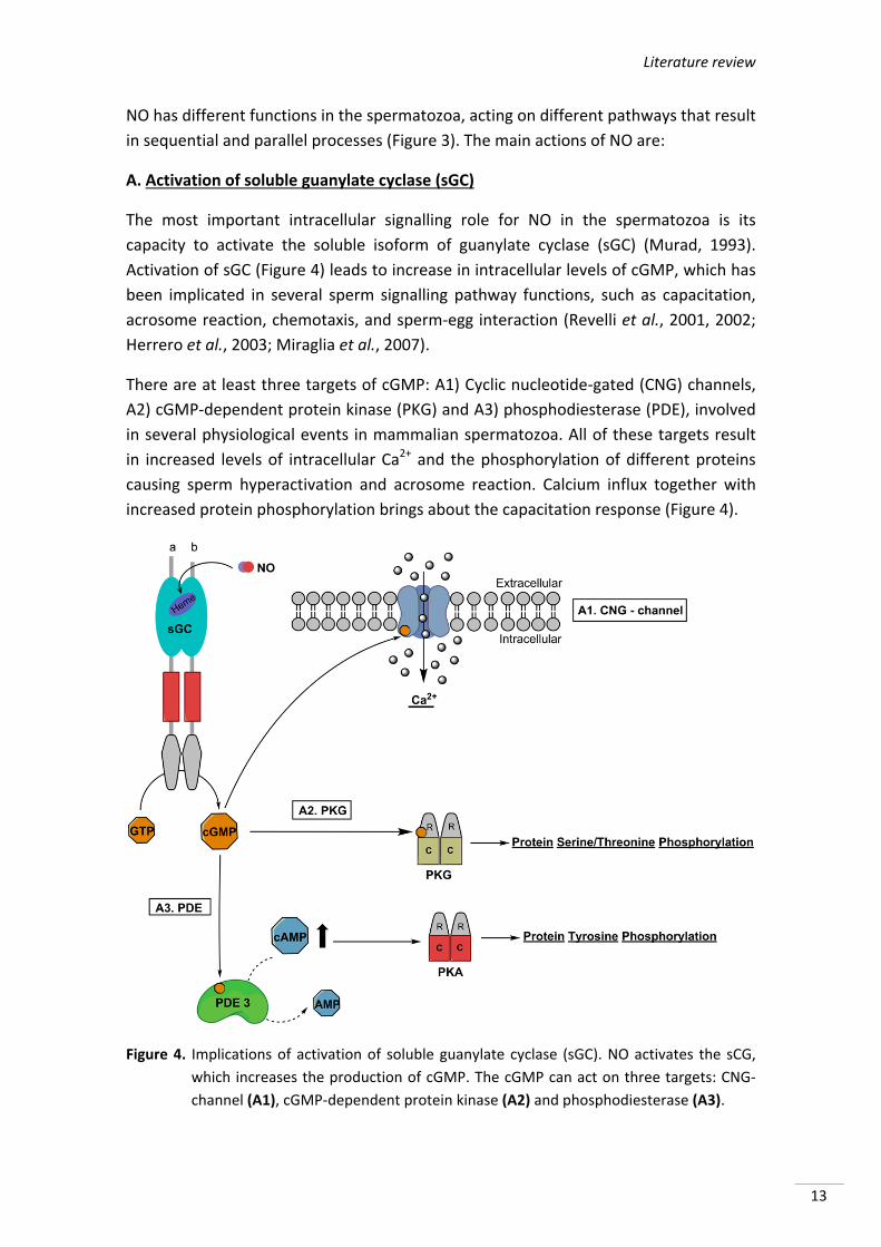
A. Activation of soluble guanylate cyclase (sGC)
The most important intracellular signalling role for NO in the spermatozoa is its
capacity to activate the soluble isoform of guanylate cyclase (sGC) (Murad, 1993).
Activation of sGC (Figure 4) leads to increase in intracellular levels of cGMP, which has
been implicated in several sperm signalling pathway functions, such as capacitation,
acrosome reaction, chemotaxis, and sperm‐egg interaction (Revelli et al., 2001, 2002;
Herrero et al., 2003; Miraglia et al., 2007).
There are at least three targets of cGMP: A1) Cyclic nucleotide‐gated (CNG) channels,
A2) cGMP‐dependent protein kinase (PKG) and A3) phosphodiesterase (PDE), involved
in several physiological events in mammalian spermatozoa. All of these targets result
in increased levels of intracellular Ca2+ and the phosphorylation of different proteins
causing sperm hyperactivation and acrosome reaction. Calcium influx together with
increased protein phosphorylation brings about the capacitation response (Figure 4).
Figure 4. Implications of activation of soluble guanylate cyclase (sGC). NO activates the sCG,
which increases the production of cGMP. The cGMP can act on three targets: CNG‐
channel (A1), cGMP‐dependent protein kinase (A2) and phosphodiesterase (A3).
Summary/Resumen
14
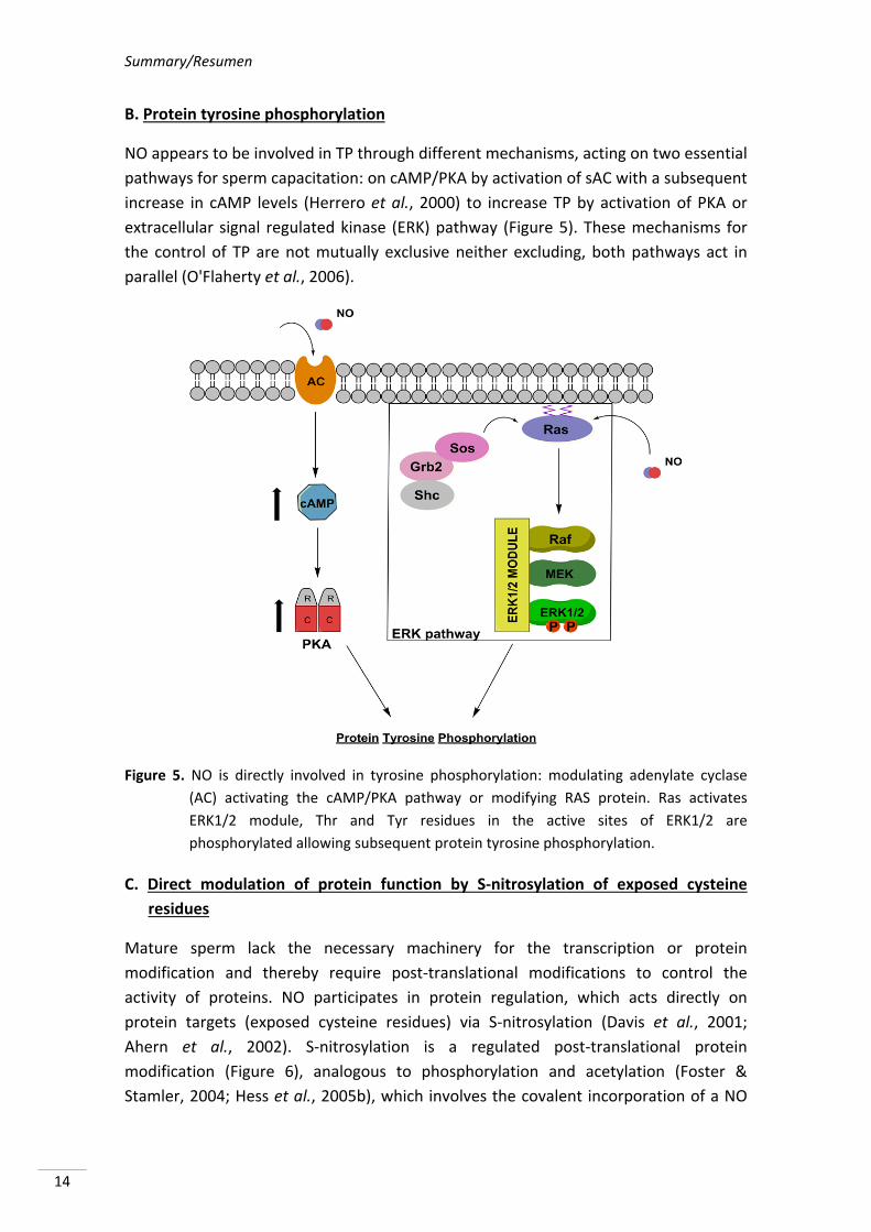
B. Protein tyrosine phosphorylation
NO appears to be involved in TP through different mechanisms, acting on two essential
pathways for sperm capacitation: on cAMP/PKA by activation of sAC with a subsequent
increase in cAMP levels (Herrero et al., 2000) to increase TP by activation of PKA or
extracellular signal regulated kinase (ERK) pathway (Figure 5). These mechanisms for
the control of TP are not mutually exclusive neither excluding, both pathways act in
parallel (O'Flaherty et al., 2006).
Figure 5. NO is directly involved in tyrosine phosphorylation: modulating adenylate cyclase
(AC) activating the cAMP/PKA pathway or modifying RAS protein. Ras activates
ERK1/2 module, Thr and Tyr residues in the active sites of ERK1/2 are
phosphorylated allowing subsequent protein tyrosine phosphorylation.
C. Direct modulation of protein function by S‐nitrosylation of exposed cysteine
residues
Mature sperm lack the necessary machinery for the transcription or protein
modification and thereby require post‐translational modifications to control the
activity of proteins. NO participates in protein regulation, which acts directly on
protein targets (exposed cysteine residues) via S‐nitrosylation (Davis et al., 2001;
Ahern et al., 2002). S‐nitrosylation is a regulated post‐translational protein
modification (Figure 6), analogous to phosphorylation and acetylation (Foster &
Stamler, 2004; Hess et al., 2005b), which involves the covalent incorporation of a NO
Literature review
15
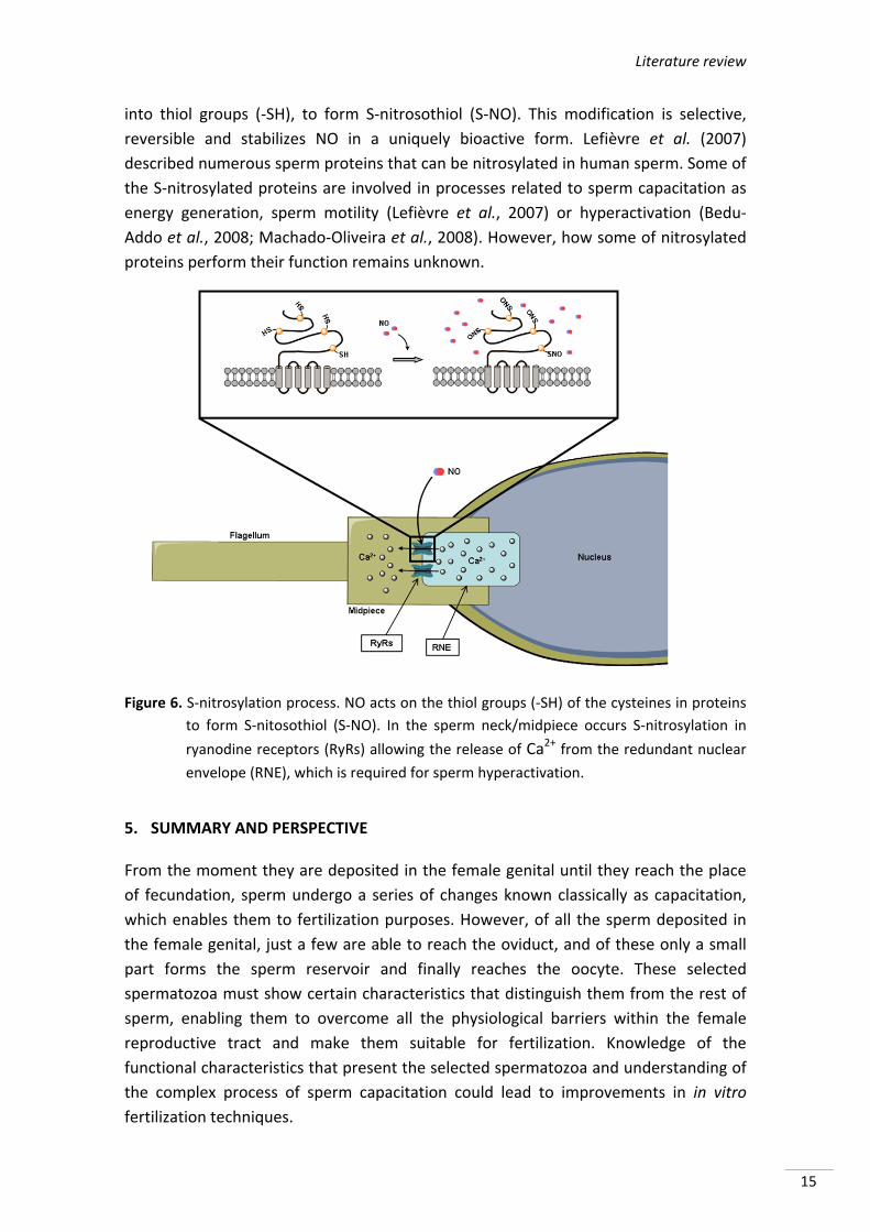
into thiol groups (‐SH), to form S‐nitrosothiol (S‐NO). This modification is selective,
reversible and stabilizes NO in a uniquely bioactive form. Lefièvre et al. (2007)
described numerous sperm proteins that can be nitrosylated in human sperm. Some of
the S‐nitrosylated proteins are involved in processes related to sperm capacitation as
energy generation, sperm motility (Lefièvre et al., 2007) or hyperactivation (Bedu‐
Addo et al., 2008; Machado‐Oliveira et al., 2008). However, how some of nitrosylated
proteins perform their function remains unknown.
Figure 6. S‐nitrosylation process. NO acts on the thiol groups (‐SH) of the cysteines in proteins
to form S‐nitosothiol (S‐NO). In the sperm neck/midpiece occurs S‐nitrosylation in
ryanodine receptors (RyRs) allowing the release of Ca2+ from the redundant nuclear
envelope (RNE), which is required for sperm hyperactivation.
5. SUMMARY AND PERSPECTIVE
From the moment they are deposited in the female genital until they reach the place
of fecundation, sperm undergo a series of changes known classically as capacitation,
which enables them to fertilization purposes. However, of all the sperm deposited in
the female genital, just a few are able to reach the oviduct, and of these only a small
part forms the sperm reservoir and finally reaches the oocyte. These selected
spermatozoa must show certain characteristics that distinguish them from the rest of
sperm, enabling them to overcome all the physiological barriers within the female
reproductive tract and make them suitable for fertilization. Knowledge of the
functional characteristics that present the selected spermatozoa and understanding of
the complex process of sperm capacitation could lead to improvements in in vitro
fertilization techniques.
Summary/Resumen
16
REFERENCES
Ahern GP, Klyachko VA & Jackson MB (2002). cGMP and S‐nitrosylation: two routes for
modulation of neuronal excitability by NO. Trends in neurosciences. 25: 510‐517. (doi:
10.1016/S0166‐2236(02)02254‐3).
Aitken RJ & Fisher H (1994). Reactive oxygen species generation and human spermatozoa: the
balance of benefit and risk. Bioessays. 16: 259‐267. (doi: 10.1002/bies.950160409).
Aitken RJ (1997). Molecular mechanisms regulating human sperm function. Molecular human
reproduction. 3: 169‐173. (doi: 10.1093/molehr/3.3.169).
Aitken RJ & Nixon B (2013). Sperm capacitation: a distant landscape glimpsed but unexplored.
Molecular human reproduction. 19: 785‐793. (doi: 10.1093/molehr/gat067).
Aquila S, Giordano F, Guido C, Rago V & Carpino A (2011). Nitric oxide involvement in the
acrosome reaction triggered by leptin in pig sperm. Reproductive Biology and
Endocrinology. 9: 133. (doi: 10.1186/1477‐7827‐9‐133).
Arnoult C, Kazam IG, Visconti PE, Kopf GS, Villaz M & Florman HM (1999). Control of the low
voltage‐activated calcium channel of mouse sperm by egg ZP3 and by membrane
hyperpolarization during capacitation. Proceedings of the National Academy of
Sciences. 96: 6757‐6762. (doi: 10.1073/pnas.96.12.6757).
Austin CR (1951). Activation and the correlation between male and female elements in
fertilization. Nature. 168: 558‐559. (doi: 10.1038/168558c0).
Austin CR (1952). The “Capacitation” of the Mammalian Sperm. Nature. 170: 326‐326.
Avilés M, Coy P & Rizos D (2015). The oviduct: a key organ for the success of early
reproductive events. Animal Frontiers. 5: 25‐31.
Bailey JL (2010). Factors regulating sperm capacitation. Systems Biology in Reproductive
Medicine. 56: 334‐348. (doi: 10.3109/19396368.2010.512377).
Baldi E, Luconi M, Bonaccorsi L & Forti G (2002). Signal transduction pathways in human
spermatozoa. Journal of reproductive immunology. 53: 121‐131. (doi: 10.1016/S0165‐
0378(01)00089‐4).
Bedu‐Addo K, Costello S, Harper C, Machado‐Oliveira G, Lefievre L, Ford C, Barratt C &
Publicover S (2008). Mobilisation of stored calcium in the neck region of human
sperm‐a mechanism for regulation of flagellar activity. International Journal of
Developmental Biology. 52: 615‐626. (doi: 10.1387/ijdb.072535kb).
Boerke A, Tsai P, Garcia‐Gil N, Brewis I & Gadella BM (2008). Capacitation‐dependent
reorganization of microdomains in the apical sperm head plasma membrane:
functional relationship with zona binding and the zona‐induced acrosome reaction.
Theriogenology. 70: 1188‐1196. (doi: 10.1016/j.theriogenology.2008.06.021).
Boerke A, Brouwers JF, Olkkonen VM, van de Lest CH, Sostaric E, Schoevers EJ, Helms JB &
Gadella BM (2013). Involvement of bicarbonate‐induced radical signaling in oxysterol
formation and sterol depletion of capacitating mammalian sperm during in vitro
fertilization. Biology of reproduction. 88: 1‐18. (doi: 10.1095/biolreprod.112.101253).
Breitbart H (2003). Signaling pathways in sperm capacitation and acrosome reaction. Cellular
and molecular biology (Noisy‐le‐Grand, France). 49: 321‐327.
Brooks D (1983). Epididymal functions and their hormonal regulation. Australian Journal of
Biological Sciences. 36: 205‐222. (doi: 10.1071/BI9830205).
Literature review
17
Brouwers JF, Boerke A, Silva PF, Garcia‐Gil N, van Gestel RA, Helms JB, van de Lest CH &
Gadella BM (2011). Mass spectrometric detection of cholesterol oxidation in bovine
sperm. Biology of reproduction. 85: 128‐136. (doi: 10.1095/biolreprod.111.091207).
Brüssow KP, Torner H, Rátky J, Manabe N & Tuchscherer A (2006). Experimental evidence for
the influence of cumulus‐oocyte‐complexes on sperm release from the porcine
oviductal sperm reservoir. Journal of Reproduction and Development. 52: 249‐257.
Brüssow KP, Ratky J & Rodriguez‐Martinez H (2008). Fertilization and early embryonic
development in the porcine fallopian tube. Reproduction in Domestic Animals. 43: 245‐
251. (doi: 10.1111/j.1439‐0531.2008.01169.x).
Bryant C, Tomlinson A, Mitchell J, Thiemermann C & Willoughby D (1995). Nitric oxide
synthase in the rat fallopian tube is regulated during the oestrous cycle. Journal of
endocrinology. 146: 149‐157. (doi: 10.1677/joe.0.1460149).
Buffone MG, Verstraeten SV, Calamera JC & Doncel GF (2009). High cholesterol content and
decreased membrane fluidity in human spermatozoa are associated with protein
tyrosine phosphorylation and functional deficiencies. Journal of andrology. 30: 552‐
558. (doi: 10.2164/jandrol.108.006551).
Carlson AE, Westenbroek RE, Quill T, Ren D, Clapham DE, Hille B, Garbers DL & Babcock DF
(2003). CatSper1 required for evoked Ca2+ entry and control of flagellar function in
sperm. Proceedings of the National Academy of Sciences. 100: 14864‐14868. (doi:
10.1073/pnas.2536658100).
Cisneros‐Mejorado A, Hernández‐Soberanis L, Islas‐Carbajal M & Sánchez D (2014).
Capacitation and Ca2+ influx in spermatozoa: role of CNG channels and protein kinase
G. Andrology. 2: 145‐154. (doi: 10.1111/j.2047‐2927.2013.00169.x).
Cohen G, Rubinstein S, Gur Y & Breitbart H (2004). Crosstalk between protein kinase A and C
regulates phospholipase D and F‐actin formation during sperm capacitation.
Developmental biology. 267: 230‐241. (doi: 10.1016/j.ydbio.2003.10.034).
Cross NL (1998). Role of cholesterol in sperm capacitation. Biology of reproduction. 59: 7‐11.
(doi: 10.1095/biolreprod59.1.7).
Chang M (1951). Fertilizing capacity of spermatozoa deposited into the fallopian tubes. Nature.
168: 697‐698 (doi: 10.1038/168697b0).
Davis B (1976). Inhibitory effect of synthetic phospholipid vesicles containing cholesterol on
the fertilizing ability of rabbit spermatozoa. Experimental Biology and Medicine. 152:
257‐261. (doi: 10.3181/00379727‐152‐39374).
Davis KL, Martin E, Turko IV & Murad F (2001). Novel effects of nitric oxide. Annual review of
pharmacology and toxicology. 41:203‐236. (doi: 10.1146/annurev.pharmtox.41.1.203).
de Lamirande E, Leclerc P & Gagnon C (1997). Capacitation as a regulatory event that primes
spermatozoa for the acrosome reaction and fertilization. Molecular human
reproduction. 3: 175‐194. (doi: 10.1093/molehr/3.3.175).
Demarco IA, Espinosa F, Edwards J, Sosnik J, de la Vega‐Beltran JL, Hockensmith JW, Kopf GS,
Darszon A & Visconti PE (2003). Involvement of a Na+/HCO3‐ cotransporter in mouse
sperm capacitation. Journal of Biological Chemistry. 278: 7001‐7009. (doi:
10.1074/jbc.M206284200).
Dobrinski I, Ignotz G, Thomas P & Ball B (1996). Role of carbohydrates in the attachment of
equine spermatozoa to uterine tubal (oviductal) epithelial cells in vitro. American
journal of veterinary research. 57: 1635‐1639.
Summary/Resumen
18
Donnelly ET, Lewis S, Thompson W & Chakravarthy U (1997). Sperm nitric oxide and motility:
the effects of nitric oxide synthase stimulation and inhibition. Molecular human
reproduction. 3: 755‐762. (doi: 10.1093/molehr/3.9.755).
Einarsson S & Viring S (1972). Effect of boar seminal plasma on the porcine uterus and the
isthmus part of oviducts in vitro. Acta veterinaria Scandinavica. 14: 639‐641.
Ekerhovd E, Brännström M, Alexandersson M & Norström A (1997). Evidence for nitric oxide
mediation of contractile activity in isolated strips of the human Fallopian tube. Human
reproduction. 12: 301‐305. (doi: 10.1093/humrep/12.2.301).
Ekerhovd E, Brännström M, Weijdegård B & Norström A (1999). Localization of nitric oxide
synthase and effects of nitric oxide donors on the human Fallopian tube. Molecular
human reproduction. 5: 1040‐1047. (doi: 10.1093/molehr/5.11.1040).
Finkel T (2001). Reactive oxygen species and signal transduction. IUBMB life. 52: 3‐6. (doi:
10.1080/15216540252774694).
Fitzpatrick JL & Lüpold S (2014). Sexual selection and the evolution of sperm quality. Molecular
human reproduction. 20: 1180‐1189. (doi: 10.1093/molehr/gau067).
Flesch FM & Gadella BM (2000). Dynamics of the mammalian sperm plasma membrane in the
process of fertilization. Biochimica et Biophysica Acta (BBA)‐Reviews on
Biomembranes. 1469: 197‐235. (doi: 10.1016/S0304‐4157(00)00018‐6).
Flesch FM, Brouwers JF, Nievelstein PF, Verkleij AJ, van Golde LM, Colenbrander B & Gadella
BM (2001a). Bicarbonate stimulated phospholipid scrambling induces cholesterol
redistribution and enables cholesterol depletion in the sperm plasma membrane.
Journal of cell science. 114: 3543‐3555.
Flesch FM, Wijnand E, Van de Lest C, Colenbrander B, van Golde LM & Gadella BM (2001b).
Capacitation dependent activation of tyrosine phosphorylation generates two sperm
head plasma membrane proteins with high primary binding affinity for the zona
pellucida. Molecular reproduction and development. 60: 107‐115. (doi:
10.1002/mrd.1067).
Florman HM, Corron ME, Kim TD‐H & Babcock DF (1992). Activation of voltage‐dependent
calcium channels of mammalian sperm is required for zona pellucida‐induced
acrosomal exocytosis. Developmental biology. 152: 304‐314. (doi: 10.1016/0012‐
1606(92)90137‐6).
Foster MW & Stamler JS (2004). New insights into protein S‐nitrosylation. Mitochondria as a
model system. Journal of Biological Chemistry. 279: 25891‐25897. (doi:
10.1074/jbc.M313853200).
Fraser LR (2010). The “switching on” of mammalian spermatozoa: molecular events involved in
promotion and regulation of capacitation. Molecular reproduction and development.
77: 197‐208. (doi: 10.1002/mrd.21124).
Funahashi H (2002). Induction of capacitation and the acrosome reaction of boar spermatozoa
by L‐arginine and nitric oxide synthesis associated with the anion transport system.
Reproduction. 124: 857‐864. (doi: 10.1530/rep.0.1240857).
Gadella BM & Harrison RA (2000). The capacitating agent bicarbonate induces protein kinase
A‐dependent changes in phospholipid transbilayer behavior in the sperm plasma
membrane. Development. 127: 2407‐2420.
Gadella BM & Harrison RA (2002). Capacitation induces cyclic adenosine 3′,5′‐
monophosphate‐dependent, but apoptosis‐unrelated, exposure of
Literature review
19
aminophospholipids at the apical head plasma membrane of boar sperm cells. Biology
of reproduction. 67: 340‐350. (doi: 10.1095/biolreprod67.1.340).
Gadella BM & Van Gestel R (2004). Bicarbonate and its role in mammalian sperm function.
Animal Reproduction Science. 82: 307‐319. (doi: 10.1016/j.anireprosci.2004.04.030).
Galantino‐Homer HL, Visconti PE & Kopf GS (1997). Regulation of protein tyrosine
phosphorylation during bovine sperm capacitation by a cyclic adenosine 3',5'‐
monophosphate‐dependent pathway. Biology of reproduction. 56: 707‐719. (doi:
10.1095/biolreprod56.3.707).
Garbers D, TUbb D & Hyne R (1982). A requirement of bicarbonate for Ca2+‐induced elevations
of cyclic AMP in guinea pig spermatozoa. Journal of Biological Chemistry. 257: 8980‐
8984.
González‐Fernández L, Macías‐García B, Velez IC, Varner DD & Hinrichs K (2012). Calcium–
calmodulin and pH regulate protein tyrosine phosphorylation in stallion sperm.
Reproduction. 144: 411‐422. (doi: 10.1530/REP‐12‐0067).
Griffith OW & Stuehr DJ (1995). Nitric oxide synthases: properties and catalytic mechanism.
Annual Review of Physiology. 57: 707‐734. (doi:
10.1146/annurev.ph.57.030195.003423).
Harrison RA, Mairet B & Miller N (1993). Flow cytometric studies of bicarbonate‐mediated
Ca2+ influx in boar sperm populations. Molecular reproduction and development. 35:
197‐208.
Harrison RA, Ashworth P & Miller N (1996). Bicarbonate/CO2, an effector of capacitation,
induces a rapid and reversible change in the lipid architecture of boar sperm plasma
membranes. Molecular reproduction and development. 45: 378‐391. (doi:
10.1002/(SICI)1098‐2795(199611)45:3<378::AID‐MRD16>3.0.CO;2‐V).
Harrison RA & Miller N (2000). cAMP‐dependent protein kinase control of plasma membrane
lipid architecture in boar sperm. Molecular reproduction and development. 55: 220‐
228. (doi: 10.1002/(SICI)1098‐2795(200002)55:2<220::AID‐MRD12>3.0.CO;2‐I).
Harrison RA (2004). Rapid PKA‐catalysed phosphorylation of boar sperm proteins induced by
the capacitating agent bicarbonate. Molecular reproduction and development. 67: 337‐
352.
Harrison RA & Gadella BM (2005). Bicarbonate‐induced membrane processing in sperm
capacitation. Theriogenology. 63: 342‐351.
Hernández‐Caravaca I, Izquierdo‐Rico MJ, Matás C, Carvajal JA, Vieira L, Abril D, Soriano‐
Úbeda C & García‐Vázquez FA (2012). Reproductive performance and backflow study
in cervical and post‐cervical artificial insemination in sows. Animal Reproduction
Science. 136: 14‐22. (doi: 10.1016/j.anireprosci.2012.10.007).
Herrero MB, Pérez Martínez S, Viggiano J & Polak J (1996). Localization by indirect
immunofluorescence of nitric oxide synthase in mouse and human spermatozoa.
Reproduction, fertility and development. 8: 931‐934. (doi: 10.1071/RD9960931).
Herrero MB, Goin J, Boquet M, Canteros M, Franchi A, Pérez Martínez S, Polak J, Viggiano J &
Gimeno M (1997). The nitric oxide synthase of mouse spermatozoa. FEBS letters. 411:
39‐42. (doi: 10.1016/S0014‐5793(97)00570‐X).
Herrero MB, de Lamirande E & Gagnon C (1999). Nitric oxide regulates human sperm
capacitation and protein‐tyrosine phosphorylation in vitro. Biology of reproduction. 61:
575‐581. (doi: 10.1095/biolreprod61.3.575).
Summary/Resumen
20
Herrero MB, Chatterjee S, Lefièvre L, de Lamirande E & Gagnon C (2000). Nitric oxide
interacts with the cAMP pathway to modulate capacitation of human spermatozoa.
Free Radical Biology and Medicine. 29: 522‐536.
Herrero MB, de Lamirande Ed & Gagnon C (2003). Nitric oxide is a signaling molecule in
spermatozoa. Current pharmaceutical design. 9: 419‐425. (doi:
10.2174/1381612033391720).
Hess KC, Jones BH, Marquez B, Chen Y, Ord TS, Kamenetsky M, Miyamoto C, Zippin JH, Kopf
GS & Suarez SS (2005a). The “soluble” adenylyl cyclase in sperm mediates multiple
signaling events required for fertilization. Developmental cell. 9: 249‐259. (doi:
10.1016/j.devcel.2005.06.007).
Hess DT, Matsumoto A, Kim S‐O, Marshall HE & Stamler JS (2005b). Protein S‐nitrosylation:
purview and parameters. Nature Reviews Molecular Cell Biology. 6: 150‐166. (doi:
10.1038/nrm1569).
Holt WV (2009). Is semen analysis useful to predict the odds that the sperm will meet the egg?
Reproduction in Domestic Animals. 44:31‐38. (doi: 10.1111/j.1439‐0531.2009.01363.x).
Hou M‐L, Huang S‐Y, Lai Y‐K & Lee W‐C (2008). Geldanamycin augments nitric oxide
production and promotes capacitation in boar spermatozoa. Animal Reproduction
Science. 104: 56‐68. (doi: 10.1016/j.anireprosci.2007.01.006).
Hunter RHF & Rodriguez‐Martinez H (2004). Capacitation of mammalian spermatozoa in vivo,
with a specific focus on events in the Fallopian tubes. Molecular reproduction and
development. 67: 243‐250. (doi: 10.1002/mrd.10390).
Hunter RHF (2008). Sperm release from oviduct epithelial binding is controlled hormonally by
peri‐ovulatory graafian follicles. Molecular reproduction and development. 75:167‐174.
(doi: 10.1002/mrd.20776).
Immler S (2008). Sperm competition and sperm cooperation: the potential role of diploid and
haploid expression. Reproduction. 135: 275‐283. (doi: 10.1530/REP‐07‐0482).
Jessup W, Gelissen IC, Gaus K & Kritharides L (2006). Roles of ATP binding cassette
transporters A1 and G1, scavenger receptor BI and membrane lipid domains in
cholesterol export from macrophages. Current opinion in lipidology. 17: 247‐257. (doi:
10.1097/01.mol.0000226116.35555.eb).
Jin M, Fujiwara E, Kakiuchi Y, Okabe M, Satouh Y, Baba SA, Chiba K & Hirohashi N (2011).
Most fertilizing mouse spermatozoa begin their acrosome reaction before contact with
the zona pellucida during in vitro fertilization. Proceedings of the National Academy of
Sciences. 108: 4892‐4896. (doi: 10.1073/pnas.1018202108).
Kalab P, Pěknicová J, Geussova G & Moos J (1998). Regulation of protein tyrosine
phosphorylation in boar sperm through a cAMP‐dependent pathway. Molecular
reproduction and development. 51: 304‐314. (doi: 10.1002/(SICI)1098‐
2795(199811)51:3<304::AID‐MRD10>3.0.CO;2‐2).
Kawano N, Araki N, Yoshida K, Hibino T, Ohnami N, Makino M, Kanai S, Hasuwa H, Yoshida
M & Miyado K (2014). Seminal vesicle protein SVS2 is required for sperm survival in
the uterus. Proceedings of the National Academy of Sciences. 111: 4145‐4150. (doi:
10.1073/pnas.1320715111).
Lapointe J, Roy M, St‐Pierre I, Kimmins S, Gauvreau D, MacLaren LA & Bilodeau J‐Fo (2006).
Hormonal and spatial regulation of nitric oxide synthases (NOS)(neuronal NOS,
Literature review
21
inducible NOS, and endothelial NOS) in the oviducts. Endocrinology. 147: 5600‐5610.
(doi: 10.1210/en.2005‐1548).
Lapointe S, Sullivan R & Sirard M‐A (1998). Binding of a bovine oviductal fluid catalase to
mammalian spermatozoa. Biology of reproduction. 58: 747‐753. (doi: 10.1095/
biolreprod58.3.747).
Lefebvre R, Lo MC & Suarez SS (1997). Bovine sperm binding to oviductal epithelium involves
fucose recognition. Biology of reproduction. 56: 1198‐1204. (doi: 10.1095/
biolreprod56.5.1198).
Lefièvre L, Chen Y, Conner SJ, Scott JL, Publicover SJ, Ford WCL & Barratt CL (2007). Human
spermatozoa contain multiple targets for protein S‐nitrosylation: An alternative
mechanism of the modulation of sperm function by nitric oxide? Proteomics. 7: 3066‐
3084. (doi: 10.1002/pmic.200700254).
Lewis S, Donnelly E, Sterling E, Kennedy M, Thompson W & Chakravarthy U (1996). Nitric
oxide synthase and nitrite production in human spermatozoa: evidence that
endogenous nitric oxide is beneficial to sperm motility. Molecular human reproduction.
2: 873‐878. (doi: 10.1093/molehr/2.11.873).
Liu D, Clarke G & Baker H (2006). Tyrosine phosphorylation on capacitated human sperm tail
detected by immunofluorescence correlates strongly with sperm–zona pellucida (ZP)
binding but not with the ZP‐induced acrosome reaction. Human reproduction. 21:
1002‐1008. (doi: 10.1093/humrep/dei435).
Loeb J (1915). On the Nature of the Conditions Which Determine or Prevent the Entrance of
the Spermatozoon Into the Egg. The American Naturalist. 49: 257‐285. (doi:
10.2307/2456166).
Machado‐Oliveira G, Lefièvre L, Ford C, Herrero MB, Barratt C, Connolly TJ, Nash K, Morales‐
Garcia A, Kirkman‐Brown J & Publicover S (2008). Mobilisation of Ca2+ stores and
flagellar regulation in human sperm by S‐nitrosylation: a role for NO synthesised in the
female reproductive tract. Development. 135: 3677‐3686. (doi: 10.1242/dev.024521).
Matthijs A, Engel B & Woelders H (2003). Neutrophil recruitment and phagocytosis of boar
spermatozoa after artificial insemination of sows, and the effects of inseminate
volume, sperm dose and specific additives in the extender. Reproduction. 125: 357‐
367. (doi: 10.1530/rep.0.1250357).
Meiser H & Schulz R (2003). Detection and localization of two constitutive NOS isoforms in bull
spermatozoa. Anatomia, histologia, embryologia. 32: 321‐325. (doi: 10.1111/j.1439‐
0264.2003.00459.x).
Miraglia E, Rullo ML, Bosia A, Massobrio M, Revelli A & Ghigo D (2007). Stimulation of the
nitric oxide/cyclic guanosine monophosphate signaling pathway elicits human sperm
chemotaxis in vitro. Fertility and sterility. 87: 1059‐1063. (doi:
10.1016/j.fertnstert.2006.07.1540).
Muñoz‐Garay C, De la Vega‐Beltrán JL, Delgado R, Labarca P, Felix R & Darszon A (2001).
Inwardly rectifying K+ channels in spermatogenic cells: functional expression and
implication in sperm capacitation. Developmental biology. 234: 261‐274. (doi:
10.1006/dbio.2001.0196).
Murad F (1993). The nitric oxide‐cyclic GMP signal transduction system for intracellular and
intercellular communication. Recent progress in hormone research. 49: 239‐248.
Summary/Resumen
22
Muratori M, Marchiani S, Tamburrino L, Forti G, Luconi M & Baldi E (2010). Markers of human
sperm functions in the ICSI era. Frontiers in bioscience (Landmark edition). 16: 1344‐
1363. (doi: 10.2741/3793).
O'Flaherty C, de Lamirande E & Gagnon C (2006). Reactive oxygen species modulate
independent protein phosphorylation pathways during human sperm capacitation.
Free Radical Biology and Medicine. 40: 1045‐1055. (doi:
10.1016/j.freeradbiomed.2005.10.055).
O’Bryan MK, Zini A, Cheng CY & Schlegel PN (1998). Human sperm endothelial nitric oxide
synthase expression: correlation with sperm motility. Fertility and sterility. 70: 1143‐
1147. (doi: 10.1016/S0015‐0282(98)00382‐3).
Okamura N, Tajima Y, Soejima A, Masuda H & Sugita Y (1985). Sodium bicarbonate in seminal
plasma stimulates the motility of mammalian spermatozoa through direct activation of
adenylate cyclase. Journal of Biological Chemistry. 260: 9699‐9705.
Parrish J, Susko‐Parrish J & First N (1989). Capacitation of bovine sperm by heparin: inhibitory
effect of glucose and role of intracellular pH. Biology of reproduction. 41: 683‐699.
(doi: 10.1095/biolreprod41.4.683).
Revelli A, Soldati G, Costamagna C, Pellerey O, Aldieri E, Massobrio M, Bosia A & Ghigo D
(1999). Follicular fluid proteins stimulate nitric oxide (NO) synthesis in human sperm: a
possible role for NO in acrosomal reaction. Journal of cellular physiology. 178: 85‐92.
(doi: 10.1002/(SICI)1097‐4652(199901)178:1<85::AID‐JCP11>3.0.CO;2‐Y).
Revelli A, Costamagna C, Moffa F, Aldieri E, Ochetti S, Bosia A, Massobrio M, Lindblom B &
Ghigo D (2001). Signaling pathway of nitric oxide‐induced acrosome reaction in human
spermatozoa. Biology of reproduction. 64: 1708‐1712. (doi:
10.1095/biolreprod64.6.1708).
Revelli A, Ghigo D, Moffa F, Massobrio M & Tur‐Kaspa I (2002). Guanylate cyclase activity and
sperm function. Endocrine reviews. 23: 484‐494. (doi: 10.1210/er.2001‐0020).
Reyes R, Vázquez M & Delgado N (2004). Detection and bioimaging of nitric oxide in bovine
oocytes and sperm cells. Systems Biology in Reproductive Medicine. 50: 303‐309. (doi:
10.1080/01485010490448471).
Rodriguez‐Martinez H, Ekstedt E & Einarsson S (1990). Acidification of epididymal fluid in the
boar. International journal of andrology. 13: 238‐243. (doi: 10.1111/j.1365‐
2605.1990.tb00982.x).
Rodriguez‐Martinez H, Tienthai P, Suzuki K, Funahashi H, Ekwall H & Johannisson A (2000).
Involvement of oviduct in sperm capacitation and oocyte development in pigs.
Reproduction (Cambridge, England) Supplement. 58: 129‐145.
Rodriguez‐Martinez H, Saravia F, Wallgren M, Tienthai P, Johannisson A, Vázquez JM,
Martínez E, Roca J, Sanz L & Calvete JJ (2005). Boar spermatozoa in the oviduct.
Theriogenology. 63: 514‐535. (doi: 10.1016/j.theriogenology.2004.09.028).
Rodriguez‐Martinez H, Saravia F, Wallgren M, Roca J & Peña F (2008). Influence of seminal
plasma on the kinematics of boar spermatozoa during freezing. Theriogenology. 70:
1242‐1250. (doi: 10.1016/j.theriogenology.2008.06.007).
Rodriguez‐Martinez H, Kvist U, Wallgren M, Johannisson A & Sanz L (2009). The physiological
roles of the boar ejaculate. Control of Pig Reproduction VIII. 66: 1‐21.
Literature review
23
Rosselli M, Imthurn B, Macas E, Keller P & Dubey RK (1994). Endogenous nitric oxide
modulates endothelin‐1 induced contraction of bovine oviduct. Biochemical and
biophysical research communications. 201: 143‐148. (doi: 10.1006/bbrc.1994.1680).
Rosselli M, Dubey RK, Rosselli MA, Macas E, Fink D, Lauper U, Keller P & Imthurn B (1996).
Identification of nitric oxide synthase in human and bovine oviduct. Molecular human
reproduction. 2: 607‐612. (doi: 10.1093/molehr/2.8.607).
Rosselli M, Keller R & Dubey RK (1998). Role of nitric oxide in the biology, physiology and
pathophysiology of reproduction. Human reproduction update. 4: 3‐24. (doi:
10.1093/humupd/4.1.3).
Rozeboom K, Troedsson M, Molitor T & Crabo B (1999). The effect of spermatozoa and
seminal plasma on leukocyte migration into the uterus of gilts. Journal of animal
science. 77: 2201‐2206. (doi: 1999.7782201x).
Salicioni A, Platt MD, Wertheimer EV, Arcelay E, Allaire A, Sosnik J & Visconti PE (2007).
Signalling pathways involved in sperm capacitation. Society of Reproduction and
Fertility supplement. 65: 245‐259.
Sengoku K, Tamate K, Yoshida T, Takaoka Y, Miyamoto T & Ishikawa M (1998). Effects of low
concentrations of nitric oxide on the zona pellucida binding ability of human
spermatozoa. Fertility and sterility. 69: 522‐527. (doi: 10.1016/S0015‐0282(97)00537‐
2).
Setchell B, Maddocks S & Brooks D (1994). Anatomy, vasculature, innervation, and fluids of
the male reproductive tract. The physiology of reproduction. 1: 1063‐1175.
Signorelli JR, Diaz ES, Fara K, Baron L & Morales P (2013). Protein phosphatases decrease their
activity during capacitation: a new requirement for this event. PloS one. 8 (doi:
10.1371/journal.pone.0081286).
Snyder SH (1995). No endothelial NO. Nature. 377: 196‐197. (doi: 10.1038/377196a0).
Soriano‐Úbeda C, Matás C & García‐Vázquez F (2013). An overview of swine artificial
insemination: retrospective, current and prospective aspects. Journal of Experimental
and Applied Animal Sciences. 1: 67‐97.
Suarez S, Redfern K, Raynor P, Martin F & Phillips D (1991). Attachment of boar sperm to
mucosal explants of oviduct in vitro: possible role in formation of a sperm reservoir.
Biology of reproduction. 44: 998‐1004. (doi: 10.1095/biolreprod44.6.998).
Suarez SS (1998). The oviductal sperm reservoir in mammals: mechanisms of formation.
Biology of reproduction. 58: 1105‐1107. (doi: 10.1095/biolreprod58.5.1105).
Suarez SS (2008). Control of hyperactivation in sperm. Human reproduction update. 14: 647‐
657. (doi: 10.1093/humupd/dmn029).
Talevi R & Gualtieri R (2010). Molecules involved in sperm‐oviduct adhesion and release.
Theriogenology. 73: 796‐801. (doi: 10.1016/j.theriogenology.2009.07.005).
Tao Y, Fu Z, Zhang M, Xia G, Yang J & Xie H (2004). Immunohistochemical localization of
inducible and endothelial nitric oxide synthase in porcine ovaries and effects of NO on
antrum formation and oocyte meiotic maturation. Molecular and cellular
endocrinology. 222: 93‐103. (doi: 10.1016/j.mce.2004.04.014).
Tardif S, Dubé C, Chevalier S & Bailey JL (2001). Capacitation is associated with tyrosine
phosphorylation and tyrosine kinase‐like activity of pig sperm proteins. Biology of
reproduction. 65: 784‐792. (doi: 10.1095/biolreprod65.3.784).
Summary/Resumen
24
Tateno H, Krapf D, Hino T, Sánchez‐Cárdenas C, Darszon A, Yanagimachi R & Visconti PE
(2013). Ca2+ ionophore A23187 can make mouse spermatozoa capable of fertilizing in
vitro without activation of cAMP‐dependent phosphorylation pathways. Proceedings
of the National Academy of Sciences. 110: 18543‐18548. (doi:
10.1073/pnas.1317113110).
Thundathil J, de Lamirande E & Gagnon C (2003). Nitric oxide regulates the phosphorylation of
the threonine‐glutamine‐tyrosine motif in proteins of human spermatozoa during
capacitation. Biology of reproduction. 68: 1291‐1298. (doi:
10.1095/biolreprod.102.008276).
Töpfer‐Petersen E, Wagner A, Friedrich J, Petrunkina AM, Ekhlasi‐Hundrieser M, Waberski D
& Drommer W (2002). Function of the mammalian oviductal sperm reservoir. Journal
of Experimental Zoology. 292: 210‐215. (doi: 10.1002/jez.1157).
Travis AJ & Kopf GS (2002). The role of cholesterol efflux in regulating the fertilization
potential of mammalian spermatozoa. Journal of Clinical Investigation. 110: 731‐736.
(doi: 10.1172/JCI16392.).
Vadnais ML, Galantino‐Homer HL & Althouse GC (2007). Current concepts of molecular
events during bovine and porcine spermatozoa capacitation. Systems Biology in
Reproductive Medicine. 53: 109‐123. (doi: 10.1080/01485010701329386).
Visconti PE, Bailey JL, Moore GD, Pan D, Olds‐Clarke P & Kopf GS (1995). Capacitation of
mouse spermatozoa. I. Correlation between the capacitation state and protein
tyrosine phosphorylation. Development. 121: 1129‐1137.
Visconti PE, Galantino‐Homer H, Moore GD, Bailey JL, Ning X, Fornes M & Kopf GS (1998).
The molecular basis of sperm capacitation. Journal of andrology. 19: 242‐248. (doi:
10.1002/j.1939‐4640.1998.tb01994.x).
Visconti PE & Kopf GS (1998). Regulation of protein phosphorylation during sperm
capacitation. Biology of reproduction. 59: 1‐6. (doi: 10.1095/biolreprod59.1.1).
Visconti PE, Ning X, Fornés MW, Alvarez JG, Stein P, Connors SA & Kopf GS (1999).
Cholesterol efflux‐mediated signal transduction in mammalian sperm: cholesterol
release signals an increase in protein tyrosine phosphorylation during mouse sperm
capacitation. Developmental biology. 214: 429‐443. (doi: 10.1006/dbio.1999.9428).
Visconti PE, Westbrook V, Chertihin O, Demarco I, Sleight S & Diekman A (2002). Novel
signaling pathways involved in sperm acquisition of fertilizing capacity. Journal of
reproductive immunology. 53: 133‐150. (doi: 10.1016/S0165‐0378(01)00103‐6).
Visconti PE (2009). Understanding the molecular basis of sperm capacitation through kinase
design. Proceedings of the National Academy of Sciences. 106: 667‐668. (doi:
10.1073/pnas.0811895106).
Vredenburgh‐Wilberg W & Parrish J (1995). Intracellular pH of bovine sperm increases during
capacitation. Molecular reproduction and development. 40: 490‐502. (doi:
10.1002/mrd.1080400413).
Wennemuth G, Carlson AE, Harper AJ & Babcock DF (2003). Bicarbonate actions on flagellar
and Ca2+‐channel responses: initial events in sperm activation. Development. 130:
1317‐1326. (doi: 10.1242/dev.00353).
Zeng Y, Clark EN & Florman HM (1995). Sperm membrane potential: hyperpolarization during
capacitation regulates zona pellucida‐dependent acrosomal secretion. Developmental
biology. 171: 554‐563. (doi: 10.1006/dbio.1995.1304).

Literature review
25
Zeng Y, Oberdorf JA & Florman HM (1996). pH regulation in mouse sperm: identification of
Na+‐, Cl‐‐, and HCO3‐‐dependent and arylaminobenzoate‐dependent regulatory
mechanisms and characterization of their roles in sperm capacitation. Developmental
biology. 173: 510‐520. (doi: 10.1006/dbio.1996.0044).

Objectives/Objetivos

Objectives/Objetivos
29
OBJECTIVES
The main objective of this thesis was the study of sperm subpopulations that have
previously been selected by different methods: in vitro (Percoll gradients in Chapter 1,
oviductal cells culture in Chapter 2 and by combining both in Chapter 3), ex vivo (in
oviductal explants, Chapter 2) and in vivo (in the oviduct of the sow after insemination,
Chapter 2), and the relationship with the functionality, sperm capacitation status and
their fertilizing capacity.
To achieve the main objective the following specific objectives were established:
CHAPTER 1
Objective 1. Characterization of sperm populations selected through three different
discontinuous Percoll gradients (45/60, 60/75 and 45/90%).
1.1 Analysis of different sperm function parameters: morphology, acrosome status,
motility and motion parameters, ROS generation, chromatin condensation,
DNA fragmentation, tyrosine phosphorylation and intracellular calcium
concentration.
1.2 Evaluation of the sperm penetrating capacity using an in vitro fertilization
system.
CHAPTER 2
Objective 2. Comparison of sperm capacitation status (by evaluation of protein
tyrosine phosphorylation) in sperm selected by oviductal epithelial cells (bound and
unbound sperm) under three different experimental conditions.
2.1 Sperm incubated with oviductal epithelial cells culture: “in vitro model”.
2.2 Sperm incubated into oviductal explants: “ex vivo model”.
2.3 Sperm found in the oviduct of the sow after insemination: “in vivo model”.
Objectives/Objetivos
30
CHAPTER 3
Objective 3. Evaluation of sperm characteristics of sperm selected by Percoll gradients
after their incubation with oviductal epithelial cells (bound vs unbound sperm).
3.1 Evaluation of their ability to fertilization purposes in an in vitro fertilization
system.
3.2 Assessment of sperm functionality (tyrosine phosphorylation, plasma
membrane phosphatidylserine translocation, DNA fragmentation and
chromatin condensation).
Objectives/Objetivos
31
OBJETIVOS
El objetivo principal de esta tesis es el estudio de las subpoblaciones espermáticas
previamente seleccionadas a través de métodos in vitro (gradientes de Percoll‐
Capítulo 1, mediante cultivo de células oviductales‐Capítulo 2 y por la combinación de
ambos‐Capítulo 3), ex vivo (en explantes oviductales‐Capítulo 2) e in vivo (en el
oviducto de la cerda tras una inseminación‐Capítulo 2), y su relación con la
funcionalidad y estado de capacitación espermática, así como su capacidad
fecundante.
Para alcanzar el objetivo principal se establecieron los siguientes objetivos específicos:
CAPÍTULO 1
Objetivo 1. Caracterización de las poblaciones espermáticas seleccionadas a través de
tres gradientes discontinuos de Percoll (45/60, 60/75 y 45/90%).
1.1 Análisis de los parámetros espermáticos funcionales: morfología, estado del
acrosoma, motilidad y parámetros mótiles, generación de ROS, condensación
de la cromatina, fragmentación del ADN, fosforilación de la tirosina y
concentración de calcio intracelular.
1.2 Evaluación de la capacidad de penetración espermática en un sistema de
fecundación in vitro.
CAPÍTULO 2
Objetivo 2. Comparación del estado de capacitación (a través de la evaluación de los
patrones de fosforilación de tirosina) en espermatozoides seleccionados por las células
epiteliales (espermatozoides unidos a las células o no unidos) bajo tres condiciones
experimentales.
2.1: Espermatozoides incubados sobre cultivos de células epiteliales del oviducto:
“modelo in vitro”.
2.2: Espermatozoides incubados en explantes de oviducto: “modelo ex vivo”.
2.3: Espermatozoides localizados en el oviducto tras una inseminación artificial:
“modelo in vivo”
Objectives/Objetivos
32
CAPÍTULO 3
Objetivo 3. Evaluación de las características de espermatozoides seleccionados a través
de gradientes de Percoll, después de su incubación sobre cultivos de células epiteliales
(espermatozoides unidos a las células o no unidos).
3.1 Evaluación de su capacidad para fecundar en un sistema de fecundación in
vitro.
3.2 Evaluación de la funcionalidad espermática (fosforilación de la tirosina,
translocación de la fosfatidilserina en la membrana plasmática, fragmentación
del ADN y condensación de la cromatina).
Chapters

35
CHAPTER 1
Effects of centrifugation through three different discontinuous Percoll
gradients on boar sperm function
ABSTRACT
In this study, different combinations of 2‐step, discontinuous gradient centrifugation
were used, consisting of three different combinations of isotonic Percoll (45/60, 60/75
and 45/90%) that allowed us to select different sperm subpopulations from fertile and
normozoospermic boars. Our objective in this study is to evaluate the effects of
centrifugation through three different discontinuous Percoll gradients on sperm
function parameters (morphology, acrosome status, motility and motion parameters,
ROS generation, chromatin condensation, DNA fragmentation, tyrosine
phosphorylation and intracellular calcium concentration) and the sperm penetrating
capacity in an IVF system. All the Percoll treatments evaluated increased the
percentage of spermatozoa with normal morphology, the proportion of undamaged
DNA, normal chromatin condensation, motion parameters measured by CASA and the
percentage of capacitated spermatozoa with tyrosine phosphorylated proteins
compared to control group. Finally, the in vitro oocyte penetrating capacity of boar
spermatozoa was significantly affected by Percoll centrifugation. All the Percoll
treatments increased the penetration rates and mean number of sperm per
penetrated oocyte. Despite the efficiency of all three of the sperm treatments tested
in selecting spermatozoa with improved sperm parameters and capacity to penetrate
oocytes in vitro, the optimum performance of this system was demonstrated after
preselecting spermatozoa by centrifugation on a discontinuous 45/90 Percoll gradient.
The P45/90 treatment leads to obtain a higher percentage of spermatozoa which
develop properly the capacitation process as it was shown measuring tyrosine
phosphorylation and intracellular calcium concentration.
Chapter 1: Effects of centrifugation through three different discontinuous Percoll gradients on boar sperm function
36
1. INTRODUCTION
Porcine semen is composed of a heterogeneous population of sperm with varying
degrees of structural and functional differentiation and normality (Thurston et al.,
2001; Quintero‐Moreno et al., 2004; Holt, 2009). In order to enhance the sperm
parameters of ejaculates with low sperm quality or to optimize porcine IVF, sperm
selection methods have been used to isolate sperm subpopulations with high fertilizing
capacity for use in animal breeding (Rodriguez‐Martinez et al., 1997; Morrell, 2006).
According to Henkel & Schill (2003), the ideal sperm separation technique should i) be
quick, easy and cost‐effective, ii) isolate as many motile spermatozoa as possible, iii)
not cause sperm damage or non‐physiological alterations of the separated sperm cells,
iv) eliminate dead spermatozoa and other cells, including leukocytes and bacteria, v)
eliminate toxic or bioactive substances such as decapacitation factors or reactive
oxygen species (ROS), and vi) allow processing of larger volumes of ejaculates. Among
these separation methods in pigs are swim‐up (Berger & Parker, 1989), Percoll
discontinuous gradient (PDG) (Berger & Horton, 1988; Berger & Parker, 1989; Mattioli
et al., 1989; Grant et al., 1994; Jeong & Yang, 2001; Matás et al., 2003; Suzuki & Nagai,
2003) and other colloid gradients (Morrell et al., 2009), Sephadex gel filtration
(Bussalleu et al., 2009) and glass wool filtration (Berger & Horton, 1988). Gradient
density centrifugation is the method most frequently used because it is reported to
meet many of these requirements (Morrell, 2006).
Percoll solution consists of silica beads coated with polyvinylpyrrolidone (PVP) to
protect the cells from the toxic action of the silica, used for the purification of cells and
isolation of subcellular particles (Pertoft, 2000). PDG has been used for separating
spermatozoa from different species (Pertoft, 2000) but the technique was withdrawn
from human clinical use in 1996, because it could damage spermatozoa (Biotech,
1996).
Since the pioneering studies in the late 1980s in porcine IVF (Berger & Horton, 1988;
Berger & Parker, 1989; Mattioli et al., 1989), PDG has been used for the selection of
different kinds of boar spermatozoa (frozen‐thawed, ejaculated or epidydimal) (Grant
et al., 1994; Jeong & Yang, 2001; Matás et al., 2003, 2010; Suzuki & Nagai, 2003).
Percoll has been used for the selection of several sperm subpopulations from
normozoospermic human seminal samples (Gomez et al., 1996; Buffone et al., 2004).
However, a lack of information is related to pig spermatozoa. In the literature different
conditions for PDG with different Percoll concentrations, number of layers and
centrifugation regimen are described. However, no data exist on the effect of such
PDG on sperm functional parameters of fertile, normozoospermic boars.
Chapter 1: Effects of centrifugation through three different discontinuous Percoll gradients on boar sperm function
37
Our objective in this study was to evaluate the effects of centrifugation through three
different discontinuous Percoll gradients on sperm function (morphology, acrosome
status, motility and motion parameters, ROS generation, chromatin condensation,
DNA fragmentation, tyrosine phosphorylation and intracellular calcium concentration).
Additionally, fertilization in vitro provides information on gamete interaction and the
ability of sperm to bind and fertilize homologous oocytes. IVF therefore gives both
qualitative and quantitative information on sperm functionality that cannot be tested
by these other laboratory tests. Our study therefore aimed at including IVF as an
important supplement to those.
2. MATERIALS AND METHODS
2.1. Ethics
This study was developed following institutional approval from the University of
Murcia, and it was performed in accordance with the Animal Welfare regulations of
that institution.
2.2. Reagents
All chemicals were obtained from Sigma‐Aldrich Química, S.A. (Madrid, Spain) unless
otherwise indicated.
2.3. Sperm collection and semen handling
Semen was collected from mature fertile boars (2‐4 years old) from an Artificial
Insemination Centre using the manual method and a dummy (King & Macpherson,
1973). We evaluated 12 ejaculates from 6 different boars. The sperm‐rich fraction was
collected in a pre‐warmed thermos, while the gel fraction was held on a gauze tissue
covering the thermos opening. The semen was then diluted 1:2 with isothermal
Beltsville Thawing Solution (BTS) extender (Pursel & Johnson, 1975) and transported to
the laboratory. Once in the laboratory, the sample was split in four fractions and added
to the different groups (control and 3 Percoll treatments).
The Percoll‐group involved layering a 0.5 ml aliquot of spermatozoa on a discontinuous
45 and 60%, 60 and 75% or 45 and 90% (v/v) Percoll (Pharmacia, Uppsala, Sweden)
gradient (Buffone et al., 2004). For Percoll 45/90 (P45/90), in a 12 ml conic centrifuge
tube, 2 ml of 45% Percoll was layered on top of 2 ml of 90% Percoll. Finally, 0.5 ml
diluted semen was added, with care taken to avoid mixing the solutions (Matás et al.,
2003). The same procedure was used for Percoll 60/75 (P60/75) and Percoll 45/60
(P45/60). The three experimental groups were then centrifuged at 700 g for 30
minutes. The sperm pellet was resuspended in TALP medium (10 ml; Rath et al., 1999)
Chapter 1: Effects of centrifugation through three different discontinuous Percoll gradients on boar sperm function
38
previously pre‐equilibrated at 38.5°C in 5% CO2 in 100% humidified air and washed by
centrifugation at 700 g for 10 minutes. For the control group, the spermatozoa were
directly diluted in TALP medium and finally sperm concentration adjusted to 2×10⁸
spermatozoa/ml in all experimental groups.
Seminal parameters were evaluated immediately after sperm treatment: morphology,
acrosome status, motility and motion parameters, ROS generation, chromatin
condensation, DNA fragmentation, tyrosine phosphorylation, intracellular calcium
concentration and in vitro oocyte penetrating capacity.
2.4. Morphologic and acrosome sperm evaluation
Wet mounts of semen fixed in buffered 2% glutaraldehyde solution were examined
under a phase‐contrast microscope (1000× magnification, Leica, Wetzlar, Germany) to
analyse morphology and acrosomes (Pursel et al., 1972). The proportion of
spermatozoa with a normal apical ridge (NAR) was determined on two slides per
sample and a total of 200 spermatozoa per slide. Four hundred spermatozoa were
categorized according to sperm morphology into those with normal morphology, cells
with attached cytoplasmic droplets, tail defects (folded tail, coiled tail) and others
(abnormal heads, etc.) (Gadea et al., 2004).
2.5. Analysis of motion parameters
Motion parameters were determined using a CASA system (ISAS®, Proiser, Valencia,
Spain). The CASA‐derived motility characteristics studied were total motility (%),
progressive motility (%), curvilinear velocity (VCL, µm/s), straight‐line velocity (VSL,
µm/s), average path velocity (VAP, µm/s), linearity of the curvilinear trajectory (LIN,
ratio of VSL/VCL,%), straightness (STR, ratio of VSL/VAP,%), wobble of the curvilinear
trajectory (WOB, ratio of VAP/VCL,%), amplitude of lateral head displacement (ALH,
µm) and beat cross‐frequency (BCF, Hz). A 7 µl drop of the sample was placed on a
warmed (37°C) slide and covered with a 24 mm × 24 mm cover slip.
The setting parameters were: 100 frames in which spermatozoa had to be present in at
least 15 in order to be counted, 50 images per second. Images were obtained at 200×
magnification using a negative contrast phase objective (Nikon, Tokyo, Japan).
Spermatozoa with a VAP <10 µm/s were considered immotile. A minimum of five fields
per sample was evaluated, counting a minimum of 200 spermatozoa per sub‐sample.
For statistical analysis the mean measurements of CASA for each field were entered
into the ANOVA model.
Chapter 1: Effects of centrifugation through three different discontinuous Percoll gradients on boar sperm function
39
2.6. Analysis of seminal parameters by flow cytometry
Flow cytometric analyses were performed on a Coulter Epics XL cytometer (Beckman
Coulter Inc., Miami, FL, USA). A 15 mW argon ion laser operating at 488 nm excited the
fluorophores. Data from 10000 events per sample were collected in list mode, and four
measures per sample were recorded. Flow cytometric data were analysed using the
program Expo32ADC (Beckman Coulter Inc.) using a gate in forward and side scatter to
exclude eventual remaining debris and aggregates from the analysis.
Production of reactive oxygen species
Production of ROS was measured by incubating the spermatozoa in TALP medium in
the presence of 2’,7’‐dichlorodihydrofluorescein diacetate (H2DCFDA) (0.5 µm) for 60
minutes at 37°C (Gadea et al., 2005a). This dye is a fluorogenic probe commonly used
to detect cellular ROS production. H2DCFDA is a stable cell‐permeable non‐fluorescent
probe. It is de‐esterified intracellularly and turns to highly fluorescent 2’,7’‐
dichlorofluorescin upon oxidation. Green fluorescence was collected with a FL1 sensor
using a 525 nm band‐pass filter. Measurements were expressed as mean green
intensity fluorescence units (mean channel in the FL1) and this was used as an index of
ROS generation.
Determination of chromatin condensation
Sperm chromatin was stained with propidium iodide (PI) for the determination of
sperm chromatin condensation (Gadea et al., 2005b). Sperm samples were centrifuged
(1200 g, 3 min) and the pellet was resuspended in a solution of ethanol and phosphate
buffered saline (PBS) (70/30, v/v) for 30 min for sperm membrane permeabilization
and stored at −20°C until analysis. After thawing, the samples were centrifuged, the
supernatant was discarded and the pellet was resuspended in a PI solution (10 mg/ml)
in PBS. Samples were maintained in darkness for 1 hour before the flow cytometric
analysis. Red PI fluorescence was collected with a FL3 sensor using a 650 nm band‐pass
filter. Measurements were expressed as mean red intensity fluorescence units (mean
channel in the FL3) and this was used as an index of the state of the chromatin
condensation, as this is directly related to the PI uptake by DNA.
Evaluation of sperm DNA fragmentation by TUNEL
Terminal deoxynucleotidyltransferase‐mediated dUTP nick‐end labeling (TUNEL)
staining was used to determine sperm DNA fragmentation according to the method
described previously (Gadea et al., 2008). In brief, the cells were concentrated by
centrifugation, fixed in a solution of ethanol and PBS (70/30, v/v) for 30 minutes for
sperm membrane permeabilization, and stored at −20°C. Cells (approximately 1×10⁶)
were washed twice with PBS and resuspended in 50 µl terminal
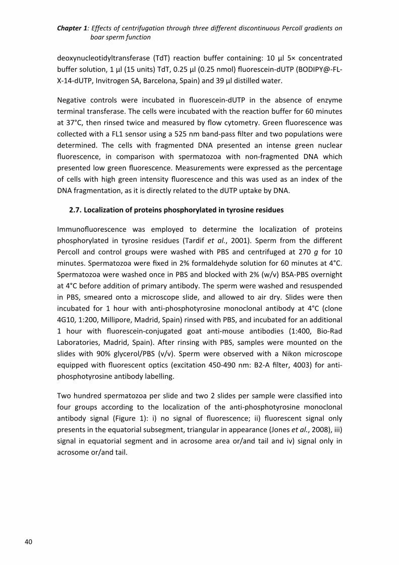
Chapter 1: Effects of centrifugation through three different discontinuous Percoll gradients on boar sperm function
40
deoxynucleotidyltransferase (TdT) reaction buffer containing: 10 µl 5× concentrated
buffer solution, 1 µl (15 units) TdT, 0.25 µl (0.25 nmol) fluorescein‐dUTP (BODIPY@‐FL‐
X‐14‐dUTP, Invitrogen SA, Barcelona, Spain) and 39 µl distilled water.
Negative controls were incubated in fluorescein‐dUTP in the absence of enzyme
terminal transferase. The cells were incubated with the reaction buffer for 60 minutes
at 37°C, then rinsed twice and measured by flow cytometry. Green fluorescence was
collected with a FL1 sensor using a 525 nm band‐pass filter and two populations were
determined. The cells with fragmented DNA presented an intense green nuclear
fluorescence, in comparison with spermatozoa with non‐fragmented DNA which
presented low green fluorescence. Measurements were expressed as the percentage
of cells with high green intensity fluorescence and this was used as an index of the
DNA fragmentation, as it is directly related to the dUTP uptake by DNA.
2.7. Localization of proteins phosphorylated in tyrosine residues
Immunofluorescence was employed to determine the localization of proteins
phosphorylated in tyrosine residues (Tardif et al., 2001). Sperm from the different
Percoll and control groups were washed with PBS and centrifuged at 270 g for 10
minutes. Spermatozoa were fixed in 2% formaldehyde solution for 60 minutes at 4°C.
Spermatozoa were washed once in PBS and blocked with 2% (w/v) BSA‐PBS overnight
at 4°C before addition of primary antibody. The sperm were washed and resuspended
in PBS, smeared onto a microscope slide, and allowed to air dry. Slides were then
incubated for 1 hour with anti‐phosphotyrosine monoclonal antibody at 4°C (clone
4G10, 1:200, Millipore, Madrid, Spain) rinsed with PBS, and incubated for an additional
1 hour with fluorescein‐conjugated goat anti‐mouse antibodies (1:400, Bio‐Rad
Laboratories, Madrid, Spain). After rinsing with PBS, samples were mounted on the
slides with 90% glycerol/PBS (v/v). Sperm were observed with a Nikon microscope
equipped with fluorescent optics (excitation 450‐490 nm: B2‐A filter, 4003) for anti‐
phosphotyrosine antibody labelling.
Two hundred spermatozoa per slide and two 2 slides per sample were classified into
four groups according to the localization of the anti‐phosphotyrosine monoclonal
antibody signal (Figure 1): i) no signal of fluorescence; ii) fluorescent signal only
presents in the equatorial subsegment, triangular in appearance (Jones et al., 2008), iii)
signal in equatorial segment and in acrosome area or/and tail and iv) signal only in
acrosome or/and tail.
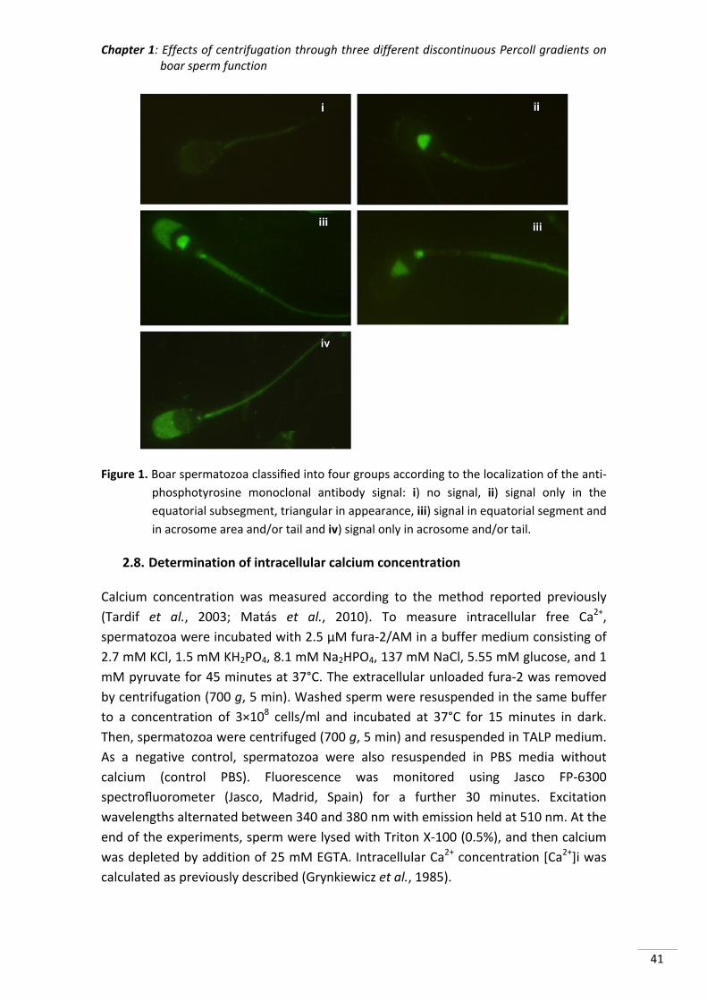
Chapter 1: Effects of centrifugation through three different discontinuous Percoll gradients on boar sperm function
41
Figure 1. Boar spermatozoa classified into four groups according to the localization of the anti‐
phosphotyrosine monoclonal antibody signal: i) no signal, ii) signal only in the
equatorial subsegment, triangular in appearance, iii) signal in equatorial segment and
in acrosome area and/or tail and iv) signal only in acrosome and/or tail.
2.8. Determination of intracellular calcium concentration
Calcium concentration was measured according to the method reported previously
(Tardif et al., 2003; Matás et al., 2010). To measure intracellular free Ca2+,
spermatozoa were incubated with 2.5 µM fura‐2/AM in a buffer medium consisting of
2.7 mM KCl, 1.5 mM KH2PO4, 8.1 mM Na2HPO4, 137 mM NaCl, 5.55 mM glucose, and 1
mM pyruvate for 45 minutes at 37°C. The extracellular unloaded fura‐2 was removed
by centrifugation (700 g, 5 min). Washed sperm were resuspended in the same buffer
to a concentration of 3×10⁸ cells/ml and incubated at 37°C for 15 minutes in dark.
Then, spermatozoa were centrifuged (700 g, 5 min) and resuspended in TALP medium.
As a negative control, spermatozoa were also resuspended in PBS media without
calcium (control PBS). Fluorescence was monitored using Jasco FP‐6300
spectrofluorometer (Jasco, Madrid, Spain) for a further 30 minutes. Excitation
wavelengths alternated between 340 and 380 nm with emission held at 510 nm. At the
end of the experiments, sperm were lysed with Triton X‐100 (0.5%), and then calcium
was depleted by addition of 25 mM EGTA. Intracellular Ca2+ concentration [Ca2+]i was
calculated as previously described (Grynkiewicz et al., 1985).
Chapter 1: Effects of centrifugation through three different discontinuous Percoll gradients on boar sperm function
42
The equation used for calculation was as follows: [Ca2+]i = Kd × (R − Rmin/Rmax − R) ×
Sf/Sb where R indicates the fluorescence ratio 340/380 nm λ; Rmin indicates the
minimum values of fluorescence; Rmax indicates the maximal fluorescence values; Sf
indicates the fluorescence intensity in the Ca2+‐free medium (excitation 380 nm λ); Sb
indicates the fluorescence intensity under Ca2+ saturation conditions and Kd (224 nmol)
is the Ca2+ constant dissociation for Fura‐2AM. For the statistical analysis the calcium
concentration (nM/l) was recorded t from 0 to 1800 s and every 30 s for every
experimental group and replicate. Finally the mean value during the incubation period
was calculated.
2.9. In vitro maturation and fertilization
Oocyte collection and in vitro maturation
Within 30 minutes of slaughter, ovaries from prepubertal gilts were transported to the
laboratory in saline (0.9% (w/v) NaCl) containing 100 µg/ml kanamycin sulphate at
37°C, washed once in 0.04% (w/v) cetrimide solution and then twice in saline.
Cumulus‐oocyte complexes were collected from non‐atretic follicles (3‐6 mm
diameter), washed twice in DPBS supplemented with 4 mg/ml polyvinyl alcohol (PVA),
and twice more in maturation medium previously equilibrated for at least 3 hours at
38.5°C under 5% CO2 in 100% humidified air. Only oocytes harvested within 2 hours of
slaughter with a complete, dense cumulus oophorus were matured (Matás et al.,
1996). The medium used for oocyte maturation was NCSU‐37 (Petters & Wells, 1993);
supplemented with 0.57 mmol/l cysteine, 1 mmol/l dibutyryl cAMP, 5 µg/ml insulin, 50
µmol/l β‐mercaptoethanol, 10 IU/ml equine chorionic gonadotrophin (Foligon, Intervet
International BV, Boxmeer), 10 IU/ml human chorionic gonadotrophin (Veterin Corion,
Divasa Farmavic, Barcelona, Spain), and 10% (v/v) pig follicular fluid. Groups of 50
oocytes were cultured in 500 µl maturation medium for 20‐22 hours at 38.5°C and 5%
CO2 in air. Once cultured, the oocytes were washed twice, transferred to fresh
maturation medium without hormones or dibutyryl cAMP, and cultured for a further
20‐22 hours. Under these conditions the efficiency of oocyte maturation is higher than
90% (Coy et al., 1999; Coy & Romar, 2002).
In vitro fertilization
After maturation oocytes were mechanically stripped of cumulus by gentle aspiration
with a pipette. Denuded oocytes were washed three times in TALP medium and groups
of 25‐30 oocytes transferred to each well of four‐well Nunc multidishes (Nunc,
Roskilde, Denmark) containing 250 µl TALP medium previously equilibrated at 38.5°C
under 5% CO2 (Matás et al., 2003, 2010). Sperm suspensions (250 µl) from each
treatment group were added to the fertilization wells to obtain a final concentration of
1×10⁵ cells/ml. At 18‐20 hours post‐insemination, putative zygotes were fixed for 30
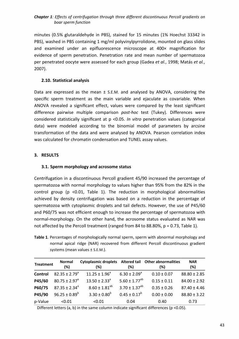
Chapter 1: Effects of centrifugation through three different discontinuous Percoll gradients on boar sperm function
43
minutes (0.5% glutaraldehyde in PBS), stained for 15 minutes (1% Hoechst 33342 in
PBS), washed in PBS containing 1 mg/ml polyvinylpyrrolidone, mounted on glass slides
and examined under an epifluorescence microscope at 400× magnification for
evidence of sperm penetration. Penetration rate and mean number of spermatozoa
per penetrated oocyte were assessed for each group (Gadea et al., 1998; Matás et al.,
2007).
2.10. Statistical analysis
Data are expressed as the mean ± S.E.M. and analysed by ANOVA, considering the
specific sperm treatment as the main variable and ejaculate as covariable. When
ANOVA revealed a significant effect, values were compared by the least significant
difference pairwise multiple comparison post‐hoc test (Tukey). Differences were
considered statistically significant at p <0.05. In vitro penetration values (categorical
data) were modeled according to the binomial model of parameters by arcsine
transformation of the data and were analysed by ANOVA. Pearson correlation index
was calculated for chromatin condensation and TUNEL assay values.
3. RESULTS
3.1. Sperm morphology and acrosome status
Centrifugation in a discontinuous Percoll gradient 45/90 increased the percentage of
spermatozoa with normal morphology to values higher than 95% from the 82% in the
control group (p <0.01, Table 1). The reduction in morphological abnormalities
achieved by density centrifugation was based on a reduction in the percentage of
spermatozoa with cytoplasmic droplets and tail defects. However, the use of P45/60
and P60/75 was not efficient enough to increase the percentage of spermatozoa with
normal‐morphology. On the other hand, the acrosome status evaluated as NAR was
not affected by the Percoll treatment (ranged from 84 to 88.80%, p = 0.73, Table 1).
Table 1. Percentages of morphologically normal sperm, sperm with abnormal morphology and
normal apical ridge (NAR) recovered from different Percoll discontinuous gradient
systems (mean values ± S.E.M.).
Treatment Normal (%)
Cytoplasmic droplets (%)
Altered tail (%)
Other abnormalities (%)
NAR (%)
Control 82.35 ± 2.79a 11.25 ± 1.96a 6.30 ± 2.09a 0.10 ± 0.07 88.80 ± 2.85
P45/60 80.75 ± 2.97a 13.50 ± 2.33a 5.60 ± 1.77ab 0.15 ± 0.11 84.00 ± 2.92
P60/75 87.35 ± 2.34a 8.60 ± 1.81ab 3.70 ± 1.37ab 0.35 ± 0.26 87.40 ± 4.46
P45/90 96.25 ± 0.89b 3.30 ± 0.80b 0.45 ± 0.17b 0.00 ± 0.00 88.80 ± 3.22
p‐Value <0.01 <0.01 0.04 0.40 0.73
Different letters (a, b) in the same column indicate significant differences (p <0.05).
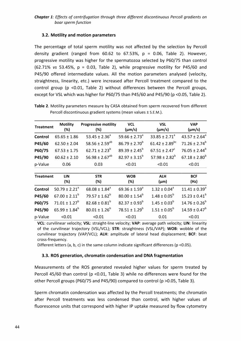
Chapter 1: Effects of centrifugation through three different discontinuous Percoll gradients on boar sperm function
44
3.2. Motility and motion parameters
The percentage of total sperm motility was not affected by the selection by Percoll
density gradient (ranged from 60.62 to 67.53%, p = 0.06, Table 2). However,
progressive motility was higher for the spermatozoa selected by P60/75 than control
(62.71% vs 53.45%, p = 0.03, Table 2), while progressive motility for P45/60 and
P45/90 offered intermediate values. All the motion parameters analysed (velocity,
straightness, linearity, etc.) were increased after Percoll treatment compared to the
control group (p <0.01, Table 2) without differences between the Percoll groups,
except for VSL which was higher for P60/75 than P45/60 and P45/90 (p <0.05, Table 2).
Table 2. Motility parameters measure by CASA obtained from sperm recovered from different
Percoll discontinuous gradient systems (mean values ± S.E.M.).
Treatment Motility (%)
Progressive motility(%)
VCL (µm/s)
VSL (µm/s)
VAP (µm/s)
Control 65.65 ± 1.86 53.45 ± 2.36a 59.66 ± 2.73a 33.85 ± 2.71a 43.57 ± 2.64a
P45/60 62.50 ± 2.04 58.56 ± 2.59ab 86.79 ± 2.70b 61.42 ± 2.89bc 71.26 ± 2.74b
P60/75 67.53 ± 1.75 62.71 ± 2.23b 89.39 ± 2.45b 67.51 ± 2.47c 76.05 ± 2.44b
P45/90 60.62 ± 2.10 56.98 ± 2.67ab 82.97 ± 3.15b 57.98 ± 2.82b 67.18 ± 2.80b
p‐Value 0.06 0.03 <0.01 <0.01 <0.01
Treatment LIN (%)
STR(%)
WOB(%)
ALH(µm)
BCF (Hz)
Control 50.79 ± 2.21a 68.08 ± 1.84a 69.36 ± 1.59a 1.32 ± 0.04a 11.41 ± 0.39a
P45/60 67.00 ± 2.11b 79.57 ± 1.62b 80.00 ± 1.54b 1.48 ± 0.05b 15.23 ± 0.41b
P60/75 71.01 ± 1.27b 82.68 ± 0.81b 82.37 ± 0.93b 1.45 ± 0.03b 14.76 ± 0.26b
P45/90 65.99 ± 1.84b 80.01 ± 1.26b 78.51 ± 1.29b 1.51 ± 0.05b 14.59 ± 0.47b
p‐Value <0.01 <0.01 <0.01 0.01 <0.01
VCL: curvilinear velocity; VSL: straight‐line velocity; VAP: average path velocity; LIN: linearity of the curvilinear trajectory (VSL/VCL); STR: straightness (VSL/VAP); WOB: wobble of the curvilinear trajectory (VAP/VCL); ALH: amplitude of lateral head displacement; BCF: beat cross‐frequency. Different letters (a, b, c) in the same column indicate significant differences (p <0.05).
3.3. ROS generation, chromatin condensation and DNA fragmentation
Measurements of the ROS generated revealed higher values for sperm treated by
Percoll 45/60 than control (p <0.01, Table 3) while no differences were found for the
other Percoll groups (P60/75 and P45/90) compared to control (p >0.05, Table 3).
Sperm chromatin condensation was affected by the Percoll treatments; the chromatin
after Percoll treatments was less condensed than control, with higher values of
fluorescence units that correspond with higher IP uptake measured by flow cytometry
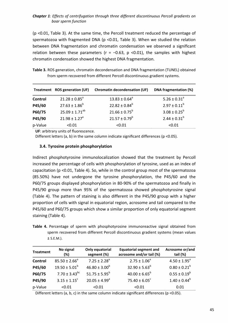
Chapter 1: Effects of centrifugation through three different discontinuous Percoll gradients on boar sperm function
45
(p <0.01, Table 3). At the same time, the Percoll treatment reduced the percentage of
spermatozoa with fragmented DNA (p <0.01, Table 3). When we studied the relation
between DNA fragmentation and chromatin condensation we observed a significant
relation between these parameters (r = −0.63, p <0.01), the samples with highest
chromatin condensation showed the highest DNA fragmentation.
Table 3. ROS generation, chromatin decondensation and DNA fragmentation (TUNEL) obtained
from sperm recovered from different Percoll discontinuous gradient systems.
Treatment ROS generation (UF) Chromatin decondensation (UF) DNA fragmentation (%)
Control 21.28 ± 0.85a 13.83 ± 0.64a 5.26 ± 0.31a
P45/60 27.63 ± 1.86b 22.82 ± 0.84b 2.97 ± 0.11b
P60/75 25.09 ± 1.71ab 21.66 ± 0.75b 3.08 ± 0.25b
P45/90 21.98 ± 1.27a 21.57 ± 0.79b 2.44 ± 0.31b
p‐Value <0.01 <0.01 <0.01
UF: arbitrary units of fluorescence. Different letters (a, b) in the same column indicate significant differences (p <0.05).
3.4. Tyrosine protein phosphorylation
Indirect phosphotyrosine immunolocalization showed that the treatment by Percoll
increased the percentage of cells with phosphorylation of tyrosine, used as an index of
capacitation (p <0.01, Table 4). So, while in the control group most of the spermatozoa
(85.50%) have not undergone the tyrosine phosphorylation, the P45/60 and the
P60/75 groups displayed phosphorylation in 80‐90% of the spermatozoa and finally in
P45/90 group more than 95% of the spermatozoa showed phosphotyrosine signal
(Table 4). The pattern of staining is also different in the P45/90 group with a higher
proportion of cells with signal in equatorial region, acrosome and tail compared to the
P45/60 and P60/75 groups which show a similar proportion of only equatorial segment
staining (Table 4).
Table 4. Percentage of sperm with phosphotyrosine immunoreactive signal obtained from
sperm recovered from different Percoll discontinuous gradient systems (mean values
± S.E.M.).
Treatment No signal
(%) Only equatorial segment (%)
Equatorial segment and acrosome and/or tail (%)
Acrosome or/and tail (%)
Control 85.50 ± 2.66a 7.25 ± 2.28a 2.75 ± 1.06a 4.50 ± 1.95a
P45/60 19.50 ± 5.01b 46.80 ± 3.00b 32.90 ± 5.63b 0.80 ± 0.21b
P60/75 7.70 ± 3.43bc 51.75 ± 5.95b 40.00 ± 6.65b 0.55 ± 0.19b
P45/90 3.15 ± 1.15c 20.05 ± 4.99a 75.40 ± 6.05c 1.40 ± 0.44b
p‐Value <0.01 <0.01 <0.01 0.01
Different letters (a, b, c) in the same column indicate significant differences (p <0.05).
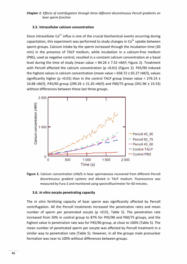
Chapter 1: Effects of centrifugation through three different discontinuous Percoll gradients on boar sperm function
46
3.5. Intracellular calcium concentration
Since intracellular Ca2+ influx is one of the crucial biochemical events occurring during
capacitation, this experiment was performed to study changes in Ca2+ uptake between
sperm groups. Calcium intake by the sperm increased through the incubation time (30
min) in the presence of TALP medium, while incubation in a calcium‐free medium
(PBS), used as negative control, resulted in a constant calcium concentration at a basal
level during the time of study (mean value = 84.26 ± 7.52 nM/l; Figure 2). Treatment
with Percoll affected the calcium concentration (p <0.01) (Figure 2). P45/90 induced
the highest values in calcium concentration (mean value = 658.72 ± 65.27 nM/l), values
significantly higher (p <0.01) than in the control TALP group (mean value = 276.14 ±
16.68 nM/l), P45/60 group (299.26 ± 21.20 nM/l) and P60/75 group (341.96 ± 23.53)
without differences between these last three groups.
Figure 2. Calcium concentration (nM/l) in boar spermatozoa recovered from different Percoll
discontinuous gradient systems and diluted in TALP medium. Fluorescence was
measured by Fura‐2 and monitored using spectrofluorimeter for 60 minutes.
3.6. In vitro oocyte penetrating capacity
The in vitro fertilizing capacity of boar sperm was significantly affected by Percoll
centrifugation. All the Percoll treatments increased the penetration rates and mean
number of sperm per penetrated oocyte (p <0.01, Table 5). The penetration rate
increased from 50% in control group to 87% for P45/90 and P60/75 groups, and the
highest value in penetration rate was for P45/90 group, at close to 100% (Table 5). The
mean number of penetrated sperm per oocyte was affected by Percoll treatment in a
similar way to penetration rate (Table 5). However, in all the groups male pronuclear
formation was near to 100% without differences between groups.
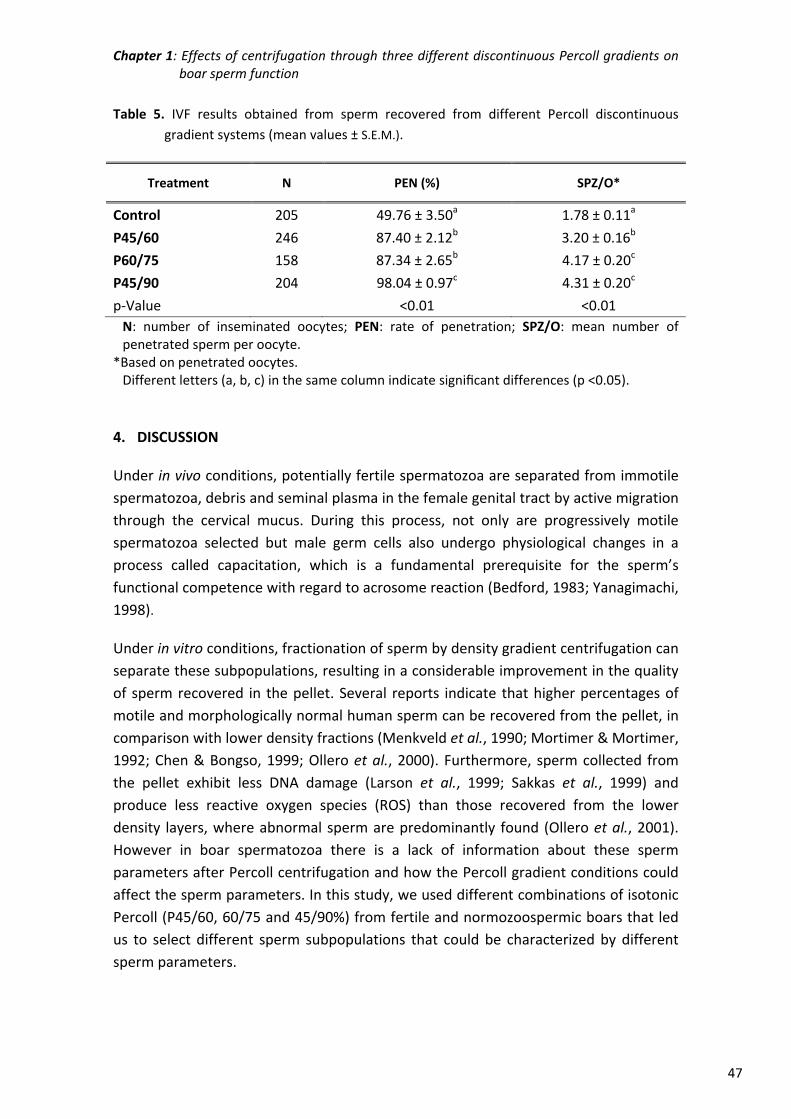
Chapter 1: Effects of centrifugation through three different discontinuous Percoll gradients on boar sperm function
47
Table 5. IVF results obtained from sperm recovered from different Percoll discontinuous
gradient systems (mean values ± S.E.M.).
Treatment N PEN (%) SPZ/O*
Control 205 49.76 ± 3.50a 1.78 ± 0.11a
P45/60 246 87.40 ± 2.12b 3.20 ± 0.16b
P60/75 158 87.34 ± 2.65b 4.17 ± 0.20c
P45/90 204 98.04 ± 0.97c 4.31 ± 0.20c
p‐Value <0.01 <0.01
N: number of inseminated oocytes; PEN: rate of penetration; SPZ/O: mean number of penetrated sperm per oocyte.
*Based on penetrated oocytes. Different letters (a, b, c) in the same column indicate significant differences (p <0.05).
4. DISCUSSION
Under in vivo conditions, potentially fertile spermatozoa are separated from immotile
spermatozoa, debris and seminal plasma in the female genital tract by active migration
through the cervical mucus. During this process, not only are progressively motile
spermatozoa selected but male germ cells also undergo physiological changes in a
process called capacitation, which is a fundamental prerequisite for the sperm’s
functional competence with regard to acrosome reaction (Bedford, 1983; Yanagimachi,
1998).
Under in vitro conditions, fractionation of sperm by density gradient centrifugation can
separate these subpopulations, resulting in a considerable improvement in the quality
of sperm recovered in the pellet. Several reports indicate that higher percentages of
motile and morphologically normal human sperm can be recovered from the pellet, in
comparison with lower density fractions (Menkveld et al., 1990; Mortimer & Mortimer,
1992; Chen & Bongso, 1999; Ollero et al., 2000). Furthermore, sperm collected from
the pellet exhibit less DNA damage (Larson et al., 1999; Sakkas et al., 1999) and
produce less reactive oxygen species (ROS) than those recovered from the lower
density layers, where abnormal sperm are predominantly found (Ollero et al., 2001).
However in boar spermatozoa there is a lack of information about these sperm
parameters after Percoll centrifugation and how the Percoll gradient conditions could
affect the sperm parameters. In this study, we used different combinations of isotonic
Percoll (P45/60, 60/75 and 45/90%) from fertile and normozoospermic boars that led
us to select different sperm subpopulations that could be characterized by different
sperm parameters.
Chapter 1: Effects of centrifugation through three different discontinuous Percoll gradients on boar sperm function
48
In relation to sperm morphology, in human spermatozoa, Percoll gradient
centrifugation reduces the percentage of morphological alterations, particularly when
teratospermia is not severe (Hall et al., 1995). In boar sperm, Percoll density gradient
has been used to isolate sperm subpopulations with cytoplasmic droplet (low‐density)
from spermatozoa without this droplet (higher density) (de Vries & Colenbrander,
1990). On the contrary, a reduction in sperm abnormalities other than cytoplasmic
droplets, such as tail loops, after Percoll 35/70 has been previously reported (Waberski
et al., 2006). Under our experimental conditions only Percoll 45/90 is able to induce a
significant reduction in both kinds of morphoanomalies, results that are in agreement
with those reported in humans (Buffone et al., 2004). Analysis of the NAR showed that
just after Percoll centrifugation no changes were detected in acrosome status, in
agreement with data obtained previously (Grant et al., 1994), although when frozen‐
thawed spermatozoa were used and showed severe acrosome damage the use of
Percoll centrifugation improved the NAR status compared to swim‐up selection (Zheng
et al., 1992).
The effect of Percoll in selecting porcine sperm with higher motility has been well
documented (Berger & Parker, 1989; Horan et al., 1991; Grant et al., 1994; Waberski et
al., 2006). However, other studies have not detected significant differences. This is
probably related to the high variability that could be present in some boars,
differences in the Percoll density procedures (Berger & Horton, 1988; Suzuki & Nagai,
2003) or differences in the system of measurement. In this study, we used a high
speed camera (50 images/s, Basler AG, Ahrensburg, Germany) and a high number of
images (100 images) per analysis, to more extensively and accurately follow the sperm
motility pattern unlike previous studies (camera 25 pictures/s, 25 pictures analysed).
As a consequence of this setting, the ALH values are lower and BCF higher than in
previous studies (Gadea et al., 2005b). On the other hand, the use of a capacitating
media as TALP medium (Rath et al., 1999), with a high level of bicarbonate and calcium
in the composition, induced an early capacitation process that implies the presence of
aggregates or pairs of spermatozoa. These sperm aggregates are not well recognized
by the software because the total area of the particles is higher than setup. In
consequence, the percentage of total and progressive motility could be
underestimated, but it did not affect the motion parameters.
Only the gradient 60/75 selected higher percentage of progressive motility
spermatozoa than control and it could be related to a selective effect on motility
produced by the top layer of Percoll 60% that is not done by Percoll 45%. However, the
differences in the pattern of movement reported in this study showed that the Percoll
selection is important to optimize recovery of a higher percentage of spermatozoa
with a fast‐linear movement which could be related to the capacitation process as a
significant increase of tyrosine phosphorylation and calcium intake was observed. This
sperm subpopulation with fast motion characteristics could be related to the fertilizing
Chapter 1: Effects of centrifugation through three different discontinuous Percoll gradients on boar sperm function
49
capacity (Davis et al., 1995; Quintero‐Moreno et al., 2004; Jedrzejczak et al., 2005).
The effect of Percoll on ROS generation could be complex. Centrifugation and
resuspension of the resulting pellet subject cells to mechanical forces due to close
packing (Abidor et al., 1994), and centrifugation could lead to sublethal damage
(Alvarez et al., 1993). Centrifugation can also be detrimental to the spermatozoa
because of the production of a burst of reactive oxygen species by a discrete
subpopulation of cells characterized by significantly diminished motility and fertilizing
capacity (Aitken & Clarkson, 1988) and the elimination of the antioxidant compounds
that are present in the seminal plasma. On the other hand, Percoll separation could
compensate by eliminating dead cells, cells with cytoplasmic droplets and debris that
generate a high proportion of ROS. These data overall suggest that the best balance is
for P45/90 and the worse results are seen for P45/60, probably because it is not the
most effective procedure for eliminating the damaged cells or because the packing is
closer and the subsequent mechanical forces higher. This is in concordance with
studies in humans that show that these fractions display higher ROS production
(Aitken & Clarkson, 1988; Ollero et al., 2001).
Another interesting point is the fact that capacitating spermatozoa produce controlled
amounts of ROS that regulate downstream events such as an increase in cAMP, protein
kinase A (PKA) activation and phosphorylation of PKA substrates (de Lamirande &
O’Flaherty, 2008). It is possible that ROS generation in spermatozoa after Percoll 45/90
could be related to the capacitation process while in the other experimental groups
ROS generation could be related to the presence of dead cells and cytoplasmic
droplets. However this hypothesis must be confirmed in further studies.
The subpopulations of spermatozoa selected by Percoll gradient differ not only in
terms of conventional criteria such as motility and morphology, but also in terms of
their DNA integrity as measured in human studies by different techniques such as the
SCSA (Ollero et al., 2001), Comet assay (Van Kooij et al., 2004) and TUNEL (Mitchell et
al., 2011). In humans the selection of spermatozoa by density gradient centrifugation
also increases the percentage of mature spermatozoa with normal chromatin
condensation compared to control (Le Lannou & Blanchard, 1988; Hammadeh et al.,
2001; Mitchell et al., 2011). We observed that chromatin condensation and DNA
fragmentation were similar among Percoll separated samples but better than in the
control group, which suggests that when Percoll density is used to select motile and
viable spermatozoa it indirectly selects spermatozoa with a nuclear normal structure
and DNA (Mitchell et al., 2011). We also reported in this study a significant relationship
between chromatin condensation and DNA fragmentation, as previously studied in
different species (Fraser & Strzeżek, 2007; Gadea et al., 2008; Mitchell et al., 2011)
that suggests that abnormally condensed spermatozoa chromatin is also related with
higher DNA fragmentation.
Chapter 1: Effects of centrifugation through three different discontinuous Percoll gradients on boar sperm function
50
Spermatozoa washed on a Percoll gradient are partly capacitated (Berger & Parker,
1989) or have undergone the early stages of the acrosome reaction (de Maistre et al.,
1996). Protein tyrosine phosphorylation has been associated with the ability of normal
sperm to undergo capacitation and fertilize an egg (Urner & Sakkas, 2003). The pattern
of tyrosine phosphorylation in boar is different from other species where the flagellum
appears to be the principal sperm compartment presenting tyrosine‐phosphorylated
proteins. In boar the flagellum is less important (Petrunkina et al., 2001; Tardif et al.,
2001) and the most important area is the equatorial area, which shows a specific
triangular pattern of tyrosine phosphorylated proteins (Jones et al., 2008).
Fluorescence is also present over the anterior acrosome, partly due to nonspecific
binding of the second layer antibody and partly due to phosphoproteins within the
acrosomal matrix (Tardif et al., 2001; Jones et al., 2008). In this study we confirmed
that Percoll increases the percentage of cells with phosphorylated tyrosine and that
the pattern of staining is different in the different Percoll procedures, which may play a
key role in sperm‐zona pellucida interaction (Flesch et al., 2001). In Percoll 45/90 the
percentage of cells with any kind of tyrosine phosphorylation is near to 100% and the
signal extends to most of the spermatozoon (acrosome, equatorial segment and tail).
In other Percoll groups (45/60 and 60/75), although the percentage of phosphorylated
cells is higher than 75%, the extension of the signal is lower than P45/90. In
accordance with other authors (Petrunkina et al., 2001; Urner & Sakkas, 2003), we
suggest that the phosphorylation‐positive spermatozoa could have a competitive
advantage for penetrating the oocytes. However, this fact must be confirmed in
further studies, including, for example, in vitro production of embryos.
Changes in intracellular calcium concentrations are associated with different aspects of
sperm function such as capacitation, acrosome reaction, sperm chemotaxis and
hyperactivation (Bedu‐Addo et al., 2008; Costello et al., 2009). In this study we
measured calcium in spermatozoa treated by Percoll density gradient and obtained
results that follow the same pattern as those obtained in the study of tyrosine‐
phosphorylated proteins. The higher values obtained in P45/90 group suggests that
the spermatozoa completed the capacitation process properly and are more capable
of proceeding with the fertilization process.
The Percoll procedure yields pure fractions of spermatozoa, whereas the washing
technique produces cell pellets that contain a heterogeneous population of
spermatozoa along with amorphous material and seminal plasma (Grant et al., 1994).
Percoll centrifugation not only selected a sperm subpopulation but also allowed some
“decapacitation” factors to be removed from the sperm membrane and/or seminal
plasma (Furimsky et al., 2005), which could facilitate subsequent sperm capacitation
and acrosome reaction (Nolan & Hammerstedt, 1997).
Chapter 1: Effects of centrifugation through three different discontinuous Percoll gradients on boar sperm function
51
Several studies have previously demonstrated that Percoll centrifugation leads to
selection of a subpopulation of sperm with a higher ability to penetrate oocytes in vitro
(Berger & Horton, 1988; Berger & Parker, 1989; Grant et al., 1994; Jeong & Yang, 2001;
Suzuki & Nagai, 2003). Previously, we have shown that fresh spermatozoa treated with
Percoll showed faster oocyte penetration and higher penetrability than control; the
time of female and male pronucleus formation was affected by the sperm treatment
and was faster for the Percoll group, while cleavage rate and rate of blastocyst
formation were also higher after Percoll treatment (Matás et al., 2003). These
differences are related to changes in some important spermatozoa parameters such as
motility parameters, calcium intake, ROS generation, lipid membrane disorder and
acrosome reaction (Matás et al., 2010). However, Percoll gradient did not improve
embryo development after ICSI injection (García‐Roselló et al., 2006).
Among all the sperm tests that have been developed, IVF tests are the most suitable
for assessing overall sperm function during fertilization. The binding and penetration of
the zona pellucida is one of the most important barriers the spermatozoa must
overcome in the fertilization process. Also, the interaction with the oocyte plasma
membrane appears to explain much of the variability in sperm fertilizing potential
(Gadea et al., 2005b). We used the IVF system to evaluate the penetration capacity
and assumption that an elevated number of sperm could penetrate the oocytes. As we
expected, the highest values for penetration were obtained in Percoll 45/90 because
all the sperm parameters were better than or equal to control. However for Percoll
45/60, where similar values for morphology, worse values for in ROS generation and
better motility, chromatin condensation, DNA and capacitation than control were
observed, there was a higher penetration rate than control.
In the present study, three combinations of Percoll gradient density were tested and as
expected, the highest penetration rates were observed after pretreatment with Percoll
45/90. Firstly, this procedure selected the highest number of morphologically normal
spermatozoa with less alteration in the chromatin structure and less DNA damage; the
motion characteristics are also improved and the percentage of capacitated
spermatozoa steadily increases throughout incubation. All these characteristics lead to
the highest proportion of penetration of oocytes in vitro.
ACKNOWLEDGEMENTS
This research was supported by MEC‐FEDER (AGL2009‐12512‐C02‐01) and Fundación
Séneca (08752/PI/08). Thanks to Ruth Tunn (University of Oxford) for help with the
language in the text.
Chapter 1: Effects of centrifugation through three different discontinuous Percoll gradients on boar sperm function
52
REFERENCES
Abidor I, Li L & Hui S (1994). Studies of cell pellets: I. Electrical properties and porosity.
Biophysical journal. 67: 418‐426. (doi: 10.1016/S0006‐3495(94)80497‐7).
Aitken R & Clarkson JS (1988). Significance of reactive oxygen species and antioxidants in
defining the efficacy of sperm preparation techniques. Journal of andrology. 9: 367‐
376. (doi: 10.1002/j.1939‐4640.1988.tb01067.x).
Alvarez JG, Lasso JL, Blasco L, Nuñez RC, Heyner S, Caballero PP & Storey BT (1993).
Centrifugation of human spermatozoa induces sublethal damage; separation of human
spermatozoa from seminal plasma by a dextran swim‐up procedure without
centrifugation extends their motile lifetime. Human reproduction. 8: 1087‐1092.
Bedford J (1983). Significance of the need for sperm capacitation before fertilization in
eutherian mammals. Biology of reproduction. 28: 108‐120. (doi: 10.1095/
biolreprod28.1.108).
Bedu‐Addo K, Costello S, Harper C, Machado‐Oliveira G, Lefievre L, Ford C, Barratt C &
Publicover S (2008). Mobilisation of stored calcium in the neck region of human
sperm‐a mechanism for regulation of flagellar activity. International Journal of
Developmental Biology. 52: 615‐626. (doi: 10.1387/ijdb.072535kb).
Berger T & Horton M (1988). Evaluation of assay conditions for the zona‐free hamster ova
bioassay of boar sperm fertility. Gamete research. 19: 101‐111. (doi:
10.1002/mrd.1120190110).
Berger T & Parker K (1989). Modification of the zona‐free hamster ova bioassay of boar sperm
fertility and correlation with in vivo fertility. Gamete research. 22: 385‐397. (doi:
10.1002/mrd.1120220405).
Biotech (1996). Important Notice: Percoll NOT to be Used in Assisted Reproduction
Techologies in Humans. Pharmacia Biotech Inc.
Buffone M, Doncel G, Briggiler CM, Vazquez‐Levin M & Calamera J (2004). Human sperm
subpopulations: relationship between functional quality and protein tyrosine
phosphorylation. Human reproduction. 19: 139‐146. (doi: 10.1093/humrep/deh040).
Bussalleu E, Pinart E, Rivera M, Briz M, Sancho S, Yeste M, Casas I, Fàbrega A, Rigau T &
Rodriguez‐Gil J (2009). Effects of matrix filtration of low‐quality boar semen doses on
sperm quality. Reproduction in Domestic Animals. 44: 499‐503. (doi: 10.1111/j.1439‐
0531.2008.01221.x).
Costello S, Michelangeli F, Nash K, Lefievre L, Morris J, Machado‐Oliveira G, Barratt C,
Kirkman‐Brown J & Publicover S (2009). Ca2+‐stores in sperm: their identities and
functions. Reproduction. 138: 425‐437. (doi: 10.1530/REP‐09‐0134).
Coy P, Ruiz S, Romar R, Campos I & Gadea J (1999). Maturation, fertilization and complete
development of porcine oocytes matured under different systems. Theriogenology. 51:
799‐812. (doi: 10.1016/S0093‐691X(99)00028‐X).
Coy P & Romar R (2002). In vitro production of pig embryos: a point of view. Reproduction,
fertility and development. 14: 275‐286. (doi: 10.1071/RD01102).
Chen MJ & Bongso A (1999). Comparative evaluation of two density gradient preparations for
sperm separation for medically assisted conception. Human reproduction. 14: 759‐764.
(doi: 10.1093/humrep/14.3.759).
Chapter 1: Effects of centrifugation through three different discontinuous Percoll gradients on boar sperm function
53
Davis R, Drobnis E & Overstreet J (1995). Application of multivariate cluster, discriminate
function, and stepwise regression analyses to variable selection and predictive
modeling of sperm cryosurvival. Fertility and sterility. 63: 1051‐1057.
de Lamirande E & O’Flaherty C (2008). Sperm activation: role of reactive oxygen species and
kinases. Biochimica et Biophysica Acta (BBA)‐Proteins and Proteomics. 1784: 106‐115.
(doi: 10.1016/j.bbapap.2007.08.024).
de Maistre E, Bene M, Foliguet B, Touati F & Faure G (1996). Centrifugation on Percoll
gradient enhances fluorescent lectin binding on human sperm: a flow cytometric
analysis. Systems Biology in Reproductive Medicine. 37: 179‐187. (doi:
10.3109/01485019608988520).
de Vries AC & Colenbrander B (1990). Isolation and characterization of boar spermatozoa with
and without a cytoplasmic droplet. International Journal of Biochemistry. 22: 519‐524.
(doi: 10.1016/0020‐711X(90)90267‐7).
Flesch F, Wijnand E, Van de Lest C, Colenbrander B, van Golde LM & Gadella BM (2001).
Capacitation dependent activation of tyrosine phosphorylation generates two sperm
head plasma membrane proteins with high primary binding affinity for the zona
pellucida. Molecular reproduction and development. 60: 107‐115. (doi:
10.1002/mrd.1067).
Fraser L & Strzeżek J (2007). Is there a relationship between the chromatin status and DNA
fragmentation of boar spermatozoa following freezing–thawing? Theriogenology. 68:
248‐257. (doi: 10.1016/j.theriogenology.2007.05.001).
Furimsky A, Vuong N, Xu H, Kumarathasan P, Xu M, Weerachatyanukul W, Khalil MB, Kates
M & Tanphaichitr N (2005). Percoll gradient‐centrifuged capacitated mouse sperm
have increased fertilizing ability and higher contents of sulfogalactosylglycerolipid and
docosahexaenoic acid‐containing phosphatidylcholine compared to washed
capacitated mouse sperm. Biology of reproduction. 72: 574‐583. (doi: 10.1095/
biolreprod.104.036095).
Gadea J, Matás C & Lucas X (1998). Prediction of porcine semen fertility by homologous in
vitro penetration (hIVP) assay. Animal Reproduction Science. 54: 95‐108. (doi:
10.1016/S0378‐4320(98)00144‐4).
Gadea J, Sellés E, Marco MA, Coy P, Matás C, Romar R & Ruiz S (2004). Decrease in
glutathione content in boar sperm after cryopreservation: effect of the addition of
reduced glutathione to the freezing and thawing extenders. Theriogenology. 62: 690‐
701. (doi: 10.1016/j.theriogenology.2003.11.013).
Gadea J, García‐Vazquez F, Matás C, Gardón JC, Cánovas S & Gumbao D (2005a). Cooling and
freezing of boar spermatozoa: supplementation of the freezing media with reduced
glutathione preserves sperm function. Journal of andrology. 26: 396‐404. (doi:
10.2164/jandrol.04155).
Gadea J, Gumbao D, Matás C & Romar R (2005b). Supplementation of the thawing media with
reduced glutathione improves function and the in vitro fertilizing ability of boar
spermatozoa after cryopreservation. Journal of andrology. 26: 749‐756. (doi:
10.2164/jandrol.05057).
Gadea J, Gumbao D, Canovas S, García‐Vázquez FA, Grullón LA & Gardón JC (2008).
Supplementation of the dilution medium after thawing with reduced glutathione
Chapter 1: Effects of centrifugation through three different discontinuous Percoll gradients on boar sperm function
54
improves function and the in vitro fertilizing ability of frozen‐thawed bull spermatozoa.
International journal of andrology. 31: 40‐49. (doi: 10.1111/j.1365‐
2605.2007.00756.x.).
García‐Roselló E, Matás C, Cánovas S, Moreira PN, Gadea J & Coy P (2006). Influence of sperm
pretreatment on the efficiency of intracytoplasmic sperm injection in pigs. Journal of
andrology. 27: 268‐275. (doi: 10.2164/jandrol.05110).
Gomez E, Buckingham DW, Brindle J, Lanzafame F, Irvine DS & Aitken RJ (1996). Development
of an image analysis system to monitor the retention of residual cytoplasm by human
spermatozoa: correlation with biochemical markers of the cytoplasmic space, oxidative
stress, and sperm function. Journal of andrology. 17: 276‐287. (doi: 10.1002/j.1939‐
4640.1996.tb01783.x).
Grant S, Long S & Parkinson T (1994). Fertilizability and structural properties of boar
spermatozoa prepared by Percoll gradient centrifugation. Journal of reproduction and
fertility. 100: 477‐483. (doi: 10.1530/jrf.0.1000477).
Grynkiewicz G, Poenie M & Tsien RY (1985). A new generation of Ca2+ indicators with greatly
improved fluorescence properties. Journal of Biological Chemistry. 260: 3440‐3450.
Hall J, Fishel S, Timson J, Dowell K & Klentzeris L (1995). Andrology: human sperm
morphology evaluation pre‐and post‐Percoll gradient centrifugation. Human
reproduction. 10: 342‐346.
Hammadeh M, Dehn C, Hippach M, Zeginiadou T, Stieber M, Georg T, Rosenbaum P &
Schmidt W (2001). Comparison between computerized slow‐stage and static liquid
nitrogen vapour freezing methods with respect to the deleterious effect on chromatin
and morphology of spermatozoa from fertile and subfertile men. International journal
of andrology. 24: 66‐72. (doi: 10.1046/j.1365‐2605.2001.00270.x).
Henkel RR & Schill W‐B (2003). Sperm preparation for ART. Reproductive Biology and
Endocrinology. 1: 108. (doi: 10.1186/1477‐7827‐1‐108).
Holt WV (2009). Is semen analysis useful to predict the odds that the sperm will meet the egg?
Reproduction in Domestic Animals. 44:31‐38. (doi: 10.1111/j.1439‐0531.2009.01363.x).
Horan R, Powell R, McQuaid S, Gannon F & Houghton J (1991). Association of foreign DNA
with porcine spermatozoa. Systems Biology in Reproductive Medicine. 26: 83‐92. (doi:
10.3109/01485019108987631).
Jedrzejczak P, Rzepczynska I, Taszarek‐Hauke G, Pawelczyk L, Kotwicka M & Warchot W
(2005). Effect of sperm subpopulation's kinetics on human fertilization in vitro.
Systems Biology in Reproductive Medicine. 51: 185‐193. (doi:
10.1080/014850190898791).
Jeong BS & Yang X (2001). Cysteine, glutathione, and Percoll treatments improve porcine
oocyte maturation and fertilization in vitro. Molecular reproduction and development.
59: 330‐335. (doi: 10.1002/mrd.1038).
Jones R, James PS, Oxley D, Coadwell J, Suzuki‐Toyota F & Howes EA (2008). The equatorial
subsegment in mammalian spermatozoa is enriched in tyrosine phosphorylated
proteins. Biology of reproduction. 79: 421‐431. (doi: 10.1095/biolreprod.107.067314).
King G & Macpherson J (1973). A comparison of two methods for boar semen collection.
Journal of animal science. 36: 563‐565. (doi: 10.2134/jas1973.363563x).
Chapter 1: Effects of centrifugation through three different discontinuous Percoll gradients on boar sperm function
55
Larson K, Brannian J, Timm B, Jost L & Evenson D (1999). Density gradient centrifugation and
glass wool filtration of semen remove spermatozoa with damaged chromatin
structure. Human reproduction. 14: 2015‐2019. (doi: 10.1093/humrep/14.8.2015).
Le Lannou D & Blanchard Y (1988). Nuclear maturity and morphology of human spermatozoa
selected by Percoll density gradient centrifugation or swim‐up procedure. Journal of
reproduction and fertility. 84: 551‐556. (doi: 10.1530/jrf.0.0840551).
Matás C, Martinez E, Vazquez J, Roca J & Gadea J (1996). In vitro penetration assay of boar
sperm fertility: effect of various factors on the penetrability of immature pig oocytes.
Theriogenology. 46: 503‐513. (doi: 10.1016/0093‐691X(96)00172‐0).
Matás C, Coy P, Romar R, Marco M, Gadea J & Ruiz S (2003). Effect of sperm preparation
method on in vitro fertilization in pigs. Reproduction. 125: 133‐141. (doi:
10.1530/rep.0.1250133).
Matás C, Decuadro G, Martinez‐Miro S & Gadea J (2007). Evaluation of a cushioned method
for centrifugation and processing for freezing boar semen. Theriogenology. 67: 1087‐
1091. (doi: 10.1016/j.theriogenology.2006.11.010).
Matás C, Sansegundo M, Ruiz S, García‐Vázquez F, Gadea J, Romar R & Coy P (2010). Sperm
treatment affects capacitation parameters and penetration ability of ejaculated and
epididymal boar spermatozoa. Theriogenology. 74: 1327‐1340. (doi:
10.1016/j.theriogenology.2010.06.002).
Mattioli M, Bacci M, Galeati G & Seren E (1989). Developmental competence of pig oocytes
matured and fertilized in vitro. Theriogenology. 31: 1201‐1207. (doi: 10.1016/0093‐
691X(89)90089‐7).
Menkveld R, Swanson R, Kotze T & Kruger T (1990). Comparison of a discontinuous Percoll
gradient method versus swim‐up method: effects on sperm morphology and other
semen parameters. Andrologia. 22: 152‐158. (doi: 10.1111/j.1439‐
0272.1990.tb01957.x).
Mitchell L, De Iuliis G & Aitken RJ (2011). The TUNEL assay consistently underestimates DNA
damage in human spermatozoa and is influenced by DNA compaction and cell vitality:
development of an improved methodology. International journal of andrology. 34: 2‐
13. (doi: 10.1111/j.1365‐2605.2009.01042.x).
Morrell JM (2006). Update on semen technologies for animal breeding. Reproduction in
Domestic Animals. 41: 63‐67. (doi: 10.1111/j.1439‐0531.2006.00621.x).
Morrell JM, Saravia F, Van Wienen M, Wallgren M & Rodriguez‐Martinez H (2009). Selection
of boar spermatozoa using centrifugation on a glycidoxypropyltrimethoxysilane‐coated
silica colloid. Journal of Reproduction and Development. 55: 547‐552. (doi:
10.1262/jrd.20243).
Mortimer D & Mortimer S (1992). Methods of sperm preparation for assisted reproduction.
Annals of the Academy of Medicine, Singapore. 21: 517‐524.
Nolan JP & Hammerstedt R (1997). Regulation of membrane stability and the acrosome
reaction in mammalian sperm. The FASEB journal. 11: 670‐682. (doi: 0892‐
6638/97/0011‐0670/$01.50).
Ollero M, Powers RD & Alvarez JG (2000). Variation of docosahexaenoic acid content in
subsets of human spermatozoa at different stages of maturation: implications for
Chapter 1: Effects of centrifugation through three different discontinuous Percoll gradients on boar sperm function
56
sperm lipoperoxidative damage. Molecular reproduction and development. 55: 326‐
334. (doi: 10.1002/(SICI)1098‐2795(200003)55:3<326::AID‐MRD11>3.0.CO;2‐A).
Ollero M, Gil‐Guzman E, Lopez MC, Sharma RK, Agarwal A, Larson K, Evenson D, Thomas AJ &
Alvarez JG (2001). Characterization of subsets of human spermatozoa at different
stages of maturation: implications in the diagnosis and treatment of male infertility.
Human reproduction. 16: 1912‐1921. (doi: 10.1093/humrep/16.9.1912).
Pertoft H (2000). Fractionation of cells and subcellular particles with Percoll. Journal of
biochemical and biophysical methods. 44:1‐30. (doi: 10.1016/S0165‐022X(00)00066‐X).
Petrunkina AM, Friedrich J, Drommer W, Bicker G, Waberski D & Töpfer‐Petersen E (2001).
Kinetic characterization of the changes in protein tyrosine phosphorylation of
membranes, cytosolic Ca2+ concentration and viability in boar sperm populations
selected by binding to oviductal epithelial cells. Reproduction. 122: 469‐480. (doi:
10.1530/rep.0.1220469).
Petters R & Wells K (1993). Culture of pig embryos. Journal of reproduction and fertility.
Supplement. 48: 61‐73.
Pursel V, Johnson L & Rampacek G (1972). Acrosome morphology of boar spermatozoa
incubated before cold shock. Journal of animal science. 34: 278‐283. (doi:
10.2134/jas1972.342278x).
Pursel V & Johnson L (1975). Freezing of boar spermatozoa: fertilizing capacity with
concentrated semen and a new thawing procedure. Journal of animal science. 40: 99‐
102.
Quintero‐Moreno A, Rigau T & Rodrıguez‐Gil JE (2004). Regression analyses and motile sperm
subpopulation structure study as improving tools in boar semen quality analysis.
Theriogenology. 61: 673‐690. (doi: 10.1016/S0093‐691X(03)00248‐6).
Rath D, Long C, Dobrinsky J, Welch G, Schreier L & Johnson L (1999). In vitro production of
sexed embryos for gender preselection: high‐speed sorting of X‐chromosome‐bearing
sperm to produce pigs after embryo transfer. Journal of animal science. 77: 3346‐3352.
(doi: 1999.77123346x).
Rodriguez‐Martinez H, Larsson B & Pertoft H (1997). Evaluation of sperm damage and
techniques for sperm clean‐up. Reproduction, fertility, and development. 9: 297‐308.
(doi: 10.1071/R96081).
Sakkas D, Mariethoz E, Manicardi G, Bizzaro D, Bianchi PG & Bianchi U (1999). Origin of DNA
damage in ejaculated human spermatozoa. Reviews of reproduction. 4: 31‐37. (doi:
10.1530/ror.0.0040031).
Suzuki K & Nagai T (2003). In vitro fertility and motility characteristics of frozen–thawed boar
epididymal spermatozoa separated by Percoll. Theriogenology. 60: 1481‐1494. (doi:
10.1016/S0093‐691X(03)00115‐8).
Tardif S, Dubé C, Chevalier S & Bailey JL (2001). Capacitation is associated with tyrosine
phosphorylation and tyrosine kinase‐like activity of pig sperm proteins. Biology of
reproduction. 65: 784‐792. (doi: 10.1095/biolreprod65.3.784).
Tardif S, Dubé C & Bailey JL (2003). Porcine sperm capacitation and tyrosine kinase activity are
dependent on bicarbonate and calcium but protein tyrosine phosphorylation is only
associated with calcium. Biology of reproduction. 68: 207‐213. (doi: 10.1095/
biolreprod.102.005082).
Chapter 1: Effects of centrifugation through three different discontinuous Percoll gradients on boar sperm function
57
Thurston L, Watson P, Mileham A & Holt WV (2001). Morphologically distinct sperm
subpopulations defined by Fourier shape descriptors in fresh ejaculates correlate with
variation in boar semen quality following cryopreservation. Journal of andrology. 22:
382‐394. (doi: 10.1002/j.1939‐4640.2001.tb02194.x).
Urner F & Sakkas D (2003). Protein phosphorylation in mammalian spermatozoa.
Reproduction. 125: 17‐26. (doi: 10.1530/rep.0.1250017).
Van Kooij R, De Boer P, Vreeden‐Elbertse D, Ganga N, Singh N & Te Velde E (2004). The
neutral comet assay detects double strand DNA damage in selected and unselected
human spermatozoa of normospermic donors. International journal of andrology. 27:
140‐146. (doi: 10.1111/j.1365‐2605.2004.00463.x).
Waberski D, Magnus F, Ardon F, Petrunkina AM, Weitze K & Töpfer‐Petersen E (2006).
Binding of boar spermatozoa to oviductal epithelium in vitro in relation to sperm
morphology and storage time. Reproduction. 131: 311‐318. (doi:
10.1530/rep.1.00814).
Yanagimachi R (1998). Mammalian fertilization. In: Knobil E, Ewing L, Markert C, Greenwald G
S and Pfaff D, The physiology of reproduction. Raven Press, New York, 135‐185.
Zheng Y, Fiser P & Sirard M (1992). The use of ejaculated boar semen after freezing in 2 or 6%
glycerol for in vitro fertilization of porcine oocytes matured in vitro. Theriogenology.
38: 1065‐1075. (doi: 10.1016/0093‐691X(92)90120‐G).

59
CHAPTER 2
Boar sperm tyrosine phosphorylation patterns in the presence of oviductal
epithelial cells: in vitro, ex vivo, and in vivo models
ABSTRACT
Spermatozoa transport through the oviduct is a controlled process that regulates
sperm capacitation. A crucial event involved in capacitation is protein tyrosine
phosphorylation (TP). This study was undertaken to determine whether similarities
exist in protein TP distribution between spermatozoa bound or unbound to oviductal
epithelial cells (OEC) in three different conditions: i) in vitro, spermatozoa coincubated
with OEC cultures; ii) ex vivo, spermatozoa deposited in porcine oviductal explants
from slaughtered animals; iii) in vivo, in which sows were inseminated and the oviduct
was recovered. The localization of phosphotyrosine protein was determined using
indirect immunofluorescence. The distribution of protein TP was significantly (p <0.05)
different between bound and unbound cell populations in all experiments. In sows
inseminated close to ovulation, spermatozoa were found mainly in the utero‐tubal
junction, where spermatozoa exhibited higher proportion of flagellum
phosphorylation. Spermatozoa not bound to OEC exhibited high levels of protein
phosphorylation (phosphorylated equatorial subsegment and acrosome and/or
phosphorylated flagellum) in the ex vivo and in vivo experiments (p <0.05). However,
unbound spermatozoa coincubated with OEC in in vitro conditions tended to show
intermediate levels of TP (equatorial subsegment with or without phosphorylated
flagellum). In spermatozoa bound to OEC, protein TP was located in the equatorial
subsegment or presented no phosphorylation (p <0.05). Although sperm capacitation
conditions in vivo were not reproducible in vitro in our experimental conditions, sperm
and OEC binding seemed to be a mechanism for selecting spermatozoa with a low level
of TP in in vivo, ex vivo, and in vitro experiments.
Chapter 2: Boar sperm tyrosine phosphorylation patterns in the presence of oviductal epithelial cells: in vitro, ex vivo, and in vivo models
60
1. INTRODUCTION
After mating, millions of sperm cells are deposited in the female reproductive tract,
where only a lesser number of these bind to oviductal epithelial cells (OEC) in the
sperm reservoir (SR; Hunter et al., 1987). This interaction is a mechanism that allows
selection of the optimal sperm population and the maintenance of fertilization
competence (Hunter & Rodriguez‐Martinez, 2004). Sperm‐OEC adhesion is mediated
by molecules exposed on the sperm rostral surface, and carbohydrates localized on the
oviductal cell plasma membrane in a species‐specific manner (Green et al., 2001).
Close to ovulation time, bound spermatozoa begin a progressive and continuous
release due to endocrine changes that produce modifications in the plasma membrane
of epithelial cells, secretory activity, and oviductal fluid composition (Fazeli et al., 1999;
Georgiou et al., 2007).
During transit through the female genital tract, spermatozoa undergo a remodelling
process known as capacitation, which provides spermatozoa with the ability to fertilize
an oocyte (Austin, 1951; Chang, 1951).
Capacitation is characterized by complex biochemical and biophysical changes that
produce structural and morphological modifications in the sperm (Gadella, 2008).
Different events accompany the capacitation of mammalian spermatozoa, such as
changes in intracellular ion concentration, plasma membrane fluidity, intracellular free
calcium concentration, and protein tyrosine phosphorylation (TP; de Lamirande &
Cagnon, 1993; Visconti et al., 1995; Matás et al., 2011). The mechanism by which
capacitation is controlled is not completely known, but the main factors that mediate
this process are sterol acceptors, bicarbonate, and calcium (Harrison et al., 1996).
Together, these signalling molecules activate the protein kinase A (PKA) and protein
tyrosine kinase (PTK) pathways controlling protein phosphorylation (Visconti et al.,
1995).
In several species, sperm capacitation is associated with an increase in protein TP
(Visconti et al., 1995; Tardif et al., 2001; Pommer et al., 2003; Grasa et al., 2006). The
presence of tyrosine‐phosphorylated proteins in the flagellum suggests changes in
motility parameters, especially in the midpiece, which has been correlated with the
acquisition of hypermotility (Nassar et al., 1999; Petrunkina et al., 2003). In addition,
during capacitation, there is a redistribution of tyrosine‐phosphorylated proteins
localized in the acrosomal region of boar sperm (Flesch et al., 2001). These proteins
may be involved in zona pellucida recognition and oocyte penetration (Dubé et al.,
2005). Inside the equatorial segment in the head of mammalian spermatozoa, an area
enriched in tyrosine‐phosphorylated proteins, called the equatorial subsegment, has
been described. It has been suggested that this region could be an organizing centre
involved in gamete fusion (Jones et al., 2008).
Chapter 2: Boar sperm tyrosine phosphorylation patterns in the presence of oviductal epithelial cells: in vitro, ex vivo, and in vivo models
61
The adhesion between sperm and OEC allows the maintenance of sperm viability,
motility, and a fertile life span because the oviduct modulates and controls sperm
capacitation until the time of ovulation (Suarez et al., 1991; Töpfer‐Petersen et al.,
2002). The main criterion for selective binding to the oviduct to ensure a suitable
number of potentially fertile spermatozoa available for fertilization appears to be the
uncapacitation status of the sperm population (Lefebvre & Suarez, 1996; Fazeli et al.,
1999). Other features, such as intact acrosomes, superior morphology, normal
chromatin structure, low internal free calcium content, and reduced membrane
protein TP, are necessary for binding (Ellington et al., 1998; Fazeli et al., 1999;
Petrunkina et al., 2001, 2003; Gualtieri & Talevi, 2003).
In vitro studies have shown that interactions between cultured OEC and sperm
suppress and modulate TP protein status (Petrunkina et al., 2001, 2003; Zumoffen et
al., 2010). We hypothesized that the sperm TP pattern is modulated by incubation with
OEC and during the transit through the different sections of the oviduct in in vitro, ex
vivo, and in vivo conditions. Our aim was to evaluate the distribution of TP protein in
spermatozoa bound and unbound to OEC in three different experimental conditions: i)
in vitro, with spermatozoa coincubated with OEC cultures during 1 hour; ii) ex vivo,
with spermatozoa deposited in porcine oviductal explants from slaughtered animals
and incubated for 1 hour; and iii) in vivo, in which sows were inseminated and the
oviduct was recovered by surgery after 24 hours.
2. MATERIALS AND METHODS
2.1. Media
Unless indicated, all chemicals used in this study were purchased from Sigma‐Aldrich
Química SA. Beltsville thawing solution (BTS) was used as seminal extender (Pursel &
Johnson, 1975). The medium used to capacitate spermatozoa was Tyrode Albumin
Lactate Pyruvate (TALP; Rath et al., 1999). The culture medium for OEC (199‐OEC)
consisted of TCM 199 supplemented with 13% FCS, 150 IU/ml penicillin, and 100
mg/ml streptomycin (Gibco BRL).
2.2. Ethics
The study was performed following approval by the Veterinary Ethical Committee of
University of Murcia. The care and use of animals were performed in accordance with
the Spanish Policy for Animal Protection RD1201/05, which meets the European Union
Directive 86/609 on the protection of animals used for experimental and other
scientific purposes.
Chapter 2: Boar sperm tyrosine phosphorylation patterns in the presence of oviductal epithelial cells: in vitro, ex vivo, and in vivo models
62
2.3. Sperm collection and preparation
The sperm‐rich fraction was collected from mature fertile boars using the gloved‐hand
method (King & Macpherson, 1973). The semen was extended 1:2 with isothermal BTS.
Sperm concentration, motility, acrosome integrity, and normal morphology were
microscopically evaluated by standard laboratory techniques (Gadea & Matás, 2000),
selecting only high‐quality semen for the experiments. After dilution in extender, a
pool of semen from four different boars was used for all the experiments to avoid the
effect of individual boars on the results.
The spermatozoa were washed through a two‐step gradient of 45 and 90% iso‐osmotic
Percoll saline (Pharmacia) and centrifuged (700 g, 30 min) (Matás et al., 2011). The
pellet was washed in TALP medium by centrifugation (700 g, 10 min) previously pre‐
equilibrated overnight at 38.5°C in 5% CO2 in 95% humidified air and with a final pH of
7.4. The remaining sperm pellet was resuspended in TALP medium. The concentration
was adjusted according to the experiment.
2.4. Sperm incubation with oviductal cells
“In vitro model”: OEC culture
Oviduct collection. Oviducts from commercial cycling sows (Landrace × Large
White) were collected in a local abattoir and transported to the laboratory in saline
solution (0.9% w/v NaCl) containing 100 µg/ml kanamycin at 37°C within 2 hours post‐
mortem. In the laboratory, the oviducts were rinsed once with 0.04% cetrimide
solution and twice with PBS supplemented with 4 mg/ml polyvinyl alcohol (PVA). The
oviducts were trimmed of surrounding connective tissue and ovaries on Petri dishes
under sterile conditions.
OEC culture. OEC were cultured as described by Romar et al. (2001). The oviducts
were closed at one end with a clip, filled with a Trypsin‐EDTA solution for endothelial
cell culture (500 BAEE units of porcine Trypsin and 180 µg EDTA), closed at the other
end, immersed in a Petri dish containing fresh solution, and incubated at 38.5°C for 45
minutes. The cells used for culture were collected in a Petri dish after a second flush
with fresh Trypsin‐EDTA solution. The cell groups were dissociated by a repeated
pipetting and transferred to a sterile conical centrifuge tube containing 199‐OEC
medium. Cell suspensions were centrifuged at 700 g for 5 minutes, the supernatant
was discarded, and the pellet was resuspended in fresh OEC culture medium at a final
concentration of 1×10⁷ cells/ml. One hundred microliters of the cell suspension were
seeded into Petri dishes previously equilibrated for 2 hours at 38.5°C under 5% CO2
containing 1900 µl of 199‐OEC medium. The culture medium was replaced every 48
hours, until confluence was reached (typically after 6‐7 days).
Chapter 2: Boar sperm tyrosine phosphorylation patterns in the presence of oviductal epithelial cells: in vitro, ex vivo, and in vivo models
63
Spermatozoa and OEC co‐culture. Petri dishes with a monolayer of OEC were
washed under sterile conditions in TALP medium and spermatozoa were added (final
concentration 1×10⁵ cells/ml). Both were coincubated for 1 hour (5% CO2, 38.5°C, and
95% humidified air). After this period, the medium with the unbound spermatozoa was
collected with a pipette. Petri dishes with sperm bound to OEC were fixed in 2% p‐
formaldehyde‐PBS for 60 minutes at 4°C. Both bound and unbound sperm were
processed and analysed by immunocytochemistry to determine TP.
“Ex vivo model”: oviductal explants
Oviductal explants and sperm coincubation. Oviducts were collected and
handled as in the in vitro model. The oviducts used showed ovaries close to ovulation
(presence of hemorrhagicum corpus and preovulatory follicles). Sperm samples (0.5
ml) were deposited in the final section of the uterine horn (close to the utero‐tubal
junction; UTJ) (1×10⁵ cells/ml), followed by clamping of the ends of the tract. Oviducts
were incubated for 1 hour at 38.5°C in a humidified atmosphere of 95% air and 5%
CO2. Then, the oviduct was cut into four parts: ampulla, ampullary‐isthmic junction
(AIJ), isthmus, and UTJ, and unbound sperm were removed by washing with warm BTS
and analysed by immunocytochemistry assay.
Tissue sections and histological protocol. After collection of unbound sperm, the
different oviductal pieces were fixed in Bouin’s solution for 24 hours. Fixed tissues
were washed repeatedly in 75% v/v methanol for 48 hours. Samples were dehydrated,
embedded in a paraffin block, and later sectioned transversely to a thickness of 10 µm.
Sections were mounted on poly‐L‐lysine coated slides and deparaffinised. The samples
were then processed by immunocytochemistry.
“In vivo model”: artificial insemination and oviduct collection
Animals. Crossbred multiparous sows (Landrace × Large White) with an average
parity number of 3 were used in the experiment. The animals were kept in individual
pens in a temperature and humidity‐controlled environment. Estrus detection was
performed twice daily by experienced workers allowing sows to contact nose‐to‐nose
with a mature boar and applying back pressure. The occurrence of estrus was defined
by the standing reflex in front of a teaser boar, reddening, and swelling of the vulva.
The sows were cervically inseminated (Import‐vet S.A., Barcelona, Spain) at 12 and 24
hours after the onset of estrus with 3×10⁹ spermatozoa per dose in 80 ml BTS.
Oviduct recovery. The sows were anesthetized and laparotomized ~24 hours after insemination (Tummaruk et al., 2007). Preoperative anesthetic and analgesic
induction of animals was carried out by a combination of ketamine (100 mg/ml) 10
mg/kg (Imalgene 1000, Merial Laboratorios S.A., Barcelona, Spain), medetomidine (1.0
mg/ml) 0.2 mg/kg (Domtor, Pfizer S.A., Madrid, Spain), midazolam (5 mg/5 ml) 0.2
Chapter 2: Boar sperm tyrosine phosphorylation patterns in the presence of oviductal epithelial cells: in vitro, ex vivo, and in vivo models
64
mg/kg (Dormicum, Roche), and morphine hydrochloride (20 mg/ml) 0.2 mg/kg
(Morphine Braun 2%, B. Braun Medical S.A., Barcelona, Spain) administered i.m. The
anesthetic level was maintained using isoflurane (Isoflo, Laboratorios Dr Esteve S.A.,
Barcelona, Spain) vaporized in oxygen at 2‐3% (García‐Vázquez et al., 2010). Ovaries
and oviducts from animals were exposed through a mid‐ventral incision. Oviducts were
dissected and collected in Petri dishes. The number of follicles and corpora
hemorrhagica for each ovary was recorded. After surgery, animals were killed by i.v.
administration of sodium thiopental (10 mg/kg) (Pentotal Lab. Abbott S.A., Madrid,
Spain).
Sperm recovery from the oviduct. The method of sperm recovery was a
modification of the method described by Kunavongkrit et al. (2003) and others
(Hunter, 1981, 1984; Mburu et al., 1996). The oviducts on each side were tied and cut
into four anatomical parts: ampulla, AIJ, isthmus, and UTJ. Every section was
intraluminally flushed with 1 ml BTS at 37°C from the cranial to the caudal section with
a pipette. The fluid recovered was processed by immunocytochemistry assay. After
flushing, every piece of the oviducts was fixed in Bouin’s solution for 24 hours for
further histological processing as described earlier.
2.5. Immunolocation of TP protein in spermatozoa
The localization of TP protein in spermatozoa was analysed by indirect
immunofluorescence, with a method described by Tardif et al. (2001) and adapted by
Matás et al. (2011). Sperm suspensions from different experimental groups were
washed with PBS and centrifuged (270 g, 10 min). Spermatozoa were fixed in 2% p‐
formaldehyde‐PBS for 60 minutes at 4°C. Samples were washed once in PBS and
blocked with 2% (w/v) BSA‐PBS and incubated overnight at 4°C. The samples were
centrifuged again, resuspended in PBS, and spread on microscope slides. Then, the
spermatozoa were incubated for 1 hour at 4°C with monoclonal anti‐phosphotyrosine
antibodies (4G10, Millipore, Madrid, Spain) at 1:200 dilution in 0.1% BSA‐PBS. The
slides were rinsed with PBS and incubated for an additional 1 hour at 4°C with FITC
goat anti‐mouse antibodies (Bio‐Rad Laboratories, Madrid, Spain) at 1:300 dilution in
0.1% BSA‐PBS. After rinsing with PBS, coverslips were mounted on the slides with
fluorescence medium (Dako, Carpinteria, CA, USA). Petri dishes and slides with
histological samples were processed following a similar protocol.
Sperm were observed with a Leica DMR microscope equipped with fluorescent optics
(excitation 450‐490 nm: I3 filter) and bright field. Images were obtained using a
microscope digital camera system (Zeis AxioCam HRc) and saved and edited using
Axiovision Release 4.8 Software.
Chapter 2: Boar sperm tyrosine phosphorylation patterns in the presence of oviductal epithelial cells: in vitro, ex vivo, and in vivo models
65
2.6. Experimental design
Three different experiments were designed to study the level and distribution of
tyrosine phosphorylated protein in spermatozoa in contact with OEC.
“In vitro model”: effect of in vitro cultured OEC on sperm TP protein location
Sperm were incubated in the presence of in vitro cultured OEC. The sperm used in this
experiment were previously selected by Percoll gradient simulating the sperm
subpopulation that reaches the oviduct under physiological conditions (spermatozoa
with normal morphology and without seminal plasma). Sperm selected by Percoll
gradient but not incubated with oviductal cells were used as control (n = 1001). A total
of eight replicates were made and 4705 sperm cells were analysed (bound = 2352,
unbound = 2353).
“Ex vivo model”: effect of oviductal explants on sperm TP protein distribution
In this second study, spermatozoa were deposited in porcine oviductal explants from
slaughtered animals to analyse the influence of the environment of the different
oviductal regions. The sperm used for this experiment were similar to those used in in
vitro model. Four oviducts were used in this experiment, analysing a total of 3574
spermatozoa (bound = 1320, unbound = 2254). Sperm selected by Percoll gradient but
not incubated with oviductal explants were used as a control group (n = 799).
“In vivo model”: sperm TP protein location in sperm collected from oviduct after
artificial insemination
Sows around ovulation were inseminated. The average parity number of sows in this
experiment was 4.0 ± 1.5. The number of ovulations per sow was 11.0 ± 3.2.
Oviducts were recovered and the effect of epithelial cells and oviductal fluid
composition on spermatozoa was studied. Sperm before insemination were used as a
control (n = 301). Eight sows were used for the experiment and a total of 2166 sperm
were analysed (bound = 19, unbound = 2147).
In all the three experiments, bound and unbound sperm were collected, processed,
analysed, and classified according to their TP protein pattern. Phosphorylation of the
sperm proteins was detected in the acrosomal region, in the equatorial subsegment
(triangular in appearance), and/or in the flagellum (Jones et al., 2008). For a better
analysis, the eight possible combinations of TP signal in the spermatozoa were
grouped according to localization into three different categories (Figure 1):
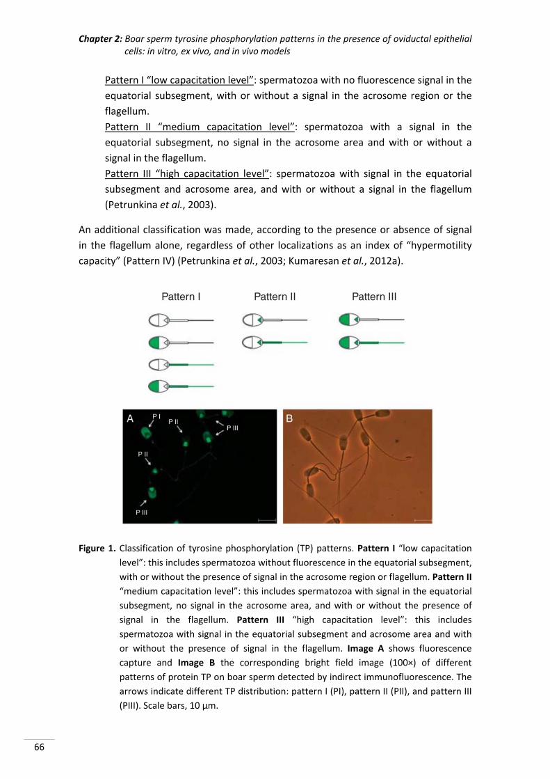
Chapter 2: Boar sperm tyrosine phosphorylation patterns in the presence of oviductal epithelial cells: in vitro, ex vivo, and in vivo models
66
Pattern I “low capacitation level”: spermatozoa with no fluorescence signal in the
equatorial subsegment, with or without a signal in the acrosome region or the
flagellum.
Pattern II “medium capacitation level”: spermatozoa with a signal in the
equatorial subsegment, no signal in the acrosome area and with or without a
signal in the flagellum.
Pattern III “high capacitation level”: spermatozoa with signal in the equatorial
subsegment and acrosome area, and with or without a signal in the flagellum
(Petrunkina et al., 2003).
An additional classification was made, according to the presence or absence of signal
in the flagellum alone, regardless of other localizations as an index of “hypermotility
capacity” (Pattern IV) (Petrunkina et al., 2003; Kumaresan et al., 2012a).
Figure 1. Classification of tyrosine phosphorylation (TP) patterns. Pattern I “low capacitation
level”: this includes spermatozoa without fluorescence in the equatorial subsegment,
with or without the presence of signal in the acrosome region or flagellum. Pattern II
“medium capacitation level”: this includes spermatozoa with signal in the equatorial
subsegment, no signal in the acrosome area, and with or without the presence of
signal in the flagellum. Pattern III “high capacitation level”: this includes
spermatozoa with signal in the equatorial subsegment and acrosome area and with
or without the presence of signal in the flagellum. Image A shows fluorescence
capture and Image B the corresponding bright field image (100×) of different
patterns of protein TP on boar sperm detected by indirect immunofluorescence. The
arrows indicate different TP distribution: pattern I (PI), pattern II (PII), and pattern III
(PIII). Scale bars, 10 µm.
Chapter 2: Boar sperm tyrosine phosphorylation patterns in the presence of oviductal epithelial cells: in vitro, ex vivo, and in vivo models
67
2.7. Statistical analysis
The data were first examined using the Shapiro‐Wilk test to assess normality
distribution. In view of the non‐Gaussian distribution of most of the data gathered, a
non‐parametric test was used. Data were expressed as mean ± S.E.M. and compared by
a non‐parametric Kruskal‐Wallis test and a Conover‐Inman test for all pairwise
comparisons was used to compare groups of samples. Differences were considered
statistically significant at p <0.05.
3. RESULTS
3.1. “In vitro model”: effect of in vitro cultured OEC on sperm TP protein location
The protein TP pattern changes when the sperm are incubated with OEC, especially as
regard patterns II and III. As Table 1 indicates, bound and unbound sperm after OEC
incubation showed less pattern II phosphorylation than non‐incubated sperm (control
group), although an increase in pattern III was observed in unbound sperm and a
decrease in the same phosphorylation pattern in bound sperm compared with the
control group. The distribution of fluorescence patterns was significantly different (p
<0.05) between bound and unbound cell populations (Table 1). The most common
pattern of TP in OEC‐bound sperm was pattern II (77.4%). OEC binding was most
prevalent in sperm with non‐phosphorylated heads and tails (Figure 2), while higher
proportions of subsegment and head and/or flagellum phosphorylated cells were
found in the unbound population (36.4%). The lowest proportion of pattern IV was
observed in sperm bound to OEC (17.9%) compared with unbound sperm (81.8%) and
sperm not incubated with OEC (84.6%).
Table 1. Immunolocation of protein tyrosine phosphorylation patterns in boar sperm bound
and unbound to OEC cultured in vitro (data shown mean ± S.E.M.).
Sperm source (N) Bound (N)
Pattern I (%)
Pattern II (%)
Pattern III (%)
Pattern IV (%)
Not incubated sperm (1001) 3.5 ± 1.8a 85.8 ± 3.5a 10.6 ± 3.1a 84.6 ± 3.6a
Incubated sperm OEC (4705) No (2352) 2.8 ± 1.6a 60.7 ± 4.8b 36.4 ± 4.8b 81.8 ± 3.9a
Yes (2353) 18.4 ± 3.8b 77.4 ± 4.1c 4.1 ± 1.9c 17.9 ± 3.8b
N: number of spermatozoa evaluated; Pattern I: non‐phosphorylated or head‐ and/or flagellum‐phosphorylated spermatozoa; Pattern II: equatorial segment or equatorial segment and flagellum phosphorylated; Pattern III: equatorial segment and head and/or flagellum phosphorylated; Pattern IV: flagellum phosphorylation independent of phosphorylation at other locations. Different letters (a, b, c) in the same column indicate significant differences (p <0.05).
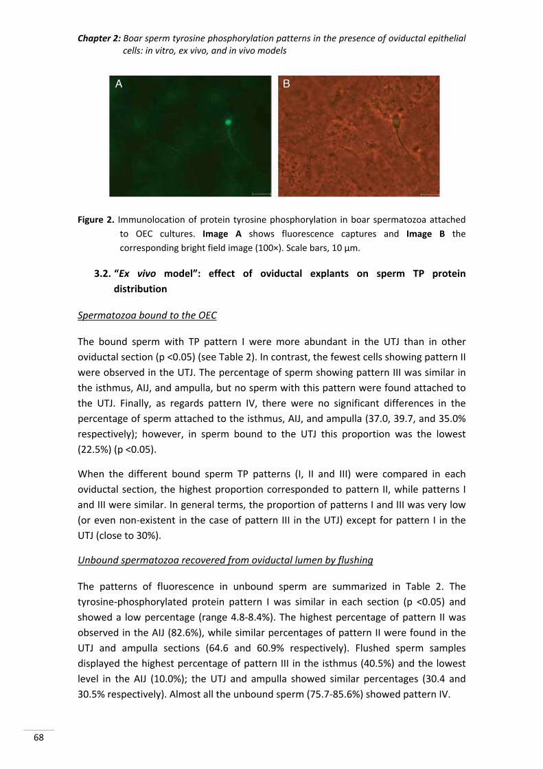
Chapter 2: Boar sperm tyrosine phosphorylation patterns in the presence of oviductal epithelial cells: in vitro, ex vivo, and in vivo models
68
Figure 2. Immunolocation of protein tyrosine phosphorylation in boar spermatozoa attached
to OEC cultures. Image A shows fluorescence captures and Image B the
corresponding bright field image (100×). Scale bars, 10 µm.
3.2. “Ex vivo model”: effect of oviductal explants on sperm TP protein
distribution
Spermatozoa bound to the OEC
The bound sperm with TP pattern I were more abundant in the UTJ than in other
oviductal section (p <0.05) (see Table 2). In contrast, the fewest cells showing pattern II
were observed in the UTJ. The percentage of sperm showing pattern III was similar in
the isthmus, AIJ, and ampulla, but no sperm with this pattern were found attached to
the UTJ. Finally, as regards pattern IV, there were no significant differences in the
percentage of sperm attached to the isthmus, AIJ, and ampulla (37.0, 39.7, and 35.0%
respectively); however, in sperm bound to the UTJ this proportion was the lowest
(22.5%) (p <0.05).
When the different bound sperm TP patterns (I, II and III) were compared in each
oviductal section, the highest proportion corresponded to pattern II, while patterns I
and III were similar. In general terms, the proportion of patterns I and III was very low
(or even non‐existent in the case of pattern III in the UTJ) except for pattern I in the
UTJ (close to 30%).
Unbound spermatozoa recovered from oviductal lumen by flushing
The patterns of fluorescence in unbound sperm are summarized in Table 2. The
tyrosine‐phosphorylated protein pattern I was similar in each section (p <0.05) and
showed a low percentage (range 4.8‐8.4%). The highest percentage of pattern II was
observed in the AIJ (82.6%), while similar percentages of pattern II were found in the
UTJ and ampulla sections (64.6 and 60.9% respectively). Flushed sperm samples
displayed the highest percentage of pattern III in the isthmus (40.5%) and the lowest
level in the AIJ (10.0%); the UTJ and ampulla showed similar percentages (30.4 and
30.5% respectively). Almost all the unbound sperm (75.7‐85.6%) showed pattern IV.
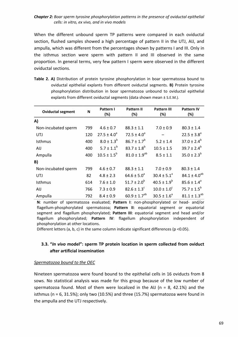
Chapter 2: Boar sperm tyrosine phosphorylation patterns in the presence of oviductal epithelial cells: in vitro, ex vivo, and in vivo models
69
When the different unbound sperm TP patterns were compared in each oviductal
section, flushed samples showed a high percentage of pattern II in the UTJ, AIJ, and
ampulla, which was different from the percentages shown by patterns I and III. Only in
the isthmus section were sperm with pattern II and III observed in the same
proportion. In general terms, very few pattern I sperm were observed in the different
oviductal sections.
Table 2. A) Distribution of protein tyrosine phosphorylation in boar spermatozoa bound to
oviductal epithelial explants from different oviductal segments. B) Protein tyrosine
phosphorylation distribution in boar spermatozoa unbound to oviductal epithelial
explants from different oviductal segments (data shown mean ± S.E.M.).
Oviductal segment N Pattern I
(%) Pattern II
(%) Pattern III
(%) Pattern IV
(%)
A)
Non‐incubated sperm 799 4.6 ± 0.7 88.3 ± 1.1 7.0 ± 0.9 80.3 ± 1.4
UTJ 120 27.5 ± 4.0a 72.5 ± 4.0a – 22.5 ± 3.8a
Isthmus 400 8.0 ± 1.3b 86.7 ± 1.7b 5.2 ± 1.4 37.0 ± 2.4b
AIJ 400 5.7 ± 1.1b 83.7 ± 1.8b 10.5 ± 1.5 39.7 ± 2.4b
Ampulla 400 10.5 ± 1.5b 81.0 ± 1.9ab 8.5 ± 1.1 35.0 ± 2.3b
B)
Non‐incubated sperm 799 4.6 ± 0.7 88.3 ± 1.1 7.0 ± 0.9 80.3 ± 1.4
UTJ 82 4.8 ± 2.3 64.6 ± 5.0a 30.4 ± 5.1a 84.1 ± 4.0ab
Isthmus 614 7.6 ± 1.0 51.7 ± 2.0b 40.5 ± 1.9b 85.6 ± 1.4a
AIJ 766 7.3 ± 0.9 82.6 ± 1.3c 10.0 ± 1.0c 75.7 ± 1.5b
Ampulla 792 8.4 ± 0.9 60.9 ± 1.7ab 30.5 ± 1.6a 81.1 ± 1.3ab
N: number of spermatozoa evaluated; Pattern I: non‐phosphorylated or head‐ and/or flagellum‐phosphorylated spermatozoa; Pattern II: equatorial segment or equatorial segment and flagellum phosphorylated; Pattern III: equatorial segment and head and/or flagellum phosphorylated; Pattern IV: flagellum phosphorylation independent of phosphorylation at other locations. Different letters (a, b, c) in the same column indicate significant differences (p <0.05).
3.3. “In vivo model”: sperm TP protein location in sperm collected from oviduct
after artificial insemination
Spermatozoa bound to the OEC
Nineteen spermatozoa were found bound to the epithelial cells in 16 oviducts from 8
sows. No statistical analysis was made for this group because of the low number of
spermatozoa found. Most of them were localized in the AIJ (n = 8, 42.1%) and the
isthmus (n = 6, 31.5%); only two (10.5%) and three (15.7%) spermatozoa were found in
the ampulla and the UTJ respectively.
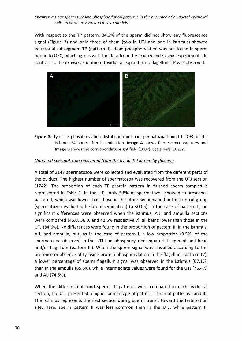
Chapter 2: Boar sperm tyrosine phosphorylation patterns in the presence of oviductal epithelial cells: in vitro, ex vivo, and in vivo models
70
With respect to the TP pattern, 84.2% of the sperm did not show any fluorescence
signal (Figure 3) and only three of them (two in UTJ and one in isthmus) showed
equatorial subsegment TP (pattern II). Head phosphorylation was not found in sperm
bound to OEC, which agrees with the data from the in vitro and ex vivo experiments. In
contrast to the ex vivo experiment (oviductal explants), no flagellum TP was observed.
Figure 3. Tyrosine phosphorylation distribution in boar spermatozoa bound to OEC in the
isthmus 24 hours after insemination. Image A shows fluorescence captures and
Image B shows the corresponding bright field (100×). Scale bars, 10 µm.
Unbound spermatozoa recovered from the oviductal lumen by flushing
A total of 2147 spermatozoa were collected and evaluated from the different parts of
the oviduct. The highest number of spermatozoa was recovered from the UTJ section
(1742). The proportion of each TP protein pattern in flushed sperm samples is
represented in Table 3. In the UTJ, only 5.8% of spermatozoa showed fluorescence
pattern I, which was lower than those in the other sections and in the control group
(spermatozoa evaluated before insemination) (p <0.05). In the case of pattern II, no
significant differences were observed when the isthmus, AIJ, and ampulla sections
were compared (46.0, 36.0, and 43.5% respectively), all being lower than those in the
UTJ (84.6%). No differences were found in the proportion of pattern III in the isthmus,
AIJ, and ampulla, but, as in the case of pattern I, a low proportion (9.5%) of the
spermatozoa observed in the UTJ had phosphorylated equatorial segment and head
and/or flagellum (pattern III). When the sperm signal was classified according to the
presence or absence of tyrosine protein phosphorylation in the flagellum (pattern IV),
a lower percentage of sperm flagellum signal was observed in the isthmus (67.1%)
than in the ampulla (85.5%), while intermediate values were found for the UTJ (76.4%)
and AIJ (74.5%).
When the different unbound sperm TP patterns were compared in each oviductal
section, the UTJ presented a higher percentage of pattern II than of patterns I and III.
The isthmus represents the next section during sperm transit toward the fertilization
site. Here, sperm pattern II was less common than in the UTJ, while pattern III
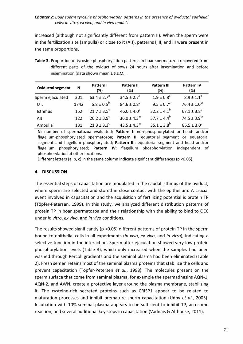
Chapter 2: Boar sperm tyrosine phosphorylation patterns in the presence of oviductal epithelial cells: in vitro, ex vivo, and in vivo models
71
increased (although not significantly different from pattern II). When the sperm were
in the fertilization site (ampulla) or close to it (AIJ), patterns I, II, and III were present in
the same proportions.
Table 3. Proportion of tyrosine phosphorylation patterns in boar spermatozoa recovered from
different parts of the oviduct of sows 24 hours after insemination and before
insemination (data shown mean ± S.E.M.).
Oviductal segment N Pattern I
(%) Pattern II
(%) Pattern III
(%) Pattern IV
(%)
Sperm ejaculated 301 63.4 ± 2.7a 34.5 ± 2.7a 1.9 ± 0.8a 8.9 ± 1.1a
UTJ 1742 5.8 ± 0.5b 84.6 ± 0.8b 9.5 ± 0.7a 76.4 ± 1.0bc
Isthmus 152 21.7 ± 3.5c 46.0 ± 4.0c 32.2 ± 4.1b 67.1 ± 3.8b
AIJ 122 26.2 ± 3.9c 36.0 ± 4.3ac 37.7 ± 4.4b 74.5 ± 3.9bc
Ampulla 131 21.3 ± 3.3c 43.5 ± 4.3ac 35.1 ± 3.8b 85.5 ± 3.0c
N: number of spermatozoa evaluated; Pattern I: non‐phosphorylated or head‐ and/or flagellum‐phosphorylated spermatozoa; Pattern II: equatorial segment or equatorial segment and flagellum phosphorylated; Pattern III: equatorial segment and head and/or flagellum phosphorylated; Pattern IV: flagellum phosphorylation independent of phosphorylation at other locations. Different letters (a, b, c) in the same column indicate significant differences (p <0.05).
4. DISCUSSION
The essential steps of capacitation are modulated in the caudal isthmus of the oviduct,
where sperm are selected and stored in close contact with the epithelium. A crucial
event involved in capacitation and the acquisition of fertilizing potential is protein TP
(Töpfer‐Petersen, 1999). In this study, we analyzed different distribution patterns of
protein TP in boar spermatozoa and their relationship with the ability to bind to OEC
under in vitro, ex vivo, and in vivo conditions.
The results showed significantly (p <0.05) different patterns of protein TP in the sperm
bound to epithelial cells in all experiments (in vivo, ex vivo, and in vitro), indicating a
selective function in the interaction. Sperm after ejaculation showed very‐low protein
phosphorylation levels (Table 3), which only increased when the samples had been
washed through Percoll gradients and the seminal plasma had been eliminated (Table
2). Fresh semen retains most of the seminal plasma proteins that stabilize the cells and
prevent capacitation (Töpfer‐Petersen et al., 1998). The molecules present on the
sperm surface that come from seminal plasma, for example the spermadhesins AQN‐1,
AQN‐2, and AWN, create a protective layer around the plasma membrane, stabilizing
it. The cysteine‐rich secreted proteins such as CRISP1 appear to be related to
maturation processes and inhibit premature sperm capacitation (Udby et al., 2005).
Incubation with 10% seminal plasma appears to be sufficient to inhibit TP, acrosome
reaction, and several additional key steps in capacitation (Vadnais & Althouse, 2011).
Chapter 2: Boar sperm tyrosine phosphorylation patterns in the presence of oviductal epithelial cells: in vitro, ex vivo, and in vivo models
72
Several authors have demonstrated that unphosphorylated spermatozoa show a
preferential binding ability to OEC after a short incubation period (Lefebvre & Suarez,
1996; Fazeli et al., 1999). Furthermore, Petrunkina et al. (2001, 2003) demonstrated
that long‐term coincubations with oviductal cells maintain sperm viability and motility
and slow the process of sperm membrane destabilization, when spermatozoa are
bound to OEC. Spermatozoa attached to epithelial cells showed constantly low levels
of protein TP (mainly pattern I), while unbound spermatozoa showed a high degree of
phosphorylation (Yeste et al., 2009). In our study, not only spermatozoa bound to OEC
cultures exhibited a low level of TP but also spermatozoa attached to the oviducts of
inseminated sows before ovulation and preovulatory porcine oviductal explants
presented this pattern (p <0.05).
We found that different TP patterns were associated with different sperm functions
(Urner & Sakkas, 2003). In spermatozoa bound to OEC, the protein TP was located in
the equatorial subsegment or did not show any fluorescence. Such labeling may
represent the first stage of phosphorylation during capacitation (Figure 2). In boar
testicular sperm, TP protein is distributed through the entire equatorial segment.
During epididymal maturation, dephosphorylation and/or redistribution of
phosphorylation occur and the cauda spermatozoa exhibit only an intense triangular
labeling in the posterior part of the acrosome and the equatorial subsegment (Jones et
al., 2008; Fàbrega et al., 2011). SPACA1 and HSPA1A proteins have been determined in
this event (Spinaci et al., 2005; Jones et al., 2008). The equatorial subsegment might be
an organizing centre that initiates membrane fusion during the fertilization process.
Phosphorylation in the acrosome, equatorial subsegment, and flagellum is related to
an advanced stage of capacitation (Petrunkina et al., 2003). The presence of tyrosine‐
phosphorylated proteins in the flagellum is a preferential pattern of sperm
capacitation in several species (Leclerc et al., 1997; Pommer et al., 2003; Grasa et al.,
2006; Jagan Mohanarao & Atreja, 2011). This phosphorylation is associated with a
fibrous sheath, suggesting that it may be involved in hyperactivation (Pommer et al.,
2003; Kumaresan et al., 2012b). Our data showed that spermatozoa washed through
Percoll gradients and spermatozoa unbound to oviductal cells had higher proportions
of flagellum phosphorylation (pattern IV, p <0.05) in all the experiments. Flagellum TP
may be related to the release of bound spermatozoa from the epithelium (Lefebvre &
Suarez, 1996). However, these findings contrast with others that point to the recovery
of faint labeling in the tail in boar spermatozoa (Petrunkina et al., 2001; Fàbrega et al.,
2011). Equatorial subsegment and/or flagellum phosphorylation (pattern II)
corresponds to an intermediate capacitation level (Petrunkina et al., 2001).
Acrosome phosphorylation was an unusual pattern and was not found in sperm bound
to OEC in any of the three experiments. Suppressed acrosome phosphorylation may be
a requirement for binding to oviductal cells (Petrunkina et al., 2003) because it
Chapter 2: Boar sperm tyrosine phosphorylation patterns in the presence of oviductal epithelial cells: in vitro, ex vivo, and in vivo models
73
represents an advanced stage of capacitation. Sp32 is a (pro) acrosin binding protein
localized on the acrosome surface. During incubation in capacitating conditions, sp32 is
phosphorylated and transformed into p32, a tyrosine phosphorylated protein (Dubé et
al., 2005). These changes may affect the sperm’s ability to bind to OEC. Moreover,
during the acrosome reaction, phosphorylation in the acrosomal region decreased due
to the redistribution of phosphotyrosine residues (Tardif et al., 2001; Dubé et al.,
2005).
In vivo, boar spermatozoa are deposited in the female genital tract, pass through the
UTJ, and accumulate in the isthmus of the oviduct, where a functional SR is established
(Hunter & Rodriguez‐Martinez, 2004). The sperm distribution results showed that
spermatozoa in sows inseminated around ovulation are mainly found in the UTJ where
they exhibit a high proportion of flagellum phosphorylation. In contrast, in oviductal
explants, spermatozoa were found along the oviduct in the same proportion. The UTJ
is a physical barrier that controls the quality and quantity of spermatozoa present in
the oviduct. Only sperm cells with an intact acrosome, low internal free calcium
content, reduced membrane protein phosphorylation, and normal chromatin structure
are able to bind to OEC (Ellington et al., 1998; Fazeli et al., 1999; Petrunkina et al.,
2001; Gualtieri & Talevi, 2003). In our study, unbound sperm exhibited a high
percentage of protein TP in the equatorial subsegment, acrosome, and flagellum (p
<0.05). Kumaresan et al. (2012a) speculated that an early increase in the population of
spermatozoa with such a phosphorylation pattern would increase the chances of
fertilization. The adhesion between sperm and OEC is mediated by molecules exposed
on the sperm rostral surface and carbohydrates localized on the oviductal cell plasma
membrane in a species‐specific manner (Green et al., 2001). Capacitated sperm lose
binding sites during capacitation, and our in vivo results showed that only sperm with
unphosphorylated heads are able to bind to cells.
At the time of ovulation, changes in the oviductal environment occur, promoting the
ascent of sperm cells toward the ampulla and the site of fertilization (Hunter, 1981).
Recent studies have shown that the arrival of spermatozoa initiates a mutual sperm‐
epithelial signaling dialog (Fazeli et al., 2004). In an in vivo experiment, a small number
of spermatozoa were seen bound to OEC around the ovulation stage. Most
spermatozoa were in the lumen of the oviduct with a high level of TP, especially when
the sperm were close to the fertilization site. Spermatozoa modified protein synthesis
and oviductal fluid composition remodeling the binding sites in oviductal cells due to
the production of a gradient of calcium during periovulatory moments (Gadella &
Harrison, 2000). In sows, several de novo synthesized proteins have been identified,
and oviduct‐specific glycoprotein is related to increased fertilization rates and
polyspermy control (Coy et al., 2008).
Chapter 2: Boar sperm tyrosine phosphorylation patterns in the presence of oviductal epithelial cells: in vitro, ex vivo, and in vivo models
74
Porcine oviductal fluid in the follicular phase promotes sperm viability and acrosomal
integrity, decreases plasma membrane fluidity, and increases zona binding and
polyspermy during IVF (Coy et al., 2010). However, other authors observed no
differences in sperm motility during incubation except at 2 hours, when spermatozoa
incubated with preovulatory fluid showed higher motility (Kumaresan et al., 2012b). In
the same study, preovulatory oviductal fluid induced TP in a higher proportion of boar
spermatozoa than post‐ovulatory fluid (Kumaresan et al., 2012b). These contradictory
results may be related to the boar in question, incubation time, or oviductal fluid
treatment. Our results showed that unbound spermatozoa exhibited high levels of
tyrosine protein phosphorylation (patterns II and III) in all the experiments.
The final objective of the capacitation process is fertilization. Spermatozoa incubated
with OEC cultures showed higher penetration and monospermy rates when short time
periods were used (Hunter, 1981). Moreover, the fertilization and cleavage rates of
spermatozoa unbound to oviductal cells culture were lower than those for
spermatozoa attached to the OEC (Kon et al., 2009). These results indicate that the
ability of spermatozoa to bind to oviductal cells is related to their fertilizing ability.
In conclusion, sperm capacitation conditions in vivo are not reproducible in in vitro
models under our experimental conditions. However, sperm‐OEC binding was seen to
be a mechanism for selecting sperm populations with a low degree of TP, as
demonstrated in the in vivo, ex vivo, and in vitro experiments. Further studies are
required to obtain more information about the mechanism by which the oviduct
modulates sperm capacitation.
ACKNOWLEDGEMENTS
This work was supported by Fundación Séneca 08752/PI/08 and MICINN‐FEDER (AGL
2009‐12512‐C02‐01).
REFERENCES
Austin C (1951). Observations on the penetration of the sperm into the mammalian egg.
Australian Journal of Biological Sciences. 4: 581‐596. (doi: 10.1071/BI9510581).
Coy P, Cánovas S, Mondéjar I, Saavedra MD, Romar R, Grullón L, Matás C & Avilés M (2008).
Oviduct‐specific glycoprotein and heparin modulate sperm–zona pellucida interaction
during fertilization and contribute to the control of polyspermy. Proceedings of the
National Academy of Sciences. 105: 15809‐15814. (doi: 10.1073/pnas.0804422105).
Coy P, Lloyd R, Romar R, Satake N, Matás C, Gadea J & Holt WV (2010). Effects of porcine pre‐
ovulatory oviductal fluid on boar sperm function. Theriogenology. 74: 632‐642. (doi:
10.1016/j.theriogenology.2010.03.005).
Chapter 2: Boar sperm tyrosine phosphorylation patterns in the presence of oviductal epithelial cells: in vitro, ex vivo, and in vivo models
75
Chang M (1951). Fertilizing capacity of spermatozoa deposited into the fallopian tubes. Nature.
168: 697‐698. (doi: 10.1038/168697b0).
de Lamirande E & Cagnon C (1993). Human sperm hyperactivation and capacitation as parts of
an oxidative process. Free Radical Biology and Medicine. 14: 157‐166. (doi:
10.1016/0891‐5849(93)90006‐G).
Dubé C, Leclerc P, Baba T, Reyes‐Moreno C & Bailey JL (2005). The proacrosin binding protein,
sp32, is tyrosine phosphorylated during capacitation of pig sperm. Journal of
andrology. 26: 519‐528. (doi: 10.2154/jandrol.04163).
Ellington JE, Evenson DP, Fleming JE, Brisbois RS, Hiss GA, Broder SJ & Wright RW (1998).
Coculture of human sperm with bovine oviduct epithelial cells decreases sperm
chromatin structural changes seen during culture in media alone. Fertility and sterility.
69: 643‐649. (doi: 10.1016/S0015‐0282(98)00023‐5).
Fàbrega A, Puigmulé M, Yeste M, Casas I, Bonet S & Pinart E (2011). Impact of epididymal
maturation, ejaculation and in vitro capacitation on tyrosine phosphorylation patterns
exhibited of boar (Sus domesticus) spermatozoa. Theriogenology. 76: 1356‐1366. (doi:
10.1016/j.theriogenology.2011.06.007).
Fazeli A, Duncan A, Watson P & Holt WV (1999). Sperm‐oviduct interaction: induction of
capacitation and preferential binding of uncapacitated spermatozoa to oviductal
epithelial cells in porcine species. Biology of reproduction. 60: 879‐886. (doi:
10.1095/biolreprod60.4.879).
Fazeli A, Affara NA, Hubank M & Holt WV (2004). Sperm‐induced modification of the oviductal
gene expression profile after natural insemination in mice. Biology of reproduction. 71:
60‐65. (doi: 10.1095/biolreprod.103.026815).
Flesch F, Wijnand E, Van de Lest C, Colenbrander B, van Golde LM & Gadella BM (2001).
Capacitation dependent activation of tyrosine phosphorylation generates two sperm
head plasma membrane proteins with high primary binding affinity for the zona
pellucida. Molecular reproduction and development. 60: 107‐115. (doi:
10.1002/mrd.1067).
Gadea J & Matás C (2000). Sperm factors related to in vitro penetration of porcine oocytes.
Theriogenology. 54: 1343‐1357. (doi: 10.1016/S0093‐691X(00)00458‐1).
Gadella BM & Harrison RA (2000). The capacitating agent bicarbonate induces protein kinase
A‐dependent changes in phospholipid transbilayer behavior in the sperm plasma
membrane. Development. 127: 2407‐2420.
Gadella BM (2008). Sperm membrane physiology and relevance for fertilization. Animal
Reproduction Science. 107: 229‐236. (doi: 10.1016/j.anireprosci.2008.05.006).
García‐Vázquez FA, Ruiz S, Matás C, Izquierdo‐Rico MJ, Grullón LA, De Ondiz A, Vieira L,
Avilés‐López K, Gutiérrez‐Adán A & Gadea J (2010). Production of transgenic piglets
using ICSI–sperm‐mediated gene transfer in combination with recombinase RecA.
Reproduction. 140: 259‐272. (doi: 10.150/REP‐10‐0129).
Georgiou AS, Snijders AP, Sostaric E, Aflatoonian R, Vazquez JL, Vazquez JM, Roca J, Martinez
EA, Wright PC & Fazeli A (2007). Modulation of the oviductal environment by
gametes. Journal of proteome research. 6: 4656‐4666. (doi: 10.1021/pr070349m).
Chapter 2: Boar sperm tyrosine phosphorylation patterns in the presence of oviductal epithelial cells: in vitro, ex vivo, and in vivo models
76
Grasa P, Cebrián‐Pérez JA & Muiño‐Blanco T (2006). Signal transduction mechanisms involved
in in vitro ram sperm capacitation. Reproduction. 132: 721‐732. (doi:
10.1530/rep.1.00770).
Green C, Bredl J, Holt WV, Watson P & Fazeli A (2001). Carbohydrate mediation of boar sperm
binding to oviductal epithelial cells in vitro. Reproduction. 122: 305‐315. (doi:
10.1530/rep.0.1250251).
Gualtieri R & Talevi R (2003). Selection of highly fertilization‐competent bovine spermatozoa
through adhesion to the Fallopian tube epithelium in vitro. Reproduction. 125: 251‐
258. (doi: 10.1530/rep.0.1220305).
Harrison RA, Ashworth P & Miller N (1996). Bicarbonate/CO2, an effector of capacitation,
induces a rapid and reversible change in the lipid architecture of boar sperm plasma
membranes. Molecular reproduction and development. 45: 378‐391. (doi:
10.1002/(SICI)1098‐2795(199611)45:3<378::AID‐MRD16>3.0.CO;2‐V).
Hunter RHF (1981). Sperm transport and reservoirs in the pig oviduct in relation to the time of
ovulation. Journal of reproduction and fertility. 63: 109‐117. (doi:
10.1530/jrf.0.0630109).
Hunter RHF (1984). Pre‐ovulatory arrest and peri‐ovulatory redistribution of competent
spermatozoa in the isthmus of the pig oviduct. Journal of reproduction and fertility. 72:
203‐211. (doi: 10.1530/jrf.0.0720203).
Hunter RHF, Fléchon B & Fléchon J‐E (1987). Pre‐and peri‐ovulatory distribution of viable
spermatozoa in the pig oviduct: a scanning electron microscope study. Tissue and Cell.
19: 423‐436. (doi: 10.1016/0040‐8166(87)90037‐1).
Hunter RHF & Rodriguez‐Martinez H (2004). Capacitation of mammalian spermatozoa in vivo,
with a specific focus on events in the Fallopian tubes. Molecular reproduction and
development. 67: 243‐250. (doi: 10.1002/mrd.10390).
Jagan Mohanarao G & Atreja S (2011). Identification of capacitation associated tyrosine
phosphoproteins in buffalo (Bubalus bubalis) and cattle spermatozoa. Animal
Reproduction Science. 123: 40‐47. (doi: 10.1016/j.anireprosci.2010.11.013).
Jones R, James PS, Oxley D, Coadwell J, Suzuki‐Toyota F & Howes EA (2008). The equatorial
subsegment in mammalian spermatozoa is enriched in tyrosine phosphorylated
proteins. Biology of reproduction. 79: 421‐431. (doi: 10.1095/biolreprod.107.067314).
King G & Macpherson J (1973). A comparison of two methods for boar semen collection.
Journal of animal science. 36: 563‐565. (doi: 10.2134/jas1973.363563x).
Kon Y, Iwata H, Shiono H, Matsubara K, Kurita A, Sakaguchi Y, Kuwayama T & Monji Y (2009).
Effect of carbohydrates on the ability of bull sperm to bind to bovine oviduct epithelial
cells. Reproduction in Domestic Animals. 44: 365‐370. (doi: 10.1111/j.1439‐
0531.2007.01013.x).
Kumaresan A, Johannisson A, Humblot P & Bergqvist AS (2012a). Oviductal fluid modulates
the dynamics of tyrosine phosphorylation in cryopreserved boar spermatozoa during
capacitation. Molecular reproduction and development. 79: 525‐540. (doi:
10.1002/mrd.22058).
Kumaresan A, Johannisson A, Saravia F & Bergqvist AS (2012b). The effect of oviductal fluid
on protein tyrosine phosphorylation in cryopreserved boar spermatozoa differs with
Chapter 2: Boar sperm tyrosine phosphorylation patterns in the presence of oviductal epithelial cells: in vitro, ex vivo, and in vivo models
77
the freezing method. Theriogenology. 77: 588‐599. (doi:
10.1016/j.theriogenology.2011.08.035).
Kunavongkrit A, Sang‐Gasanee K, Phumratanaprapin C, Tantasuparuk W & Einarsson S
(2003). A study on the number of recovered spermatozoa in the uterine horns and
oviducts of gilts, after fractionated or non‐fractionated insemination. Journal of
veterinary medical science. 65: 63‐67. (doi: 10.1292/jvms.65.63).
Leclerc P, De Lamirande E & Gagnon C (1997). Regulation of protein‐tyrosine phosphorylation
and human sperm capacitation by reactive oxygen derivatives. Free Radical Biology
and Medicine. 22: 643‐656. (doi: 10.1016/S0891‐5849(96)00379‐6).
Lefebvre R & Suarez SS (1996). Effect of capacitation on bull sperm binding to homologous
oviductal epithelium. Biology of reproduction. 54: 575‐582. (doi:
10.1095/biolreprod54.3.575).
Matás C, Vieira L, García‐Vázquez F, Avilés‐López K, López‐Úbeda R, Carvajal J & Gadea J
(2011). Effects of centrifugation through three different discontinuous Percoll
gradients on boar sperm function. Animal Reproduction Science. 127: 62‐72. (doi:
10.1016/S0015‐0282(99)00013‐8).
Mburu J, Einarsson S, Lundeheim N & Rodriguez‐Martinez H (1996). Distribution, number and
membrane integrity of spermatozoa in the pig oviduct in relation to spontaneous
ovulation. Animal Reproduction Science. 45: 109‐121. (doi: 10.1016/S0378‐
4320(96)01566‐7).
Nassar A, Mahony M, Morshedi M, Lin M‐H, Srisombut C & Oehninger S (1999). Modulation
of sperm tail protein tyrosine phosphorylation by pentoxifylline and its correlation
with hyperactivated motility. Fertility and sterility. 71: 919‐923. (doi: 10.1016/S0015‐
0282(99)00013‐8).
Petrunkina AM, Friedrich J, Drommer W, Bicker G, Waberski D & Töpfer‐Petersen E (2001).
Kinetic characterization of the changes in protein tyrosine phosphorylation of
membranes, cytosolic Ca2+ concentration and viability in boar sperm populations
selected by binding to oviductal epithelial cells. Reproduction. 122: 469‐480. (doi:
10.1530/rep.0.1220469).
Petrunkina AM, Simon K, Günzel‐Apel AR & Töpfer‐Petersen E (2003). Specific order in the
appearance of protein tyrosine phosphorylation patterns is functionally coordinated
with dog sperm hyperactivation and capacitation. Journal of andrology. 24: 423‐437.
(doi: 10.1002/j.1939‐4640.2003.tb02691.x).
Pommer AC, Rutllant J & Meyers SA (2003). Phosphorylation of protein tyrosine residues in
fresh and cryopreserved stallion spermatozoa under capacitating conditions. Biology of
reproduction. 68: 1208‐1214. (doi: 10.1095/biolreprod.102.011106).
Pursel V & Johnson L (1975). Freezing of boar spermatozoa: fertilizing capacity with
concentrated semen and a new thawing procedure. Journal of animal science. 40: 99‐
102.
Rath D, Long C, Dobrinsky J, Welch G, Schreier L & Johnson L (1999). In vitro production of
sexed embryos for gender preselection: high‐speed sorting of X‐chromosome‐bearing
sperm to produce pigs after embryo transfer. Journal of animal science. 77: 3346‐3352.
(doi: 1999.77123346x).
Chapter 2: Boar sperm tyrosine phosphorylation patterns in the presence of oviductal epithelial cells: in vitro, ex vivo, and in vivo models
78
Romar R, Coy P, Campos I, Gadea J, Matás C & Ruiz S (2001). Effect of co‐culture of porcine
sperm and oocytes with porcine oviductal epithelial cells on in vitro fertilization.
Animal Reproduction Science. 68: 85‐98. (doi: 10.1016/S0378‐4320(01)00133‐6).
Spinaci M, Volpe S, Bernardini C, De Ambrogi M, Tamanini C, Seren E & Galeati G (2005).
Immunolocalization of heat shock protein 70 (Hsp 70) in boar spermatozoa and its role
during fertilization. Molecular reproduction and development. 72: 534‐541. (doi:
10.1002/mrd.20367).
Suarez S, Katz D, Owen D, Andrew J & Powell R (1991). Evidence for the function of
hyperactivated motility in sperm. Biology of reproduction. 44: 375‐381. (doi:
10.1095/biolreprod44.2.375).
Tardif S, Dubé C, Chevalier S & Bailey JL (2001). Capacitation is associated with tyrosine
phosphorylation and tyrosine kinase‐like activity of pig sperm proteins. Biology of
reproduction. 65: 784‐792. (doi: 10.1095/biolreprod65.3.784).
Töpfer‐Petersen E, Romero A, Varela P, Ekhlasi‐Hundrieser M, Dostalova Z, Sanz L & Calvete J
(1998). Spermadhesins: a new protein family. Facts, hypotheses and perspectives.
Andrologia. 30: 217‐224. (doi: 10.1111/j.1439‐0272.1998.tb02263.x).
Töpfer‐Petersen E (1999). Molecules on the sperm's route to fertilization. Journal of
Experimental Zoology. 285: 259‐266. (doi: 10.1002/(SICI)1097‐
010X(19991015)285:3<259::AID‐JEZ9<3.0.CO;2‐B).
Töpfer‐Petersen E, Wagner A, Friedrich J, Petrunkina AM, Ekhlasi‐Hundrieser M, Waberski D
& Drommer W (2002). Function of the mammalian oviductal sperm reservoir. Journal
of Experimental Zoology. 292: 210‐215. (doi: 10.1002/jez.1157).
Tummaruk P, Sumransap P, Techakumphu M & Kunavongkrit A (2007). Distribution of
spermatozoa and embryos in the female reproductive tract after unilateral deep intra
uterine insemination in the pig. Reproduction in Domestic Animals. 42: 603‐609. (doi:
10.1111/j.1439‐0531.2006.00830.x).
Udby L, Bjartell A, Malm J, Egesten A, Lundwall Å, Cowland JB, Borregaard N & Kjeldsen L
(2005). Characterization and localization of cysteine‐rich secretory protein 3 (CRISP‐3)
in the human male reproductive tract. Journal of andrology. 26: 333‐342. (doi:
10.2164/jandrol.04132).
Urner F & Sakkas D (2003). Protein phosphorylation in mammalian spermatozoa.
Reproduction. 125: 17‐26. (doi: 10.1530/rep.0.1250017).
Vadnais ML & Althouse GC (2011). Characterization of capacitation, cryoinjury, and the role of
seminal plasma in porcine sperm. Theriogenology. 76: 1508‐1516. (doi:
10.1016/j.theriogenology.2011.06.021).
Visconti PE, Bailey JL, Moore GD, Pan D, Olds‐Clarke P & Kopf GS (1995). Capacitation of
mouse spermatozoa. I. Correlation between the capacitation state and protein
tyrosine phosphorylation. Development. 121: 1129‐1137.
Yeste M, Lloyd R, Badia E, Briz M, Bonet S & Holt WV (2009). Direct contact between boar
spermatozoa and porcine oviductal epithelial cell (OEC) cultures is needed for optimal
sperm survival in vitro. Animal Reproduction Science. 113: 263‐278. (doi:
10.1016/j.anireprosci.2008.08.018).

Chapter 2: Boar sperm tyrosine phosphorylation patterns in the presence of oviductal epithelial cells: in vitro, ex vivo, and in vivo models
79
Zumoffen C, Caille A, Munuce M, Cabada M & Ghersevich S (2010). Proteins from human
oviductal tissue‐conditioned medium modulate sperm capacitation. Human
reproduction. 25: 1504‐1512. (doi: 10.1093/humrep/deq063).

81
CHAPTER 3
Sperm selection by oviductal epithelial cells is not merely a
morphological‐based phenomenon
ABSTRACT
The interaction of oviductal epithelial cells with the spermatozoa has beneficial effects
on the sperm functions. The objective of this study was to evaluate the in vitro
fertilizing capacity of incubating spermatozoa previously selected by density gradient
in oviductal epithelial cells (OEC) and determinate some sperm characteristics that
could explain the results obtained. In this study, we assessed in vitro fertilization (IVF),
tyrosine phosphorylation (TP), phosphatidylserine translocation (PS), nuclear DNA
fragmentation and chromatin decondensation. Three sets of sperm, previously
selected by density gradient, were used in the different experiments: i) washed
spermatozoa (W30 group): sperm incubated for 30 minutes without oviductal
epithelial cells; ii) unbound (NB group): spermatozoa not bound to OEC after 30
minutes of coincubation; iii) bound (B group): spermatozoa bound to OEC after 30
minutes of coincubation. We found that sperm from the NB group led to a statistically
significant lower IVF yield, accompanied by low penetration rates and problems of
nuclear decondensation. Moreover, significantly higher levels of TP and PS were
observed in the NB group compared with the W30 and B groups. These results
demonstrate that the OEC exert a rigorous degree of sperm selection, even within an
already highly selected population of spermatozoa, and are able to capture the best
functional spermatozoa for fertilization.
Chapter 3: Sperm selection by oviductal epithelial cells is not merely a morphological‐based phenomenon
82
1. INTRODUCTION
Once deposited in the female genital tract, sperm undergo a selection process that
starts in the lower part of the uterus (cervix), continues in the uterus horns and
finishes in the upper part of the female genital tract, in the oviduct, where fertilization
takes place. Sperm reaching the oviduct are selected according to their morphological
intactness (Waberski et al., 2006), maturity (Petrunkina et al., 2001), uncapacitated
status (Fazeli et al., 1999; Luño et al., 2013) and high quality chromatin (Ardón et al.,
2008). When sperm come into contact with oviductal epithelial cells that line the
female tract and its secretions, some spermatozoa are stored (Hunter, 1981), allowing
the selection of sperm with certain qualities (Talevi & Gualtieri, 2010). A highly fertile
subpopulation of the original ejaculate will bind to oviductal epithelial cells, mainly in
the spermatic reservoir (Gualtieri & Talevi, 2003). Therefore, two functionally distinct
sperm subpopulations with the ability to reach and enter into the oviduct can be
discerned: i) those bound to the epithelium, which are considered to have been
selected because of their high quality (Talevi & Gualtieri, 2010) and found at the
bottom of the crypts of the oviductal folds, where they form a reservoir; and ii) those
found in the lumen, not bound to the epithelium, and showing membrane
modifications or poor vitality (Mburu et al., 1996, 1997; Rodriguez‐Martinez et al.,
2001; Tienthai et al., 2004). The binding of sperm to oviductal cells is mediated by
carbohydrates on the oviductal cell apical membranes and lectin‐like molecules on the
sperm rostral surface (Gualtieri & Talevi, 2000). During the capacitation of
spermatozoa, these lectin‐like molecules may be released from the sensitive
acrosomal region of the sperm head, allowing the sperm to abandon the epithelium
(Töpfer‐Petersen et al., 2002) in a sequential process after ovulation (Brüssow et al.,
2008) and swim into the ampulla region, where fertilization occurs (Fazeli et al., 1999).
The interaction of oviductal epithelial cells with the spermatozoa has beneficial effects
on the sperm functions. Several authors have shown that the coincubation of
spermatozoa with oviductal epithelial cells or their conditioned media maintains sperm
viability and motility (Zhu et al., 1994; Kervancioglu et al., 2000; Quintero et al., 2005;
Munuce et al., 2009; Zumoffen et al., 2010), enhances the fertilization potential by
protecting them from oxidative stress (Huang et al., 2013) and even, in some species,
stabilizes the sperm chromatin structure (Ellington et al., 1998).
Numerous in vitro techniques have been developed for sperm selection and
capacitation in an attempt to obtain the best spermatozoa for fertilization (viable and
motile) (Matás et al., 2003, 2011; Buffone et al., 2004). All these systems base their
selection on sperm motility and morphology, and have certainly improved the yields of
IVF. But with the progress of assisted reproduction technologies (ART), these
techniques are insufficient to identify the most suitable spermatozoa for fertilization
since they do not select sperm populations free of phosphatidylserine translocation.
Chapter 3: Sperm selection by oviductal epithelial cells is not merely a morphological‐based phenomenon
83
All the above led as to believe that if an initial selection of sperm could be made in a
physical medium such as a Percoll gradient (mimicking the uterus), subsequent
selection by means of a biological system such as oviductal cell culture (mimicking the
oviduct’s action) might provide a more suitable sperm subpopulation for use in ART,
particularly when semen quality is poor or when intracytoplasmic sperm injection
(ICSI) is to be performed. To develop this hypothesis, IVF was carried out using sperm
bound and unbound to oviductal cells. Besides, to determine putative differences
between these kinds of spermatozoa, sperm functionality parameters such as tyrosine
phosphorylation (TP), phosphatidylserine translocation (PS), DNA fragmentation and
chromatin condensation, were be determined.
2. MATERIALS AND METHODS
2.1. Culture media
Unless otherwise indicated, all the chemicals used in this study were purchased from
Sigma‐Aldrich Química S.A. (Madrid, Spain). The medium used for oocyte maturation
was NCSU‐37 supplemented with 0.57 mM cysteine, 1 mM dibutyryl cAMP, 5 µg/ml
insulin, 50 µM β‐mercaptoethanol, 1 mM glutamine, 10 IU/ml eCG (Folligon, Intervet
International B.V., Boxmeer, Holland), 10 IU/ml hCG (Chorulon, Intervet International
B.V., Boxmeer, Holland), 10 ng/ml EGF and 10% (v/v) porcine follicular fluid (Matás et
al., 2003). The fertilization medium was modified TALP supplemented with 3 mg/ml
fatty acid‐free BSA and 1.10 mM Na‐pyruvate (Matás et al., 2003). Oviductal epithelial
cells were cultured in TCM 199 with Earle’s salts, l‐glutamine and NaHCO3
supplemented with 13% (v/v) of foetal calf serum, 150 IU/ml penicillin and 100 µg/ml
streptomycin (Gibco BRL, Paisley, UK).
2.2. Ethics
The study was carried out in strict accordance with the recommendations in the
Guiding Principles for the Care and Use of Animals (DHEW Publication, NIH, 80‐23) and
was performed following approval by the Veterinary Ethical Committee of University of
Murcia. The care and the use of animals were performed in accordance with the
Spanish Policy for Animal Protection RD1201/05, which meets the European Union
Directive 86/609 on the protection of animals used for experimental and other
specifics purposes.
2.3. Culture of porcine oviductal epithelial cells (OEC)
The procedure used to culture OEC was described previously by Ouhibi et al. (1991)
with the following minor modifications. Oviducts from commercial cycling gilts were
recovered from slaughterhouse material. They were rinsed once in saline and twice in
Chapter 3: Sperm selection by oviductal epithelial cells is not merely a morphological‐based phenomenon
84
phosphate buffered saline (PBS) at 37°C in the laboratory. In a Petri dish, fat pads and
connective tissues were mechanically removed from the oviducts with sterile forceps
and fine scissors. The oviducts were closed at one end with a clip, filled with a trypsin‐
EDTA solution for endothelial cell culture (500 BAAE units of porcine trypsin and 180
µg EDTA), before being closed and incubated for 45 minutes at 38.5°C (Romar et al.,
2005).
After incubation, the wall of the oviduct was gently squeezed and its contents flushed
into a Petri dish containing pre‐equilibrated cell culture medium. The epithelial cell
clusters were dissociated by gentle, repeated pipetting followed by centrifugation at
700 g for 5 minutes. The supernatant was discarded and the pellet resuspended with
fresh cell culture medium and seeded at a final concentration of approximately 1×10⁵
cells/ml into Nunc plates (Nunc, Roskilde, Denmark) and cultured at 38.5°C under 5%
CO2. The medium was changed 72 hours after the beginning of the incubation and
every 2 days until cell confluence was observed (6‐7 days after initial seeding).
2.4. Preparation of spermatozoa
Semen was collected from mature fertile boars (2‐4 years old) in an artificial
insemination centre using the manual method and a dummy (King & Macpherson,
1973). The sperm‐rich fraction was collected in a pre‐warmed thermo, while the gel
fraction was held on a gauze tissue covering the thermos opening. The semen was
then diluted 1:2 with isothermal Beltsville Thawing Solution (BTS) extender (Pursel &
Johnson, 1975) and transported to the laboratory. Only high‐quality semen samples
were used: more than 80% motility, <10% abnormal morphology, <5% damaged
acrosomes. Once in the laboratory, sperm cells were selected by centrifugation on a
Percoll (Pharmacia, Uppsala, Sweden) discontinuous gradient (45‐90%) in a 12 ml conic
centrifuge tube, with 2 ml of 45% Percoll layered on top of 2 ml of 90% Percoll. Finally,
0.5 ml diluted semen was added, taking care to avoid mixing the solutions (Matás et
al., 2003), and centrifuged at 700 g for 30 minutes. The sperm pellet was resuspended
in 10 ml TALP medium (Rath et al., 1999) previously pre‐equilibrated at 38.5°C in 5%
CO2 in 100% humidified air and later washed by centrifugation at 700 g for 10 minutes.
Finally, the sperm concentration was adjusted to 1×10⁶ spermatozoa/ml.
2.5. Coincubation of spermatozoa with porcine OEC
Nunc plates, with OEC monolayers cultured for 7 days, were washed twice with culture
medium. Then, the culture medium was replaced by 2 ml TALP medium, and 1×10⁶
washed sperm cells/ml were added. Washed spermatozoa were coincubated with OEC
monolayers at 38.5°C and 5% CO2 in air for 30 minutes. After coincubation, Nunc plates
were washed several times with TALP medium to retrieve all the sperm not bound to
OEC (NB). The sperm in the supernatant (NB) were collected and replaced with fresh
TALP medium.
Chapter 3: Sperm selection by oviductal epithelial cells is not merely a morphological‐based phenomenon
85
The number of spermatozoa from the NB group was calculated using a counting
chamber. The difference between added and collected sperm was used to estimate
the number of sperm remaining bound to the OEC (B group) and was subsequently
used to calculate the sperm concentration for IVF.
2.6. In vitro maturation and fertilization
Oocyte collection and in vitro maturation
Ovaries of prepubertal gilts were transported at 37°C from a local abattoir to the
laboratory in physiological saline containing 100 mg/ml kanamycin. Cumulus‐oocyte
complexes were collected from follicles (3‐6 mm in diameter) by aspiration, and
washed twice in modified PBS supplemented with 4 mg/ml polyvinyl alcohol (PVA).
Oocytes with evenly granulated cytoplasm and several layers of cumulus oophorus cells
were selected and rinsed twice in maturation medium previously equilibrated for at
least 3 hours under 5% CO2 in maximally humidified air at 38.5°C. Groups of 50 oocytes
were matured in 500 µl maturation medium for 22 hours under 5% CO2 in air at 38.5°C.
After the maturation period, the oocytes were washed three times and transferred to
hormone‐free maturation medium for another 22 hours (Matás et al., 2003).
In vitro fertilization
After maturation, oocytes were mechanically stripped of cumulus by gentle aspiration
with a pipette. Denuded oocytes were washed three times in TALP medium and groups
of 25‐30 oocytes were transferred to a Nunc plate containing 2 ml TALP medium
previously equilibrated at 38.5°C under 5% CO2 (Matás et al., 2003, 2010) with or
without OEC according to the experiment. The sperm concentration used for IVF was
approximately the same in all the experimental groups (W30, B and NB). Among
different replicates, the concentration ranged from 3.5 to 5×10⁵ cells/ml, depending
on the number of sperm bound to OEC in each one.
At 18‐20 hours post‐insemination, putative zygotes were fixed for 30 minutes (0.5%
glutaraldehyde in PBS), stained for 15 minutes (1% Hoechst 33342 in PBS), washed in
PBS containing 1 mg/ml polyvinylpyrrolidone, mounted on glass slides and examined
under an epifluorescence microscope at 400× magnification for evidence of sperm
penetration.
Chapter 3: Sperm selection by oviductal epithelial cells is not merely a morphological‐based phenomenon
86
2.7. Tyrosine phosphorylation (TP) evaluated by indirect immunofluorescence and
Western blot
Immunolocation by indirect immunofluorescence
An immunofluorescence technique was used to determine the localization of proteins
phosphorylated in tyrosine residues (Tardif et al., 2001; Matás et al., 2011; Luño et al.,
2013). Sperm from the different groups were washed with PBS and centrifuged at 270
g for 10 minutes and then fixed in 2% formaldehyde solution for 60 minutes at 4°C.
Spermatozoa were washed once in PBS and blocked with 2% (w/v) BSA‐PBS and
incubated overnight at 4°C before addition of the primary antibody. The sperm were
washed and resuspended in PBS, smeared onto a microscope slide, and allowed to dry
in air. Slides were then incubated for 1 hour with anti‐phosphotyrosine monoclonal
antibody at 4°C (clone 4G10, 1:200, Millipore, Madrid, Spain) rinsed with PBS, and
incubated for an additional hour with fluorescein‐conjugated goat anti‐mouse
antibody (1:400, Bio‐Rad Laboratories, Madrid, Spain). After rinsing with PBS, samples
were mounted on the slides with 90% glycerol/PBS (v/v). Sperm were observed with a
microscope equipped with fluorescent optics (Leica DMLS, Barcelona, Spain) for
antibody detection. After removing the supernatant, the Petri dish samples were
processed following a similar protocol (PBS, formaldehyde, primary antibody and
secondary antibody added in an amount sufficient to cover the plate and under
continuous stirring).
The state of the proteins phosphorylated in the tyrosine residues of sperm bound to
the zona pellucida of oocytes was also assessed by indirect immunofluorescence 3
hours after IVF. The protocol employed for this purpose was the same as that used to
evaluate TP in spermatozoa, but conjugating the secondary antibody with Alexa Fluor
568 (1:400, Life technologies, Madrid, Spain).
Western blot
Samples were resuspended in lysis buffer and boiled for 5 minutes. Protein extracts
equivalent to 1×10⁶ sperm were loaded per lane (Jha et al., 2006) and resolved on a
10% SDS‐PAGE gel and transferred to a PVDF membrane (Millipore, CA, USA).
Nonspecific binding sites were blocked by incubation in TBS with 0.1% Tween 20 (TBS‐
T) and 5% BSA (A‐9647, Sigma‐Aldrich, Madrid, Spain) for 1 hour, washed in TBS‐T and
then incubated with anti‐phosphotyrosine antibody (4G10, Millipore, Madrid, Spain,
1:10,000) at 4°C overnight. Then, the membrane was washed in TBS‐T and incubated
with peroxidase conjugate secondary antibody (1721011, Bio‐Rad Laboratories, CA,
USA, 1:10,000) for 1 hour at room temperature. Protein bands were visualised using an
enhanced chemiluminescence detection kit (ECL Plus, Amersham, GE Healthcare)
according to the manufacturer’s instructions. The Precision Plus ProteinTM Dual Color
Standards (Bio‐Rad Laboratories, CA, USA) was used as a molecular weight standard.
Chapter 3: Sperm selection by oviductal epithelial cells is not merely a morphological‐based phenomenon
87
2.8. Evaluation of membrane phosphatidylserine translocation (PS)
Translocation of PS residues to the outer leaflet of the plasma membrane was
detected by Annexin V‐Cy3TM Apoptosis Detection Kit (Sigma, Madrid, Spain). For this
assay, 1μl Annexin V with 5 μl CFDA in 450 μl of binding buffer (commercial kit) was
mixed with 50 μl of each sperm sample. After 10 minutes of incubation in the dark, at
room temperature, samples were fixed with 10 μl formaldehyde (10% in PBS) (Marti et
al., 2008).
2.9. Evaluation of sperm DNA fragmentation by TUNEL
Terminal deoxynucleotidyl transferase‐mediated BrdUTP nick‐end labelling (TUNEL)
was used to determine sperm DNA fragmentation, following the method described
previously (Matás et al., 2011). In brief, the cells were concentrated by centrifugation,
fixed in a solution of ethanol and PBS (70/30, v/v) for 30 minutes to induce sperm
membrane permeabilization, and stored at ‐20°C until analysis. Cells (approximately
1×10⁶) were washed twice with PBS and resuspended in 50 μl of DNA‐labelling solution
containing 10 μl of reaction buffer, 0.75 μl TdT enzyme, 8.0 μl of BrdUTP (5‐Bromo‐2’‐
deoxyuridine 5’‐triphosphate) (APO‐BrdU™ TUNEL Assay Kit, Invitrogen SA, Barcelona,
Spain) and 31.25 μl of dH2O. The cells were incubated in the DNA‐labelling solution for
120 minutes at 37°C. At the end of incubation 1 ml of rinse buffer was added and the
mixture was centrifuged twice. Finally, the pellet was incubated with anti‐BrdU
antibody for 30 minutes at 37°C in a temperature‐controlled bath. Negative controls
were incubated in the absence of enzyme terminal transferase.
Samples were measured by flow cytometry (Epics XL, Beckman Coulter, L’Hospitalet de
Llobregat, Barcelona, Spain). Green fluorescence was collected with an FL1 sensor
using a 525 nm band‐pass filter, determining two populations.
2.10. Determination of chromatin condensation
Sperm chromatin was stained with propidium iodide (PI) to determine sperm
chromatin condensation (Matás et al., 2011). Seminal samples were centrifuged (1200
g, 3 min) and the pellet was resuspended in a solution of ethanol and phosphate
buffered saline (PBS) (70/30 v/v) for 30 minutes to induce sperm membrane
permeabilization. The samples were then centrifuged, the supernatant was discarded
and the pellet was resuspended in a PI solution (10 mg/ml) in PBS. Samples were
maintained in darkness for 1 hour before flow cytometric analysis. Red PI fluorescence
was collected with an FL3 sensor using a 650 nm band‐pass filter.
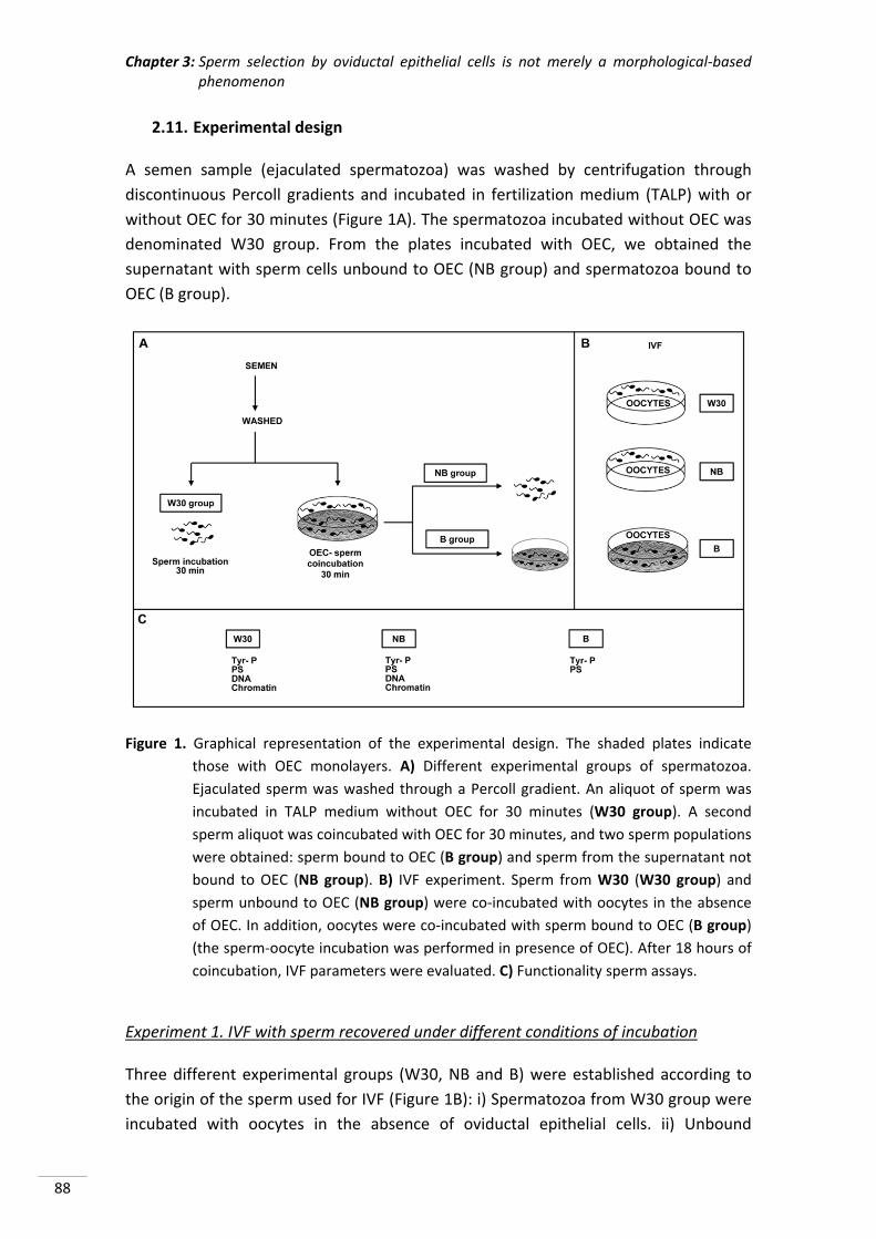
Chapter 3: Sperm selection by oviductal epithelial cells is not merely a morphological‐based phenomenon
88
2.11. Experimental design
A semen sample (ejaculated spermatozoa) was washed by centrifugation through
discontinuous Percoll gradients and incubated in fertilization medium (TALP) with or
without OEC for 30 minutes (Figure 1A). The spermatozoa incubated without OEC was
denominated W30 group. From the plates incubated with OEC, we obtained the
supernatant with sperm cells unbound to OEC (NB group) and spermatozoa bound to
OEC (B group).
Figure 1. Graphical representation of the experimental design. The shaded plates indicate
those with OEC monolayers. A) Different experimental groups of spermatozoa.
Ejaculated sperm was washed through a Percoll gradient. An aliquot of sperm was
incubated in TALP medium without OEC for 30 minutes (W30 group). A second
sperm aliquot was coincubated with OEC for 30 minutes, and two sperm populations
were obtained: sperm bound to OEC (B group) and sperm from the supernatant not
bound to OEC (NB group). B) IVF experiment. Sperm from W30 (W30 group) and
sperm unbound to OEC (NB group) were co‐incubated with oocytes in the absence
of OEC. In addition, oocytes were co‐incubated with sperm bound to OEC (B group)
(the sperm‐oocyte incubation was performed in presence of OEC). After 18 hours of
coincubation, IVF parameters were evaluated. C) Functionality sperm assays.
Experiment 1. IVF with sperm recovered under different conditions of incubation
Three different experimental groups (W30, NB and B) were established according to
the origin of the sperm used for IVF (Figure 1B): i) Spermatozoa from W30 group were
incubated with oocytes in the absence of oviductal epithelial cells. ii) Unbound
Chapter 3: Sperm selection by oviductal epithelial cells is not merely a morphological‐based phenomenon
89
spermatozoa (NB) were incubated with oocytes, in the absence of OEC. iii) Bound
spermatozoa (B) were incubated with oocytes in the OEC plates (IVF carried out on
OEC).
Oocytes were collected 18 hours post insemination and were fixed for subsequent
evaluation of the IVF parameters. The penetration rate, the number of spermatozoa
bound to the zona pellucida, the number of spermatozoa per penetrated oocyte, the
number of swollen spermatozoa and male pronuclear formation were assessed for
each group (Matás et al., 2003). A total of 5 repetitions were performed, using 689
oocytes.
Experiment 2. Assessment of sperm functionality selected by OEC
Tyrosine protein phosphorylation (TP), plasma membrane phosphatidylserine
translocation (PS), sperm DNA fragmentation and chromatin condensation were
evaluated in the three experimental sperm groups (W30, NB and B) (Figure 1C). For the
sperm groups in suspension (W30 and NB) protein tyrosine phosphorylation (by
indirect immunofluorescence and Western blot), PS translocation, DNA fragmentation
and chromatin condensation were studied. In the case of sperm from the B group, due
to the impossibility of unbinding them, phosphorylation by indirect
immunofluorescence and PS translocation were evaluated in situ, but it was not
possible to perform the rest of the assays.
TP of the sperm proteins evaluated by indirect inmunofluorescence, was classified and
grouped into three different categories (Figure 2) according to the patterns given by
Luño et al. (2013), following a hierarchical sequence of capacitation events, which
includes phosphorylation in the different regions of the sperm (acrosome, equatorial
subsegment and flagellum). Pattern I includes spermatozoa with no fluorescence signal
in the equatorial subsegment (Figure 2‐A1), with phosphorylated acrosome (Figure 2‐
A2), with phosphorylated tail (Figure 2‐A3) or with both acrosome and tail
phosphorylated (Figure 2‐A4). Pattern II includes spermatozoa with a signal in the
equatorial subsegment, no signal in the acrosome area and with (Figure 2‐B1) or
without (Figure 2‐B2) a signal in the flagellum. Finally, pattern III includes spermatozoa
with signal in the equatorial subsegment and acrosome area, and with (Figure 2‐C1) or
without (Figure 2‐C2) signal in the flagellum. Four repetitions were performed using
1650 spermatozoa.
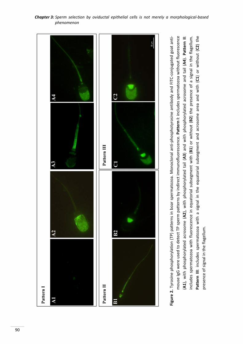
Chapter 3: Sperm selection by oviductal epithelial cells is not merely a morphological‐based phenomenon
90
Figure 2. Tyrosine phosphorylation (TP) patterns in boar spermatozoa. M
onoclonal anti‐phosphotyrosine antibody and FITC‐conjugated goat anti‐
mouse IgG
were used to detect TP
sperm patterns by indirect im
munofluorescen
ce. Pattern I: includes spermatozoa without fluorescen
ce
(A1), w
ith phosphorylated acrosome (A2), w
ith phosphorylated tail (A3) and w
ith phosphorylated acrosome and tail (A4). Pattern II:
includes spermatozoa with fluorescen
ce in equatorial subsegm
ent with (B1) or without (B2) the presence of a signal in the flagellum.
Pattern III: includes spermatozoa with a signal in the eq
uatorial subsegm
ent and acrosome area
and w
ith (C1) or without (C2) the
presence of signal in
the flagellum.
Chapter 3: Sperm selection by oviductal epithelial cells is not merely a morphological‐based phenomenon
91
To check the maximum level of sperm capacitation, which takes place moments before
fertilization (Sakkas et al., 2003), we also analysed the localization of proteins
phosphorylated in tyrosine residues of sperm bound to the zona pellucida 3 hours
after IVF by indirect immunofluorescence (Figure 3B). Three repetitions were
performed using 60 oocytes.
In the evaluation of TP proteins by Western blot eight repetitions were performed.
Dual sperm staining was used for PS and the samples were examined by
epifluorescence microscopy. Sperm with functional esterases (CFDA+) were visualized
in green fluorescence (fluorescein filter) and sperm with PS exposed (Annexin +) in red
fluorescence (rhodamine filter) (Marti et al., 2008). Five repetitions were performed
using 4800 spermatozoa.
For the DNA fragmentation assay, cells were classified into two categories according to
the intensity of fluorescence: low green fluorescence (low DNA damage) and high
green fluorescence (high DNA damage). In addition, measurements were expressed as
the mean green intensity fluorescence units, which were used as an index of the DNA
fragmentation (Matás et al., 2011). A total of 8 repetitions were performed.
For the chromatin condensation assay, measurements were expressed as the mean
red intensity fluorescence units, which were used as index of the state of the
chromatin condensation, as this is directly related to the PI uptake by DNA. A total of 8
repetitions were performed.
2.12. Statistical analysis
Data are expressed as the mean ± S.E.M. and analysed by ANOVA, considering the
specific sperm treatment as the main variable. When ANOVA revealed a significant
effect, values were compared by the least significant difference pairwise multiple
comparison post hoc test (Tukey). Differences were considered statistically significant
at p <0.05.
3. RESULTS
3.1. Experiment 1. IVF with sperm recovered under different conditions of
incubation
IVF provides information on gamete interaction and is the ultimate goal for many
reproductive studies. For this reason, IVF was performed using sperm from different
incubation conditions (Figure 1B).
Chapter 3: Sperm selection by oviductal epithelial cells is not merely a morphological‐based phenomenon
92
The studied IVF parameters changed depending on the origin of the sperm used (W30,
NB or B) (Table 1). The penetration rate (PEN) significantly differed among the three
study groups, being highest for the sperm bound to oviductal cells (B group: 94.9%),
followed by the W30 group (62.9%) and finally the NB group (19.6%) (p <0.05).
The number of spermatozoa bound to the zona pellucida (SPZ/ZP) followed the same
trend (B group: 52.9%, W30: 25.7% and NB: 12.4%; p <0.05). However, although
binding to the zona pellucida was greatest in the group B, the number of spermatozoa
per penetrated oocyte (SPZ/O) was similar to that observed in the washed
spermatozoa (W30), and higher than in the NB group. Although male pronuclear
formation was observed in all groups studied, the number of swollen spermatozoa
(SWO) was lower in the NB group than in the others (Table 1, p <0.05).
Table 1. IVF outcomes using different sperm sources: W30 group: sperm after 30 minutes of
incubation without oviductal epithelial cells (OEC); NB group: sperm not bound to
OEC after previous coincubation of 30 minutes. For W30 and NB groups IVF was
performed in the absence of OEC; B group: sperm bound to OEC after a previous
coincubation of 30 minutes; IVF was performed in the presence of OEC (data shown
mean ± S.E.M.).
Sperm source N PEN (%) SPZ/ZP SPZ/O* SWO*
W30 200 62.9 ± 4.2a 25.7 ± 1.8a 6.1 ± 0.5a 4.1 ± 0.5a
NB 270 19.6 ± 2.4b 12.4 ± 0.7b 1.5 ± 0.1b 0.4 ± 0.1b
B 219 94.9 ± 1.5c 52.9 ± 2.7c 6.1 ± 0.4a 4.4 ± 0.4a
N: number of oocytes used for fertilization ability (five replicates); PEN: rate of penetration;
SPZ/ZP: mean number of sperm bound to zona pellucida; SPZ/O: mean number of
spermatozoa per penetrated oocyte; SWO: swollen spermatozoa.
*Based on penetrated oocytes.
Different subscript letters (a, b, c) in the same column indicate significant differences (p
<0.05).
3.2. Experiment 2. Assessment of sperm functionality selected by OEC
A great difference was found in the phosphorylation patterns (patterns II and III)
(measured by indirect immunofluorescence) between the sperm subpopulations of the
bound (B) and unbound (NB) groups (Figure 3A) (p <0.05).
When sperm were incubated in the absence of OEC (W30) the principal pattern
exhibited was pattern II, followed by pattern III. However, pattern II was much more
prevalent and pattern III much less prevalent in B than in NB and W30. The unbound
sperm group (NB) showed a high level of TP in pattern III. This pattern III was
significantly reduced when sperm cells bound to OEC (group B). The localization of
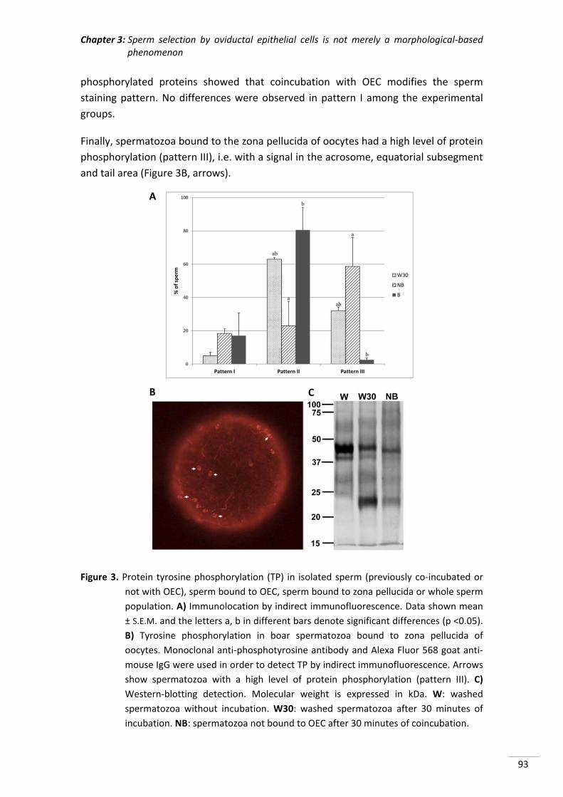
Chapter 3: Sperm selection by oviductal epithelial cells is not merely a morphological‐based phenomenon
93
phosphorylated proteins showed that coincubation with OEC modifies the sperm
staining pattern. No differences were observed in pattern I among the experimental
groups.
Finally, spermatozoa bound to the zona pellucida of oocytes had a high level of protein
phosphorylation (pattern III), i.e. with a signal in the acrosome, equatorial subsegment
and tail area (Figure 3B, arrows).
Figure 3. Protein tyrosine phosphorylation (TP) in isolated sperm (previously co‐incubated or
not with OEC), sperm bound to OEC, sperm bound to zona pellucida or whole sperm
population. A) Immunolocation by indirect immunofluorescence. Data shown mean
± S.E.M. and the letters a, b in different bars denote significant differences (p <0.05).
B) Tyrosine phosphorylation in boar spermatozoa bound to zona pellucida of
oocytes. Monoclonal anti‐phosphotyrosine antibody and Alexa Fluor 568 goat anti‐
mouse IgG were used in order to detect TP by indirect immunofluorescence. Arrows
show spermatozoa with a high level of protein phosphorylation (pattern III). C)
Western‐blotting detection. Molecular weight is expressed in kDa. W: washed
spermatozoa without incubation. W30: washed spermatozoa after 30 minutes of
incubation. NB: spermatozoa not bound to OEC after 30 minutes of coincubation.
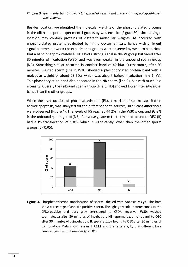
Chapter 3: Sperm selection by oviductal epithelial cells is not merely a morphological‐based phenomenon
94
Besides location, we identified the molecular weights of the phosphorylated proteins
in the different sperm experimental groups by western blot (Figure 3C), since a single
location may contain proteins of different molecular weights. As occurred with
phosphorylated proteins evaluated by immunocytochemistry, bands with different
signal patterns between the experimental groups were observed by western blot. Note
that a band of approximately 45 kDa had a strong signal in the W group but faded after
30 minutes of incubation (W30) and was even weaker in the unbound sperm group
(NB). Something similar occurred in another band of 40 kDa. Furthermore, after 30
minutes, washed sperm (line 2, W30) showed a phosphorylated protein band with a
molecular weight of about 23 kDa, which was absent before incubation (line 1, W).
This phosphorylation band also appeared in the NB sperm (line 3), but with much less
intensity. Overall, the unbound sperm group (line 3, NB) showed lower intensity/signal
bands than the other groups.
When the translocation of phosphatidylserine (PS), a marker of sperm capacitation
and/or apoptosis, was analysed for the different sperm sources, significant differences
were observed (Figure 4). The levels of PS reached 44.2% in the W30 group and 93.8%
in the unbound sperm group (NB). Conversely, sperm that remained bound to OEC (B)
had a PS translocation of 5.8%, which is significantly lower than the other sperm
groups (p <0.05).
Figure 4. Phosphatidylserine translocation of sperm labelled with Annexin V‐Cy3. The bars
show percentage of annexin positive sperm. The light grey colour corresponds to the
CFDA positive and dark grey correspond to CFDA negative. W30: washed
spermatozoa after 30 minutes of incubation. NB: spermatozoa not bound to OEC
after 30 minutes of coincubation. B: spermatozoa bound to OEC after 30 minutes of
coincubation. Data shown mean ± S.E.M. and the letters a, b, c in different bars
denote significant differences (p <0.01).
Chapter 3: Sperm selection by oviductal epithelial cells is not merely a morphological‐based phenomenon
95
The number of spermatozoa (CFDA +/annexin +) compared with the total number of
spermatozoa (annexin +) decreased slightly with the time of incubation (W30: 48.6%;
Figure 4). However this distribution was greatly impaired in groups of sperm
coincubated with OEC, while 91.4% of the sperm were CFDA and annexin positive in
group B, a value that fell to 39.2% in unbound sperm (NB).
The degree of chromatin condensation measured by PI staining was higher in the
unbound group (NB) than in W30 (17.8 ± 0.9 vs 23.3 ± 0.8; p <0.05), and inversely
related with PI uptake by DNA. When DNA damage was measured by TUNEL in these
groups, no differences were found (5.60 ± 0.69 vs 4.46 ± 0.75).
4. DISCUSSION
The insemination of large numbers of sperm translates into a selection of a few highly
fertile sperm able to reach and interact with the oviduct and subsequently with
oocytes. For this, complex natural sperm selection mechanisms, beside the well known
barriers in the female tract, are essential. However, although some factors have been
identified as fundamental for spermatozoa to reach the sperm reservoir (revised by
Soriano‐Úbeda et al., 2013), how exactly oviductal cells select the best sperm for
successful fertilization remains unknown.
When sperm are prepared for use in IVF for ART, seminal plasma is removed in order
to select the most suitable subpopulation of sperm for fertilization purposes (Matás et
al., 2011). Although this process attempts to mimic what happens in vivo, the final
performance, in terms of the quality of embryos produced, is far lower.
Various investigators have characterized some aspects of the superior sperm quality
associated with oviductal binding (revised by Holt & Fazeli, 2010), concluding that
sperm‐oviduct binding plays a key role in the maintenance of sperm fertilizing ability.
So, an IVF system based on OEC incubation and further collection of the sperm bound
to OEC should be of great use for selecting the best spermatozoa for ART, an ICSI
technique widely used in human infertility clinics. In this study, we aim to evaluate the
fertilizing capacity of spermatozoa, previously selected by Percoll gradient, incubated
in OEC. IVF was carried out with sperm bound and unbound to the OEC and some
characteristics of sperm that could explain the fertility results obtained were
determined.
The IVF results show that sperm bound to the OEC (B group) were able to fertilize
oocytes to a greater extent than those not are in contact with the OEC (W30 group).
This was expected because other authors had already mentioned the same (Romar et
al., 2005). However, we did not expect the unbound sperm group (NB) to have such
low levels of fertilization. We believe that sperm not bound to OEC are less capable of
Chapter 3: Sperm selection by oviductal epithelial cells is not merely a morphological‐based phenomenon
96
fertilizing due to the high levels of capacitation, as seen in experiment 2, and
subsequent apoptosis (Aitken, 2011). The number of spermatozoa bound to the zona
pellucida showed the same trend as the penetration rate (highest values in the B
group). The ability of sperm to bind to the zona pellucida is one of the most important
characteristics in the sperm fertilizing capacity (Oehninger et al., 1997). Defective
binding of spermatozoa to the zona pellucida reflects multiple sperm dysfunctions
(Larsson & Rodriguez‐Martinez, 2000), which would be consistent with decreased
penetration rates. Spermatozoa with high levels of capacitation (Oehninger et al.,
1997) or loss of plasma membrane functions (Barroso et al., 2000), as shown here by
NB sperm, could contribute to a low incidence of sperm‐zona pellucida binding, which
would affect the whole process of fertilization. In the same way, it seems to be a
prerequisite for such sperm‐zona pellucida interaction that spermatozoa must bind to
the OEC (Waberski et al., 2005), because this enables the spermatozoa to penetrate
the oocytes investments (Hunter, 1984).
Although male pronuclear formation was evident in all the groups studied, the number
of swollen spermatozoa (SWO) was lower in the NB group than in the others. Despite
problems with binding to the zona pellucida and penetrating oocytes, some NB sperm
are able to penetrate oocytes and can form male pronucleus because the oocyte has
the capacity to repair pre‐existing damage in the sperm (Evenson et al., 2002).
However, in some cases, this oocyte repair capacity may be insufficient to repair highly
damaged sperm, resulting in non‐swollen spermatozoa. The IVF results obtained
support the hypothesis that OEC select the best spermatozoa for fertilization, although
many of the qualities which spermatozoa must present to be selected by the OEC
remain unknown.
It is well documented that sperm protein phosphorylation is associated with
capacitation (Visconti et al., 1995) and that sperm treatment prior to IVF increases the
percentage of cells with phosphorylated tyrosine (Matás et al., 2011). Besides, the
sperm areas that show tyrosine phosphorylation depend on their capacitation status.
In boar sperm, the equatorial area is the most important area for tyrosine
phosphorylation (Jones et al., 2008), but it is the phosphorylation of the acrosome
which reflects high levels of sperm capacitation (Luño et al., 2013). Our results show
that an important factor in sperm selection by the OEC is the capacitation status of the
sperm population, as mentioned by other authors (Lefebvre & Suarez, 1996; Fazeli et
al., 1999; Luño et al., 2013). The sperm population bound to the epithelial cell
population showed a degree of fluorescence in the equatorial segment but no
fluorescence in the acrosomal area. However, in sperm unbound to the cells (NB
group), this pattern was reversed, i.e. they showed fluorescence in the acrosomal area.
In human spermatozoa, it has been established that a high degree of TP is considered a
prerequisite for fertilization (Sakkas et al., 2003). In this way, sperm must exhibit the
Chapter 3: Sperm selection by oviductal epithelial cells is not merely a morphological‐based phenomenon
97
highest degree of phosphorylation at the moment prior to contact with the zona
pellucida of the oocyte to activate the acrosome reaction. Conversely, if these levels
are reached earlier, far from the oocyte, sperm initiate the apoptosis pathway.
In this paper, the sperm with the highest level of protein TP (pattern III) were those in
the NB group, which, nevertheless, was the group with the lowest percentage of
penetration, precisely because they reached a high level of phosphorylation very early.
However, when we analyzed the pattern of TP in sperm bound to the zona pellucida,
like Sakkas et al. (2003), the highest level of phosphorylation was exhibited (pattern III:
acrosome, equatorial subsegment and flagellum).
On the one hand, during the capacitation process, there is a redistribution of tyrosine‐
phosphorylated proteins localized in the acrosomal region of boar sperm (Flesch et al.,
2001) and, on the other, the oviductal epithelium secretes proteins capable of
reversing sperm capacitation (Ghersevich et al., 2015). This redistribution is also
evident in the western blot, where the incubation of washed spermatozoa leads to the
appearance of new bands due to increased levels of phosphorylation of the principal
sperm pieces (Harayama et al., 2004), mainly the equatorial subsegment. The same
bands were also found in the NB group, indicating that phosphorylation levels were
also increased. This protein redistribution may help explain the change in the intensity
of the observed bands. However, we cannot affirm this because only localization
patterns were analysed without distinguishing the intensities of fluorescence, so the
amount of phosphorylated protein in each piece was not ascertained. We suggest that
the changes observed in sperm phosphorylation after coincubation with OEC are not
only due to the redistribution of proteins but also to modifications in phosphorylation‐
dephosphorylation in the proteins. It is still unknown whether OEC select the sperm
bound to them based on other criteria and then adjust their levels of phosphorylation,
or whether only sperm with a low capacitation pattern are bound.
It is still a matter of controversy too, whether PS translocation is due to capacitation
(Barroso et al., 2000; Duru et al., 2001; de Vries et al., 2003; Martin et al., 2005), the
apoptosis process (Aitken & Baker, 2013) or both (Aitken, 2011). Our results showed
significant differences in levels of PS translocation between the different experimental
groups of sperm (the lowest values were observed in the B group), indicating that OEC
exercise a selective function and that binding does not occur randomly. It is not
surprising that after 30 minutes of incubation, the percentage of spermatozoa with PS
translocation increased (Kotwicka et al., 2002) due to an increase in the capacitated
sperm population in the sample, which leads to destabilization of the membrane and
thus the translocation of PS (Vermes et al., 1995). Accordingly, if OEC bind mainly
uncapacitated spermatozoa (Fazeli et al., 1999), this population in our study would
correspond to spermatozoa without PS translocation (annexin ‐), while sperm with PS
translocation (annexin +) showed not be able to bind to OEC, remaining in the
Chapter 3: Sperm selection by oviductal epithelial cells is not merely a morphological‐based phenomenon
98
supernatant after coincubation, because they are capacitated or apoptotic. OEC would
also be able to discriminate between annexin negative spermatozoa, with their
morphologically superior quality, and annexin positive sperm (Hoogendijk et al., 2009).
Moreover, PS translocation is associated with a decrease in the adenosine
triphosphate (ATP) level of spermatozoa (Kotwicka et al., 2013) and with a lower
mitochondrial potential (Barroso et al., 2006), and so the OEC do not bind these
spermatozoa because they have insufficient energy reserves to remain in the sperm
reservoir and perform fertilization. Another explanation is that phospholipids
(including PS) are incorporated into the sperm membrane by OEC (Evans et al., 1980)
as a selection mechanism. This mechanism would result in a negative selection, similar
to that reported previously (Teijeiro et al., 2011), for detecting early phases of
disturbed membrane functions and marking for elimination those sperm which
probably have a reduced life span (Watson, 1995, 2000), and hence are less suitable
for storage in the oviduct because they have ceased to be functional. This could
explain the increase in PS translocation observed in NB sperm. Nevertheless, the
mechanisms that lead OEC to select and bind with live spermatozoa without
phosphatidylserine translocation in the plasma membrane (annexin ‐) remains
unknown.
When reproduction assisted techniques are used, it is necessary to select sperm in an
attempt to eliminate sperm with poor quality and seminal plasma, despite the
prejudicial effects this may have. In this sense, it has been shown that during
centrifugation to remove seminal plasma ROS are generated and these, in turn, induce
capacitation, apoptosis and DNA damage (Aitken & Koppers, 2011). In this study, we
observed that washing sperm through a Percoll gradient induced chromatin
decondensation. After 30 minutes of incubating sperm in a medium with OEC, the
sperm that had not been bound to the OEC had the same chromatin condensation as
ejaculated spermatozoa (S) even though the sperm had been selected through a
Percoll gradient, leaving only sperm of the highest quality (Matás et al., 2011). It has
been shown that during sperm capacitation chromatin decondensation occurs, which
prepares the sperm for the subsequent formation of pronucleus after fertilization (de
Lamirande et al., 2012). The reason for the greater compactness of the unbound sperm
is unknown although it has been attributed to defects in the disulphide bonds of
proteins to prevent chromatin decondensation. This hypothesis is reinforced by the IVF
results since the unbound sperm showed the lowest level of nucleus decondensation
in oocyte (SWO). In this regard, it has been shown that alterations in the protamine
packing of sperm DNA may contribute to a delay in the initiation of the zygotic S‐phase
(Eid et al., 1994), increase the length of the zygotic G2‐phase (Eid & Parrish, 1995), and
block blastocyst formation (Fatehi et al., 2006). On the other hand, Ardón et al. (2008)
suggested that chromatin instability may be associated with plasma membrane
characteristics that hinder the binding of such sperm to the oviduct.
Chapter 3: Sperm selection by oviductal epithelial cells is not merely a morphological‐based phenomenon
99
Another important factor related to the nucleus is DNA fragmentation and its effect on
fertility. Although there is still controversy regarding this, it has been shown that DNA
fragmentation in sperm and poor quality samples increase and are correlated with
failed fertilization in intracytoplasmic sperm injection (Lopes et al., 1998). However, in
normozoospermic samples fertility failure has been observed caused by spermatozoa
with damaged DNA (Aitken & De Iuliis, 2007). The results presented in this paper show
that spermatozoa selected by Percoll gradient showed less DNA fragmentation.
However, after 30 minutes of incubation with or without OEC, DNA fragmentation was
in fact unaffected, so selection with Percoll could be efficient because the selective
effect by the OEC was not evident in the case of DNA fragmentation.
Although sperm selection by density gradient initially selects the best spermatozoa
from a sperm population, such selection is insufficient to identify the most suitable
spermatozoon for the subsequent fertilization process. This paper shows that oviductal
epithelial cells (OEC) exert a more rigorous second selection. We conclude that it is
OEC that are responsible for real spermatic selection. Such selection by OEC could in
the future represent a solution to obtaining the best spermatozoa for fertilization and
embryo development. Using a non‐invasive technique that does not damage or impair
the sperm for later use would avoid the subjective selection that currently exists in
conventional sperm selection techniques.
ACKNOWLEDGEMENTS
The authors thank Virginia Reig for her assistance at the start of the experiments. We
also thank Rodrigo Diego for his technical support with the use of Annexin V‐Cy3TM and
Juan Carvajal for technical assistance. This work was supported by the Spanish Ministry
of Economy and Competitiveness (MINECO) and the European Regional Development
Fund (FEDER), Grant AGL2012‐40180‐C03‐01.
REFERENCES
Aitken RJ & De Iuliis GN (2007). Origins and consequences of DNA damage in male germ cells.
Reproductive BioMedicine Online. 14: 727‐733. (doi: 10.1016/S1472‐6483(10)60676‐1).
Aitken RJ (2011). The capacitation‐apoptosis highway: oxysterols and mammalian sperm
function. Biology of reproduction. 85: 9‐12. (doi: 10.1095/biolreprod.111.092528).
Aitken RJ & Koppers AJ (2011). Apoptosis and DNA damage in human spermatozoa. Asian
Journal of Andrology. 13: 36‐42. (doi: 10.1038/aja.2010.68).
Aitken RJ & Baker MA (2013). Causes and consequences of apoptosis in spermatozoa;
contributions to infertility and impacts on development. The international journal of
developmental Biology. 57: 265‐272. (doi: 10.1387/ijdb.130146ja).
Chapter 3: Sperm selection by oviductal epithelial cells is not merely a morphological‐based phenomenon
100
Ardón F, Helms D, Sahin E, Bollwein H, Töpfer‐Petersen E & Waberski D (2008). Chromatin‐
unstable boar spermatozoa have little chance of reaching oocytes in vivo.
Reproduction. 135: 461‐470. (doi: 10.1530/REP‐07‐0333).
Barroso G, Morshedi M & Oehninger S (2000). Analysis of DNA fragmentation, plasma
membrane translocation of phosphatidylserine and oxidative stress in human
spermatozoa. Human reproduction. 15: 1338‐1344. (doi: 10.1093/humrep/15.6.1338).
Barroso G, Taylor S, Morshedi M, Manzur F, Gaviño F & Oehninger S (2006). Mitochondrial
membrane potential integrity and plasma membrane translocation of
phosphatidylserine as early apoptotic markers: a comparison of two different sperm
subpopulations. Fertility and sterility. 85: 149‐154. (doi:
10.1016/j.fertnstert.2005.06.046).
Brüssow KP, Ratky J & Rodriguez‐Martinez H (2008). Fertilization and early embryonic
development in the porcine fallopian tube. Reproduction in Domestic Animals. 43: 245‐
251. (doi: 10.1111/j.1439‐0531.2008.01169.x).
Buffone M, Doncel G, Briggiler CM, Vazquez‐Levin M & Calamera J (2004). Human sperm
subpopulations: relationship between functional quality and protein tyrosine
phosphorylation. Human reproduction. 19: 139‐146. (doi: 10.1093/humrep/deh040).
de Lamirande E, Gabriel M & Zini A (2012). Human sperm chromatin undergoes physiological
remodeling during in vitro capacitation and acrosome reaction. Journal of andrology.
33: 1025‐1035. (doi: 10.2164/jandrol.111.015982).
de Vries K, Wiedmer T, Sims P & Gadella BM (2003). Caspase‐independent exposure of
aminophospholipids and tyrosine phosphorylation in bicarbonate responsive human
sperm cells. Biology of reproduction. 68: 2122‐2134. (doi:
10.1095/biolreprod.102.012500).
Duru NK, Morshedi MS, Schuffner A & Oehninger S (2001). Cryopreservation‐thawing of
fractionated human spermatozoa is associated with membrane phosphatidylserine
externalization and not DNA fragmentation. Journal of andrology. 22: 646‐651. (doi:
10.1002/j.1939‐4640.2001.tb02225.x).
Eid L, Lorton S & Parrish J (1994). Paternal influence on S‐phase in the first cell cycle of the
bovine embryo. Biology of reproduction. 51: 1232‐1237. (doi:
10.1095/biolreprod51.6.1232).
Eid L & Parrish J (1995). Duration of G2‐phase and onset of M‐phase during the first cell cycle
of the bovine embryo is dependent on bull in vivo fertility. Theriogenology. 43: 205.
(doi: 10.1016/0093‐691X(95)92359‐H).
Ellington JE, Evenson DP, Fleming JE, Brisbois RS, Hiss GA, Broder SJ & Wright RW (1998).
Coculture of human sperm with bovine oviduct epithelial cells decreases sperm
chromatin structural changes seen during culture in media alone. Fertility and sterility.
69: 643‐649. (doi: 10.1016/S0015‐0282(98)00023‐5).
Evans RW, Weaver DE & Clegg ED (1980). Diacyl, alkenyl, and alkyl ether phospholipids in
ejaculated, in utero‐, and in vitro‐incubated porcine spermatozoa. Journal of lipid
research. 21: 223‐228.
Evenson DP, Larson KL & Jost LK (2002). Sperm chromatin structure assay: its clinical use for
detecting sperm DNA fragmentation in male infertility and comparisons with other
Chapter 3: Sperm selection by oviductal epithelial cells is not merely a morphological‐based phenomenon
101
techniques. Journal of andrology. 23: 25‐43. (doi: 10.1002/j.1939‐
4640.2002.tb02599.x).
Fatehi A, Bevers M, Schoevers E, Roelen B, Colenbrander B & Gadella BM (2006). DNA
damage in bovine sperm does not block fertilization and early embryonic development
but induces apoptosis after the first cleavages. Journal of andrology. 27: 176‐188. (doi:
10.2164/jandrol.04152).
Fazeli A, Duncan A, Watson P & Holt WV (1999). Sperm‐oviduct interaction: induction of
capacitation and preferential binding of uncapacitated spermatozoa to oviductal
epithelial cells in porcine species. Biology of reproduction. 60: 879‐886. (doi:
10.1095/biolreprod60.4.879).
Flesch F, Wijnand E, Van de Lest C, Colenbrander B, van Golde LM & Gadella BM (2001).
Capacitation dependent activation of tyrosine phosphorylation generates two sperm
head plasma membrane proteins with high primary binding affinity for the zona
pellucida. Molecular reproduction and development. 60: 107‐115. (doi:
10.1002/mrd.1067).
Ghersevich S, Massa E & Zumoffen C (2015). Oviductal secretion and gamete interaction.
Reproduction. 149: R1‐R14. (doi: 10.1530/REP‐14‐0145).
Gualtieri R & Talevi R (2000). In vitro‐cultured bovine oviductal cells bind acrosome‐intact
sperm and retain this ability upon sperm release. Biology of reproduction. 62: 1754‐
1762. (doi: 10.1095/biolreprod62.6.1754).
Gualtieri R & Talevi R (2003). Selection of highly fertilization‐competent bovine spermatozoa
through adhesion to the Fallopian tube epithelium in vitro. Reproduction. 125: 251‐
258. (doi: 10.1530/rep.0.1220305).
Harayama H, Muroga M & Miyake M (2004). A cyclic adenosine 3′,5′‐monophosphate‐induced
tyrosine phosphorylation of syk protein tyrosine kinase in the flagella of boar
spermatozoa. Molecular reproduction and development. 69: 436‐447. (doi:
10.1002/mrd.20176).
Holt WV & Fazeli A (2010). The oviduct as a complex mediator of mammalian sperm function
and selection. Molecular reproduction and development. 77: 934‐943. (doi:
10.1002/mrd.21234).
Hoogendijk CF, Kruger TF, Bouic PJ & Henkel RR (2009). A novel approach for the selection of
human sperm using annexin V‐binding and flow cytometry. Fertility and sterility. 91:
1285‐1292. (doi: 10.1016/j.fertnstert.2008.01.042).
Huang VW, Zhao W, Lee C‐L, Lee CY, Lam KK, Ko JK, Yeung WS, Ho P‐C & Chiu PC (2013). Cell
membrane proteins from oviductal epithelial cell line protect human spermatozoa
from oxidative damage. Fertility and sterility. 99: 1444‐1452. e1443. (doi:
10.1016/j.fertnstert.2012.11.056).
Hunter RHF (1981). Sperm transport and reservoirs in the pig oviduct in relation to the time of
ovulation. Journal of reproduction and fertility. 63: 109‐117. (doi:
10.1530/jrf.0.0630109).
Hunter RHF (1984). Pre‐ovulatory arrest and peri‐ovulatory redistribution of competent
spermatozoa in the isthmus of the pig oviduct. Journal of reproduction and fertility. 72:
203‐211. (doi: 10.1530/jrf.0.0720203).
Chapter 3: Sperm selection by oviductal epithelial cells is not merely a morphological‐based phenomenon
102
Jha K, Salicioni A, Arcelay E, Chertihin O, Kumari S, Herr J & Visconti P (2006). Evidence for
the involvement of proline‐directed serine/threonine phosphorylation in sperm
capacitation. Molecular human reproduction. 12: 781‐789. (doi:
10.1093/molehr/gal085).
Jones R, James PS, Oxley D, Coadwell J, Suzuki‐Toyota F & Howes EA (2008). The equatorial
subsegment in mammalian spermatozoa is enriched in tyrosine phosphorylated
proteins. Biology of reproduction. 79: 421‐431. (doi: 10.1095/biolreprod.107.067314).
Kervancioglu ME, Saridogan E, Aitken RJ & Djahanbakhch O (2000). Importance of sperm‐to‐
epithelial cell contact for the capacitation of human spermatozoa in fallopian tube
epithelial cell cocultures. Fertility and sterility. 74: 780‐784. (doi: 10.1016/S0015‐
0282(00)01514‐4).
King G & Macpherson J (1973). A comparison of two methods for boar semen collection.
Journal of animal science. 36: 563‐565. (doi: 10.2134/jas1973.363563x).
Kotwicka M, Jendraszak M & Warchoł J (2002). Plasma membrane translocation of
phosphatidylserine in human spermatozoa. Folia histochemica et cytobiologica/Polish
Academy of Sciences, Polish Histochemical and Cytochemical Society. 40: 111‐112.
Kotwicka M, Jendraszak M, Skibinska I, Jedrzejczak P & Pawelczyk L (2013). Decreased
motility of human spermatozoa presenting phosphatidylserine membrane
translocation‐cells selection with the swim‐up technique. Human cell. 26: 28‐34. (doi:
10.1007/s13577‐011‐0024‐1).
Larsson B & Rodriguez‐Martinez H (2000). Can we use in vitro fertilization tests to predict
semen fertility? Animal Reproduction Science. 60: 327‐336. (doi: 10.1016/S0378‐
4320(00)00089‐0).
Lefebvre R & Suarez SS (1996). Effect of capacitation on bull sperm binding to homologous
oviductal epithelium. Biology of reproduction. 54: 575‐582. (doi:
10.1095/biolreprod54.3.575).
Lopes S, Sun J‐G, Jurisicova A, Meriano J & Casper RF (1998). Sperm deoxyribonucleic acid
fragmentation is increased in poor‐quality semen samples and correlates with failed
fertilization in intracytoplasmic sperm injection. Fertility and sterility. 69: 528‐532.
(doi: 10.1016/S0015‐0282(97)00536‐0).
Luño V, López‐Úbeda R, García‐Vázquez FA, Gil L & Matás C (2013). Boar sperm tyrosine
phosphorylation patterns in the presence of oviductal epithelial cells: in vitro, ex vivo,
and in vivo models. Reproduction. 146: 315‐324. (doi: 10.1530/REP‐13‐0159).
Marti E, Perez‐Pe R, Colas C, Muino‐Blanco T & Cebrian‐Perez J (2008). Study of apoptosis‐
related markers in ram spermatozoa. Animal Reproduction Science. 106: 113‐132. (doi:
10.1016/j.anireprosci.2007.04.009).
Martin G, Sabido O, Durand P & Levy R (2005). Phosphatidylserine externalization in human
sperm induced by calcium ionophore A23187: relationship with apoptosis, membrane
scrambling and the acrosome reaction. Human reproduction. 20: 3459‐3468. (doi:
10.1093/humrep/dei245).
Matás C, Coy P, Romar R, Marco M, Gadea J & Ruiz S (2003). Effect of sperm preparation
method on in vitro fertilization in pigs. Reproduction. 125: 133‐141. (doi:
10.1530/rep.0.1250133).
Chapter 3: Sperm selection by oviductal epithelial cells is not merely a morphological‐based phenomenon
103
Matás C, Sansegundo M, Ruiz S, García‐Vázquez F, Gadea J, Romar R & Coy P (2010). Sperm
treatment affects capacitation parameters and penetration ability of ejaculated and
epididymal boar spermatozoa. Theriogenology. 74: 1327‐1340. (doi:
10.1016/j.theriogenology.2010.06.002).
Matás C, Vieira L, García‐Vázquez F, Avilés‐López K, López‐Úbeda R, Carvajal J & Gadea J
(2011). Effects of centrifugation through three different discontinuous Percoll
gradients on boar sperm function. Animal Reproduction Science. 127: 62‐72. (doi:
10.1016/S0015‐0282(99)00013‐8).
Mburu J, Einarsson S, Lundeheim N & Rodriguez‐Martinez H (1996). Distribution, number and
membrane integrity of spermatozoa in the pig oviduct in relation to spontaneous
ovulation. Animal Reproduction Science. 45: 109‐121. (doi: 10.1016/S0378‐
4320(96)01566‐7).
Mburu J, Rodriguez‐Martinez H & Einarsson S (1997). Changes in sperm ultrastructure and
localisation in the porcine oviduct around ovulation. Animal Reproduction Science. 47:
137‐148. (doi: 10.1016/S0378‐4320(96)01631‐4).
Munuce MJ, Serravalle A, Caille AM, Zumoffen C, Botti G, Cabada M & Ghersevich S (2009).
Human tubal secretion can modify the affinity of human spermatozoa for the zona
pellucida. Fertility and sterility. 91: 407‐413. (doi: 10.1016/j.fertnstert.2007.11.077).
Oehninger S, Mahony M, Özgür K, Kolm P, Kruger T & Franken D (1997). Clinical significance
of human sperm‐zona pellucida binding. Fertility and sterility. 67: 1121‐1127. (doi:
10.1016/S0015‐0282(97)81449‐5).
Ouhibi N, Benet G & Menezo Y (1991). Fetal bovine oviduct epithelial cell monolayers: method
of culture and identification. Journal of tissue culture methods. 13: 289‐294.
Petrunkina AM, Gehlhaar R, Drommer W, Waberski D & Töpfer‐Petersen E (2001). Selective
sperm binding to pig oviductal epithelium in vitro. Reproduction. 121: 889‐896. (doi:
10.1530/rep.0.1210889).
Pursel V & Johnson L (1975). Freezing of boar spermatozoa: fertilizing capacity with
concentrated semen and a new thawing procedure. Journal of animal science. 40: 99‐
102.
Quintero I, Ghersevich S, Caille A, Munuce MJ, Daniele S & Morisoli L (2005). Effects of
human oviductal in vitro secretion on spermatozoa and search of sperm–oviductal
proteins interactions. International journal of andrology. 28: 137‐143. (doi:
10.1111/j.1365‐2605.2005.00525.xv).
Rath D, Long C, Dobrinsky J, Welch G, Schreier L & Johnson L (1999). In vitro production of
sexed embryos for gender preselection: high‐speed sorting of X‐chromosome‐bearing
sperm to produce pigs after embryo transfer. Journal of animal science. 77: 3346‐3352.
(doi: 1999.77123346x).
Rodriguez‐Martinez H, Tienthai P, Suzuki K, Funahashi H, Ekwall H & Johannisson A (2001).
Involvement of oviduct in sperm capacitation and oocyte development in pigs.
Reproduction (Cambridge, England) Supplement. 58: 129‐145.
Romar R, Coy P, Gadea J & Rath D (2005). Effect of oviductal and cumulus cells on zona
pellucida and cortical granules of porcine oocytes fertilized in vitro with epididymal
spermatozoa. Animal Reproduction Science. 85: 287‐300. (doi:
10.1016/j.anireprosci.2004.04.040).
Chapter 3: Sperm selection by oviductal epithelial cells is not merely a morphological‐based phenomenon
104
Sakkas D, Leppens‐Luisier G, Lucas H, Chardonnens D, Campana A, Franken D & Urner F
(2003). Localization of tyrosine phosphorylated proteins in human sperm and relation
to capacitation and zona pellucida binding. Biology of reproduction. 68: 1463‐1469.
(doi: 10.1095/biolreprod.102.011023).
Soriano‐Úbeda C, Matás C & García‐Vázquez F (2013). An overview of swine artificial
insemination: retrospective, current and prospective aspects. Journal of Experimental
and Applied Animal Sciences. 1: 67‐97.
Talevi R & Gualtieri R (2010). Molecules involved in sperm‐oviduct adhesion and release.
Theriogenology. 73: 796‐801. (doi: 10.1016/j.theriogenology.2009.07.005).
Tardif S, Dubé C, Chevalier S & Bailey JL (2001). Capacitation is associated with tyrosine
phosphorylation and tyrosine kinase‐like activity of pig sperm proteins. Biology of
reproduction. 65: 784‐792. (doi: 10.1095/biolreprod65.3.784).
Teijeiro JM, Dapino DG & Marini PE (2011). Porcine oviduct sperm binding glycoprotein and its
deleterious effect on sperm: a mechanism for negative selection of sperm? Biological
research. 44: 329‐337.
Tienthai P, Johannisson A & Rodriguez‐Martinez H (2004). Sperm capacitation in the porcine
oviduct. Animal Reproduction Science. 80: 131‐146. (doi: 10.1016/S0378‐
4320(03)00134‐9).
Töpfer‐Petersen E, Wagner A, Friedrich J, Petrunkina AM, Ekhlasi‐Hundrieser M, Waberski D
& Drommer W (2002). Function of the mammalian oviductal sperm reservoir. Journal
of Experimental Zoology. 292: 210‐215. (doi: 10.1002/jez.1157).
Vermes I, Haanen C, Steffens‐Nakken H & Reutellingsperger C (1995). A novel assay for
apoptosis flow cytometric detection of phosphatidylserine expression on early
apoptotic cells using fluorescein labelled annexin V. Journal of immunological methods.
184: 39‐51. (doi: 10.1016/0022‐1759(95)00072‐I).
Visconti PE, Bailey JL, Moore GD, Pan D, Olds‐Clarke P & Kopf GS (1995). Capacitation of
mouse spermatozoa. I. Correlation between the capacitation state and protein
tyrosine phosphorylation. Development. 121: 1129‐1137.
Waberski D, Magnus F, Ferreira FM, Petrunkina AM, Weitze K & Töpfer‐Petersen E (2005).
Importance of sperm‐binding assays for fertility prognosis of porcine spermatozoa.
Theriogenology. 63: 470‐484. (doi: 10.1016/j.theriogenology.2004.09.025).
Waberski D, Magnus F, Ardon F, Petrunkina AM, Weitze K & Töpfer‐Petersen E (2006).
Binding of boar spermatozoa to oviductal epithelium in vitro in relation to sperm
morphology and storage time. Reproduction. 131: 311‐318. (doi:
10.1530/rep.1.00814).
Watson P (1995). Recent developments and concepts in the cryopreservation of spermatozoa
and the assessment of their post‐thawing function. Reproduction, fertility and
development. 7: 871‐891. (doi: 10.1071/RD9950871).
Watson P (2000). The causes of reduced fertility with cryopreserved semen. Animal
Reproduction Science. 60: 481‐492. (doi: 10.1016/S0378‐4320(00)00099‐3).
Zhu J, Barratt C, Lippes J, Pacey A & Cooke I (1994). The sequential effects of human cervical
mucus, oviductal fluid, and follicular fluid on sperm function. Fertility and sterility. 61:
1129‐1135.

Chapter 3: Sperm selection by oviductal epithelial cells is not merely a morphological‐based phenomenon
105
Zumoffen C, Caille A, Munuce M, Cabada M & Ghersevich S (2010). Proteins from human
oviductal tissue‐conditioned medium modulate sperm capacitation. Human
reproduction. 25: 1504‐1512. (doi: 10.1093/humrep/deq063).

Conclusions/
Conclusiones

Conclusions/Conclusiones
109
CONCLUSIONS
CHAPTER 1
1. Different Percoll gradients can be used to separate sperm subpopulation based on
their functional parameters.
2. The spermatozoa selected through a 45/90% Percoll gradient exhibit higher
penetration levels when they are used for in vitro fertilization, due to the
acquisition of a greater degree of capacitation compared with 45/60 and 60/75
gradients.
CHAPTER 2
3. Sperm‐oviductal epithelial cell binding can be considered a mechanism for selecting
sperm populations with a low degree of protein tyrosine phosphorylation, as
demonstrated in in vivo, ex vivo, and in vitro conditions.
CHAPTER 3
4. The sperm selected by oviductal epithelial cells led to better output when used in in
vitro fertilization systems.
5. Oviductal epithelial cells exert a high degree of sperm selection, binding
spermatozoa of better quality based on different functional parameters.
GENERAL CONCLUSION
Boar semen is composed of different sperm populations, of which those with greater
functionality can be selected in vitro by Percoll gradients or through a more rigorous
selection carried out by the epithelial cells of the oviduct, which are able to distinguish
between a higher sperm fertilizing capacity.
Conclusions/Conclusiones
110
CONCLUSIONES
CAPÍTULO 1
1. El uso de diferentes gradientes de Percoll nos permiten separar subpoblaciones
espermáticas en base a sus características funcionales.
2. Los espermatozoides seleccionados a través del gradiente de Percoll 45/90%
presentan mayores niveles de penetración cuando son utilizados en fecundación in
vitro debido a la adquisición de un mayor grado de capacitación comparado con los
gradientes 45/60 y 60/75.
CAPÍTULO 2
3. Las células epiteliales del oviducto, bajo condiciones in vivo, ex vivo e in vitro,
ejercen un mecanismo de selección espermática permitiendo la unión de aquellos
espermatozoides que presentan un bajo grado de fosforilación en tirosina.
CAPÍTULO 3
4. Los espermatozoides seleccionados mediante células epiteliales del oviducto dan lugar a mejores resultados cuando son empleados en sistemas de fecundación in vitro.
5. Las células epiteliales del oviducto ejercen un alto grado de selección espermática,
uniendo aquellos espermatozoides de mayor calidad en base a diferentes parámetros funcionales.
CONCLUSIÓN GENERAL
El semen porcino se encuentra compuesto por diferentes poblaciones espermáticas,
de las cuales, aquellas con una mayor funcionalidad pueden ser seleccionadas in vitro
mediante gradientes de Percoll o bien a través de una selección más rigurosa llevada a
cabo por las células epiteliales de oviducto, siendo capaces de distinguir entre
espermatozoides con una capacidad fecundante superior.
Appendix

Appendix
113
APPENDIX
Publications derived from the Doctoral Thesis
The results obtained in the present Doctoral Thesis have been included in the following
publications:
1. ARTICLES IN JOURNALS INCLUDED IN THE SCIENCE CITATION INDEX (SCI) OF THE
INSTITUTE FOR SCIENTIFIC INFORMATION (ISI):
López‐Úbeda R, García‐Vázquez FA, Gadea J, Matás C. (2015). Sperm selection
by oviductal epithelial cells is not merely a morphological‐based phenomenon.
Under review.
Luño V, López‐Úbeda R, García‐Vázquez FA, Gil L, Matás C. (2013). Boar sperm
tyrosine phosphorylation patterns in the presence of oviductal epithelial cells:
in vitro, ex vivo, and in vivo models. Reproduction. 146 (4): 315‐24. (doi:
10.1530/REP‐13‐0159).
Matás C, Vieira L, García‐Vázquez FA, Avilés‐López K, López‐Úbeda R, CarvajaL
JA, Gadea J. (2011). Effects of centrifugation through three different
discontinuous Percoll gradients on boar sperm function. Animal Reproduction
Science. 127 (1‐2): 62‐72. (doi: 10.1016/j.anireprosci.2011.06.009).
2. OTHER ARTICLES:
López‐Úbeda R, Matás C. (2015). An approach to the factors related to sperm
capacitation process. Andrology‐Open Access. 4: 128. (doi: 10.4172/2167‐
0250.1000128).
3. ABSTRACTS IN JOURNALS INCLUDED IN THE SCIENCE CITATION INDEX (SCI):
Luño V, López‐Úbeda R, Lefievre L, Matás C. (2012). Protein tyrosine
phosphorylation pattern of sperm bound to porcine oviduct during
periovulation stage. 39th Annual conference of International Embryo Transfer
Society (IETS), Hannover, Germany. January 19‐23, 2013. Reproduction, Fertility
and Development. 25 (1): 270. (doi: 10.1071/RDv25n1Ab244).
López‐Úbeda R, Matás C. (2012). Effect of incubation in oviductal epithelial
cells and co‐culture with oocytes in the protein tyrosine phosphorylation
pattern in boar sperm. 39th Annual conference of International Embryo
Appendix
114
Transfer Society (IETS), Hannover, Germany. January 19‐23, 2013.
Reproduction, Fertility and Development. 25 (1): 270. (doi:
10.1071/RDv25n1Ab245).
Luño V, López‐Úbeda R, Gadea J, Matás C. (2011). Protein tyrosine
phosphorylation in boar sperm during co‐culture with oviductal epithelial cells.
37th Annual conference of International Embryo Transfer Society (IETS),
Orlando, Florida, EEUU, January 9‐12, 2011. Reproduction, Fertility and
Development. 23 (1): 216. (doi: 10.1071/RDv23n1Ab236).
López‐Úbeda R, Luño V, Vieira L, Gadea J, Matás C. (2011). Protein‐tyrosine
phosphorylation and calcium uptake in boar sperm subpopulations after
different discontinuous Percoll gradient centrifugations. 37th Annual
conference of International Embryo Transfer Society (IETS), Orlando, Florida,
EEUU, January 9‐12, 2011. Reproduction, Fertility and Development. 23 (1): 217.
(doi: 10.1071/RDv23n1Ab237).
4. COMMUNICATIONS TO CONGRESS
López‐Úbeda R, Diego R, Matás C. (2015). Bajo condiciones in vitro, la adhesión
de los espermatozoides a las células del oviducto se ve facilitada si no tienen
exteriorizada la fosfatidilserina. XVI Jornadas Sobre Producción Animal.
Zaragoza. 19 y 20 de Mayo de 2015. Tomo II. P: 364‐366. (ISBN: 978‐84‐606‐
7971‐4).
López‐Úbeda R, Matás C. (2013). The sperm bound to oviductal cells change
their proteins phosphorylated. 1st COST Action FA1201 EPICONCEPT, General
Conference “Epigenetics and Periconception Environment”, Antalya (Turkey), 24
y 25 de April de 2013. P: 34. 2013. (ISBN: 978‐975‐7636‐95‐3).
López‐Úbeda R, Matás C. (2013). La selección de espermatozoides por las
células oviductales modifica los parámetros de fecundación in vitro. XV
Jornadas sobre Producción Animal, Zaragoza 14 y 15 de Mayo 2013. Tomo I. P:
383‐385. (ISBN: 978‐84‐695‐7684‐7).
Luño V, López‐Úbeda R, García‐Vázquez F.A, Matás C. (2013). Fosforilación de
tirosina en espermatozoides incubados en explantes oviductales en la especie
porcina: efecto del lavado espermático. XV Jornadas sobre Producción Animal,
Zaragoza 14 y 15 de Mayo 2013. Tomo I. P: 386‐388. (ISBN: 978‐84‐695‐7684‐
7).
