understanding retinitis pigmentosa
TRANSCRIPT

Understanding
retinitispigmentosa
RCOphth

The Understanding series is designed to help you, yourfriends and family understand a little bit more about youreye condition.
Other titles in the series include:Understanding age-related macular degenerationUnderstanding cataractsUnderstanding Charles Bonnet syndromeUnderstanding dry eyeUnderstanding eye conditions related to diabetesUnderstanding glaucomaUnderstanding nystagmusUnderstanding posterior vitreous detachmentUnderstanding retinal detachment
All these leaflets are available in audio, print and brailleformats. To order please contact our Helpline on 0303 123 9999 (all calls charged at local rate), [email protected] or visit rnib.org.uk/shop
RNIB’s Understanding series
2

About retinitis pigmentosa . . . . . . . . . . . . . . . . . . 4
How your eye works . . . . . . . . . . . . . . . . . . . . . . . . 5
Causes . . . . . . . . . . . . . . . . . . . . . . . . . . . . . . . . . . . . . 6
Symptoms . . . . . . . . . . . . . . . . . . . . . . . . . . . . . . . . . 10
Eye tests . . . . . . . . . . . . . . . . . . . . . . . . . . . . . . . . . . 12
Hospital investigation. . . . . . . . . . . . . . . . . . . . . . 14
Other eye conditions with RP. . . . . . . . . . . . . . . 18
Treatment . . . . . . . . . . . . . . . . . . . . . . . . . . . . . . . . . 19
Coping . . . . . . . . . . . . . . . . . . . . . . . . . . . . . . . . . . . . 22
Useful contacts . . . . . . . . . . . . . . . . . . . . . . . . . . . . 24
Contents
3

4
Retinitis pigmentosa (RP) is the name given to a diversegroup of inherited eye disorders. These eye conditionsaffect a part of your eye called the retina. RP causespermanent changes to your vision but how quickly thishappens and how it changes differs between people.These changes may include difficulty with vision in dimlight or the dark and loss of your side or peripheral vision.
If you have RP, sight loss is gradual but progresses over a period of many years. Some people with RP mightbecome blind but most people with RP keep some usefulvision well into old age.
New research is constantly changing our understandingof RP. The British Retinitis Pigmentosa Society (RPFighting Blindness) website and leaflets offer currentupdates with detailed explanations of these issues.
About retinitis pigmentosa
cornea
pupil
retina
macula
iris
to the brain
optic nerve
lens

When you look at something, light passes through thefront of your eye, and is focused by the lens onto yourretina. The retina is a delicate tissue that is sensitive tolight. It converts the light into electrical signals thattravel along the optic nerve to your brain. The brain theninterprets these signals to “see” the world around you.
Your retina has two main layers, a thin one called thepigment epithelium and a thicker one, called the neuralretina. The neural retina contains many millions of cellscalled photoreceptors and these cells convert light intoelectrical signals that travel to your brain.
Light is focused onto a tiny area of the central retinacalled the macula. This specialised area of your retina isabout the size of a pinhead. The macula contains a fewmillion specialised photoreceptors called cone cells. Thesecone cells work best in bright light levels and allow youto see fine detail for activities such as reading and writingand to recognise colours.
The rest of your retina, called the peripheral retina, ismostly made up of the other type of photoreceptorcalled rod cells. Rod cells enable you to see when light isdim and provide your peripheral vision. Peripheral visionis what you can see to the sides and above and belowwhen you are looking at something straight ahead.
5
How your eye works

6
All types of RP affect the retina. The retinal cellsgradually stop working and eventually die. In most cases,the peripheral rod cells are affected first and RP lateraffects the central cone cells. The symptoms youexperience depend on the way your retina is affected byRP and can be very different from person to person.
Almost all types of RP are inherited, caused by a fault inthe genetic information passed down from a parent. Thegenes you inherit contain the instructions that tell ourbody how to grow, repair and renew. When a gene isfaulty these instructions are faulty and the cells usingthose instructions do not work as they should.
In RP, the faulty genes cause the retinal cells to stopworking and eventually die off. Researchers have foundmany of the genes which, when faulty, cause RP butthere is still work to be done to discover them all.
As there are many genes that can cause the retinal cellsto stop working, there are many different types of RP.This is why RP is described as a group of inherited retinaldisorders. RP is often mentioned alongside other eyeconditions with similar genetic causes and effects onvision, such as Leber’s Congential Amaurosis, cone andcone-rod dystrophies, and Choroideremia.
Causes

RP can also be associated with other problems such ashearing loss. These rare conditions are referred to as RPsyndromes.
RP syndromes
In most cases, the inherited gene defect only affects theeyes. Sometimes, other parts of the body are alsoaffected. One example of this is Usher syndrome, wherepeople develop both hearing loss and sight loss. Othersinclude Refsum, Alström, and Laurence-Moon-Bardet-Biedl (LMBB) syndromes.
How is RP inherited?
RP often runs in families and is classified by the way it ispassed from generation to generation. The type ofinheritance can tell you who in your family has had thecondition, how severely your vision could be affected byRP and the chances of your children being affected. RP can be inherited in three different ways:
Autosomal dominant inheritanceAutosomal dominant RP affects men and women equallyand there tends to be a known history of the condition inthe family. This form of RP is less severe than the other
7

8
two listed below and the first signs of it tend to appearat around 30 years of age.
Autosomal recessive inheritanceAutosomal recessive RP also affects men and womenequally but there may be little or no known history of thecondition in either family in the past. This form of RPtends to show first signs between 30 and 40 years andtends to cause more severe sight loss.
X-linked inheritanceThis is a pattern of inheritance that affects mostly men.Female members of a family are carriers of the faultygene but rarely develop the full condition, although somecarriers can develop a mild form of RP. If there have beenno boys in the family in the last few generations thenthere may be no history of the condition. This type of RPaffects vision severely and can result in very poor visionby the age of 40.
No known relativeIn about half of diagnosed cases of RP there does notseem to be any previously affected relatives. Relativeswill have passed on the faulty genetic information butmay have not developed symptoms themselves. In suchcases it may not be possible to determine which of thethree types of inheritance have caused the RP.

Genetic counsellingGenetic counselling aims to help you understand the type of RP you have, how it is likely to affect you in thelong term and the risks of passing on the condition toany children you may have. A genetic counsellor asksabout your family tree in detail to try and understandhow RP has been inherited through the generations.Genetic counselling is a free NHS service. It may beprovided by a specialist RP eye clinic or a medicalgenetics department. You can ask your GP orophthalmologist to refer you to your local geneticcounselling service.
Genetic testingYou may also be offered genetic tests to try and work out which genes are faulty. Testing is carried out inregional genetics centres and your eye specialist(ophthalmologist) could refer you to one. Testing for RP is complex and is not useful or possible yet for alltypes of RP. Ask your ophthalmologist or geneticcounsellor to discuss testing with you.
9

Early symptoms
In most of the more common forms of RP, the firstsymptoms occur between childhood and the age of 30.The first symptom you usually notice is that you find itdifficult to see in poor light, such as outdoors at dusk, orin a dimly lit room. This is often referred to as “nightblindness”. While most people find it takes their eyesabout 20 minutes to adapt to dim light, if you have RP itwill either take much longer or it won’t happen at all.
A second symptom is the loss of some of your peripheralvision or peripheral visual field. This means that whenyou’re looking straight ahead you become less able to seethings either to the side, above or below. Difficultyseeing in low light and loss of peripheral vision are a signthat the peripheral rod cells are being affected by RP.
For some people, the early loss of peripheral vision maymean it is no longer safe for you to drive. You arerequired by law to report a condition which might affectyour sight and you need to report a diagnosis of RP tothe DVLA so they can carry out regular tests.
In some RP-related conditions, central vision is lost firstbecause the central cone cells are affected first. You
Symptoms
10

might find it difficult reading print or carrying outdetailed work at this time. In these types of RP,peripheral vision is affected in the later stages.
Later symptoms
All RP conditions are progressive, but the speed andpattern of deterioration of sight varies from one personto another. For most people, the first effect of RP is thegradual loss of peripheral vision. This means that you canstart to miss things slightly to the side of you or trip overor bump into things you would have seen in the past.Most people with RP eventually have a very restrictedvisual field, leaving only a narrow tunnel of vision.
Most people with RP retain useful central vision throughtheir twenties, which means the ability to read andrecognise faces is not greatly affected. By 50 years of agemost people’s central vision is affected to the extent thatreading is a problem without the help of a magnifier.
Many people who have RP find the glare from brightlights and sunlight becomes an increasing problem. Theretinal cells become less able to adapt to changing lightlevels and it becomes more difficult to use your visionwhen you move between a light and a dark room.
11

12
Most people first experience problems in low light levelsand this may prompt them to see their optometrist(optician) or general practitioner (GP). Because the earlysymptoms can vary from person to person, some peoplemay have their condition diagnosed at an early stagewhile other people’s RP may go undetected for many years.
An optometrist can examine your retina to detect RP. By looking into your eye with a piece of equipment called an ophthalmoscope, an optometrist can examinethe back of the eye. Normally, they would see the orange red of the healthy retina and the blood vesselsthat supply it. When someone has RP, the shape of theblood vessels is affected and the orange surface isinterrupted by tiny clumps of black or brown pigment.This is quite an early sign.
The types of RP which cause loss of central vision tendto be detected by a routine eye test at an early stage.Loss of central vision makes the letter chart harder tosee. The more common symptom of peripheral field loss,or loss of side vision, is not so obvious and this can onlybe detected by a field of vision test. Most optometristscan carry out this test but may not do so routinely.
Eye tests

13
If you have any concerns about your peripheral visionthen you should ask your optometrist for a field of vision test.
If you have a family history of RP or you have hadproblems with your vision in the dark, or when movingfrom light to dark, you need to make this clear to theperson testing your eyes. This will help them to devisethe most appropriate set of tests for you. If after an eyetest there is cause for concern the optometrist can referyou to a specialist eye doctor (ophthalmologist) at ahospital for more detailed testing.

14
If you have been referred to the ophthalmologist, a set of tests can be done to diagnose RP. The testing processvaries from person to person and may take more thanone visit. The ophthalmologist may be able to say thatyou have RP after the first few sets of tests but it isoften not possible in the early stages of the condition todefine exactly what form of RP you have or what thelikely long-term effects will be on your vision.
It is important to ask your ophthalmologist to talk youthrough the tests and the results at each stage. None ofthe tests are painful but they can take a long time andbe repetitive. You may be asked to have some or all ofthe following tests:
Examining the back of your eye
Your retina will be examined each time you visit thehospital. You will be given eye drops to dilate your pupilsto allow the ophthalmologist to see the back of your eyeclearly. The dilating drops take about 30 minutes to work.They will make you sensitive to light and cause yourvision to be blurry. The effects of the drops usually wearoff in about six hours though sometimes it can takeovernight. It is not safe to drive until the effects haveworn off.
Hospital investigation

15
Retinal photographs and fluoresceinangiograms
You may have photographs taken of your retina using aspecial camera. This photograph of the retina can be usedfor comparison during future visits as an additional wayof tracking the progress of your RP. Your ophthalmologistmay ask for a more specialised set of photographs to betaken using fluorescein dye. The yellow fluorescein dye isinjected into a vein. It travels into the tiny blood vesselsin your retina, and a series of photographs are taken. Thedye in the blood vessels shows up changes in the retinathat are not visible with normal photography. Thefluorescein dye can make your skin look yellow for up to24 hours. The dye is passed through the urine, which willbe a deep yellow colour for about 24 hours.
Visual field test
A visual field test checks whether your peripheral visionhas been affected. You look straight ahead at a particularpoint in the bowl-shaped screen of a visual field machinein a darkened room. Each time you spot dots of light youclick a button. The test takes about 10 minutes for eacheye and tests what you notice to the sides, above andbelow when you are looking straight ahead.

16
Colour vision
Your colour vision may be tested. You will be asked tolook at a booklet that shows numbers composed ofdifferent coloured dots. The numbers are printed withindifferent coloured dotty backgrounds. This quick andstraight forward test shows which colours you are able todistinguish from each other.
Electro-diagnostic tests
Electro-diagnostic tests may be needed to investigatehow your retina is working. The electrical activity of theretina is measured under different lighting conditions andthis then identifies layers of the retina that are notworking properly.
These tests include the Electroretinogram (ERG), Electro-oculogram (EOG) and the PatternElectroretinogram (PERG). Each test records electricalactivity producing a trace or plot. When these plots arecompared to the plot from a retina without RP your eyespecialist can see which layers of your retina have beenaffected.

17
These tests are usually carried out by the electro-diagnostics department of the eye department. Each testhas a specific procedure and you should ask the staff toexplain exactly what will happen before you start. Thetests are painless and straight forward but may involvehaving your eyes dilated and/or numbed, a tiny electrodebeing placed on your eye and a sensor on your skin.
18
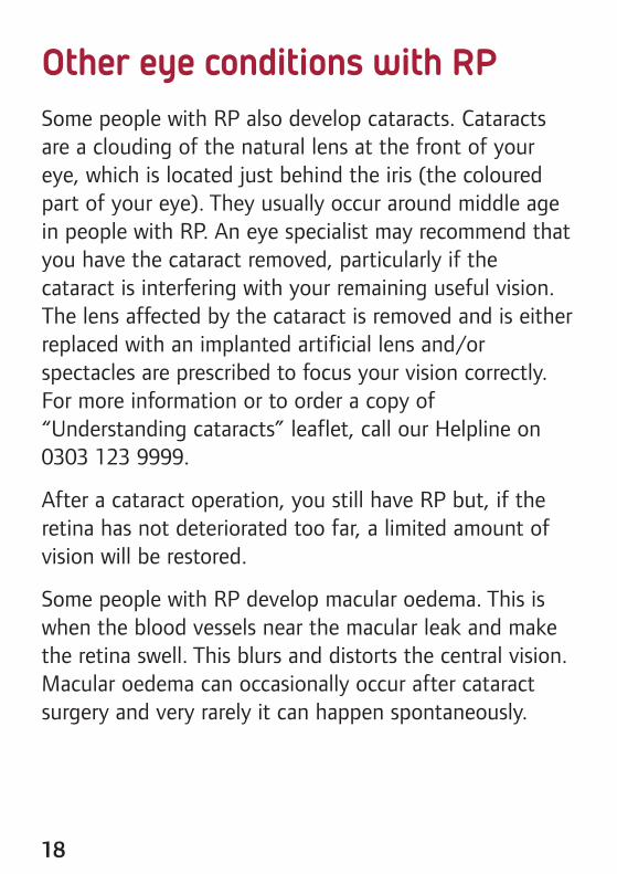
Some people with RP also develop cataracts. Cataractsare a clouding of the natural lens at the front of youreye, which is located just behind the iris (the colouredpart of your eye). They usually occur around middle agein people with RP. An eye specialist may recommend thatyou have the cataract removed, particularly if thecataract is interfering with your remaining useful vision.The lens affected by the cataract is removed and is eitherreplaced with an implanted artificial lens and/orspectacles are prescribed to focus your vision correctly.For more information or to order a copy of“Understanding cataracts” leaflet, call our Helpline on0303 123 9999.
After a cataract operation, you still have RP but, if theretina has not deteriorated too far, a limited amount ofvision will be restored.
Some people with RP develop macular oedema. This iswhen the blood vessels near the macular leak and makethe retina swell. This blurs and distorts the central vision.Macular oedema can occasionally occur after cataractsurgery and very rarely it can happen spontaneously.
Other eye conditions with RP
19
Currently there is no known cure or treatment for RP orassociated retinal disorders. Many research groups aroundthe world are working on different aspects of thecondition with the aim of developing treatments. Manyof the genes causing RP and related conditions are beingdiscovered (or mapped) and it is this understanding ofwhere the faults occur in the genetic information thatmay enable potential treatments to be devised. It ispossible that eventually one or more treatments may bedevised which will combine the knowledge gained fromsome of the following avenues of research. Most of thework so far has only been carried out in the laboratory.
Gene therapy
Once a faulty gene causing RP has been identified, genetherapy aims to replace the faulty gene within theaffected retinal cells with new genes that work properly.The new genetic material, usually carried by a harmlessvirus, is injected directly into the affected area of theretina. The hope is that the cells then begin to workcorrectly and the damage is either stopped or reversed.This method relies on the gene causing the problembeing known but in many cases of RP the faulty gene orgenes are yet to be discovered.
Treatment

20
Stem cell therapy
The body contains many different types of cells, andsome are more specialised than others. The retinal cellsaffected by RP are very specialised cells that the bodycannot easily replace. Stem cells are cells that can divide(differentiate) into other cell types and they have thepotential to replace damaged or missing retinal cells. Theaim of research into stem cell therapy is to see if stemcells injected into the retina can be persuaded todifferentiate into retinal cells.
Growth factors
Growth factors are chemicals that support cells to growand repair themselves. Research groups are working onthe potential uses of growth factors in the treatment ofretinal disease in the hope that damaged cells can berepaired or protected from damage.
Nutrition
It has been suggested by some research that vitamin Amay have a beneficial effect for people with RP. Thisresearch has been questioned and the positive effectsobserved were very slight. This type of treatment is not
21
currently being prescribed by most specialists. Takingvitamin A can be bad for your health and should bediscussed with your GP and ophthalmologist. Otherstudies are investigating the benefits of mixtures ofnutritional supplements which have an antioxidant effect.
Refsum syndrome is one of these rare situations whereRP is known to be affected by nutrition. Strictly adheringto a diet that excludes or is low in phytanic acid isbeneficial in Refsum syndrome. Phytanic acid is in dairyproducts, beef and lamb, and fatty fish such as tuna,cod, and haddock. Ask to be referred to a dietician if it isrecommended that you follow a restricted diet, so thatyou can be sure to get the nutrients you need.
Research updates
Because research and new theories change quickly,stories about potential cures or treatments for sight lossoften appear in the newspapers, on television and on theinternet. Such stories are often over-simplified andoccasionally misleading. Regular authoritative updates oncurrent research are available on the RP FightingBlindness website rpfightingblindness.org.uk

22
Being diagnosed with an eye condition can be veryupsetting. You may find that you are worried about thefuture and how you will manage with a change in yourvision. All these feelings are natural.
Some people may want to talk over some of thesefeelings with someone outside their circle of friends orfamily. RNIB can help you with our telephone Helplineand our emotional support service. Your GP or socialworker may also be able to help you find a counsellor if you think this would help you.
RP Fighting Blindness also offers information and advicefor people affected by RP and is an excellent source ofthe latest research regarding RP and support for peoplediagnosed with RP and their families. Their national RPHelpline number is 0845 123 2354.
Help to see things better
Having RP can cause serious changes to vision in thelong term, but much can be done to help you make themost of your remaining vision and adapt to any changes.
Coping

23
There are lots of things that you can do to make themost of the vision you have. This may mean makingthings bigger, using brighter lighting or using colour tomake things easier to see. Ask your ophthalmologist,optometrist or GP about low vision aids, such as amagnifier, and ask for a referral to your local low visionservice. You should also ask whether you are eligible toregister as “sight impaired” (partially sighted) or “severelysight impaired” (blind). Registration can act as your“passport” to expert help and sometimes to financialconcessions. Even if you aren’t registered a lot of thissupport is still available to you.
Local social services should also be able to offer youinformation on staying safe in your home and getting outand about safely. They should also be able to offer yousome practical mobility training to give you moreconfidence when you are out.
Our Helpline can also give you information about lowvision clinics and the help available from social serviceson 0303 123 9999. They can also offer advice if you haveany difficulties accessing these services. Our websiternib.org.uk offers lots of practical information aboutadapting to changes in your vision and products thatmake everyday tasks easier.

24
Royal National Institute of Blind People105 Judd Street, London WC1H 9NEt: 0303 123 [email protected]
RP Fighting Blindness(The British Retinitis Pigmentosa Society)PO Box 350, Buckingham MK18 1GZt: 01280 821 334RP Helpline 0845 123 2354info@rpfightingblindness.org.ukwww.rpfightingblindness.org.uk
Royal College of Ophthalmologists17 Cornwall Terrace, London NW1 4QWt: 020 7935 0702www.rcophth.ac.uk
Sense101 Pentonville Road, London N1 9LGt: 0845 127 0060Text 0845 127 0062Fax 0845 127 [email protected]
Useful contacts

25
Please help us improve the information we supply bysharing your comments on this publication.
Please complete the form and return to:FREEPOST RSCB-GJHJ-HLXGRNIB Publishing105 Judd StreetLondon WC1H 9NE(There is no need to use a stamp.)
Alternatively, you can email [email protected]
1. Where did you receive your copy of this leaflet?
2. Did you find that the information was presented in away that was easy to read and easy to understand?Please give details of anything you feel could beimproved.
We value your feedback
✁
3. Is there any information you would have found helpful,or was expecting to find, that was missing?
4. Further comments. Please use the space below for anyother comments you have on the information in thisleaflet or any aspect of your contact with RNIB.
26 10879/05/13

We do all we can to ensure that the information we supplyis accurate, up to date and in line with the latest researchand expertise.
The information used in RNIB’s Understanding series ofleaflets uses:l Royal College of Ophthalmologists guidelines fortreatment
l clinical research and studies obtained through literaturereviews
l information published by specific support groups forindividual conditions
l information from text booksl information from RNIB publications and research.
For a full list of references and information sources used inthe compilation of this leaflet email [email protected]
Information sources
27

This leaflet has been produced jointly by the RoyalCollege of Ophthalmologists and Royal NationalInstitute of Blind People.
© RNIB and RCOphth RNIB reg charity no. 226227RCOphth reg charity no. 299872
Printed May 2013. Review date May 2014.
ISBN: 978 1 85878 768 8 PR10879
If you, or someone you know, is living with sight loss, we’re here to help.
RNIB Helpline
0303 123 9999