ultrastructural changes in avocado leaf tissue infected with avocado sunblotch
TRANSCRIPT
Phytopath. Z., 102, 185—194 (1981)@ 1981 Verlag Paul Parey, Berlin und HamburgISSN0031-9481 / InterCode: PHYZA3
Department of Microbiology and Plant Pathology,University of Natal, Pietermaritzburgy South Africa
Ultrastructural Changes in Avocado Leaf TissueInfected with Avocado Sunblotch
By
J. V. DA GRA9A and M. M. MARTIN
With 13 figures
Received January 5, 1981
Very little investigation of avocado sunblotch-infected plant tissue byeither light or electron microscopy appears to have been done. In the onlypublished light microscope study, SCHROEDER (1935) reported that there wasa lack of development and differentiation of stem vascular tissue in the regionswhere external grooves were visible. ALPER, LOEBENSTEIN, BAR-JOSEPH andCOHN (1975) observed during an electron microscope study two parenchymacells in bark tissue which contained virus-like particles, while DESJARDINS,
DRAKE and SWIECKI (1980) mention that during EM examinations they didnot observe any micro-organisms. Neither paper contains electron micrographsnor comments on any ultrastructural changes, but DESJARDINS (pers. comm.,1979) observed gross disorganization making organelle identification difficult.
An electron microscope study on avocado sunblotch-infected tissue wasconducted. The following is a report on infected leaf tissue; a preliminar)report having already been published (DA GRA9A 1979).
Materials and Methods
Samples from the following leaf tissues of Persea americana cv. Hass seedlings weretaken for electron microscopy:
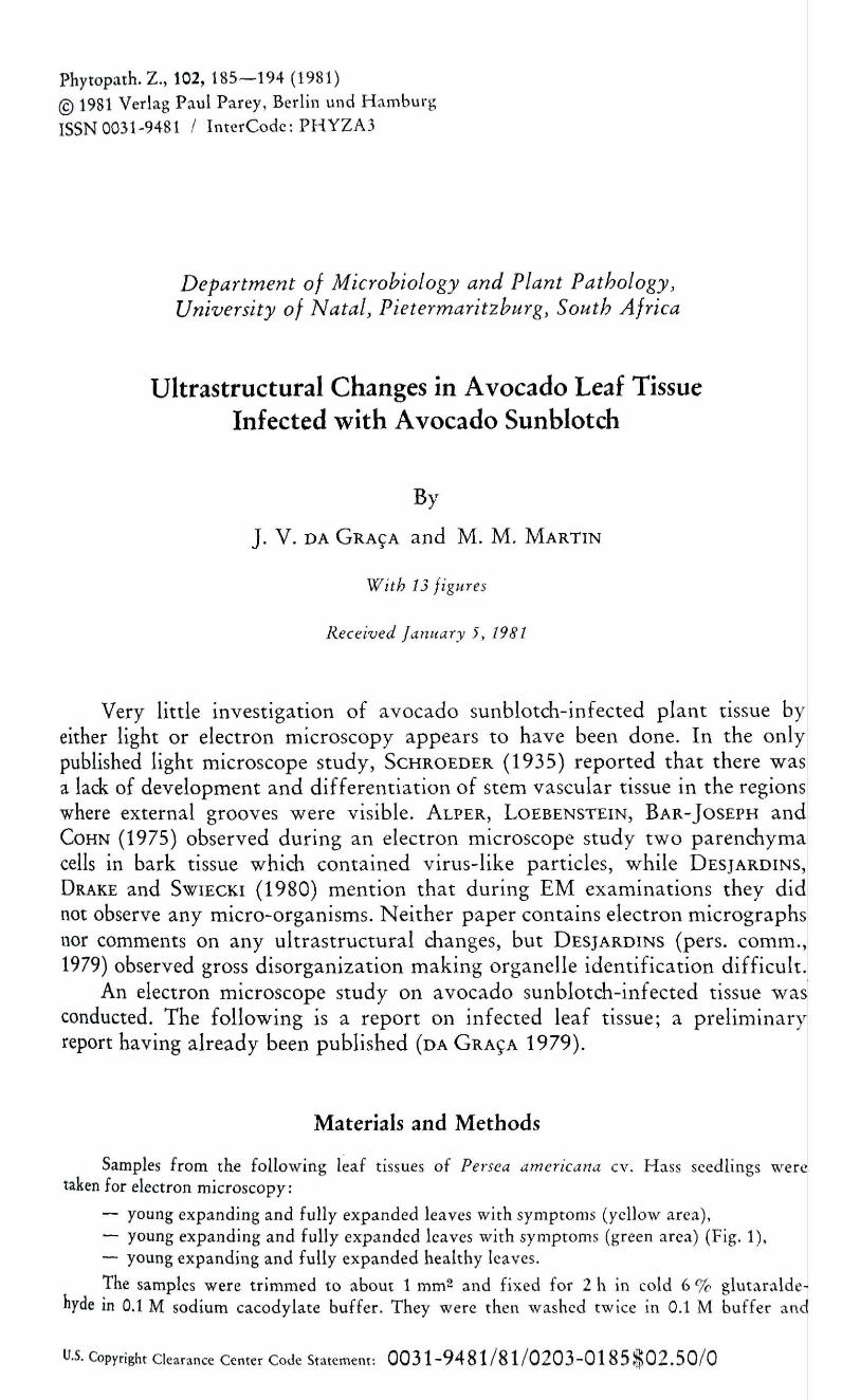
— young expanding and fully expanded leaves with symptoms (yellow area),— young expanding and fully expanded leaves with symptoms (green area) (Fig. 1),— young expanding and fully expanded healthy leaves.
The samples were trimmed to about 1 mm^ and fixed for 2 h in cold 6 % glutaralde-nyde in 0,1 M sodium cacodylate buffer. They were then washed twice in 0.1 M buffer and
l̂ .S. Copyright Clearance Center Code Statemem: 0 0 3 1 - 9 4 8 1 / 8 l / 0 2 0 3 - 0 1 8 5 $ 0 2 . 5 0 / 0
186 DA GRA9A and MARTIN
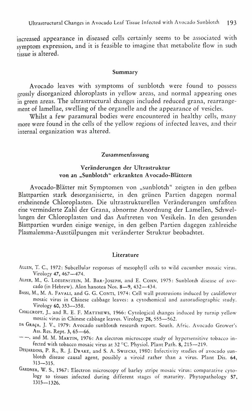
Fig. 1. Avocado leaveswith sunblotch symptoms
post-fixed overnight in 2 % osmium tetroxide. After post-fixation the pieces of tissue werewashed in three 20-min dhanges of deionised water, and dehydrated in two 15-min changesof 2,2-dimethoxypropane acidified with 0.5 % HCl (THORPE and HARVEY 1979). They werethen embedded in Spurr's resin (SPURR 1969).
The embedded samples were sectioned with a diamond knife on an LKB UM III ultra-microtome, stained with 2 '/r uranyl acetate and lead citrate, and then examined in an HitadiiHU- l lE electron microscope.
Results
Two obvious ultrastructural changes were observed in infected tissue,a major disorganization of the chloroplasts, and a significant increase in num-ber and size of paramural bodies (plasmalemmasome-like structures) in in-fected cells.
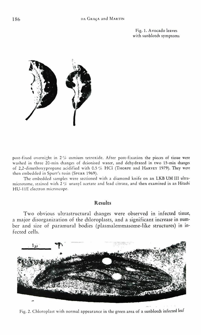
Fig. 2. Chloroplast with normal appearance in the green area of a sunblotdi infected leaf
ultrastructural Changes in Avocado Leaf Tissue Infected with Avocado Sunblotch 187
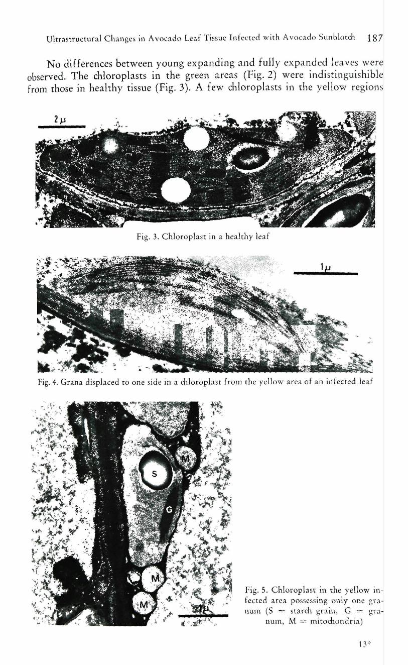
No differences between young expanding and fully expanded leaves wereobserved. The chloroplasts in the green areas (Fig. 2) were indistinguishiblefrom those in healthy tissue (Fig. 3). A few chloroplasts in the yellow regions
Fig, 3, Chloroplast in a healthy leaf
Fig. 4. Grana displaced to one side in a chloroplast from the yellow area of an infected leaf
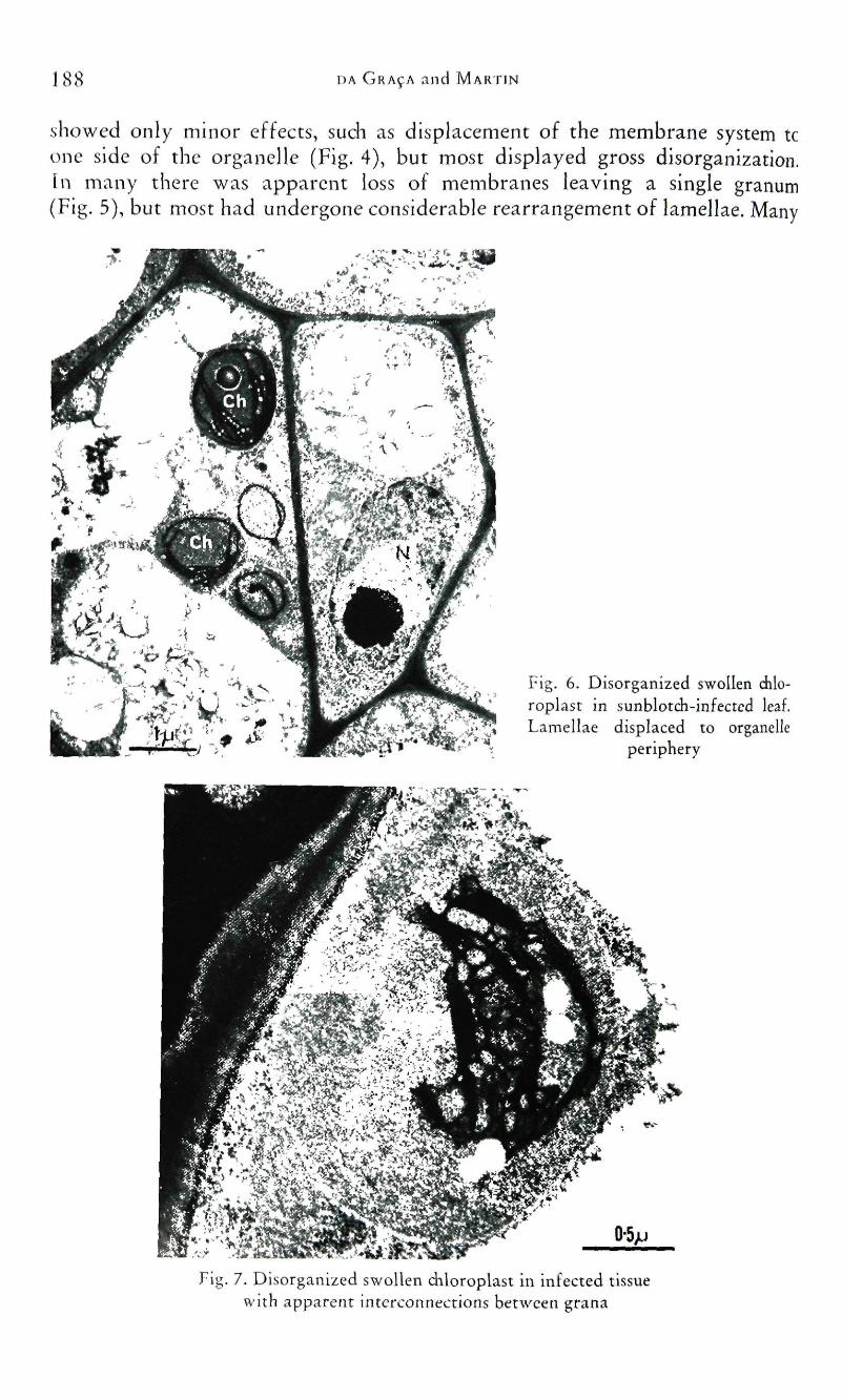
Fig. 5. Chloroplast in the yellow in-fected area possessing only ovie granum (S = starch grain, G — gra
num, M = mitodiondria)
188 DA GRA9A and MARTIN
showed only minor effects, such as displacement of the membrane system tcone side of the organelle (Fig. 4), but most displayed gross disorganization,[n many there was apparent loss of membranes leaving a single granum(Fig. 5), but most had undergone considerable rearrangement of lamellae. Many
Fig. 6. Disorganized swollen dilo-roplast in sunblotch-infected leaf.Lamellae displaced to organelle
periphery
Fig. 7. Disorganized swollen chloroplast in infected tissuewith apparent interconnections between grana
Ultrastructural Changes in Avocado Leaf Tissue Infected with Avocado Sunblocch j g9
had their lamellae forming a ring near the periphery of the organelles (Fig. 6),while some had unusual interconnections between membranes (Fig. 7). Mostdisorganized chloroplasts were round in shape.
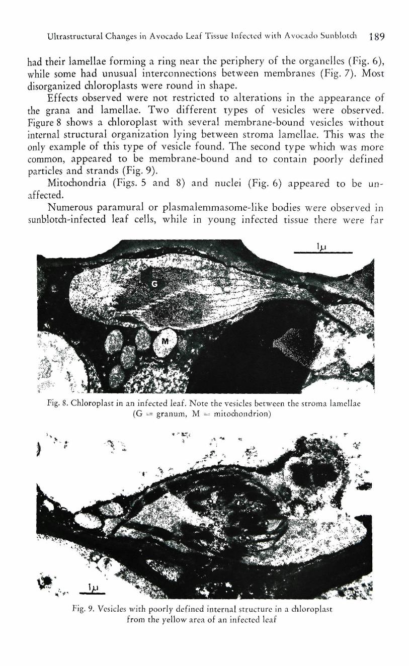
Effects observed were not restricted to alterations in the appearance ofthe grana and lamellae. Two different types of vesicles were observed.Figure 8 shows a chloroplast with several membrane-bound vesicles withoutinternal structural organization lying between stroma lamellae. This was theonly example of this type of vesicle found. The second type which was morecommon, appeared to be membrane-bound and to contain poorly definedparticles and strands (Fig. 9).
Mitochondria (Figs. 5 and 8) and nuclei (Fig. 6) appeared to be un-affected.
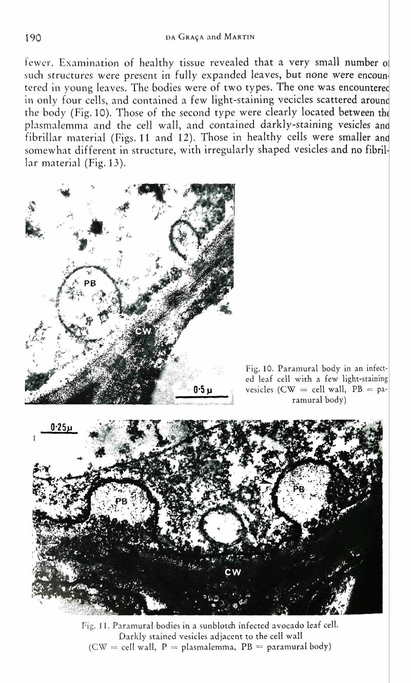
Numerous paramural or plasmalemmasome-like bodies were observed insunblotch-infected leaf cells, while in young infected tissue there were far
Fig. 8. Chloroplast in an infected leaf. Note the vesicles between the stroma lamellae(G = granum, M — mitochondrion)
Fig. 9. Vesicles with poorly defined internal structure in a chloroplastfrom the yellow area of an infected leaf
190 DA GRA9A and MARTIN
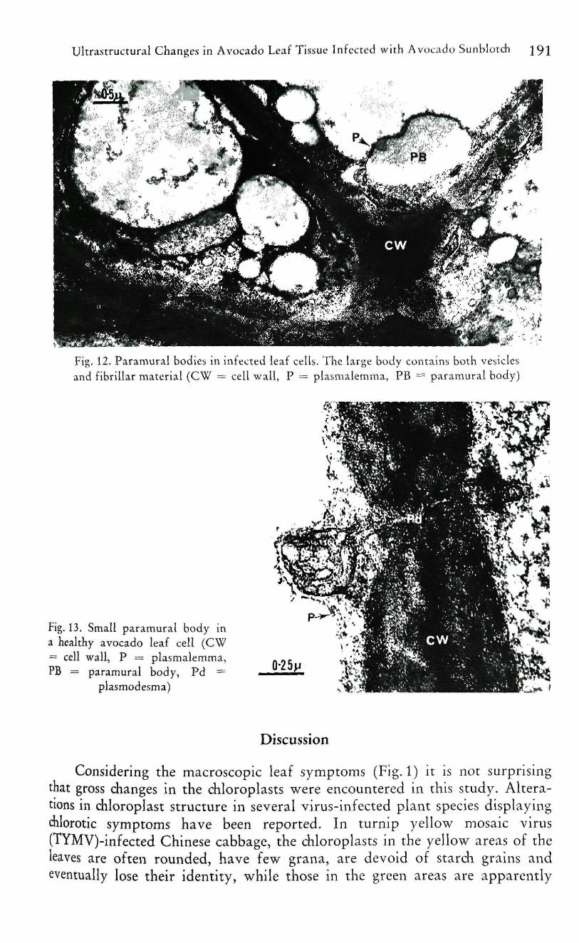
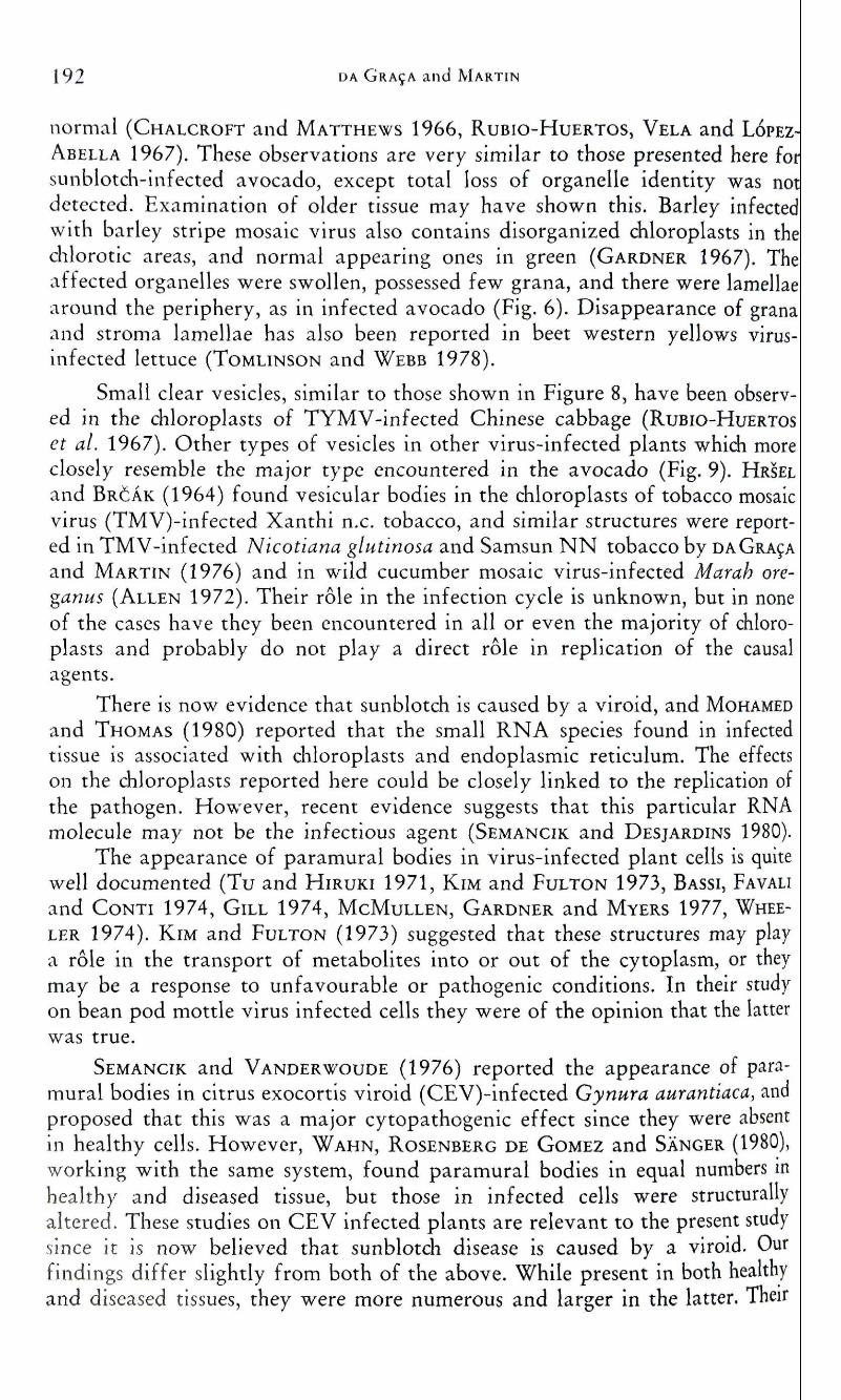
fewer. Examination of healthy tissue revealed that a very small number 0such structures were present in fully expanded leaves, but none were encoun-tered in young leaves. The bodies were of two types. The one was encounterecin only four cells, and contained a few light-staining vecicles scattered arouncthe body (Fig. 10). Those of the second type were clearly located between thiplasmalemma and the cell wall, and contained darkly-staining vesicles ancfibrillar material (Figs. 11 and 12). Those in healthy cells were smaller andsomewhat different in structure, with irregularly shaped vesicles and no fibril-lar material (Fig. 13).
Fig, 10. Paramural body in an infect-ed leaf cell with a few light-stainingvesicles (CW = cell wall /PB = pa-
ramural body)
0-25ii
Fig. 11. Paramural bodies in a sunblotch infected avocado leaf cell.Darkly stained vesicles adjacent to the cell wall
(CW = cell wall, P == plasmalcmma, PB ~ paramural body)
Ultrastructural Changes in Avocado Leaf Tissue Infected with Avocado Sunblotch
Fig. 12. Paramural bodies in infected leaf cells. The large body contains both vesiclesand fibrillar material (CW — cell wall, P = plasmalemma, PB = paramural body)
Fig. 13. Small paramural body ina healthy avocado leaf cell (CW= cell wall, P = plasmalemma,PB = paramural body, Pd =
plasmodesma)
Discussion
Considering the macroscopic leaf symptoms (Fig. 1) it is not surprisingthat gross changes in the chloroplasts were encountered in this study. Altera-tions in chloroplast structure in several virus-infected plant species displayingchlorotic symptoms have been reported. In turnip yellow mosaic virus(TYMV)-infected Chinese cabbage, the chloroplasts in the yellow areas of theleaves are often rounded, have few grana, are devoid of starch grains andeventually lose their identity, while those in the green areas are apparently
192 i>A GRA^A and MARTIN
normal (CHALCROFT and MATTHEWS 1966, RUBIO-HUERTOS, VELA and LOPEZ-
ABELLA 1967). These observations are very similar to those presented here forsunblotch-infected avocado, except total loss of organelle identity was notdetected. Examination of older tissue may have shown this. Barley infectedwith barley stripe mosaic virus also contains disorganized chloroplasts in thechlorotic areas, and normal appearing ones in green (GARDNER 1967). Theaffected organelles were swollen, possessed few grana, and there were lamellaearound the periphery, as in infected avocado (Fig. 6). Disappearance of granaand stroma lamellae has also been reported in beet western yellows virus-infected lettuce (ToMLiNSON and WEBB 1978).
Small clear vesicles, similar to those shown in Figure 8, have been observ-ed in the chloroplasts of TYMV-infected Chinese cabbage (RUBIO-HUERTOS
ct al. 1967). Other types of vesicles in other virus-infected plants which moreclosely resemble the major type encountered in the avocado (Fig. 9). HRSEL
and BRCAK (1964) found vesicular bodies in the chloroplasts of tobacco mosaicvirus (TMV)-infected Xanthi n.c. tobacco, and similar structures were report-ed in TMV-infected Nicotiana glutinosa and Samsun N N tobacco by DAGRAfAand MARTIN (1976) and in wild cucumber mosaic virus-infected Marah ore-ganus (ALLEN 1972). Their role in the infection cycle is unknown, but in noneof the cases have they been encountered in all or even the majority of chloro-plasts and probably do not play a direct role in replication of the causalagents.
There is now evidence that sunblotch is caused by a viroid, and MOHAMED
and THOMAS (1980) reported that the small RNA species found in infectedtissue is associated with chloroplasts and endoplasmic reticulum. The effectson the chloroplasts reported here could be closely linked to the replication ofthe pathogen. However, recent evidence suggests that this particular RNAmolecule may not be the infectious agent (SEMANCIK and DESJARDINS 1980).
The appearance of paramural bodies in virus-infected plant cells is quitewell documented (Tu and HIRUKI 1971, KIM and FULTON 1973, BASSI, FAVALI
and CONTI 1974, GILL 1974, MCMULLEN, GARDNER and MYERS 1977, WHEE-
LER 1974). KIM and FULTON (1973) suggested that these structures may playa role in the transport of metabolites into or out of the cytoplasm, or theymay be a response to unfavourable or pathogenic conditions. In their studyon bean pod mottle virus infected cells they were of the opinion that the latterwas true.
SEMANCIK and VANDERWOUDE (1976) reported the appearance of para-mural bodies in citrus exocortis viroid (CEV)-infected Gynura aurantiaca, andproposed that this was a major cytopathogenic effect since they were absentin healthy cells. However, WAHN, ROSENBERG DE GOMEZ and SANGER (1980),working with the same system, found paramural bodies in equal numbers mhealthy and diseased tissue, but those in infected cells were structurallyaltered. These studies on CEV infected plants are relevant to the present studysince it is now believed that sunblotch disease is caused by a viroid. Ourfindings differ slightly from both of the above. While present in both healthyand diseased tissues, they were more numerous and larger in the latter. Their
Ultrastructural Changes in Avocado Leaf Tissue Infected with Avocado Sunblotch 193
increased appearance in diseased cells certainly seems to be associated withsymptom expression, and it is feasible to imagine that metabolite flow in suchtissue is altered.
Summary
Avocado leaves with symptoms of sunblotch were found to possessgrossly disorganized chloroplasts in yellow areas, and normal appearing onesin green areas. The ultrastructural changes included reduced grana, rearrange-ment of lamellae, swelling of the organelle and the appearance of vesicles.
Whilst a few paramural bodies were encountered m healthy cells, manymore were found in the cells of the yellow regions of infected leaves, and theirinternal organization was altered.
Zusammenfassung
Veranderungen der Ultrastrukturvon an ,,Sunblotch** erkrankten Avocado-Blattern
Avocado-Blatter mit Symptomen von ,,sunblotch" zeigten in den gelbenBlattpartien stark desorganisierte, m den griinen Partien dagegen normalerscheinende Chloroplasten. Die ultrastrukturellen Veranderungen umfafiteneme vermmderte Zahl der Grana, abnorme Anordnung der Lamellen, Schwel-lungen der Ghloroplasten und das Auftreten von Vesikeln. In den gesundenBlattpartien wurden emige wenige, m den gelben Partien dagegen zahlreichePlasmalemma-Ausstiilpungen mit veranderter Struktur beobachtet.
Literature
ALLEN, T. C , 1972; Subcellular responses of mesophyll cells to wild cucumber mosaic virus.Virology 47, 467—474.
ALPER, M., G. LOEDENSTEIN, M . BAR-JOSEPH, and E. COHN, 1975; Sunblotch disease of avo-cado (in Hebrew). Alon hanotea Nos. 8—9, 432—434.
BASSI, M., M. A. FAVALI, and G. G. CONTI, 1974: Cell wall protrusions induced by cauliflowermosaic virus in Chinese cabbage leaves: a cytochemical and auioradiographic study.Virology 60, 353—358.
CHALCROFT, J., and R, E. F. MATTHEWS, 1966: Cytological changes induced by turnip yellowmosaic virus in Chinese cabbage leaves. Virology 28, 555—562.
DA GRA9A, J. V,, 1979: Avocado sunblotch research report. South. Afric. Avocado Grower'sAss. Res. Rept. 3, 65—66.
, and M. M. MARTIN, 1976: An electron microscope study of hypersensitive tobacco in-fected with tobacco mosaic virus at 32 "^C. Physiol. Plant Path. 8, 215—219.
DESJARDINS, P. R., R. J. DR\KE, and S. A. SWIECKI, 1980: Infectivity studies of avocado sun-blotch disease causal agent, possibly a viroid rather than a virus. Plant Dis, 64,313—315.
, W. S., 1967: Electron microscopy of barley stripe mosaic virus: comparative cyto-logy to tissues infected during different stages of maturity. Phytopathology 57,1315—1326.
194 ^^ GRA^A/MARMN, Uhrastructural Changes In Avocado Leaf Tissue
GILL, C . C , 1974: Inclusions and wall deposits in cells of plants infected with oat necroumottle virus. Canad. J. Bot. 52, 621—626.
HRSEL, I., and J. BRCAK, 1964: Ukrastructural changes in chloroplasts and cytoplasm causecby local infection of tobacco mosaic virus and cucumber virus 4. Virology 53, 252—258
KiM, S. K., and J. P. FULTON, 1973: Plant Virus-induced cell wall overgrowth and associatecmembrane elaboration. J. Ultrastruct. Res. 45, 328—342.
McMuLLEN, C. R., W. S. GARDNFR, and G. A. MYERS, 1977; Ultrastructure of cell walthickenings and paramural bodies induced by barley stripe mosaic virus. Phyto-pathology 67, 462—467.
MoHAMED, N. A., and W. THOMAS, 1980: Vjroid-like properties of aa RNA species associated•with sunblotch disease of avocados. J. Gen. Virol. 46, 157—167.
RuBio-HuERTOS, M., A. VELA, and D. LOPEZ-ABELLA, 1967: Crystalline arrays of sphericalparticles in turnip yellow mosaic virus-infected cells. Virology 32, 438—444.
ScHROEDER, C. A., 1935: Effects of sunblotch on the anatomy o{ the avocado stem. Calif.Avocado Assoc. Yearbook 1935, 125—129.
SEMANCIK, J . S., and P. R. DESJARDINS, 1980: Multiple small RNA species and the viroidh)'pothesis for the sunblotch disease of avocado. Virology 104, 117—121.
, and W. J. VANDERWOUDE, 1976: Exocortis viroid; cytopathic effects at the plasmamembrane in association with pathogenic RNA. Virology 69, 719—726.
SPURR. A. R., 1969: A low viscosity epoxy resin embedding medium for electron microscopy.J. Ultrastruct. Res. 26, 31—43.
THORPE, J. R., and D. M. R. HARVEY, 1979: Optimization and investigation of the use of 2,2-dimethoxypropane as a dehydration agent for plant tissues in transmission electronmicroscopy. J. Ultrastruct. Res. 68, 186—194.
ToMLiNSON, J. A., and M. J, W. WEBB, 1978: Ultrastructural dianges in diloroplasts of lettuceinfected with beet western yellows virus. Physiol. Plant Path. 12, 13—18.
Tu, J. C , and C. HIRDKI. 1971: Electron microscopy of cell wall thickening in local lesions ofpotato virus M infected red kidney bean. Phytopathology 61, 862—868.
WAHN, K., F. ROSENBERG DE GOMEZ, und H. L. SANGER, 1980: Cytopathologie von viroid-infiziertem Pflanzengewebe. I. Veranderungen des Plasmalemmas und der Zellwandbei Gynura aiiruntiaca nach Infektion mit dem Viroid der Citrus-Exocortis-KrankheitfCEV). Phytopathol. Z. 98, 1 — 18.
WHEELER, H. , 1974: Cell wall and plasmalemma modification in diseased and injured planttissue. Canad. J. Bot. 52, 1005—1009.
Authors address: Department of Microbiology and Plant Pathology, University ofNatal, P.O. Box 375, Pietermaritzburg, 3200 (South Africa).