ultrastructural analysis of the junctional zone of vocal ... · ultrastructural analysis of the...
TRANSCRIPT
Journal of Otolaryngology-ENT Research
Ultrastructural Analysis of the Junctional Zone of Vocal Fold Polyps and Nodules
Submit Manuscript | http://medcraveonline.com
Volume 6 Issue 6 - 2017
1Department of Otorhinolaryngology, Ain Shams University, Cairo, Egypt2Department of Pathology, Ain Shams University, Cairo, Egypt
*Corresponding author: Badr E Mostafa, 75 El Nozha Street, Heliopolis 11361, Cairo, Egypt, Tel: +202 24198118; +2012 27461137; Fax: +202 26708390; Email:
Received: August 10, 2016 | Published: April 17, 2017
Research Article
J Otolaryngol ENT Res 2017, 6(6): 00182
Abstract
Objectives: To study the ultrastructural changes of the junctional zone [the apparently normal mucosa] of vocal fold polyps and nodules.
Methods: 15 lesions (nodules and polyps) by standard microscopy (Hx. and eosin and PAS) and by electron microscopy
Results: The junctional zone showed basement membrane thickening and the vessels showed endothelial interruptions. Some endothelial cells were degenerated and a few were apoptotic. Endothelial cells showed increased microfilaments with foci of detachment from the underlying basement membrane. Larger blood vessels showed large plump endothelial cells alternating with attenuated ones. There was also widening of the intercellular junctions between the endothelial cells.
Conclusion: Our findings indicate that the pathological changes in the substance of vocal fold polyps and nodules extend beyond the gross visible edges of the lesion. This may have implications in the planning of surgical excision to remove all potentially unstable tissue to prevent recurrences.
Keywords: Larynx; Dysphonia; Vocal nodules; Vocal polyps; Phonosurgery; Ultrastructural; Junctional; Anteroposterior; Food; Surgical.
IntroductionVocal nodules and polyps are common causes of voice
disorders. The differentiation between vocal nodules and polyps is not clear cut, and the issue is further confused by differences in the terminology used by the laryngologist and pathologist [1]. Polyps and nodules of vocal cords are considered to be tissue reactions to chronic mechanical irritations in the form of vocal misuse or abuse, [2] The main difference is usually unclear [3] but may be in the time course with polyps being considered younger lesions and nodules of more long standing nature [4]. The etiology and pathogenesis of the vocal disorders have not been elucidated. Various histological and biochemical mechanisms have been reported including the involvement of nitric oxide, hormones, or vascular endothelial disturbances [1,5-7]. Therapy for such lesions is usually a combination of voice therapy and surgical excision. The basic principle of phonosurgery is to maintain or improve the functional structure of the vocal fold by respecting its layered structure. This is achieved by means of minimal tissue excision, minimal disruption of the superficial layer of the lamina propria and preservation of the epithelium, especially at the vibrating edge [8].
The aim of this study is to determine the changes of the transitional zone (the angle between apparently normal mucosa and the base of the polyp or nodule) to determine how far do the pathological changes extend along the free edge of the vocal fold.
Material and MethodsThe study was conducted in a tertiary referral University
Hospital. It included 15 lesions (seven nodules and 8 polyps). The lesions were microsurgically excised from 15 patients (7 females and 8 males) with a median age of 36 years. The specimens included the nodule or the polyp and the adjacent mucosa (transitional zone). The transitional zone mucosa was specifically studied. The specimens were bisected. One half was preserved in formalin, then processed and embedded in paraffin for haematoxylin and eosin and PAS staining. The second half was preserved in F/G solution then processed and embedded in epoxy resin. Ultrathin sections were cut and stained by uranyl acetate and lead citrate and then examined by Transmission Electron Microscope Philips 400 (at 80 K.V.)
Results
Histological findings
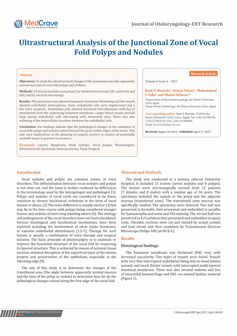
The basement membrane was thickened (PAS +ve), with increased vascularity. Two types of vessels were found. Vessels with very thin interrupted endothelial lining and no basal lamina around; and much thicker vessels with interrupted multi-layered basement membrane. There was also stromal oedema and foci of interstitial haemorrhage and PAS +ve stained hyaline material (Figure 1).
Ultrastructural Analysis of the Junctional Zone of Vocal Fold Polyps and Nodules 2/4Copyright:
©2017 Mostafa et al.
Citation: Mostafa BE, Fawaz S, Taha MS, Salman M (2017) Ultrastructural Analysis of the Junctional Zone of Vocal Fold Polyps and Nodules. J Otolaryngol ENT Re 6(6): 00182. DOI: 10.15406/joentr.2017.06.00182
Electron microscopic findings
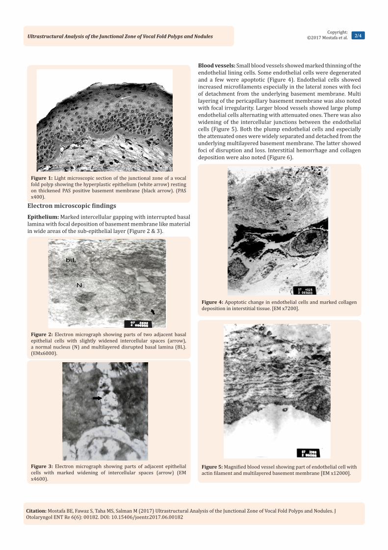
Epithelium: Marked intercellular gapping with interrupted basal lamina with focal deposition of basement membrane like material in wide areas of the sub-epithelial layer (Figure 2 & 3).
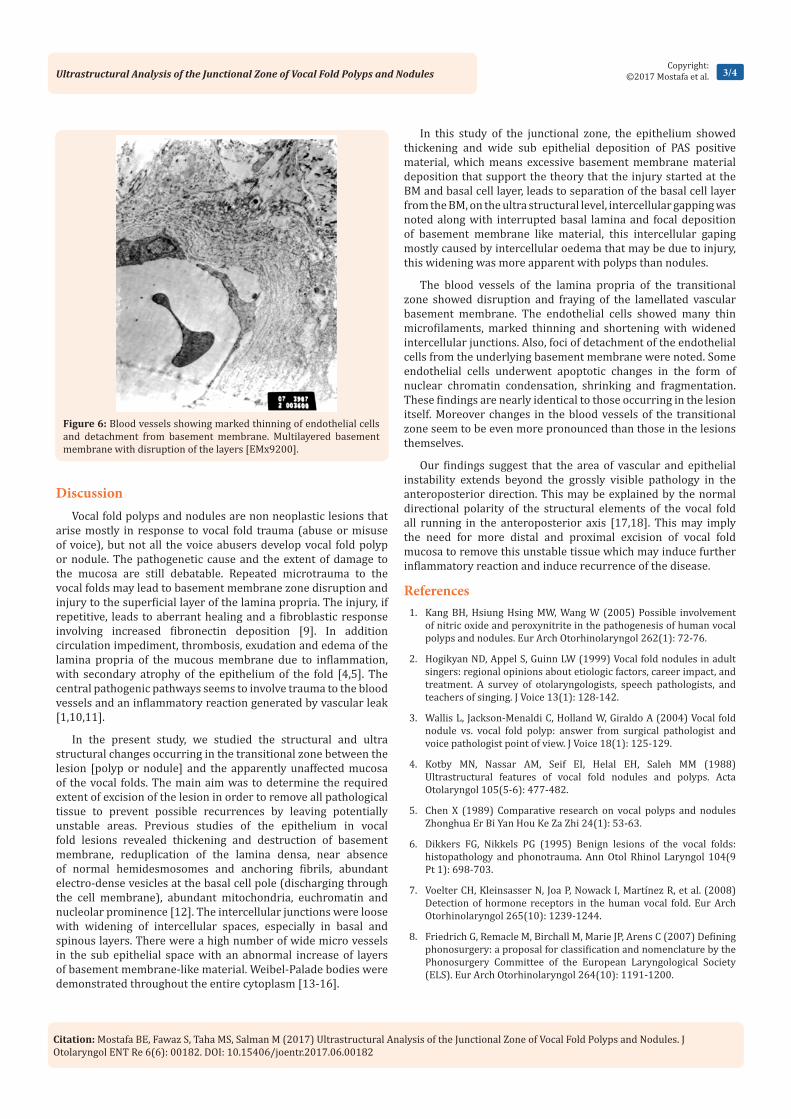
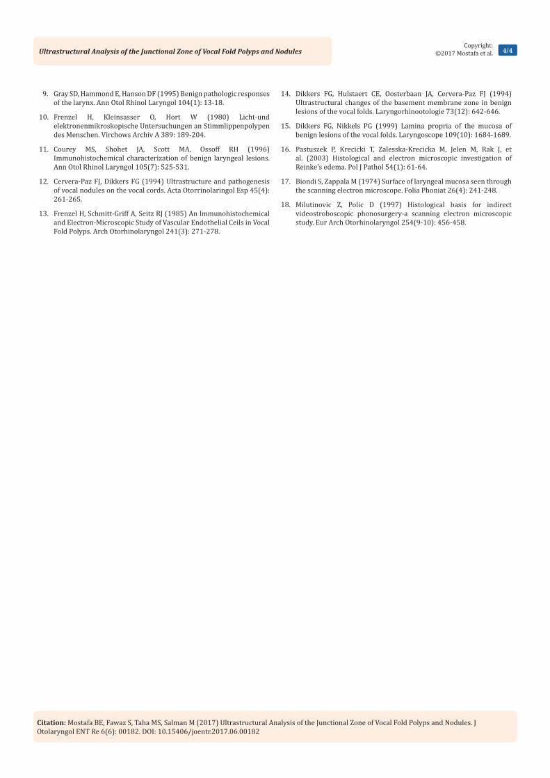
Blood vessels: Small blood vessels showed marked thinning of the endothelial lining cells. Some endothelial cells were degenerated and a few were apoptotic (Figure 4). Endothelial cells showed increased microfilaments especially in the lateral zones with foci of detachment from the underlying basement membrane. Multi layering of the pericapillary basement membrane was also noted with focal irregularity. Larger blood vessels showed large plump endothelial cells alternating with attenuated ones. There was also widening of the intercellular junctions between the endothelial cells (Figure 5). Both the plump endothelial cells and especially the attenuated ones were widely separated and detached from the underlying multilayered basement membrane. The latter showed foci of disruption and loss. Interstitial hemorrhage and collagen deposition were also noted (Figure 6).
Figure 1: Light microscopic section of the junctional zone of a vocal fold polyp showing the hyperplastic epithelium (white arrow) resting on thickened PAS positive basement membrane (black arrow). (PAS x400).
Figure 2: Electron micrograph showing parts of two adjacent basal epithelial cells with slightly widened intercellular spaces (arrow), a normal nucleus (N) and multilayered disrupted basal lamina (BL). (EMx6000).
Figure 3: Electron micrograph showing parts of adjacent epithelial cells with marked widening of intercellular spaces (arrow) (EM x4600).
Figure 4: Apoptotic change in endothelial cells and marked collagen deposition in interstitial tissue. [EM x7200].
Figure 5: Magnified blood vessel showing part of endothelial cell with actin filament and multilayered basement membrane [EM x12000].
Ultrastructural Analysis of the Junctional Zone of Vocal Fold Polyps and Nodules 3/4Copyright:
©2017 Mostafa et al.
Citation: Mostafa BE, Fawaz S, Taha MS, Salman M (2017) Ultrastructural Analysis of the Junctional Zone of Vocal Fold Polyps and Nodules. J Otolaryngol ENT Re 6(6): 00182. DOI: 10.15406/joentr.2017.06.00182
Discussion Vocal fold polyps and nodules are non neoplastic lesions that
arise mostly in response to vocal fold trauma (abuse or misuse of voice), but not all the voice abusers develop vocal fold polyp or nodule. The pathogenetic cause and the extent of damage to the mucosa are still debatable. Repeated microtrauma to the vocal folds may lead to basement membrane zone disruption and injury to the superficial layer of the lamina propria. The injury, if repetitive, leads to aberrant healing and a fibroblastic response involving increased fibronectin deposition [9]. In addition circulation impediment, thrombosis, exudation and edema of the lamina propria of the mucous membrane due to inflammation, with secondary atrophy of the epithelium of the fold [4,5]. The central pathogenic pathways seems to involve trauma to the blood vessels and an inflammatory reaction generated by vascular leak [1,10,11].
In the present study, we studied the structural and ultra structural changes occurring in the transitional zone between the lesion [polyp or nodule] and the apparently unaffected mucosa of the vocal folds. The main aim was to determine the required extent of excision of the lesion in order to remove all pathological tissue to prevent possible recurrences by leaving potentially unstable areas. Previous studies of the epithelium in vocal fold lesions revealed thickening and destruction of basement membrane, reduplication of the lamina densa, near absence of normal hemidesmosomes and anchoring fibrils, abundant electro-dense vesicles at the basal cell pole (discharging through the cell membrane), abundant mitochondria, euchromatin and nucleolar prominence [12]. The intercellular junctions were loose with widening of intercellular spaces, especially in basal and spinous layers. There were a high number of wide micro vessels in the sub epithelial space with an abnormal increase of layers of basement membrane-like material. Weibel-Palade bodies were demonstrated throughout the entire cytoplasm [13-16].
In this study of the junctional zone, the epithelium showed thickening and wide sub epithelial deposition of PAS positive material, which means excessive basement membrane material deposition that support the theory that the injury started at the BM and basal cell layer, leads to separation of the basal cell layer from the BM, on the ultra structural level, intercellular gapping was noted along with interrupted basal lamina and focal deposition of basement membrane like material, this intercellular gaping mostly caused by intercellular oedema that may be due to injury, this widening was more apparent with polyps than nodules.
The blood vessels of the lamina propria of the transitional zone showed disruption and fraying of the lamellated vascular basement membrane. The endothelial cells showed many thin microfilaments, marked thinning and shortening with widened intercellular junctions. Also, foci of detachment of the endothelial cells from the underlying basement membrane were noted. Some endothelial cells underwent apoptotic changes in the form of nuclear chromatin condensation, shrinking and fragmentation. These findings are nearly identical to those occurring in the lesion itself. Moreover changes in the blood vessels of the transitional zone seem to be even more pronounced than those in the lesions themselves.
Our findings suggest that the area of vascular and epithelial instability extends beyond the grossly visible pathology in the anteroposterior direction. This may be explained by the normal directional polarity of the structural elements of the vocal fold all running in the anteroposterior axis [17,18]. This may imply the need for more distal and proximal excision of vocal fold mucosa to remove this unstable tissue which may induce further inflammatory reaction and induce recurrence of the disease.
References1. Kang BH, Hsiung Hsing MW, Wang W (2005) Possible involvement
of nitric oxide and peroxynitrite in the pathogenesis of human vocal polyps and nodules. Eur Arch Otorhinolaryngol 262(1): 72-76.
2. Hogikyan ND, Appel S, Guinn LW (1999) Vocal fold nodules in adult singers: regional opinions about etiologic factors, career impact, and treatment. A survey of otolaryngologists, speech pathologists, and teachers of singing. J Voice 13(1): 128-142.
3. Wallis L, Jackson-Menaldi C, Holland W, Giraldo A (2004) Vocal fold nodule vs. vocal fold polyp: answer from surgical pathologist and voice pathologist point of view. J Voice 18(1): 125-129.
4. Kotby MN, Nassar AM, Seif EI, Helal EH, Saleh MM (1988) Ultrastructural features of vocal fold nodules and polyps. Acta Otolaryngol 105(5-6): 477-482.
5. Chen X (1989) Comparative research on vocal polyps and nodules Zhonghua Er Bi Yan Hou Ke Za Zhi 24(1): 53-63.
6. Dikkers FG, Nikkels PG (1995) Benign lesions of the vocal folds: histopathology and phonotrauma. Ann Otol Rhinol Laryngol 104(9 Pt 1): 698-703.
7. Voelter CH, Kleinsasser N, Joa P, Nowack I, Martínez R, et al. (2008) Detection of hormone receptors in the human vocal fold. Eur Arch Otorhinolaryngol 265(10): 1239-1244.
8. Friedrich G, Remacle M, Birchall M, Marie JP, Arens C (2007) Defining phonosurgery: a proposal for classification and nomenclature by the Phonosurgery Committee of the European Laryngological Society (ELS). Eur Arch Otorhinolaryngol 264(10): 1191-1200.
Figure 6: Blood vessels showing marked thinning of endothelial cells and detachment from basement membrane. Multilayered basement membrane with disruption of the layers [EMx9200].
Ultrastructural Analysis of the Junctional Zone of Vocal Fold Polyps and Nodules 4/4Copyright:
©2017 Mostafa et al.
Citation: Mostafa BE, Fawaz S, Taha MS, Salman M (2017) Ultrastructural Analysis of the Junctional Zone of Vocal Fold Polyps and Nodules. J Otolaryngol ENT Re 6(6): 00182. DOI: 10.15406/joentr.2017.06.00182
9. Gray SD, Hammond E, Hanson DF (1995) Benign pathologic responses of the larynx. Ann Otol Rhinol Laryngol 104(1): 13-18.
10. Frenzel H, Kleinsasser O, Hort W (1980) Licht-und elektronenmikroskopische Untersuchungen an Stimmlippenpolypen des Menschen. Virchows Archiv A 389: 189-204.
11. Courey MS, Shohet JA, Scott MA, Ossoff RH (1996) Immunohistochemical characterization of benign laryngeal lesions. Ann Otol Rhinol Laryngol 105(7): 525-531.
12. Cervera-Paz FJ, Dikkers FG (1994) Ultrastructure and pathogenesis of vocal nodules on the vocal cords. Acta Otorrinolaringol Esp 45(4): 261-265.
13. Frenzel H, Schmitt-Griff A, Seitz RJ (1985) An lmmunohistochemical and Electron-Microscopic Study of Vascular Endothelial Ceils in Vocal Fold Polyps. Arch Otorhinolaryngol 241(3): 271-278.
14. Dikkers FG, Hulstaert CE, Oosterbaan JA, Cervera-Paz FJ (1994) Ultrastructural changes of the basement membrane zone in benign lesions of the vocal folds. Laryngorhinootologie 73(12): 642-646.
15. Dikkers FG, Nikkels PG (1999) Lamina propria of the mucosa of benign lesions of the vocal folds. Laryngoscope 109(10): 1684-1689.
16. Pastuszek P, Krecicki T, Zalesska-Krecicka M, Jelen M, Rak J, et al. (2003) Histological and electron microscopic investigation of Reinke’s edema. Pol J Pathol 54(1): 61-64.
17. Biondi S, Zappala M (1974) Surface of laryngeal mucosa seen through the scanning electron microscope. Folia Phoniat 26(4): 241-248.
18. Milutinovic Z, Polic D (1997) Histological basis for indirect videostroboscopic phonosurgery-a scanning electron microscopic study. Eur Arch Otorhinolaryngol 254(9-10): 456-458.