ultrasound in abdominal trauma
TRANSCRIPT
Emerg Med Clin N Am
22 (2004) 581–599
Ultrasound in abdominal trauma
John S. Rose, MDDepartment of Emergency Medicine, University of California Davis Medical Center,
2315 Stockton Blvd., PSSB 2100, Sacramento, CA 95817, USA
Ultrasound in the evaluation of abdominal trauma has evolved over thepast 30 years. The use of ultrasound for abdominal trauma was describedinitially by Kristensen and colleagues [1] in 1971. In 1976, Ascher andcolleagues [2] first reported the accuracy of ultrasound in Radiology, with80% sensitivity for the detection of splenic injury. In a study of 808 patients,Tiling and colleagues [3] in 1990 reported a sensitivity of 89%, a specificityof 100%, and an accuracy of 98%. This same group also was first tocomment on the effect of training and experience and reported that surgeonswith extensive ultrasound experience could diagnose intra-abdominal fluidwith a sensitivity of 96% and an accuracy of 99%. Interest and experiencewith ultrasound for trauma grew steadily around the world among surgeonsand emergency physicians during the early 1990s [4–7]. During this period,ultrasound technology was improving with regard to price, portability, andresolution, allowing its use during resuscitation. At the same time, in theUnited States, there was continuing reliance on diagnostic peritoneal lavage(DPL) and CT and much less interest in sonography for abdominal trauma.This all changed when emergency physicians and surgeons in the UnitedStates began to publish their experience with ultrasound [4,8,9]. The termFocused Assessment with Sonography for Trauma (FAST) was coined byRozycki et al [10] in 1996 and has persisted as the accepted acronym for thetrauma ultrasound evaluation. The basic four-view examination (perihe-patic, perisplenic, pelvic, and pericardial views) has become the foundationof the FAST examination. The rapid, noninvasive, and practical nature ofultrasound for bedside evaluation of critically injured patients has changedthe evaluation of blunt abdominal trauma.
E-mail address: [email protected]
0733-8627/04/$ - see front matter � 2004 Elsevier Inc. All rights reserved.
doi:10.1016/j.emc.2004.04.007
582 J.S. Rose / Emerg Med Clin N Am 22 (2004) 581–599
Technique
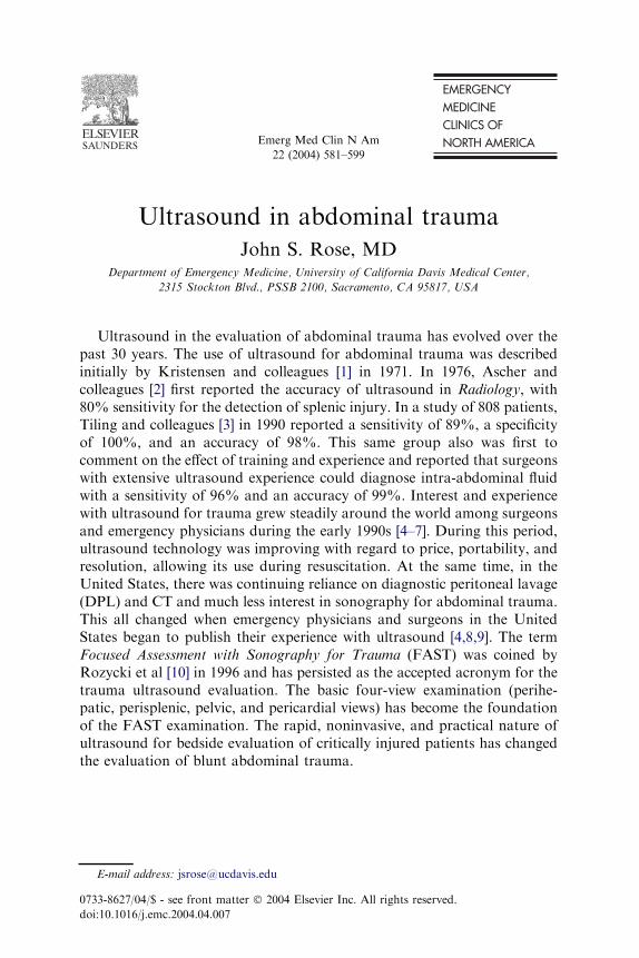
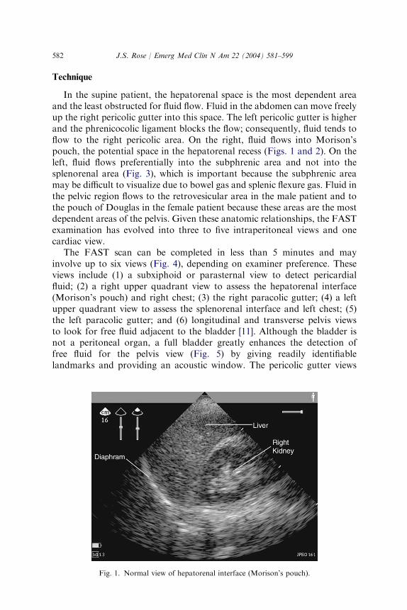
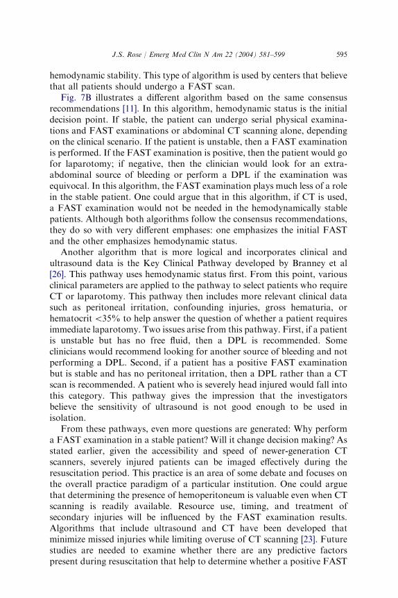
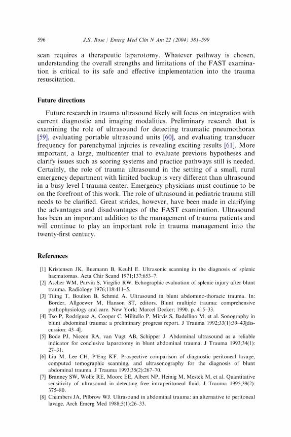
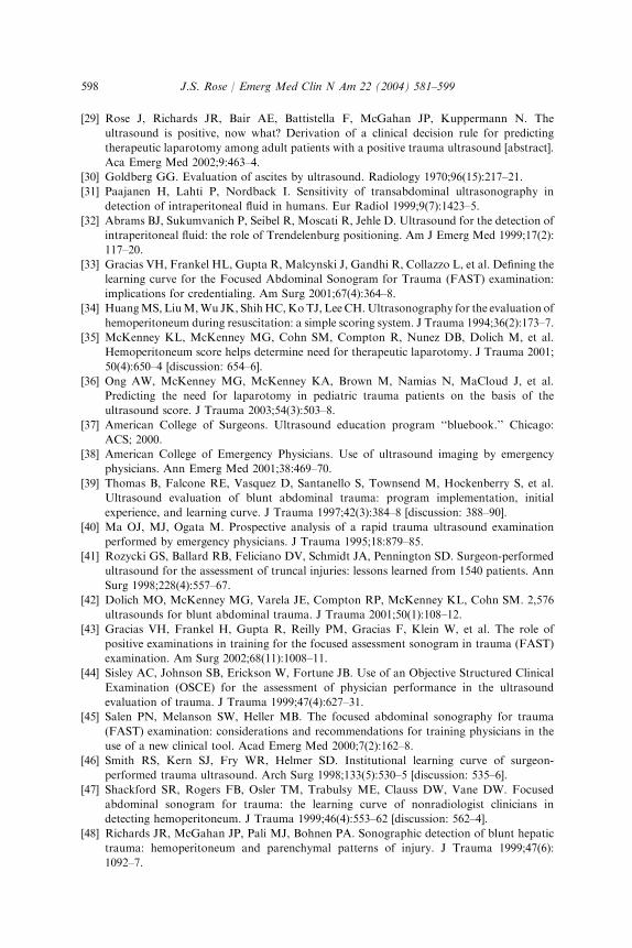
In the supine patient, the hepatorenal space is the most dependent areaand the least obstructed for fluid flow. Fluid in the abdomen can move freelyup the right pericolic gutter into this space. The left pericolic gutter is higherand the phrenicocolic ligament blocks the flow; consequently, fluid tends toflow to the right pericolic area. On the right, fluid flows into Morison’spouch, the potential space in the hepatorenal recess (Figs. 1 and 2). On theleft, fluid flows preferentially into the subphrenic area and not into thesplenorenal area (Fig. 3), which is important because the subphrenic areamay be difficult to visualize due to bowel gas and splenic flexure gas. Fluid inthe pelvic region flows to the retrovesicular area in the male patient and tothe pouch of Douglas in the female patient because these areas are the mostdependent areas of the pelvis. Given these anatomic relationships, the FASTexamination has evolved into three to five intraperitoneal views and onecardiac view.
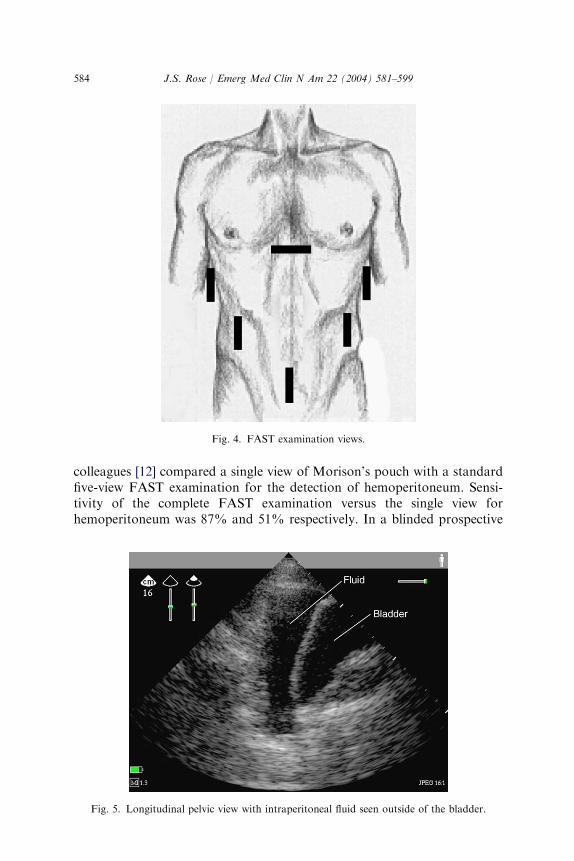
The FAST scan can be completed in less than 5 minutes and mayinvolve up to six views (Fig. 4), depending on examiner preference. Theseviews include (1) a subxiphoid or parasternal view to detect pericardialfluid; (2) a right upper quadrant view to assess the hepatorenal interface(Morison’s pouch) and right chest; (3) the right paracolic gutter; (4) a leftupper quadrant view to assess the splenorenal interface and left chest; (5)the left paracolic gutter; and (6) longitudinal and transverse pelvis viewsto look for free fluid adjacent to the bladder [11]. Although the bladder isnot a peritoneal organ, a full bladder greatly enhances the detection offree fluid for the pelvis view (Fig. 5) by giving readily identifiablelandmarks and providing an acoustic window. The pericolic gutter views
Fig. 1. Normal view of hepatorenal interface (Morison’s pouch).
583J.S. Rose / Emerg Med Clin N Am 22 (2004) 581–599
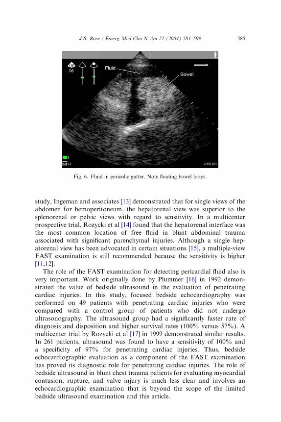
are optional views that some providers use (Fig. 6). Their role inimproving the accuracy of the FAST scan has not been studied. Thefocus of the FAST examination is the detection of free fluid; however,during the procedure, specific organs occasionally may be visualized,providing potential injury localization.
Given the flow of fluid in the abdomen and free flow toward the right,the utility of a single right-sided view has been examined. Ma and
Fig. 2. Free fluid in hepatorenal interface.
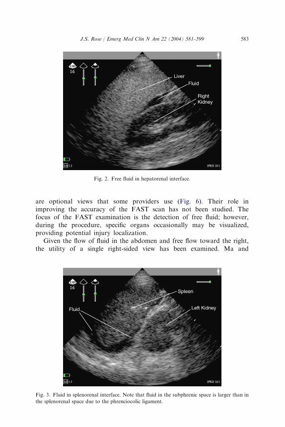
Fig. 3. Fluid in splenorenal interface. Note that fluid in the subphrenic space is larger than in
the splenorenal space due to the phrenciocolic ligament.
584 J.S. Rose / Emerg Med Clin N Am 22 (2004) 581–599
colleagues [12] compared a single view of Morison’s pouch with a standardfive-view FAST examination for the detection of hemoperitoneum. Sensi-tivity of the complete FAST examination versus the single view forhemoperitoneum was 87% and 51% respectively. In a blinded prospective
Fig. 4. FAST examination views.
Fig. 5. Longitudinal pelvic view with intraperitoneal fluid seen outside of the bladder.
585J.S. Rose / Emerg Med Clin N Am 22 (2004) 581–599
study, Ingeman and associates [13] demonstrated that for single views of theabdomen for hemoperitoneum, the hepatorenal view was superior to thesplenorenal or pelvic views with regard to sensitivity. In a multicenterprospective trial, Rozycki et al [14] found that the hepatorenal interface wasthe most common location of free fluid in blunt abdominal traumaassociated with significant parenchymal injuries. Although a single hep-atorenal view has been advocated in certain situations [15], a multiple-viewFAST examination is still recommended because the sensitivity is higher[11,12].
The role of the FAST examination for detecting pericardial fluid also isvery important. Work originally done by Plummer [16] in 1992 demon-strated the value of bedside ultrasound in the evaluation of penetratingcardiac injuries. In this study, focused bedside echocardiography wasperformed on 49 patients with penetrating cardiac injuries who werecompared with a control group of patients who did not undergoultrasonography. The ultrasound group had a significantly faster rate ofdiagnosis and disposition and higher survival rates (100% versus 57%). Amulticenter trial by Rozycki et al [17] in 1999 demonstrated similar results.In 261 patients, ultrasound was found to have a sensitivity of 100% anda specificity of 97% for penetrating cardiac injuries. Thus, bedsideechocardiographic evaluation as a component of the FAST examinationhas proved its diagnostic role for penetrating cardiac injuries. The role ofbedside ultrasound in blunt chest trauma patients for evaluating myocardialcontusion, rupture, and valve injury is much less clear and involves anechocardiographic examination that is beyond the scope of the limitedbedside ultrasound examination and this article.
Fig. 6. Fluid in pericolic gutter. Note floating bowel loops.
586 J.S. Rose / Emerg Med Clin N Am 22 (2004) 581–599
Clinical implications of the focused assessment with sonography for
trauma examination results
Much of the initial research into the use of ultrasound in trauma focusedon the test characteristics of ultrasound for detecting hemoperitoneum andon establishing its role in blunt abdominal trauma. Indeed, when describingthe history of the FAST examination, sensitivity and specificity define itsusefulness compared with DPL and CT. From a purely statistical sense,sensitivity and specificity calculations rely on all injuries being confirmed orexcluded by a ‘‘gold standard’’ test; however, this is impractical in much ofclinical research. A significant number of trauma ultrasound studies haveused these clinical outcomes and, thus, sensitivity and specificity have beencalculated using different outcomes and ‘‘gold standards,’’ depending on thestudy. For example, a false negative in one study may be a true negative inanother if the patient required no operative intervention for the injury. Thissituation is exemplified by a study performed by McGahan et al [18] thatevaluated the use of ultrasound in 500 blunt trauma patients. This groupcompared ultrasound findings to CT or laparotomy findings as the ‘‘goldstandard,’’ not clinical observation. They reported a sensitivity of 63%,a specificity of 95%, and an accuracy of 85% for the detection ofhemoperitoneum and organ injury. This calculation was based on veryconservative positive and negative criteria, defined as the presence of anyinjury regardless of whether a therapeutic procedure was performed. Ifa study endpoint like clinical outcome or observation was used, however,then their reported sensitivity would have increased to greater than 90%.When assessing ultrasound in the evaluation of the blunt trauma patient,calculations such a sensitivity and specificity may be misleading. Conse-quently, the question should be, How does this ultrasound result affect thepatient for whom I am caring right now?
In detecting intra-abdominal injuries in trauma patients, the governingparadigm always has been to quickly recognize those patients who requirelaparotomy and to prevent further morbidity or mortality. Underlying thisparadigm is the understanding that ongoing evaluation through serialexaminations or other imaging will be needed to determine whether a patienthas any intra-abdominal injury. Most experts would concur that ultrasoundhas performed best when limited to detecting free intraperitoneal fluid[10,11,19]. Historically, the presence of any intraperitoneal fluid indicateda significant intra-abdominal injury and warranted an immediate explor-atory laparotomy. Over the past decade, however, the practice of non-operative management for intra-abdominal injuries in adults and childrenhas increased [20–22]. This changing practice has relied on the increasing useof high-performance CT to image the abdomen. The availability of rapidCT scanners has allowed many trauma centers to develop criteria forobtaining abdominal CT scans during the resuscitative phase [23,24].Thus, patients who previously would have had an ultrasound and serial
587J.S. Rose / Emerg Med Clin N Am 22 (2004) 581–599
examinations as the primary means of detecting abdominal injuries now aretaken quickly to a high-speed CT scanner and undergo extensive bodyimaging. Given this practice, the question of sensitivity and specificity isa question not only of detecting an intra-abdominal injury with ultrasoundbut also of detecting intra-abdominal injuries that require laparotomy. Themore important question is, What is the role of ultrasound in determiningthe need for laparotomy? The presence of free fluid is only one surrogateindicator of serious intra-abdominal injury [25]. A small amount of fluid ina stable patient with a small liver laceration is much different than a largeamount of fluid in an unstable patient with a high-grade splenic disruption.Both examinations are positive, but each patient falls into a very differentmanagement category.
A 1999 consensus conference on the performance of ultrasound intrauma examined the question of the positive and negative studies and howthey apply to clinical practice. This report suggested the following guidelines[11]: in hemodynamically unstable patients, a positive FAST examinationgenerally should be followed by a laparotomy, and a negative FASTexamination warrants examination for an extra-abdominal source ofhemorrhage. In hemodynamically stable patients, a positive FAST exam-ination should be followed by an abdominal CT scan to better define theinjury, and a negative FAST examination should be followed with serialexams for 6 hours and a follow-up ultrasound or abdominal CT scanning,depending on the clinical scenario.
Although this practice paradigm has not been validated, it representsa consensus among experts. Variations in this practice, however, arecommonplace. For example, given a significant mechanism and a negativeFAST examination, a stable patient may still undergo CT scanning [25]. Anunconscious patient with severe head trauma requiring operative interven-tion who has a negative FAST examination still may undergo CT scanningto provide preoperative clearance. Suffice it to say that stable patients witha negative FAST examination define a more ambiguous population.Patients can have significant intra-abdominal injuries and not have freefluid or they may have delayed fluid accumulation, especially if theexamination is performed shortly after the injury. Does the FASTexamination add much if the patient is going to undergo an abdominalCT anyway? Given the speed of the average FAST scan, it likely would notdelay care. One could make an argument that the FAST examination allowsfor better resource use in multipatient scenarios or in situations wherea single emergency physician is managing the trauma patients alone. Inaddition, smaller hospitals may not have CT as readily available; conse-quently, ultrasound would be important. Branney et al [26] developed a keyclinical decision pathway using ultrasound in blunt trauma patients thatreduced CT and DPL use and cost, without an increase in missed injuries. Inaddition, recent evidence in a randomized trial demonstrated that whenultrasound is absent, more abdominal CT scans were ordered, suggesting
588 J.S. Rose / Emerg Med Clin N Am 22 (2004) 581–599
that some abdominal CT scans are being performed as ‘‘screening’’ tests,especially in patients with a low clinical probability of intra-abdominalinjury [27].
Likewise, a positive FAST examination in a stable patient also issomewhat ambiguous. The consensus document recommends CT imagingin this population, which is somewhat different from the 1995 AmericanCollege of Surgeons Committee on Trauma recommendation [28] for a DPL,abdominal CT, or laparotomy to further evaluate for intra-abdominalinjuries. The amount of fluid and its location become more important whenexamining this population. In addition, there is some preliminary evidencethat there may be criteria in this population that allow for the developmentof a clinical decision rule, including right upper quadrant fluid, hypotension,femur fracture, abdominal tenderness, and age[60 years [29].
Fluid volume and scoring systems
Fluid in the abdomen appears as an anechoic signal. Fluids such asunclotted blood, ascites, urine, and bowel fluid may have a similarappearance. In 1970, Goldberg [30] demonstrated that with ultrasound, aslittle as 100 mL of intraperitoneal fluid could be visualized in a right lateraldecubitus position. Using the single view of Morison’s pouch, Branney andcoworkers [7] scanned supine patients undergoing DPL and found, onaverage, that a minimum of 619 mL was needed before free fluid could bevisualized in Morison’s pouch by most examiners. When 1 L of crystalloidwas infused, the sensitivity was 97% using the single view in their study. It islikely that most of the infused fluid localized to the gravity-dependent pelvis.Current evidence suggests that in lower volumes, fluid accumulates in thepelvis or near the site of injury. The acoustic window created by a fullbladder enhances detection of fluid in the pelvis [18]. It is not until there arelarger intraperitoneal fluid volumes (>500 mL) that fluid is detectable in theperihepatic and perisplenic spaces [31]. A study by Abrams and colleagues[32] confirmed these findings and, further, determined that 5( of Trendelen-berg positioning resulted in the detection of free fluid in Morison’s pouch(668 mL in the supine position and 443 mL in Trendelenberg’s position).The available data suggest that the average volume of fluid detectable by theFAST scan ranges from approximately 250 mL to 620 mL [7,33]. Aconfounder associated with fluid detection is the learning curve. Asexaminers gain more experience, their sensitivity improves. Gracias et al[33] demonstrated that examiners who had performed over 100 examina-tions were significantly better at diagnosing smaller fluid volumes.
Despite these results, a discussion of the minimal detectable fluid volumealone is not helpful to the clinician for decision making. As statedpreviously, a small amount of fluid may not change clinical decision makingif the patient is stable and able to undergo an abdominal CT scan.Determining which volume of intraperitoneal fluid will require surgical
589J.S. Rose / Emerg Med Clin N Am 22 (2004) 581–599
intervention is the next step. Applying a semiquantitative measure such as‘‘small,’’ ‘‘moderate,’’ or ‘‘large’’ generally is not helpful clinically and issubject to significant inter-rater variability. Development of a more stan-dardized scoring system would allow for improved transfer of clinical dataand overall care.
Two scoring systems currently exist for the FAST examination. Huangand associates [34] in 1994 created a scoring system to estimate the amountof hemoperitoneum detected by ultrasound and applied this system to 442patients in a prospective study. One point was assigned to each anatomic sitein which free fluid was detected during the FAST scan, with a score rangingfrom 0 to 8. Fluid of more than 2 mm in depth in the hepatorenal or thesplenorenal space was given 2 points instead of 1. Floating loops of bowelwere given 1 point. Ninety-six percent of patients with scores �3 requiredexploratory laparotomy; however, 38% of patients with scores \3 stillrequired surgery. This system was 84% sensitive and 71% specific forquantifying hemoperitoneum greater or less than 1 L compared withintraoperative findings.
McKenney et al [35,36] developed and prospectively evaluated a scoringsystem that measured the depth of fluid in the deepest pocket, and 1 pointwas added for fluid in each of the other areas (four areas maximum.) In thisstudy, 85% of patients with a score[3 required a therapeutic laparotomy,whereas 15% of patients with a score of �2 required surgery. Theseinvestigators concluded that their scoring system was better than systolicblood pressure and base deficit in determining the need for therapeuticlaparotomy.
These scoring systems are relatively reproducible and easy to apply buthave yet to be validated by other centers. In addition, they rely exclusivelyon fluid volume scoring, without considering any clinical criteria. Forexample, taking the systems to an extreme, a patient could have anultrasound score of 2 and be in hemorrhagic shock but not meet criteriafor laparotomy. Although a simple scoring system would allow for reliableinformation transfer and could be an objective measure for serial examina-tions, a large trial is needed to validate such a scoring system. In addition,any useful scoring system should be easy to apply and be combined withreliable clinical variables.
Training and credentialing
The training and proficiency in performing a FAST examination has beenan area of increasing research. Ultrasound training has been required inGermany since the 1970s. More recently, ultrasound training has beenrequired in emergency medicine residencies approved by the AccreditationCouncil for Graduate Medical Education. In addition, the FAST examina-tion has been adopted as a modular component of the American College of

590 J.S. Rose / Emerg Med Clin N Am 22 (2004) 581–599
Surgeons ultrasound training curriculum [37]. Given the increasing educa-tional requirements of the FAST examination, data are slowly emergingoutlining required experience.
The 1999 consensus document addresses this issue; its major recommen-dation was a 4-hour didactic component, a 4-hour practical component, and200 supervised examinations [11]. The document referred to alternativerecommendations, including a similar 1-day course followed by 50 exami-nations. The recent American College of Emergency Physicians UltrasoundGuideline recommendations published in 2001 recommended 25 to 40supervised examinations [38]. As evidenced by this discrepancy, the truerequired number for proficiency seems ill-defined.
Assessing ultrasound competency and proficiency is important. Initialtraining and ongoing review is critical to the safe and effective implementa-tion of a FAST program. Proficiency requires an adequate number ofsupervised examinations with a significant number of positive findings.After an initial 1-day course, which includes didactic and hands-on training,the reported sensitivity is 81% and specificity is 91%, with an overallaccuracy of 98% [39]. Sensitivity is the parameter most affected by increasedpractice. The first 30 to 50 examinations demonstrate a rapid improvementin overall sensitivity. The learning curve appears to level off after 50examinations, and improvement is more gradual until 200 examinations areperformed. Little measured improvement in sensitivity or specificity is seenafter 200 examinations [33].
The overall number of examinations is not the only issue in FASTtraining and proficiency. A critical part of training is the presence of anadequate number and variability of positive examinations. The positiveFAST rate is reported to be 9% to 13% [27,40–42]. Thus, with 50 sequentialexaminations, a provider may have less than 10 positive examinations. TheAmerican College of Emergency Physicians Ultrasound Guidelines specifythat 50% of examinations should be positive [38]. In 2002, Gracias et al [43]demonstrated the importance of positive examinations in overall trainingand proficiency with FAST scanning. They found that training withperitoneal dialysis patients increased sensitivity from 43% in their controlgroup to 87% in the study group after a 2-hour practicum. Thus, theinclusion of peritoneal dialysis models is a recommended adjunct in FASTtraining.
Standardizing the training experience also is important. ObjectiveStructured Clinical Examinations (OSCEs) are used in several areas ofsurgical education. Sisley et al [44] used an OSCE developed for FASTtraining to assess knowledge and interpretation skills. The OSCE waseffective in measuring improvement after a standardized FAST trainingcourse. The OSCE best measured factual knowledge improvement.Tools such as OSCEs allow for the comparison of different instructionstyles and can be used to determine the efficacy of these styles in FASTeducation.
591J.S. Rose / Emerg Med Clin N Am 22 (2004) 581–599
Salen et al [45] summarized many of the important issues surroundingFAST proficiency and training. Their review demonstrates the lack ofconsensus in current FAST training. After appropriate training withdidactic, practical, and experiential curriculum, they recommended 25 to50 proctored examinations. This recommendation is very different from theFAST consensus document that recommended 200 examinations [11]. Toadd more confusion, one series [46] could find no learning curve in a 24-month study of surgical residents.
It appears that most investigators find that sensitivity and specificitybegin to plateau after 25 to 50 examinations [33,39,47]. Whether it isappropriate to label this as proficient or wait until maximal experience isachieved at 200 examinations is unclear. Central to this discussion ofexamination numbers is that experience with an adequate number ofpositive examinations in actual clinical situations is critical to true pro-ficiency. Nothing takes the place of scanning during an actual resuscitation.Anechoic signals are seen very differently in a trauma patient who may beintubated and have a fractured femur. Seeing fluid in positive FASTexaminations may be subtle and is learned only through supervised practice.This clinical experience is central to technical proficiency and reducing false-negative examinations.
Parenchymal and bowel injuries
The reported sensitivities and specificities of ultrasound for detectingparenchymal intra-abdominal injuries are much lower than for hemoper-itoneum [11,48,49]. Isolated solid-organ injuries without hemoperitoneumare much more difficult to detect. Brown et al [50] reported on 2693 blunttrauma patients and found that 26% of the patients with injuries had nohemoperitoneum. They also found that they could detect subtle findings ofinjury in 46% of those patients, including parenchymal injuries andretroperitoneal fluid. They used very experienced sonographers and didnot separate therapeutic interventions from all injuries.
Severe solid-organ injury may not produce sonographically detectablequantities of free intraperitoneal blood if the capsule remains intact [51].These injuries, however, may be detected by ultrasound as aberrations in thenormal parenchymal architecture of the spleen, liver, and kidney. Hemato-mas may be identified as cystic or mixed echogenic areas in a subcapsular orintraparenchymal distribution. Minor injuries may be isoechoic or presentas a geographic hyperechoic pattern. Some injuries, such as subcapsularhematomas or bowel perforations, may not result in appreciable hemoper-itoneum. These potentially lethal occult injuries may be missed with FASTexamination alone. Detection of solid-organ injury requires greater skill inimage interpretation that goes beyond simply searching for free fluid [48].
Splenic injuries have a variable appearance. The parenchymal architec-ture may have a disorganized appearance, with cystic or hypoechoic regions
592 J.S. Rose / Emerg Med Clin N Am 22 (2004) 581–599
visualized. Subcapsular hematomas may appear as an echogenic orhypoechoic rim. The utility of the ultrasound for detecting free fluid hasbeen well established, but less is known about its accuracy for detectingsolid-organ injuries. Richards and associates [49] recently evaluatedultrasound for detecting splenic injuries. Free fluid and parenchymalabnormalities were assessed during FAST examinations, and the sono-graphic patterns of splenic injuries were reported. The overall sensitivity forall injuries was 69% but rose to 86% for grade III or higher splenic injuries.This series used radiologists for retrospective interpretation and theexaminations were not read in real time during the resuscitation; conse-quently, the true practicality of evaluating for parenchymal injuries isuncertain from this data. It was concluded that inclusion of parenchymalabnormality without accompanying free fluid improved the sensitivity of theFAST examination. The most common sonographic parenchymal findingwas a diffuse hyper- and hypoechoic pattern within the spleen. Thisexamination, however, can be very difficult because the stomach lies closeto the acoustic window of the spleen and can give a hyperechoic signal thatappears to be within the spleen. This examination takes significant practice,and its real role in the era of CT scanning is unclear.
Hepatic injuries may be difficult to detect when the liver capsule remainsintact. The liver has a greater volume than spleen; thus, small intra-parenchymal lesions may be missed with the rapid FAST technique. Ina recent study by Ohta and colleagues [52], a geographic hyperechoic liverpattern was detected with ultrasound in 33 patients and believed to representa mild form of liver injury not requiring surgical repair.
Bowel injuries from blunt trauma notoriously are difficult to diagnose,even with CT. Ultrasound is inaccurate for detecting bowel injuries, and themost common finding is free fluid [53]. Clinical suspicion for this injurymandates further observation and laboratory tests.
Despite research into the value of ultrasound for detecting parenchymalinjuries, most of the studies have used radiologists in retrospective reviews.The added time and clinical value, given the advent of high-speed CTimaging, makes for a less compelling argument for using the FASTexamination to detect parenchymal organ injuries.
The pediatric trauma patient
Most advocates of FAST evaluation of pediatric trauma patients stillrecommend abdominal CT scans in hemodynamically stable children withpositive ultrasound examinations [22,51,54]. As with adults, a CT scan isthought to be necessary to obtain detailed information regarding specificorgan injuries that FAST examinations do not reliably provide. Althougha negative FAST examination does not obviate the need for a subsequentabdominal CT scan, it likely provides enough extra information to decrease
593J.S. Rose / Emerg Med Clin N Am 22 (2004) 581–599
the use of abdominal CT in children at low risk of intraperitoneal injury[55]. The FAST examination may be useful for the evaluation of pediatrictrauma patients in several circumstances, although these circumstances havenot been studied in a controlled fashion as yet. For hemodynamicallyunstable pediatric trauma patients, the FAST examination can help toidentify rapidly the source of hypotension and assist decision making withregard to timing of diagnostic testing versus operative intervention [56]. Forchildren with head and abdominal trauma, the FAST examination offersimportant clinical information to assist in making decisions regarding thetiming of head CT and abdominal CT versus laparotomy. The FASTexamination also may be of use in evaluating alert pediatric trauma patientswithout abdominal tenderness who otherwise would not routinely undergoradiographic imaging and who, on occasion, may have intra-abdominalinjuries. The FAST examination also may allow for prioritization ofimaging studies after initial evaluation and resuscitation are complete.Patients with intraperitoneal fluid on ultrasound could be triaged toabdominal CT with greater expediency than those patients with nointraperitoneal fluid detected on FAST examination. The additional in-formation offered by a negative FAST examination also may be sufficientfor the clinician to decide against abdominal CT in pediatric patients withan already low likelihood of intra-abdominal injury. Thus, although theFAST examination routinely may not replace abdominal CT scanning ofpediatric trauma patients, there are specific clinical scenarios in whichultrasound likely is useful and may enhance clinical efficiency [55,56].
Controversies regarding the use of ultrasound technology in the setting oftrauma were highlighted in a recent survey of general emergency physicians,pediatric emergency physicians, and trauma surgeons [57]. In the case ofadult trauma patients, 91% of the respondents considered ultrasound to be‘‘somewhat to extremely useful.’’ In the case of pediatric trauma patients,however, 73% of respondents considered ultrasound to be useful and only57% of pediatric emergency physicians considered it useful. Only 14% ofpediatric emergency departments routinely used the FAST examination fortheir trauma patients [57]. The investigators recommended further studies toevaluate the clinical utility of the FAST examination for pediatric traumapatients. These data suggest that the role of ultrasound in pediatric traumapatients generally is less clear than for adults. With most intra-abdominalinjuries in children being managed through nonoperative management, thetrue role of ultrasound in children is yet to be fully clarified.
Clinical algorithms
The development of clinical algorithms for the use of ultrasound intrauma is a logical extension of its growing application. The FASTexamination must be performed in the context of the resuscitation and
594 J.S. Rose / Emerg Med Clin N Am 22 (2004) 581–599
guided by a given clinical scenario. Luks [58] described it accurately, stating,‘‘ultrasound does not have to surpass other diagnostic modalities as long asit identifies the life-threatening conditions.’’ Emphasis has moved away fromultrasound replacing other diagnostic modalities and moved toward in-corporating it into the resuscitation of trauma patients. Clinical algorithmsattempt to integrate ultrasound into trauma care.
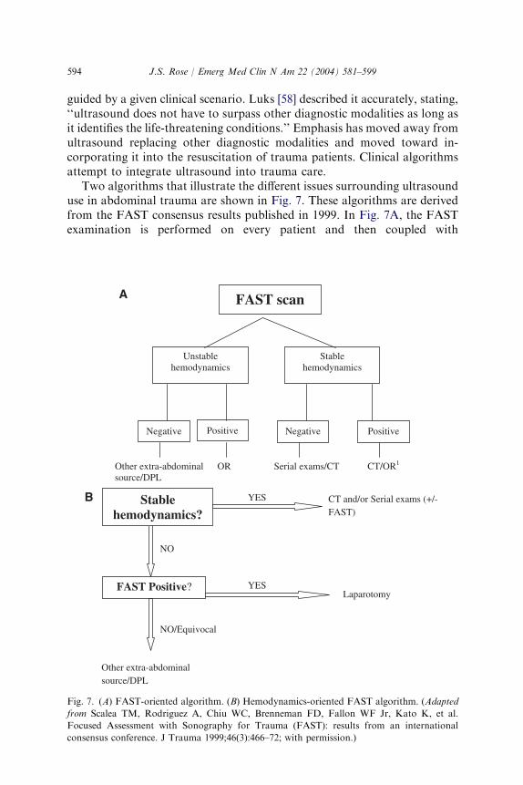
Two algorithms that illustrate the different issues surrounding ultrasounduse in abdominal trauma are shown in Fig. 7. These algorithms are derivedfrom the FAST consensus results published in 1999. In Fig. 7A, the FASTexamination is performed on every patient and then coupled with
Negative Positive
Unstable hemodynamics
Negative Positive
CT/OR1OR Serial exams/CT Other extra-abdominal source/DPL
Stable hemodynamics
FAST scan A
Stable hemodynamics?
FAST Positive?
CT and/or Serial exams (+/- FAST)
YES
YESLaparotomy
NO
NO/Equivocal
Other extra-abdominal source/DPL
B
Fig. 7. (A) FAST-oriented algorithm. (B) Hemodynamics-oriented FAST algorithm. (Adapted
from Scalea TM, Rodriguez A, Chiu WC, Brenneman FD, Fallon WF Jr, Kato K, et al.
Focused Assessment with Sonography for Trauma (FAST): results from an international
consensus conference. J Trauma 1999;46(3):466–72; with permission.)
595J.S. Rose / Emerg Med Clin N Am 22 (2004) 581–599
hemodynamic stability. This type of algorithm is used by centers that believethat all patients should undergo a FAST scan.
Fig. 7B illustrates a different algorithm based on the same consensusrecommendations [11]. In this algorithm, hemodynamic status is the initialdecision point. If stable, the patient can undergo serial physical examina-tions and FAST examinations or abdominal CT scanning alone, dependingon the clinical scenario. If the patient is unstable, then a FAST examinationis performed. If the FAST examination is positive, then the patient would gofor laparotomy; if negative, then the clinician would look for an extra-abdominal source of bleeding or perform a DPL if the examination wasequivocal. In this algorithm, the FAST examination plays much less of a rolein the stable patient. One could argue that in this algorithm, if CT is used,a FAST examination would not be needed in the hemodynamically stablepatients. Although both algorithms follow the consensus recommendations,they do so with very different emphases: one emphasizes the initial FASTand the other emphasizes hemodynamic status.
Another algorithm that is more logical and incorporates clinical andultrasound data is the Key Clinical Pathway developed by Branney et al[26]. This pathway uses hemodynamic status first. From this point, variousclinical parameters are applied to the pathway to select patients who requireCT or laparotomy. This pathway then includes more relevant clinical datasuch as peritoneal irritation, confounding injuries, gross hematuria, orhematocrit\35% to help answer the question of whether a patient requiresimmediate laparotomy. Two issues arise from this pathway. First, if a patientis unstable but has no free fluid, then a DPL is recommended. Someclinicians would recommend looking for another source of bleeding and notperforming a DPL. Second, if a patient has a positive FAST examinationbut is stable and has no peritoneal irritation, then a DPL rather than a CTscan is recommended. A patient who is severely head injured would fall intothis category. This pathway gives the impression that the investigatorsbelieve the sensitivity of ultrasound is not good enough to be used inisolation.
From these pathways, even more questions are generated: Why performa FAST examination in a stable patient? Will it change decision making? Asstated earlier, given the accessibility and speed of newer-generation CTscanners, severely injured patients can be imaged effectively during theresuscitation period. This practice is an area of some debate and focuses onthe overall practice paradigm of a particular institution. One could arguethat determining the presence of hemoperitoneum is valuable even when CTscanning is readily available. Resource use, timing, and treatment ofsecondary injuries will be influenced by the FAST examination results.Algorithms that include ultrasound and CT have been developed thatminimize missed injuries while limiting overuse of CT scanning [23]. Futurestudies are needed to examine whether there are any predictive factorspresent during resuscitation that help to determine whether a positive FAST
596 J.S. Rose / Emerg Med Clin N Am 22 (2004) 581–599
scan requires a therapeutic laparotomy. Whatever pathway is chosen,understanding the overall strengths and limitations of the FAST examina-tion is critical to its safe and effective implementation into the traumaresuscitation.
Future directions
Future research in trauma ultrasound likely will focus on integration withcurrent diagnostic and imaging modalities. Preliminary research that isexamining the role of ultrasound for detecting traumatic pneumothorax[59], evaluating portable ultrasound units [60], and evaluating transducerfrequency for parenchymal injuries is revealing exciting results [61]. Moreimportant, a large, multicenter trial to evaluate previous hypotheses andclarify issues such as scoring systems and practice pathways still is needed.Certainly, the role of trauma ultrasound in the setting of a small, ruralemergency department with limited backup is very different than ultrasoundin a busy level I trauma center. Emergency physicians must continue to beon the forefront of this work. The role of ultrasound in pediatric trauma stillneeds to be clarified. Great strides, however, have been made in clarifyingthe advantages and disadvantages of the FAST examination. Ultrasoundhas been an important addition to the management of trauma patients andwill continue to play an important role in trauma management into thetwenty-first century.
References
[1] Kristensen JK, Buemann B, Keuhl E. Ultrasonic scanning in the diagnosis of splenic
haematomas. Acta Chir Scand 1971;137:653–7.
[2] Ascher WM, Parvin S, Virgilio RW. Echographic evaluation of splenic injury after blunt
trauma. Radiology 1976;118:411–5.
[3] Tiling T, Boulion B, Schmid A. Ultrasound in blunt abdomino-thoracic trauma. In:
Border, Allgoewer M, Hanson ST, editors. Blunt multiple trauma: comprehensive
pathophysiology and care. New York: Marcel Decker; 1990. p. 415–33.
[4] Tso P, Rodriguez A, Cooper C, Militello P, Mirvis S, Badellino M, et al. Sonography in
blunt abdominal trauma: a preliminary progress report. J Trauma 1992;33(1):39–43[dis-
cussion: 43–4].
[5] Bode PJ, Niezen RA, van Vugt AB, Schipper J. Abdominal ultrasound as a reliable
indicator for conclusive laparotomy in blunt abdominal trauma. J Trauma 1993;34(1):
27–31.
[6] Liu M, Lee CH, P’Eng KF. Prospective comparison of diagnostic peritoneal lavage,
computed tomographic scanning, and ultrasonography for the diagnosis of blunt
abdominal trauma. J Trauma 1993;35(2):267–70.
[7] Branney SW, Wolfe RE, Moore EE, Albert NP, Heinig M, Mestek M, et al. Quantitative
sensitivity of ultrasound in detecting free intraperitoneal fluid. J Trauma 1995;39(2):
375–80.
[8] Chambers JA, Pilbrow WJ. Ultrasound in abdominal trauma: an alternative to peritoneal
lavage. Arch Emerg Med 1988;5(1):26–33.

597J.S. Rose / Emerg Med Clin N Am 22 (2004) 581–599
[9] Jehle D, Guarino J, Karamanoukian H. Emergency department ultrasound in the
evaluation of blunt abdominal trauma. Am J Emerg Med 1993;11(4):342–6.
[10] Rozycki GS, Ochsner MG, Schmidt JA, Frankel HL, Davis TP, Wang D, et al. A
prospective study of surgeon-performed ultrasound as the primary adjuvant modality for
injured patient assessment. J Trauma 1995;39(3):492–8 [discussion: 498–500].
[11] Scalea TM, Rodriguez A, Chiu WC, Brenneman FD, Fallon WF Jr, Kato K, et al.
Focused Assessment with Sonography for Trauma (FAST): results from an international
consensus conference. J Trauma 1999;46(3):466–72.
[12] Ma OJ, Kefer MP, Mateer JR, Thoma B. Evaluation of hemoperitoneum using a single- vs
multiple-view ultrasonographic examination [comments]. Acad Emerg Med 1995;2(7):581–6.
[13] Ingeman JE, Plewa MC, Okasinski RE, King RW, Knotts FB. Emergency physician use of
ultrasonography in blunt abdominal trauma. Acad Emerg Med 1996;3(10):931–7.
[14] Rozycki GS, Ochsner MG, Feliciano DV, Thomas B, Boulanger BR, Davis FE, et al.
Early detection of hemoperitoneum by ultrasound examination of the right upper
quadrant: a multicenter study. J Trauma 1998;45(5):878–83.
[15] Rose JS, Bair AE, Mandavia D, Kinser DJ. The UHP ultrasound protocol: a novel
ultrasound approach to the empiric evaluation of the undifferentiated hypotensive patient.
Am J Emerg Med 2001;19(4):299–302.
[16] Plummer D, Brunett D. Emergency department echocardiography improves outcome in
penetrating cardiac injury. Ann Emerg Med 1992;21(5):709–12.
[17] Rozycki GS, Feliciano DV, Ochsner MG, Knudson MM, Hoyt DB, Davis F, et al. The
role of ultrasound in patients with possible penetrating cardiac wounds: a prospective
multicenter study. J Trauma 1999;46(4):543–51 [discussion: 551–2].
[18] McGahan JP, Rose J, Coates TL, Wisner DH, Newberry P. Use of ultrasonography in the
patient with acute abdominal trauma. J Ultrasound Med 1997;16(10):653–62 [quiz: 663–4].
[19] Yoshii H, Sato M, Yamamoto S, Motegi M, Okusawa S, Kitano M, et al. Usefulness and
limitations of ultrasonography in the initial evaluation of blunt abdominal trauma.
J Trauma 1998;45(1):45–50 [discussion: 50–1].
[20] Cogbill TH, Moore EE, Jurkovich GJ, Morris JA, Mucha P Jr, Shackford SR, et al.
Nonoperative management of blunt splenic trauma: a multicenter experience. J Trauma
1989;29(10):1312–7.
[21] Bose SM, Mazumdar A, Gupta R, Giridhar M, Lal R, Praveen BV. Expectant
management of haemoperitoneum. Injury 1999;30(4):269–73.
[22] Minarik L, Slim M, Rachlin S, Brudnicki A. Diagnostic imaging in the follow-up of non-
operative management of splenic trauma in children. Pediatr Surg Int 2002;18(5–6):429–31.
[23] Jacobs DG, Sarafin JL, Marx JA. Abdominal CT scanning for trauma: how low can we
go? Injury 2000;31(5):337–43.
[24] Navarrete-Navarro P, Vazquez G, Bosch JM, Fernandez E, Rivera R, Carazo E.
Computed tomography vs clinical and multidisciplinary procedures for early evaluation of
severe abdomen and chest trauma—a cost analysis approach. Intensive Care Med 1996;
22(3):208–12.
[25] Chiu WC, Cushing BM, Rodriguez A, Ho SM, Mirvis SE, Shanmuganathan K, et al.
Abdominal injuries without hemoperitoneum: a potential limitation of focused abdominal
sonography for trauma (FAST) [comments]. J Trauma 1997;42(4):617–22 [discussion:
623–5].
[26] Branney SW, Moore EE, Cantrill SV, Burch JM, Terry SJ. Ultrasound based key clinical
pathway reduces the use of hospital resources for the evaluation of blunt abdominal
trauma. J Trauma 1997;42(6):1086–90.
[27] Rose JS, Levitt MA, Porter J, Hutson A, Greenholtz J, Nobay F, et al. Does the presence
of ultrasound really affect computed tomographic scan use? A prospective randomized trial
of ultrasound in trauma. J Trauma 2001;51(3):545–50.
[28] American College of Surgeons. Advanced trauma life support for physicians. Chicago:
ACS; 1997.
598 J.S. Rose / Emerg Med Clin N Am 22 (2004) 581–599
[29] Rose J, Richards JR, Bair AE, Battistella F, McGahan JP, Kuppermann N. The
ultrasound is positive, now what? Derivation of a clinical decision rule for predicting
therapeutic laparotomy among adult patients with a positive trauma ultrasound [abstract].
Aca Emerg Med 2002;9:463–4.
[30] Goldberg GG. Evaluation of ascites by ultrasound. Radiology 1970;96(15):217–21.
[31] Paajanen H, Lahti P, Nordback I. Sensitivity of transabdominal ultrasonography in
detection of intraperitoneal fluid in humans. Eur Radiol 1999;9(7):1423–5.
[32] Abrams BJ, Sukumvanich P, Seibel R, Moscati R, Jehle D. Ultrasound for the detection of
intraperitoneal fluid: the role of Trendelenburg positioning. Am J Emerg Med 1999;17(2):
117–20.
[33] Gracias VH, Frankel HL, Gupta R, Malcynski J, Gandhi R, Collazzo L, et al. Defining the
learning curve for the Focused Abdominal Sonogram for Trauma (FAST) examination:
implications for credentialing. Am Surg 2001;67(4):364–8.
[34] HuangMS, LiuM,Wu JK, ShihHC,KoTJ, LeeCH.Ultrasonography for the evaluation of
hemoperitoneum during resuscitation: a simple scoring system. J Trauma 1994;36(2):173–7.
[35] McKenney KL, McKenney MG, Cohn SM, Compton R, Nunez DB, Dolich M, et al.
Hemoperitoneum score helps determine need for therapeutic laparotomy. J Trauma 2001;
50(4):650–4 [discussion: 654–6].
[36] Ong AW, McKenney MG, McKenney KA, Brown M, Namias N, MaCloud J, et al.
Predicting the need for laparotomy in pediatric trauma patients on the basis of the
ultrasound score. J Trauma 2003;54(3):503–8.
[37] American College of Surgeons. Ultrasound education program ‘‘bluebook.’’ Chicago:
ACS; 2000.
[38] American College of Emergency Physicians. Use of ultrasound imaging by emergency
physicians. Ann Emerg Med 2001;38:469–70.
[39] Thomas B, Falcone RE, Vasquez D, Santanello S, Townsend M, Hockenberry S, et al.
Ultrasound evaluation of blunt abdominal trauma: program implementation, initial
experience, and learning curve. J Trauma 1997;42(3):384–8 [discussion: 388–90].
[40] Ma OJ, MJ, Ogata M. Prospective analysis of a rapid trauma ultrasound examination
performed by emergency physicians. J Trauma 1995;18:879–85.
[41] Rozycki GS, Ballard RB, Feliciano DV, Schmidt JA, Pennington SD. Surgeon-performed
ultrasound for the assessment of truncal injuries: lessons learned from 1540 patients. Ann
Surg 1998;228(4):557–67.
[42] Dolich MO, McKenney MG, Varela JE, Compton RP, McKenney KL, Cohn SM. 2,576
ultrasounds for blunt abdominal trauma. J Trauma 2001;50(1):108–12.
[43] Gracias VH, Frankel H, Gupta R, Reilly PM, Gracias F, Klein W, et al. The role of
positive examinations in training for the focused assessment sonogram in trauma (FAST)
examination. Am Surg 2002;68(11):1008–11.
[44] Sisley AC, Johnson SB, Erickson W, Fortune JB. Use of an Objective Structured Clinical
Examination (OSCE) for the assessment of physician performance in the ultrasound
evaluation of trauma. J Trauma 1999;47(4):627–31.
[45] Salen PN, Melanson SW, Heller MB. The focused abdominal sonography for trauma
(FAST) examination: considerations and recommendations for training physicians in the
use of a new clinical tool. Acad Emerg Med 2000;7(2):162–8.
[46] Smith RS, Kern SJ, Fry WR, Helmer SD. Institutional learning curve of surgeon-
performed trauma ultrasound. Arch Surg 1998;133(5):530–5 [discussion: 535–6].
[47] Shackford SR, Rogers FB, Osler TM, Trabulsy ME, Clauss DW, Vane DW. Focused
abdominal sonogram for trauma: the learning curve of nonradiologist clinicians in
detecting hemoperitoneum. J Trauma 1999;46(4):553–62 [discussion: 562–4].
[48] Richards JR, McGahan JP, Pali MJ, Bohnen PA. Sonographic detection of blunt hepatic
trauma: hemoperitoneum and parenchymal patterns of injury. J Trauma 1999;47(6):
1092–7.
599J.S. Rose / Emerg Med Clin N Am 22 (2004) 581–599
[49] Richards JR, McGahan JP, Jones CD, Zhan S, Gerscovich EO. Ultrasound detection of
blunt splenic injury. Injury 2001;32(2):95–103.
[50] Brown MA, Casola G, Sirlin CB, Hoyt DB. Importance of evaluating organ parenchyma
during screening abdominal ultrasonography after blunt trauma. J Ultrasound Med 2001;
20(6):577–83[quiz: 585].
[51] Krupnick AS, Teitelbaum DH, Geiger JD, Strouse PJ, Cox CS, Blane CE, et al. Use of
abdominal ultrasonography to assess pediatric splenic trauma. Potential pitfalls in the
diagnosis. Ann Surg 1997;225(4):408–14.
[52] Ohta S, Hagiwara A, Yukioka T. Hyperechoic appearance of hepatic parenchyma on
ultrasound examination of patients with blunt hepatic injury. J Trauma 1998;44:135–8.
[53] Brown MA, Casola G, Sirlin CB, Patel NY, Hoyt DB. Blunt abdominal trauma: screening
us in 2,693 patients. Radiology 2001;218(2):352–8.
[54] Katz S, Lazar L, Rathaus V, Erez I. Can ultrasonography replace computed tomography
in the initial assessment of children with blunt abdominal trauma? J Pediatr Surg 1996;
31(5):649–51.
[55] Thourani VH, Pettitt BJ, Schmidt JA, Cooper WA, Rozycki GS. Validation of surgeon-
performed emergency abdominal ultrasonography in pediatric trauma patients. J Pediatr
Surg 1998;33(2):322–8.
[56] Holmes JF, Brant WE, Bond WF, Sokolove PE, Kuppermann N. Emergency department
ultrasonography in the evaluation of hypotensive and normotensive children with blunt
abdominal trauma. J Pediatr Surg 2001;36(7):968–73.
[57] Boulanger BR, Kearney PA, Brenneman FD, Tsuei B, Ochoa J. Utilization of FAST
(Focused Assessment with Sonography for Trauma) in 1999: results of a survey of North
American trauma centers. Am Surg 2000;66(11):1049–55.
[58] Luks F, Lemiere A. Blunt abdominal trauma in children: the practical value of
ultrasonography. J Trauma 1993;34(5):607–10.
[59] Chan SS. Emergency bedside ultrasound to detect pneumothorax. Acad Emerg Med 2003;
10(1):91–4.
[60] Kirkpatrick AW, Simons RK, Brown R, Nicolaou S, Dulchavsky S. The hand-held FAST:
experience with hand-held trauma sonography in a level-I urban trauma center. Injury
2002;33(4):303–8.
[61] Stengel D, Bauwens K, Sehouli J, Nantke J, Ekkernkamp A. Discriminatory power of 3.5
MHz convex and 7.5 MHz linear ultrasound probes for the imaging of traumatic splenic
lesions: a feasibility study. J Trauma 2001;51(1):37–43.