tyrosine phosphorylation of the human glutathione … · -1- tyrosine phosphorylation of the human...
TRANSCRIPT
-1-
TYROSINE PHOSPHORYLATION OF THE HUMAN GLUTATHIONE S-TRANSFERASE P1 BY EPIDERMAL GROWTH FACTOR RECEPTOR
Tatsunori Okamura1, Simendra Singh1, John Buolamwini4, Timothy Haystead2, Henry Friedman1, Darell Bigner3, and Francis Ali-Osman1
From Departments of Surgery1, Pharmacology2, Pathology3, Duke Comprehensive Cancer Center and the Preston Robert Tisch Brain Tumor Center, Duke University, Durham, North Carolina 27710 and
Department of Pharmaceutical Sciences4, College of Pharmacy, University of Tennessee Health Sciences Center, Memphis, Tennessee 38163
Running head: GSTP1 phosphorylated by EGFR
Corresponding Author: Francis Ali-Osman, 421 Medical Science Research Building I, Duke University Medical Center, Durham, NC 27710. Tel: 919-681-5769, Fax: 919-684-5483, E-mail: [email protected]
Epidermal growth factor receptor (EGFR) gene amplification, mutations and/or aberrant activation are frequent abnormalities in malignant gliomas and other human cancers, and have been associated with an aggressive clinical course and a poor therapeutic outcome. Elevated glutathione S-transferase P1 (GSTP1), a major drug metabolizing and stress response signaling protein, is also associated with drug resistance and poor clinical outcome in gliomas and other cancers. Here, we provide evidence that GSTP1 is a downstream EGFR target and that EGFR binds to and phosphorylates tyrosine residues in the GSTP1 protein in vitro and in vivo. Mass spectrometry and mutagenesis analyses in a cell-free system and in gliomas cells identified Tyr7 and Tyr198 as major EGFR-specific phospho-acceptor residues in the GSTP1 protein. The phosphorylation increased GSTP1 enzymatic activity significantly and computer-based modeling showed a corresponding increase in electronegativity of the GSTP1 active site. In human glioma and breast cancer cells, EGF-stimulation rapidly increased GSTP1 tyrosine phosphorylation and decreased cisplatin sensitivity. Lapatinib, a clinically active EGFR inhibitor, significantly reversed the EGF-induced cisplatin resistance. These data define phosphorylation and activation of GSTP1 by EGFR as a novel, heretofore, unrecognized component of the EGFR signaling network and a novel mechanism of tumor drug resistance,
particularly, in tumors with elevated GSTP1 and/or activated EGFR.
INTRODUCTION
Epidermal growth factor receptor (EGFR), a 170 kDa receptor-type tyrosine kinase, mediates diverse signaling pathways and cellular processes, including, proliferation, differentiation, motility and survival (1-4). Ligand binding and activation of EGFR result in receptor dimerization, autophosphorylation, and activation of downstream effector pathways, such as, Phosphatidylinositol 3-kinase (PI3K)/AKT, Janus kinases (JAK)/Signal Transducers and Activators of Transcription (STAT), and Ras/Raf/mitogen-activated protein kinase (MAPK) (1, 2). The cellular signaling cascades initiated and transduced by EGFR have been implicated in oncogenesis and functional dysregulation of EGFR and its downstream pathways are frequently observed in malignant gliomas and other human cancers, and have been shown to regulate features of the malignant phenotype, such as, tumor progression, adhesion, invasion, angiogenesis, and apoptosis (3, 5).
EGFR gene amplification is a hallmark of glioblastoma multiforme (GBM), the most aggressive and most common intrinsic malignant brain tumor (6). In primary (de novo) GBM, which accounts for approx. 95% of all GBM (7, 8), EGFR amplification follows LOH of chromosome
http://www.jbc.org/cgi/doi/10.1074/jbc.M808153200The latest version is at JBC Papers in Press. Published on March 2, 2009 as Manuscript M808153200
Copyright 2009 by The American Society for Biochemistry and Molecular Biology, Inc.
by guest on October 10, 2018
http://ww
w.jbc.org/
Dow
nloaded from

-2-
10q as the most frequently observed genetic alteration (8). In preclinical studies, a strong correlation has been reported between high aberrant EGFR signaling and ligand-dependent GBM cell proliferation (9) and the resistance of GBM to chemotherapy and radiation therapy (10). Consistent with this, clinical studies of GBM have shown EGFR gene amplification to be a significant negative predictor of patient survival (4), and EGFR overexpression has been associated with failure to respond to radiation therapy (11). In addition to its amplification and overexpression, a mutant EGFR, EGFRvIII, characterized by deletion of exons 2-7, is present in almost half of GBMs with amplified EGFR (4, 12). EGFRvIII is unique in that the loss of a large portion of the extracellular ligand-binding domain, leads to its constitutive and ligand-independent activation (13). In GBM, EGFRvIII has been associated with increased tumor growth, cell proliferation and drug resistance, which, similar to wild-type EGFR, occurs via constitutive activation of downstream EGFR pathways (14, 15).
Glutathione S-transferase P1 (GSTP1), a major phase II metabolizing enzyme, encoded in a polymorphic gene locus (16), catalyzes the S-conjugation of endogenous and exogenous electrophiles, including, many genotoxins, carcinogens and anticancer agents, to the nucleophilic thiol group of reduced glutathione, GSH (17). In addition, GSTP1 is a major regulator of cell signaling in response to stress, hypoxia, growth factors and other stimuli. This results, in part, from its ability to inhibit downstream MAP kinase signaling, notably, that mediated by c-jun N-terminal kinase (JNK) (18, 19, 20). GSTP1 also regulates important normal cellular functions through interaction with a number of critical cellular proteins, including, transglutaminase 2 (TGM2), apoptosis signal-regulating kinase 1 (ASK1) and Fanconi anemia group C protein (FANCC) (21). Recently, we reported that GSTP1 is a substrate for two Ser/Thr protein kinases, viz., cAMP-dependent protein kinase (PKA) and protein kinase C (PKC) (22). In many human cancers, including, gliomas, leukemias, lymphomas, melanoma and carcinomas of the breast, ovary, colorectum, lung, liver etc (23, 24, 25), GSTP1 is frequently over-expressed and the high expression is associated with a more aggressive tumor biology and poor patient survival.
Given the roles that both EGFR and GSTP1 play in cell signaling and in both normal and neoplastic biology, we investigated, using GBM and inflammatory breast cancer cell lines, the possibility and consequences of the interaction between the two proteins in vitro and in GBM xenografts growing in vivo. The nature of the interaction was characterized structurally and functionally by a combination of mass spectrometry and other biochemical analyses and its effects on the response of the tumor cells to chemotherapy was investigated. Our findings support EGFR-mediated tyrosine phosphorylation of GSTP1 to be a, heretofore, unrecognized component of the EGFR cellular network and constitutes an important mechanism of cellular protection and drug resistance, particularly, in tumors and/or tissues with activated EGFR and/or elevated GSTP1 expression.
EXPERIMENTAL PROCEDURES
Chemicals and Antibodies- Recombinant human GSTP1-1 protein was purchased from Invitrogen Corporation (Carlsbad, CA), recombinant human EGFR active kinase domain and normal mouse IgG from Upstate Biotechnology (Lake Placid, NY). [γ-32P]ATP and Protein A sepharose were from Amersham Corporation (Piscataway, NJ). Anti-human GSTP1-1 rabbit polyclonal antibody was from Oxford Biomedical Research (Oxford, MI), anti-human GSTP1 mouse monoclonal antibody from BIODESIGN International (Saco, ME). Anti-phosphotyrosine (P-Tyr-100) and anti-phospho-EGFR (Tyr1068) monoclonal antibodies were from Cell Signaling Technology (Danvers, MA). Anti-GRB2 and HRP-conjugated secondary antibodies were from Santa Cruz Biotechnology (Santa Cruz, CA). The EGFR inhibitor, lapatinib, was purchased from LC Laboratories (Woburn, MA), and prepared in DMSO stock solution. All other chemicals and biochemicals were purchased from Sigma-Aldrich Corporation, unless otherwise stated.
Cell Lines- The human cell lines, MGR1 (anaplastic astrocytoma) and MGR3 (GBM) were established in our laboratory from primary specimens (22). The high EGFR expressing human GBM U87MG.wtEGFR was derived by stable transfection of the parental U87MG cells with wild-type EGFR (26). SUM 149 (Asterand PLC, Detroit, MI) is a human inflammatory breast
by guest on October 10, 2018
http://ww
w.jbc.org/
Dow
nloaded from
-3-
cancer cell line. Cells were cultured in DMEM with 10% FCS (MGR1, MGR3), Improved MEM Zinc Option with 10% FCS (U87MG, U87MG.wtEGFR) or Ham’s F-12 with 5% FCS (SUM 149).
GSTP1 Phosphorylation by EGFR in Cell-free System- To mimic intracellular conditions in which the GSTP1 protein exists in equilibrium with GSH bound to its GSH-binding site (22, 27), 1 µg of human recombinant GSTP1 was preincubated with 5 mM GSH for 20 min at 37°C and then added to a reaction mixture containing EGFR active kinase domain (25 ng) and [γ-32P]ATP in Mn2+, Mg2+-containing kinase buffer. After 1 hr-incubation at 30°C, the reaction terminated by boiling and resolved by SDS-PAGE, followed by Coomassie Blue staining and autoradiography. For stoichiometry of the GSTP1 phosphorylation by EGFR, the phosphorylation reaction was set up, containing 1 µg GSTP1, 0.05 µM EGFR, and a saturating (100 µM) ATP concentration. Over 0 to 4 hours, aliquots were removed and subjected to SDS-PAGE, the phosphorylated GSTP1 bands excised, solubilized and the radioactivity counted by beta-scintillation. The incorporated 32P phosphate was computed from the specific activity of the [γ-32P]ATP, expressed per mole of the dimeric GSTP1 protein and plotted against time.
Analysis of GSTP1 Phosphorylated Amino Acid Residues by Thin Layer Electrophoresis- GSTP1 was 32P-phosphorylated by EGFR, subjected to SDS-PAGE and transferred to PVDF membranes. After autoradiography, the phospho-GSTP1 bands were excised, hydrolyzed at 110°C in 5M HCl for 1 hr, vacuum-dried and resuspended in a loading buffer of acetic acid:pyridine:water (10:1:189), containing phosphoserine, phosphothreonine and phosphotyrosine standards, electrophoresed at pH 3.5 on a cellulose TLC plate. The plate was dried, stained with ninhydrin and autoradiographed.
Western Blotting for GSTP1 Tyrosine Phosphorylation in Cell-free System- A mixture of GSTP1 pre-incubated with or without 5 mM GSH was applied to the GSTP1 phosphorylation assay with 50 nM EGFR and 200 µM ATP, followed by SDS-PAGE/western blotting with anti-phosphotyrosine (pTyr) antibody. After stripping, the membrane was reprobed with anti-GSTP1 antibody to control for loading. To inhibit the
EGFR activation, a 50 nM EGFR pre-incubated for 30 mins with 0-1 mM lapatinib, an EGFR inhibitor, was used for the GSTP1 phosphorylation assay. Tyrosine phosphorylated GSTP1 was normalized against total GSTP1 protein.
EGFR-dependent GSTP1 Phosphorylation in Glioma and Breast Cancer Cells- Tumor cells grown in serum-free medium for 24 hrs were treated with 100 ng/ml EGF for 10 mins, rinsed with ice-cold PBS and lysed in 50 mM Tris-HCl (pH 7.5) containing 1% Triton X-100, protease and phosphatase inhibitors cocktail (Pierce, Rockford, IL). The lysates were centrifuged at 20,000 rpm for 15 mins Supernatants were subjected to western blotting with anti-pTyr, phospho-EGFR (pEGFR), and GSTP1 antibodies. For a combined immunoprecipitation-western (IP-Western) blotting, supernatants (1 mg total protein) were incubated (4oC; overnight) with anti-GSTP1, or pEGFR antibodies, or normal mouse IgG (as a negative control). The protein A-sepharose beads were incubated with immunocomplexes for 1 hr and washed four times with the lysis buffer, and the immunoprecipitates were subjected to western blotting. To examine the effect of EGFR inhibition, tumor cells with activated EGFR were treated with 2.5 µM lapatinib for 30 mins, as per experimental protocol before lysis and western blotting.
Enzyme Kinetic Analysis of EGFR-phosphorylated GSTP1- These were performed as we had previously described (22). Briefly, 0.1 unit each of unphosphorylated recombinant GSTP1 and GSTP1 phosphorylated by EGFR as described earlier, were used to set up reactions containing 0.05 to 0.5 mM of the GSTP1 specific substrate, ethacrynic acid (EA) and 0.25 mM GSH in a 0.1mM potassium phosphate buffer (pH 6.8). The rate of formation of the reaction product between EA and GSH was monitored at 270 nm. Reaction rates, normalized against that of the non-enzymatic reaction, were computed and used to generate double reciprocal plots from which the enzyme kinetic constants, Km, Vmax, Kcat, and Kcat/Km, were computed for phosphorylated and unphosphorylated GSTP1 (28) and the results presented as the mean ± 1 SD of triplicate experiments.
In Vivo EGFR Phosphorylation of GSTP1 in GBM Xenografts- Xenografts (approximately, 200 mm2
by guest on October 10, 2018
http://ww
w.jbc.org/
Dow
nloaded from
-4-
in diameter) of U87MG and U87MG.wtEGFR growing subcutaneously in the flanks of 4-week-old male athymic BALB/c nu/nu mice were excised, minced and sonicated on ice in the lysis buffer. Supernatants were subjected to the IP (anti-GSTP1)-Western (anti-pTyr) procedure as described earlier.
Phosphorylation Sites Analysis in EGFR Phosphorylated GSTP1 by Mass Spectrometry (MS)- Recombinant GSTP1 was EGFR-phosphorylated GSTP1, reduced and alkylated and second dimension acrylamide gel electrophoresis performed. SYPRO Ruby stained protein spots were robotically excised, reduced with DTT, alkylated with iodoacetamide, digested with trypsin and subjected to LC-MS/MS. MS/MS data were analyzed using the MASCOT MS/MS Ions Search (www.matrixscience.com) and de novo sequence analysis performed with the Scaffold Software (Proteome Software Inc., Portland, OR). Details of the protocols used are available at www.prsproteomics.com and in Supplemental Data.
EGFR Phosphorylation of GSTP1 Peptides- Peptides containing each of the twelve tyrosine residues in the GSTP1 protein, namely, Tyr3, Tyr7, Tyr49, Tyr63, Tyr79, Tyr103, Tyr108, Tyr111, Tyr118, Tyr153, Tyr179, and Tyr198, as well as, human angiotensin II peptide (DRVYIHPF as a positive control; Calbiochem, La Jolla, CA) and crosstide (GRPRTSSFAEG as a negative control; AnaSpec Inc., San Jose, CA) were EGFR phosphorylated using 32P-ATP, spotted on Whatman P81 cellulose phosphate filters, acetone-washed and air-dried. The radioactivity was quantitated by β-scintillation counting and used to compute the incorporated phosphate in each peptide. To better ascertain the GSTP1 phospho-acceptor residues, Tyr3/Tyr7, Tyr63, Tyr118 and Tyr198, in six peptides, selected from the LC-MS/MS analysis, were mutated to aspartic acid (BioSynthesis, Louisville, TX) and the level of EGFR phosphorylation determined, as described above. Peptide information is available in the Supplemental Data Table S1.
Mutagenesis and Phosphorylation of GSTP1 in Gliomas Cells- Mutant GSTP1 cDNAs were created by PCR on a template plasmid vector pBK-CMV/GSTP1A (29), using GSTP1-specific primers containing tyrosine to phenylalanine
mutations at Tyr3, 7 and 198. All mutations were verified by DNA sequencing. Cloning was performed using the Gateway technology (Invitrogen, Carlsbad, CA) with the pcDNA-DEST40 destination vector to allow C-terminal fusions with a 6x histidine tag. Transient transfections were performed with FuGENE HD (Roche, Basel, Switzerland), according to the manufacturer’s instructions. Briefly, 5 x 10⁵ U87MG.wtEGFR cells were plated in 6-well plates and transfected with 2 µg pcDNA-DEST40 expression vector carrying the wild type GSTP1 (WT), the single tyrosine to phenylalanine mutants Y3F, Y7F and Y198F, the double mutants, Y3F/Y7F, Y3F/Y198F and Y7F/Y198F, the triple mutant Y3F/Y7F/Y198F, and the empty vector (negative control). After 48 hrs, the cells were treated with 100 ng/ml of EGF for 10 mins and lysed. The histidine-tagged GSTP1 proteins were immunoprecipitated with TALON cobalt Dynabeads (Invitrogen, Carlsbad, CA), according to the manufacturer’s instructions, followed by SDS-PAGE and Western blotting with anti-pTyr antibody, as described earlier. The relative levels of tyrosine phosphorylation of the mutant GSTP1 proteins relative to the wild-type GSTP1 were quantified by densitometry using ImageJ version 1.34s software. Primer information is available in the Supplemental Data Table S2.
Molecular Dynamics Simulations- X-ray crystallographic data were imported from the Brookhaven Protein DataBank and using the Insight II modeling program (Accelerys Software, San Diego), the 3D structures of the GSH-bound GSTP1 monomer with and without the hydroxyl group of Tyr7 phosphorylated were created. The modeled structures were soaked in a cubic box of water molecules and subjected to energy minimization and long-duration molecular dynamics simulation, using the NAMD program 2.5 running on a 5 node Scyld Beowulf linux cluster. The coordinate and parameter files for input were generated using the `psfgen’ utility in the CHARMM PARAM 22 topology file, while the all atom CHARMM PARAM 22 force field was used to describe the potential energy. The results of the analyses of energies and structure frames of the simulated system were extracted using the VMD software and illustrations produced with both the VMD and SYBYL software. Details of the simulation procedure are provided in the Supplemental Data.
by guest on October 10, 2018
http://ww
w.jbc.org/
Dow
nloaded from
-5-
Effect of EGFR modulation on GSTP1 Enzymatic Activity in Tumor Cells- EGFR was activated in exponentially growing tumor cells by a 10 min treatment with 100 ng/ml EGF. Extracts from cells, with and without subsequent treatment with 2.5 µM of the EGFR inhibitor, lapatinib for 30 mins, were assayed for specific GSTP1 activity, as we described earlier (22).
Effect of EGFR activation on glutathione-platinum metabolite formation in tumor cells- These studies were performed with the GSTP1- and EGF-over-expressing human inflammatory breast cancer cell line, SUM 149. Approximately, 5 x 10⁶ cells in exponential growth were pre-treated with 100 ng/ml EGF for 10 mins in triplicate, after which the medium was replaced with fresh medium containing 100 µM cisplatin. After an additional 2 hrs at 37oC, the cells were washed twice, harvested and homogenized in 500 µl PBS. Supernatants after centrifugation at 15,000 x G for 20 mins were removed and protein precipitated by adding trichloroacetic acid (TCA) to 10% (final concentration) and incubating at 4oC for 3 hrs. After final centrifugation, the supernatants (normalized for equal protein content) were used for glutathionylplatinum (GS-Pt) metabolite quantitation, as we previously described (30). Briefly, aliquots of the supernatant were diluted 1:10 with 10% TCA and the absorbance measured by scanning spectrophotometry over a wavelength range of 240-400 nm (Beckman DU-70 spectrophotometer). The absorbance at 265 nm, A265, (peak absorbance of the GS-Pt conjugate), of the supernatants, with and without EGF pretreatment, were normalized against that of control without cisplatin treatment and the resulting ∆A265 was used as a measure of the level of glutathionylcisplatin in the cells.
Effect of EGFR activation on cisplatin sensitivity of tumor cells- Tumor cells with activated EGFR were treated with and without 2.5 nM lapatinib for 30 mins, followed by 0-50 µM cisplatin for 3 hrs. The cells were washed and cell survival examined after 48 hrs, using the CellTiter-Blue Assay (Promega, Madison, WI), according to the manufacturer’s instructions.
Effect of siRNA-mediated GSTP1 down-regulation on EGFR-induced cisplatin resistance in glioma cells- MGR3 and SUM 149 cells were plated at 1 x 10³ to 104 cells in 100 µl DMEM containing
10% FCS in a flat-bottomed microtiter plate. After 24 hrs at 370C, the cells were transfected with siRNA with the sequence (5’-ACCAGAUCUCCUUCGCUGACUACAA-3’) targeting the N-terminal region of GSTP1 mRNA, using Lipofectamine™ (Invitrogen, Carlsbad, CA), according to the manufacturer’s instructions. After 6 hrs, the cultures were refed with fresh medium and incubated at 370C for a further 24 hrs. Untransfected cells and cells transfected with scrambled siRNA served as controls. After 24 hrs, the cells were treated with and without 100 ng/ml EGF for 10 mins, followed by 0-50 µM cisplatin. Cell survival was determined as described earlier. Cisplatin sensitivity (surviving fraction) after each treatment relative to controls (normalized against scrambled siRNA) was determined and IC50 values were computed. Replicate cells after siRNA treatment were lysed and subjected to western blotting for GSTP1 expression to monitor the level of GSTP1 knockdown.
RESULTS
GSTP1 is phosphorylated by EGFR in a cell-free system. Figures 1A-C summarizes the results of the cell-free analysis of the phosphorylation of GSTP1 by EGFR, performed with recombinant GSTP1 and EGFR proteins. The 32P-labeling results (Fig.1A) show that, following its pre-equilibration with GSH, GSTP1 undergoes dose-dependent phosphorylation by EGFR. The western blots with an anti-phosphotyrosine (pTyr) specific antibody (Fig. 1B) show that while for GSTP1, tyrosine-phosphorylation required GSH and was significantly reduced in its absence, the presence of GSH, resulted in a slight reduction in the level of EGFR autophosphorylation, consistent with previous reports that EGFR is a redox-regulated protein and that its intracellular activation is suppressed by reducing agents (31). Equal loading of GSTP1 in the lanes is shown by the western blots for GSTP1 (lowest panel in Fig. 1B). To determine which amino acids in the GSTP1 protein undergo phosphorylation by EGFR, recombinant GSTP1 was [32P]-phosphorylated by EGFR, acid-hydrolyzed and subjected to thin layer electrophoresis and autoradiography. The results (Fig. 1C) show tyrosine to be the only amino acid residue phosphorylated by EGFR in the GSTP1 protein. No phosphoserines or phosphothreonines were detected. In the cell-free system and at
by guest on October 10, 2018
http://ww
w.jbc.org/
Dow
nloaded from
-6-
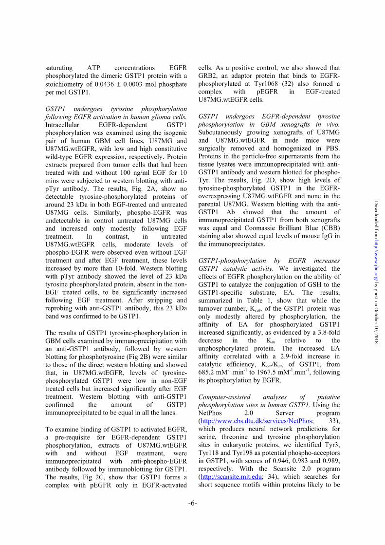
saturating ATP concentrations EGFR phosphorylated the dimeric GSTP1 protein with a stoichiometry of 0.0436 ± 0.0003 mol phosphate per mol GSTP1.
GSTP1 undergoes tyrosine phosphorylation following EGFR activation in human glioma cells. Intracellular EGFR-dependent GSTP1 phosphorylation was examined using the isogenic pair of human GBM cell lines, U87MG and U87MG.wtEGFR, with low and high constitutive wild-type EGFR expression, respectively. Protein extracts prepared from tumor cells that had been treated with and without 100 ng/ml EGF for 10 mins were subjected to western blotting with anti-pTyr antibody. The results, Fig. 2A, show no detectable tyrosine-phosphorylated proteins of around 23 kDa in both EGF-treated and untreated U87MG cells. Similarly, phospho-EGFR was undetectable in control untreated U87MG cells and increased only modestly following EGF treatment. In contrast, in untreated U87MG.wtEGFR cells, moderate levels of phospho-EGFR were observed even without EGF treatment and after EGF treatment, these levels increased by more than 10-fold. Western blotting with pTyr antibody showed the level of 23 kDa tyrosine phosphorylated protein, absent in the non-EGF treated cells, to be significantly increased following EGF treatment. After stripping and reprobing with anti-GSTP1 antibody, this 23 kDa band was confirmed to be GSTP1.
The results of GSTP1 tyrosine-phosphorylation in GBM cells examined by immunoprecipitation with an anti-GSTP1 antibody, followed by western blotting for phosphotyrosine (Fig 2B) were similar to those of the direct western blotting and showed that, in U87MG.wtEGFR, levels of tyrosine-phosphorylated GSTP1 were low in non-EGF treated cells but increased significantly after EGF treatment. Western blotting with anti-GSTP1 confirmed the amount of GSTP1 immunoprecipitated to be equal in all the lanes.
To examine binding of GSTP1 to activated EGFR, a pre-requisite for EGFR-dependent GSTP1 phosphorylation, extracts of U87MG.wtEGFR with and without EGF treatment, were immunoprecipitated with anti-phospho-EGFR antibody followed by immunoblotting for GSTP1. The results, Fig 2C, show that GSTP1 forms a complex with pEGFR only in EGFR-activated
cells. As a positive control, we also showed that GRB2, an adaptor protein that binds to EGFR-phosphorylated at Tyr1068 (32) also formed a complex with pEGFR in EGF-treated U87MG.wtEGFR cells.
GSTP1 undergoes EGFR-dependent tyrosine phosphorylation in GBM xenografts in vivo. Subcutaneously growing xenografts of U87MG and U87MG.wtEGFR in nude mice were surgically removed and homogenized in PBS. Proteins in the particle-free supernatants from the tissue lysates were immunoprecipitated with anti-GSTP1 antibody and western blotted for phospho-Tyr. The results, Fig. 2D, show high levels of tyrosine-phosphorylated GSTP1 in the EGFR-overexpressing U87MG.wtEGFR and none in the parental U87MG. Western blotting with the anti-GSTP1 Ab showed that the amount of immunoprecipitated GSTP1 from both xenografts was equal and Coomassie Brilliant Blue (CBB) staining also showed equal levels of mouse IgG in the immunoprecipitates.
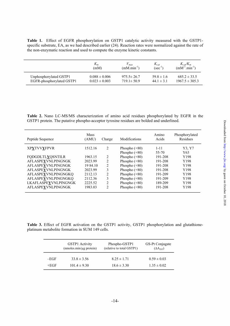
GSTP1-phosphorylation by EGFR increases GSTP1 catalytic activity. We investigated the effects of EGFR phosphorylation on the ability of GSTP1 to catalyze the conjugation of GSH to the GSTP1-specific substrate, EA. The results, summarized in Table 1, show that while the turnover number, Kcat, of the GSTP1 protein was only modestly altered by phosphorylation, the affinity of EA for phosphorylated GSTP1 increased significantly, as evidenced by a 3.8-fold decrease in the Km relative to the unphosphorylated protein. The increased EA affinity correlated with a 2.9-fold increase in catalytic efficiency, Kcat/Km, of GSTP1, from 685.2 mM-1.min-1 to 1967.5 mM-1.min-1, following its phosphorylation by EGFR.
Computer-assisted analyses of putative phosphorylation sites in human GSTP1. Using the NetPhos 2.0 Server program (http://www.cbs.dtu.dk/services/NetPhos; 33), which produces neural network predictions for serine, threonine and tyrosine phosphorylation sites in eukaryotic proteins, we identified Tyr3, Tyr118 and Tyr198 as potential phospho-acceptors in GSTP1, with scores of 0.946, 0.983 and 0.989, respectively. With the Scansite 2.0 program (http://scansite.mit.edu; 34), which searches for short sequence motifs within proteins likely to be
by guest on October 10, 2018
http://ww
w.jbc.org/
Dow
nloaded from
-7-
phosphorylated by specific protein kinases, however, only Tyr198 was identified as a residue phosphorylated by EGFR with high stringency (0.3734; 0.191%). Finally, using the PhosphoMotif Finder (http://www.hprd.org/PhosphoMotif_finder; 35), which contains known published kinase/phosphatase substrate and binding motifs, we identified Tyr118 and Tyr198 to be in regions homologous with a reported EGFR phosphorylation motif, X[E/D]pY[I/L/V], where X is any amino acid (36). Taken together, these results predict Tyr3, Tyr118 and Tyr198 as residues in the GSTP1 protein with a potential of being phosphorylated by EGFR. This information was used to design experiments to determine the phospho-acceptor residues in the GSTP1 protein.
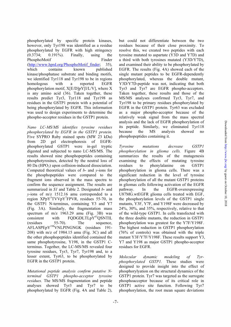
Nano LC-MS/MS identifies tyrosine residues phosphorylated by EGFR in the GSTP1 protein. Five SYPRO Ruby stained spots (MW 23 kDa) from 2D gel electrophoresis of EGFR-phosphorylated GSTP1 were in-gel tryptic digested and subjected to nano LC-MS/MS. The results showed nine phosphopeptides containing phosphotyrosines, detected by the neutral loss of 80 Da (HPO3) upon collision-induced dissociation. Computed theoretical values of b- and y-ions for the phosphopeptides were compared to the fragment ions observed in the mass spectra to confirm the sequence assignment. The results are summarized in S1 and Table 2. Designated b- and y-ions of m/z 1512.16 amu corresponded to a region XPpY3TVVpY7FPVR, residues 55-70, in the GSTP1 N-terminus, containing Y3 and Y7 (Fig. 3A). Similarly, the fragmentation mass spectrum of m/z 1963.29 amu (Fig. 3B) was consistent with FQDGDLTLpY63QSNTIL (residues 55-70). The sequence, AFLASPEpY198VNLPINGNGK (residues 191-208) with m/z of 1984.15 amu (Fig. 3C) and all the other phosphopeptides identified contained the same phosphotyrosine, Y198, in the GSTP1 C-terminus. Together, the LC-MS/MS revealed four tyrosine residues, Tyr3, Tyr7, Tyr198 and, to a lesser extent, Tyr63, to be phosphorylated by EGFR in the GSTP1 protein.
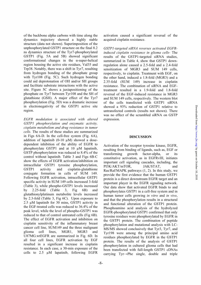
Mutational peptide analysis confirm putative N-terminal GSTP1 phospho-acceptor tyrosine residues. The MS/MS fragmentation and peptide analyses showed Tyr3 and Tyr7 to be phosphorylated by EGFR (Fig. 4A and Table 2),
but could not differentiate between the two residues because of their close proximity. To resolve this, we created two peptides with each tyrosine mutated to aspartate (Y3D and Y7D) and a third with both tyrosines mutated (Y3D/Y7D), and examined their ability to be phosphorylated by EGFR. The results (Fig. 4A) showed each of the single mutant peptides to be EGFR-dependently phosphorylated, whereas the double mutant, Y3D/Y7D-peptide was not, indicating that both Tyr3 and Tyr7 are EGFR phospho-acceptors. Taken together, these results and those of the MS/MS analyses confirmed Tyr3, Tyr7, and Tyr198 to be primary residues phosphorylated by EGFR in the GSTP1 protein. Tyr63 was excluded as a major phospho-acceptor because of the relatively weak signal from the mass spectral analysis and the lack of EGFR phosphorylation of its peptide. Similarly, we eliminated Tyr118 because the MS analysis showed no phosphopeptides containing it.
Tyrosine mutations decrease GSTP1 phosphorylation in glioma cells. Figure 4B summarizes the results of the mutagenesis examining the effects of mutating tyrosine residues to phenylalanine on GSTP1 phosphorylation in glioma cells. There was a significant reduction in the level of tyrosine phosphorylation of all the mutant GSTP1 proteins in gliomas cells following activation of the EGFR pathway. In the EGFR-overexpressing U87MG.wtEGFR glioma cells treated with EGF, the phosphorylation levels of the GSTP1 single mutants, Y3F, Y7F, and Y198F were decreased by 24%, 30%, and 35%, respectively, relative to that of the wild-type GSTP1. In cells transfected with the three double mutants, the reduction in GSTP1 phosphorylation was greatest for the Y7F/Y198F. The highest reduction in GSTP1 phosphorylation (76% of controls) was obtained with the triple mutant Y3F/Y7F/Y198F. These results support Y3, Y7 and Y198 as major GSTP1 phospho-acceptor residues for EGFR.
Molecular dynamic modeling of Tyr-phosphorylated GSTP1. These studies were designed to provide insight into the effect of phosphorylyation on the structural dynamics of the GSTP1 protein. Tyr7 was targeted as the surrogate phosphoacceptor because of its critical role in GSTP1 active site function. Following Tyr7 phosphorylation, the root mean square deviations
by guest on October 10, 2018
http://ww
w.jbc.org/
Dow
nloaded from
-8-
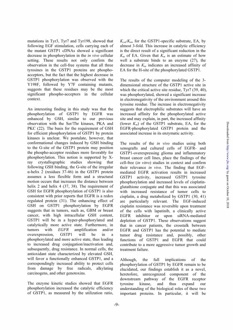
of the backbone alpha carbons with time along the dynamics trajectory showed a highly stable structure (data not shown). Superimposition of the unphosphorylated GSTP1 structure on the final 8.2 ns dynamics structure of the Tyr7-phosphorylated GSTP1 (Fig. 5A and 5B) showed significant conformational changes in the α-super-helical region housing the active site residues, Val35 and Trp38. Notably, there was a shift in Tyr7, resulting from hydrogen bonding of the phosphate group with Tyr108 (Fig. 5C). Such hydrogen bonding could aid deprotonation of OH and/or SH groups and facilitate substrate interactions with the active site. Figure 5C shows a juxtapositioning of the phosphate on Tyr7 between Tyr108 and the SH of glutathione (GSH). A major effect of the Tyr7 phosphorylation (Fig. 5D) was a dramatic increase in electronegativity of the GSTP1 active site region.
EGFR modulation is associated with altered GSTP1 phosphorylation and enzymatic activity, cisplatin metabolism and drug resistance in tumor cells. The results of these studies are summarized in Figs 6A-D. In the cell-free system (Fig. 6A), addition of lapatinib (0-10 µM) showed a dose-dependent inhibition of the ability of EGFR to phosphorylate GSTP1 and at 10 µM lapatinib, GSTP phosphorylation was reduced to 8.4% of the control without lapatinib. Table 3 and Figs 6B-C show the effects of EGFR activation/inhibition on intracellular GSTP1 tyrosine phosphorylation, GSTP1 activity and cisplatin-glutathione conjugate formation in cells of SUM 149. Following EGFR activation, intracellular GSTP1 specific activity in SUM 149 cells increased 3-fold (Table 3), while phospho-GSTP1 levels increased by 2.25-fold (Table 3, Fig 6B) and glutathionylplatinum metabolite levels increased by 2.3-fold (Table 3, Fig 6C). Upon exposure to 2.5 µM lapatinib for 30 mins, GSTP1 activity in the EGF-treated cells was reduced to 36.4% of the peak level, while the level of phospho-GSTP1 was reduced to that of control untreated cells (Fig 6B). The effect of EGFR activation and inhibition on cisplatin sensitivity of the inflammatory breast cancer cell line, SUM149 and the three malignant glioma cell lines, MGR1, MGR3 and U87MG.wtEGFR are summarized in Fig. 6D. In all four cell lines, EGFR activation by EGF resulted in a significant increase in cisplatin resistance. In each case, a 30-min exposure of the cells to 2.5 µM lapatinib, following EGFR
activation caused a significant reversal of the acquired cisplatin resistance.
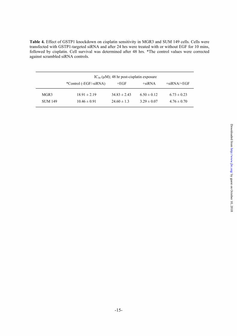
GSTP1-targeted siRNA reverses activated EGFR-induced cisplatin resistance in glioma cells- The results of the GSTP1-targeted siRNA studies, summarized in Table 4, show that GSTP1 down-regulation alone caused a 2.5-fold and a 2.4-fold sensitization of MGR3 and SUM 149 cells, respectively, to cisplatin. Treatment with EGF, on the other hand, induced a 1.8-fold (MGR3) and a 2.35-fold (SUM 149) increase in cisplatin resistance. The combination of siRNA and EGF-treatment resulted in a 1.9-fold and 1.8-fold reversal of the EGF-induced resistance in MGR3 and SUM 149 cells, respectively. The western blot of the cells transfected with GSTP1 siRNA showed a 93% reduction of GSTP1 relative to untransfected controls (results not shown). There was no effect of the scrambled siRNA on GSTP expression.
DISCUSSION
Activation of the receptor tyrosine kinase, EGFR, resulting from binding of ligands, such as, EGF or transforming growth factor-alpha, or its constitutive activation, as in EGFRvIII, initiates important cell signaling cascades, including, the PI3K/AKT/mTOR, JAK/STAT, and Ras/Raf/MAPK pathways (1, 2). In this study, we provide the first evidence that the human GSTP1 protein is a direct downstream EGFR target and an important player in the EGFR signaling network. Our data show that activated EGFR binds to and phosphorylates GSTP1 in a cell-free system and in human tumor cells growing in vitro and in vivo, and that the phosphorylation results in a structural and functional alteration of the GSTP1 protein. Phosphoamino acid analysis of the hydrolyzed EGFR-phosphorylated GSTP1 confirmed that only tyrosine residues were phosphorylated by EGFR in the GSTP1 protein. The combination of peptide phosphorylation and mutational analysis with LC-MS/MS showed conclusively that Tyr3, Tyr7, and Tyr198 were among the principal amino acid residues phosphorylated by EGFR in the GSTP1 protein. The results of the analysis of GSTP1 phosphorylation in cultured glioma cells that had been transfected with full-length GSTP1 cDNAs carrying Tyr→Phe single, double and triple
by guest on October 10, 2018
http://ww
w.jbc.org/
Dow
nloaded from
-9-
mutations in Tyr3, Tyr7 and Tyr198, showed that following EGF stimulation, cells carrying each of the mutant GSTP1 cDNAs showed a significant decrease in phosphorylation in the in vivo cellular setting. These results not only confirm the observation in the cell-free systems that all three tyrosines in the GSTP1 proteins are phospho-acceptors, but the fact that the highest decrease in GSTP1 phosphorylation was observed with the Y198F, followed by Y7F containing mutants, suggests that these residues may be the most significant phospho-acceptors in the cellular context.
An interesting finding in this study was that the phosphorylation of GSTP1 by EGFR was enhanced by GSH, similar to our previous observation with the Ser/Thr kinases, PKA and PKC (22). The basis for the requirement of GSH for efficient phosphorylation of GSTP1 by protein kinases is unclear. We postulate, however, that conformational changes induced by GSH binding to the G-site of the GSTP1 protein may position the phospho-acceptor residues more favorably for phosphorylation. This notion is supported by X-ray crystallographic studies showing that following GSH binding, the G-site of the irregular α-helix 2 (residues 37-46) in the GSTP1 protein assumes a less flexible form and a structural motion occurs that increases the distance between helix 2 and helix 4 (37, 38). The requirement of GSH for EGFR phosphorylation of GSTP1 is also consistent with prior reports that EGFR is a redox regulated protein (31). The enhancing effect of GSH on GSTP1 phosphorylation by EGFR suggests that in tumors, such as, GBM or breast cancer, with high intracellular GSH content, GSTP1 will be in a hyper-phosphorylated and catalytically more active state. Furthermore, in tumors with EGFR amplification and/or overexpression, GSTP1 will be in a phosphorylated and more active state, thus leading to increased drug conjugation/inactivation and, subsequently, drug resistance. In normal cells, the antioxidant state characterized by elevated GSH, will favor a functionally enhanced GSTP1, and a correspondingly increased ability to protect cells from damage by free radicals, alkylating carcinogens, and other genotoxins.
The enzyme kinetic studies showed that EGFR phosphorylation increased the catalytic efficiency of GSTP1, as measured by the utilization ratio,
Kcat/Km, for the GSTP1-specific substrate, EA, by almost 3-fold. This increase in catalytic efficiency is the direct result of a significant reduction in the Km of EA. Given that Km is an estimate of how well a substrate binds to an enzyme (27), the decrease in Km indicates an increased affinity of EA for the H-site of the phosphorylated GSTP1.
The results of the computer modeling of the 3-dimensional structure of the GSTP1 active site in which the critical active site residue, Tyr7 (39, 40), was phosphorylated, showed a significant increase in electronegativity of the environment around this tyrosine residue. The increase in electronegativity suggests that electrophilic substrates will have an increased affinity for the phosphorylated active site and may explain, in part, the increased affinity (lower Km) of the GSTP1 substrate, EA, for the EGFR-phosphorylated GSTP1 protein and the associated increase in its enzymatic activity.
The results of the in vivo studies using both xenografts and cultured cells of EGFR- and GSTP1-overexpressing glioma and inflammatory breast cancer cell lines, place the findings of the cell-free (in vitro) studies in context and confirm their relevance in vivo. The finding that EGF-mediated EGFR activation results in increased GSTP1 activity, increased GSTP1 tyrosine phosphorylation and increased levels of cisplatin-glutathione conjugate and that this was associated with increased resistance of tumor cells to cisplatin, a drug metabolized by GSTP1 (30, 41) are particularly relevant. The EGF-induced cisplatin resistance was reversible upon treatment of the cells with lapatinib, a clinically active EGFR inhibitor or upon siRNA-mediated depletion of GSTP1. These observations suggest that in cancer patients, the crosstalk between EGFR and GSTP1 has the potential to mediate tumor drug resistance and, possibly, other functions of GSTP1 and EGFR that could contribute to a more aggressive tumor growth and treatment failure.
Although, the full implications of the phosphorylation of GSTP1 by EGFR remain to be elucidated, our findings establish it as a novel, heretofore, unrecognized component of the downstream pathway of the EGFR receptor tyrosine kinase, and thus expand our understanding of the biological roles of these two important proteins. In particular, it will be
by guest on October 10, 2018
http://ww
w.jbc.org/
Dow
nloaded from
-10-
important to establish whether the stability, ability to metabolize other anticancer drugs and known carcinogens and/or to interact with other known important GSTP binding, such as, JNK, ASK1, TGM2 or FANCC, of GSTP1 are altered upon phosphorylation by EGFR. Taken together, the
findings in this study, suggest that in cancer therapy, the dual targeting of EGFR and GSTP1 could, potentially, be more effective than the current strategy of targeting either protein individually.
REFERENCES
1. Hackel, P.O., Zwick, E., Prenzel, N., and Ullrich, A. (1999) Curr Opin Cell Biol 11, 184-189 2. Ali-Osman, F., Friedman, H.S., Antoun, G.R., Readon, D., Bigner, D.D., and Buolamwini, J.K. (2005) Brain Tumors, Humana Press, Totowa, NJ, pp 359-381 3. Arteaga, C.L. (2001) J Clin Oncol 19, 32S-40S 4. Shinojima, N., Tada, K., Shiraishi, S., Kamiryo, T., Kochi, M., Nakamura, H., Makino, K., Saya, H., Hirano, H., Kuratsu, J., Oka, K., Ishimaru, Y., and Ushio, Y. (2003) Cancer Res 63, 6962-6970 5. Yang, Z., Bagheri-Yarmand, R., Wang, R.A., Adam, L., Papadimitrakopoulou, V.V., Clayman, G.L., El-Naggar, A., Lotan, R., Barnes, C.J., Hong, W.K., and Kumar, R. (2004) Clin Cancer Res 10, 658-667 6. Gurney, J.G., and Kadan-Lottick, N. (2001) Curr Opin Oncol 13, 160-166 7. Kleihues, P., and Cavenee, W.K. (2000) WHO classification of tumors: pathology and genetics of tumours of the nervous system, IARC Press, Lyon. 8. Ohgaki, H., Dessen, P., Jourde, B., Horstmann, S., Nishikawa, T., Di Patre, P.L., Burkhard, C., Schüler, D., Probst-Hensch, N.M., Maiorka, P.C., Baeza, N., Pisani, P., Yonekawa, Y., Yasargil, M.G., Lütolf, U.M., and Kleihues, P. (2004) Cancer Res 64, 6892-6899 9. Halatsch, M.E., Gehrke, E., Borhani, F.A., Efferth, T., Werner, C., Nomikos, P., Schmidt, U., and Buchfelder, M. (2003) Anticancer Res 23, 2315-2320 10. Chakravarti, A., Chakladar, A., Delaney, M.A., Latham, D.E., and Loeffler, J.S. (2002) Cancer Res 62, 4307-4315 11. Barker, F.G. 2nd, Simmons, M.L., Chang, S.M., Prados, M.D., Larson, D.A., Sneed, P.K., Wara, W.M., Berger, M.S., Chen, P., Israel, M.A., and Aldape, K.D. (2001) Int J Radiat Oncol Biol Phys 51, 410-418 12. Aldape, K.D., Ballman, K., Furth, A., Buckner, J.C., Giannini, C., Burger, P.C., Scheithauer, B.W., Jenkins, R.B., and James, C.D. (2004) J Neuropathol Exp Neurol 63, 700-707 13. Batra, S.K., Castelino-Prabhu, S., Wikstrand, C.J., Zhu, X., Humphrey, P.A., Friedman, H.S., and Bigner, D.D. (1995) Cell Growth Differ 6, 1251-1259 14. Raizer, J.J. (2005) J Neurooncol 74, 77-86 15. Narita, Y., Nagane, M., Mishima, K., Huang, H.J., Furnari, F.B., and Cavenee, W.K. (2002) Cancer Res 62, 6764-6769 16. Ali-Osman, F., Akande, O., Antoun, G., Mao, J.X., and Buolamwini, J. (1997) J Biol Chem 272, 10004-10012 17. Chasseaud, L.F. (1979) Adv Cancer Res 29, 175-274 18. Adler, V., Yin, Z., Fuchs, S.Y., Benezra, M., Rosario, L., Tew, K.D., Pincus, M.R., Sardana, M., Henderson, C.J., Wolf, C.R., Davis, R.J., and Ronai, Z. (1999) EMBO J 18, 1321-1334 19. Wang, T., Arifoglu, P., Ronai, Z., Tew, K.D. (2001) J Biol Chem 276, 20999-21003 20. Ranganathan, P.N., Whalen, R., and Boyer, T.D. (2005) Biochem J 386, 525-533 21. Lo, H.W. and Ali-Osman, F. (2007) Curr Opin Pharmacol 7, 367-374 22. Lo, H.W., Antoun, G., and Ali-Osman, F. (2004) Cancer Res 64, 9131-9138 23. Ali-Osman, F., Brunner, J.M., Kutluk, T.M., and Hess, K (1997) Clin Cancer Res 3, 2253-2261 24. Okamura, T., Kurisu, K., Yamamoto, W., Takano, H., and Nishiyama, M. (2000) Int J Oncol 16, 295-303 25. Nishiyama, M., Suzuki, K., Kumazaki, T., Yamamoto, W., Toge, T., Okamura, T., and Kurisu, K. (1997) Int J Cancer 72, 649-656 26. Huang, H.S., Nagane, M., Klingbeil, C.K., Lin, H., Nishikawa, R., Ji, X.D., Huang, C.M., Gill, G.N., Wiley, H.S., and Cavenee, W.K. (1997) J Biol Chem 272, 2927-2935
by guest on October 10, 2018
http://ww
w.jbc.org/
Dow
nloaded from
-11-
27. Caccuri, A.M., Antonini, G., Ascenzi, P., Nicotra, M., Nuccetelli, M., Mazzetti, A.P., Federici, G., Lo Bello, M., and Ricci, G. (1999) J Biol Chem 274, 19276–19280 28. Segel, I.H. (1976) Biochemical Calculations, John Wiley and Sons, New York, pp 208-318 29. Ali-Osman, F., Akande, O., Antoun, G., Mao, J.X., and Buolamwini, J. (1997) J Biol Chem 272, 10004-10012 30. Ishikawa, T. and Ali-Osman, F. (1993) J Biol Chem 268, 20116–20125 31. Kamata, H., Shibukawa, Y., Oka, S.I., and Hirata, H. (2000) Eur J Biochem 267, 1933-1944 32. Rojas, M., Yao, S., and Lin, Y.Z. (1996) J Biol Chem 271, 27456-27461 33. Blom, N., Gammeltoft, S., and Brunak, S. (1999) J Mol Biol 294, 1351-1362 34. Obenauer, J.C., Cantley, L.C., and Yaffe, M.B. (2003) Nucleic Acids Res 31, 3635-3641 35. Peri S, Navarro JD, Amanchy R, Kristiansen TZ, Jonnalagadda CK, Surendranath V, Niranjan V, Muthusamy B, Gandhi TK, Gronborg M, Ibarrola N, Deshpande N, Shanker K, Shivashankar HN, Rashmi BP, Ramya MA, Zhao Z, Chandrika KN, Padma N, Harsha HC, Yatish AJ, Kavitha MP, Menezes M, Choudhury DR, Suresh S, Ghosh N, Saravana R, Chandran S, Krishna S, Joy M, Anand SK, Madavan V, Joseph A, Wong GW, Schiemann WP, Constantinescu SN, Huang L, Khosravi-Far R, Steen H, Tewari M, Ghaffari S, Blobe GC, Dang CV, Garcia JG, Pevsner J, Jensen ON, Roepstorff P, Deshpande KS, Chinnaiyan AM, Hamosh A, Chakravarti A, Pandey A. (2003). Genome Res 13, 2363-2371 36. Songyang, Z. and Cantley, L.C. (1995) Trends Biochem Sci 20, 470-475 37. Ricci, G., Caccuri, A.M., Lo Bello, M., Rosato, N., Mei, G., Nicotra, M., Chiessi, E., Mazzetti, A.P., and Federici, G. (1996) J Biol Chem 271, 16187–16192 38. Lo Bello, M, Nuccetelli, M, Chiessi, E, Lahm, A, Mazzetti, AP, Battistoni, A, Caccuri, AM, Oakley, AJ, Parke,r MW, Tramontano, A, Federici, G, Ricci G. (1998) J Mol Biol 284, 1717–1725 39. Kong, K.H., Nishida, M., Inoue, H., and Takahashi, K. (1992) Biochem Biophys Res Commun 182, 1122-1129 40. Orozco, M., Vega, C., Parraga, A., García-Sáez, I., Coll, M., Walsh, S., Mantle, T.J., and Javier Luque, F. (1997) Proteins 28, 530-542 41. Goto, S., Iida, T., Cho, S., Oka, M., Kohno, S., and Kondo, T. (1999) Free Radic Res 31, 549-558
FOOTNOTES
This work was supported by NIH grants RO1 CA127872, RO1 CA 112519, P50CA108786 and P30-CA14236.
The abbreviations used are: GBM, glioblastoma multiforme; GSTP1, glutathione S-transferase P1; GSH, glutathione; EGFR, epidermal growth factor receptor; CDNB, 1-chloro-2,4-dinitrobenzene; EA, ethacrynic acid; pTyr, phosphotyrosine; pSer, phosphoserine; pThr, phosphothreonine, pGSTP1, phospho-GSTP1; pEGFR, phospho-EGFR; GS-Pt, glutathionylplatinum.
by guest on October 10, 2018
http://ww
w.jbc.org/
Dow
nloaded from
-12-
FIGURE LEGENDS
Figure 1. Phosphorylation of GSTP1 by EGFR in a cell-free system. A) Autoradiograph showing tyrosine phosphorylated GSTP1 and autophosphorylated EGFR. B) Western blotting demonstrating GSH-dependence of EGFR phosphorylation of GSTP1. C) Thin layer cellulose electrophoresis showing tyrosine residues to be the only amino acids phosphorylated by EGFR in GSTP1 (Left: Phospho-amino acid standards, Right: [32P]-labeled phospho-GSTP1 hydroxylate).
Figure 2. In vivo EGFR-mediated phosphorylation of GSTP1 in glioma cell cultures and xenografts. A) Western blot showing Tyr-phosphorylation of a 23 kDa protein in cultured U87MG.wtEGFR, but not in parental U87MG. After stripping and reprobing with anti-GSTP1 antibody, this 23 kDa band was confirmed to be GSTP1. B) IP/Western blotting showing an enhancement of a 23 kDa tyrosine phosphorylated GSTP1 in U87MG.wtEGFR cells after EGF stimulation, whereas no altered expression of a 24 kDa IgG light chain C) Phospho-EGFR in complex with GSTP1 and with GRB2 (positive control) in U87MG.wtEGFR cells. D) IP-Western blotting showing presence of phospho-GSTP1 in xenografts of U87MG.wtEGFR, but not in parental U87MG growing in nude mice. CBB gel staining showing an equal loading (IgG heavy chain).
Figure 3. Isolation and identification of GSTP1 phosphorylated peptides by nano LC-MS/MS. Fragmentation mass spectra of the three phosphopeptides from human recombinant GSTP1 phosphorylated by EGFR tyrosine kinase in cell free systems. Only the positively identified b and y ions are labeled. The sequence analysis was performed using the Scaffold Software distributed by Proteome Software Inc., Portland, OR, followed by manual validation. The peptide sequence corresponding to the b and y ions labeled in the spectra is shown above the spectra. The fragment ions produced due to the neutral loss of HPO3 (mass=80) are marked with asterisks. A) 1XPpYTVVpYFPVR11 (mass = 1512.16 amu, +2H) B) 55FQDGDLTLpYQSNTILR70 (mass = 1963.29 amu, +2H) C) 191AFLASPEpYVNLPINGNGK208 (mass = 1984.15 amu, +2H)
Figure 4. A) EGFR-mediated 32P-phosphorylation of GSTP1 peptides containing putative phospho-acceptor tyrosines. Each of the single mutant peptides (Y3D and Y7D) was EGFR-dependently phosphorylated, whereas the double mutant peptide (Y3D/Y7D) was not. Angiotensin II peptide (DRVYIHPF) used as a positive control. B) Analysis of EGFR-mediated tyrosine phosphorylation of GSTP1 mutants in cultured glioma cells. Exponentially growing U87MG.wtEGFR cells were transiently transfected with the indicated expression vectors containing single, double and triple tyrosines 3, 7 and 198 mutated to phenylalanine. After 48 hrs, the cells were exposed to EGF for 10 mins, cell lysates were prepared and the His-tagged GSTP1 mutant proteins pulled down with TALON cobalt beads and western blotted with anti-pTyr and anti-His antibodies. The tyrosine phosphorylation levels in the GSTP1 mutants were determined relative to that of wild-type GSTP1.
Figure 5. Computer modeling of the effect of tyrosine phosphorylation on GSTP1 protein structure. A) Ribbon representation of superimposed structures of unphosphorylated GSTP1 (cyan) and Tyr7-phosphorylated GSTP1 (magenta), and B) tube representation of structures of unphosphorylated GSTP1 (left) and Tyr7-phosphorylated GSTP1 (right) showing significant conformational changes in the active site region. C) Superimposition of key GSTP1 active site residues showing shifts after phosphorylation of Tyr7. Residues are colored by atom type (C, gray; O, red; N, blue; S, yellow; P, orange). Corresponding residues in the native protein are colored cyan. Hydrogens are omitted for clarity. D) Electrostatic potential surface maps of the active site regions of unphosphorylated (left panel) and Tyr7-phosphorylated GSTP1 (right panel). Red is the most positive electrostatic potential, while purple is the most negative.
Figure 6. Effect of EGFR modulation on GSTP1 phosphorylation, glutathione-cisplatin conjugate formation and tumor cell survival. A) Western blotting showing dose-dependent inhibition of GSTP1 phosphorylation by lapatinib in a cell-free system. The tyrosine phosphorylated GSTP1 bands were
by guest on October 10, 2018
http://ww
w.jbc.org/
Dow
nloaded from
-13-
normalized against those of total GSTP1 protein. B) IP-Western blotting of SUM 149 inflammatory breast cancer cells with and without EGF or lapatinib treatment showing EGF-induced GSTP1 tyrosine phosphorylation and lapatinib-mediated inhibition of the EGF-induced GSTP1 phosphorylation. The tyrosine-phosphorylated GSTP1 is present only in immunoprecipitates of the EGF-treated cells, while the IgG light chain is present in all immunoprecipitates. C) Analysis of glutathione-cisplatin conjugate in vivo (cultured tumor cells). SUM 149 cells were treated with EGF to activate EGFR, after which they were treated with 100 µM cisplatin. After 2 hrs, the cells were harvested and used to quantitate the level of glutathionylplatinum (GS-Pt). D) Survival curves of control, EGF- and EGF and lapatinib-treated cells, following by exposure to cisplatin.
by guest on October 10, 2018
http://ww
w.jbc.org/
Dow
nloaded from
-14-
Table 1. Effect of EGFR phosphorylation on GSTP1 catalytic activity measured with the GSTP1-specific substrate, EA, as we had described earlier (24). Reaction rates were normalized against the rate of the non-enzymatic reaction and used to compute the enzyme kinetic constants. Km Vmax Kcat Kcat/Km (mM) (mM.min-1) (sec-1) (mM-1.min-1)
Unphosphorylated GSTP1 0.088 ± 0.006 975.5± 26.7 59.8 ± 1.6 685.2 ± 33.5 EGFR-phosphorylated GSTP1 0.023 ± 0.003 719.1± 50.9 44.1 ± 3.1 1967.5 ± 305.3
Table 2. Nano LC-MS/MS characterization of amino acid residues phosphorylated by EGFR in the GSTP1 protein. The putative phospho-acceptor tyrosine residues are bolded and underlined. Mass Amino Phosphorylated Peptide Sequence (AMU) Charge Modifications Acids Residues XPYTVVYFPVR 1512.16 2 Phospho (+80) 1-11 Y3, Y7 Phospho (+80) 55-70 Y63 FQDGDLTLYQSNTILR 1963.15 2 Phospho (+80) 191-208 Y198 AFLASPEYVNLPINGNGK 2023.99 2 Phospho (+80) 191-208 Y198 AFLASPEYVNLPINGNGK 19 84.10 2 Phospho (+80) 191-208 Y198 AFLASPEYVNLPINGNGK 2023.99 3 Phospho (+80) 191-208 Y198 AFLASPEYVNLPINGNGKQ 2112.13 2 Phospho (+80) 191-209 Y198 AFLASPEYVNLPINGNGKQ 2112.36 3 Phospho (+80) 191-209 Y198 LKAFLASPEYVNLPINGNGK 2225.52 2 Phospho (+80) 189-209 Y198 AFLASPEYVNLPINGNGK 1983.03 2 Phospho (+80) 191-208 Y198 Table 3. Effect of EGFR activation on the GSTP1 activity, GSTP1 phosphorylation and glutathione-platinum metabolite formation in SUM 149 cells. GSTP1 Activity Phospho-GSTP1 GS-Pt Conjugate (nmoles.min/µg protein) (relative to total GSTP1) (∆A265)
–EGF 33.8 ± 3.56 8.25 ± 1.71 0.59 ± 0.03
+EGF 101.4 ± 9.30 18.6 ± 3.30 1.35 ± 0.02
by guest on October 10, 2018
http://ww
w.jbc.org/
Dow
nloaded from
-15-
Table 4. Effect of GSTP1 knockdown on cisplatin sensitivity in MGR3 and SUM 149 cells. Cells were transfected with GSTP1-targeted siRNA and after 24 hrs were treated with or without EGF for 10 mins, followed by cisplatin. Cell survival was determined after 48 hrs. *The control values were corrected against scrambled siRNA controls.
IC50 (µM); 48 hr post-cisplatin exposure
*Control (-EGF/-siRNA) +EGF +siRNA +siRNA/+EGF
MGR3 18.91 ± 2.19 34.83 ± 2.43 6.50 ± 0.12 6.73 ± 0.23
SUM 149 10.46 ± 0.91 24.60 ± 1.3 3.29 ± 0.07 4.76 ± 0.70
by guest on October 10, 2018
http://ww
w.jbc.org/
Dow
nloaded from

Figure 1
C
pTyr pTyr
pThr
pSer
A EGFR (nM)
25
pEGFR
pGSTP1
0 50
EGFR
GSTP1
CBB gel staining
32P-Autorad
pGSTP1/total GSTP1
B
WB:GSTP1
WB:pTyr Ab
GSHEGFR
GSTP1
pEGFR
pGSTP1
Total GSTP1
+ + + +
- + +-
+ +- -
0 0.15 0 1.4
by guest on October 10, 2018
http://ww
w.jbc.org/
Dow
nloaded from
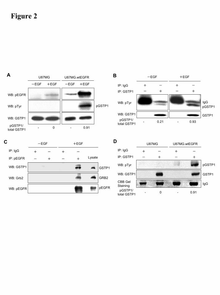
Figure 2
C
IP: pEGFR
IP: IgG
WB: GSTP1
WB: Grb2
WB: pEGFR
-EGF +EGF
-
+
+
-
-
+
+
- Lysate
pEGFR
GRB2
GSTP1
A
WB: pTyr
WB: GSTP1
WB: pEGFR
U87MG U87MG.wtEGFR
-EGF +EGF-EGF +EGF
- 0 - 0.91pGSTP1/total GSTP1
pGSTP1
B
WB: GSTP1
WB: pTyr
IP: GSTP1
IP: IgG
-EGF +EGF
+ - + -
+ - +-
pGSTP1/total GSTP1 - 0.21 - 0.93
IgGpGSTP1
GSTP1
D
WB: GSTP1
WB: pTyr
IP: GSTP1
IP: IgG + - + -
+ - +-
CBB GelStaining
U87MG U87MG.wtEGFR
pGSTP1/total GSTP1 - 0 - 0.91
IgG
GSTP1
pGSTP1
by guest on October 10, 2018
http://ww
w.jbc.org/
Dow
nloaded from
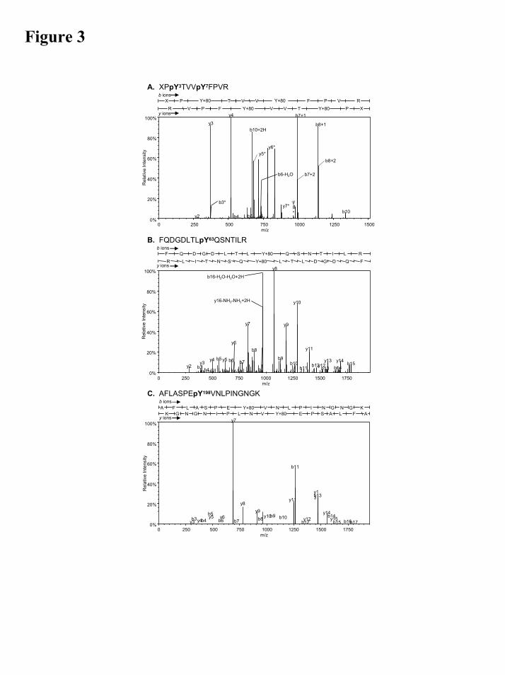
Figure 3
A. XPpY3TVVpY7FPVR
b10+2H
b6-H2O
b7+1
b8+1
y2
y3
y4
b4 b5b10
m/z
Rel
ativ
e In
tens
ity
0%
20%
40%
60%
80%
100%
0 250 500 750 1000 1250 1500
b7+2
b8+2
X P Y+80 T V V Y+80 F P V RR V P F Y+80 V V T Y+80 P X
b ions
y ions
y5*y6*
y7*y8*
b3*
B. FQDGDLTLpY63QSNTILR
b16-H2O-H2O+2H
y16-NH3-NH3+2H
y2 b3y3
b4
y4 b5 y5 b6
y6
b7
y7
b8
y8
b9
y9
b10
y10
b11
y11
b12y12b13
y13b14y14 b15
m/z
Rel
ativ
e In
tens
ity
0%
20%
40%
60%
80%
100%
0 250 500 750 1000 1250 1500 1750
F Q D G D L T L Y+80 Q S N T I L RR L I T N S Q Y+80 L T L D G D Q F
b ions
y ions
C. AFLASPEpY198VNLPINGNGK
y3b3y4b4y5b5
b6y6
y7
b7
y8y9
b8 y10b9 b10
y11
b11
b12y12
y13b13
y14b14y15
b15 b16b17
m/z
Rel
ativ
e In
tens
ity
0%
20%
40%
60%
80%
100%
0 250 500 750 1000 1250 1500 1750
A F L A S P E Y+80 V N L P I N G N G KK G N G N I P L N V Y+80 E P S A L F A
y ions
b ions
by guest on October 10, 2018
http://ww
w.jbc.org/
Dow
nloaded from
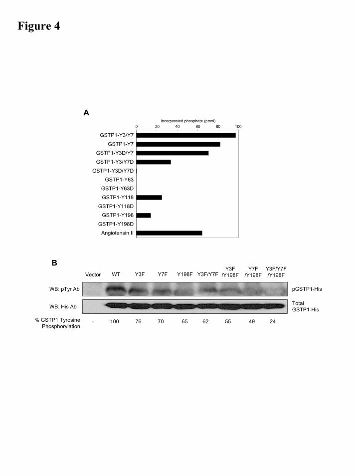
Figure 4
0 20 40 60 80 100
1
2
3
4
5
6
7
8
9
10
11
12
0 20 40 60 80 100Incorporated phosphate (pmol)
GSTP1-Y3/Y7
GSTP1-Y7
GSTP1-Y3D/Y7
GSTP1-Y3/Y7D
GSTP1-Y3D/Y7DGSTP1-Y63
GSTP1-Y63D
GSTP1-Y118
GSTP1-Y118D
GSTP1-Y198
GSTP1-Y198D
Angiotensin II
A
% GSTP1 TyrosinePhosphorylation
pGSTP1-His
Total GSTP1-HisWB: His Ab
WB: pTyr Ab
WT Y3F Y7F Y198FVector Y3F/Y7FY3F
/Y198FY7F
/Y198FY3F/Y7F/Y198F
- 100 76 70 65 62 55 49 24
B
by guest on October 10, 2018
http://ww
w.jbc.org/
Dow
nloaded from

Figure 5
A B
DC
by guest on October 10, 2018
http://ww
w.jbc.org/
Dow
nloaded from
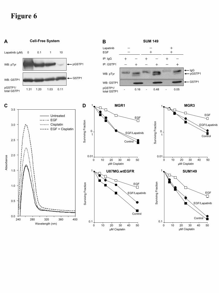
Figure 6
C
Wavelength (nm)240 280 320 360 400
Abs
orba
nce
0.0
0.5
1.0
1.5
2.0
2.5
3.0
3.5
Untreated
EGF + Cisplatin
EGFCisplatin
A
Lapatinib (µM)
Cell-Free System
1010.10
WB: pTyr
WB: GSTP1
pGSTP1
pGSTP1/total GSTP1 1.31 1.20 1.03 0.11
GSTP1
B
WB: GSTP1
WB: pTyr
IP: GSTP1
IP: IgG + - + -
+ - +-
pGSTP1/total GSTP1
- 0.16 - 0.48 - 0.05
+ -
- +
SUM 149
EGFLapatinib
-- -
+++
IgGpGSTP1
GSTP1
D MGR1
Sur
vivi
ng F
ract
ion
Control
EGF/Lapatinib
EGF
µM Cisplatin0 10 20 30 40 50
0.01
0.1
1
Sur
vivi
ng F
ract
ion
MGR3
Control
EGF/Lapatinib
EGF
0 10 20 30 40 500.01
0.1
1
µM Cisplatin
EGF
U87MG.wtEGFR
Sur
vivi
ng F
ract
ion
EGF/Lapatinib
µM Cisplatin
Control
0 10 20 30 40 500.1
1 SUM149
EGF
EGF/Lapatinib
Control
0 10 20 30 40 500.1
1
µM Cisplatin
Sur
vivi
ng F
ract
ion
by guest on October 10, 2018
http://ww
w.jbc.org/
Dow
nloaded from
Friedman, Darell D. Bigner and Francis Ali-OsmanTatsunori Okamura, Simendra Singh, John Buolamwini, Timothy A. Haystead, Henry S.
growth factor receptorTyrosine phosphorylation of the human glutathione S-transferase p1 by epidermal
published online March 2, 2009J. Biol. Chem.
10.1074/jbc.M808153200Access the most updated version of this article at doi:
Alerts:
When a correction for this article is posted•
When this article is cited•
to choose from all of JBC's e-mail alertsClick here
Supplemental material:
http://www.jbc.org/content/suppl/2009/03/04/M808153200.DC1
by guest on October 10, 2018
http://ww
w.jbc.org/
Dow
nloaded from