tumornecrosisfactor-a inhibits albumin gene expression...
TRANSCRIPT
Tumor Necrosis Factor-a Inhibits Albumin GeneExpression in a Murine Model of CachexiaD. A. Brenner, M. Buck, S. P. Feitelberg, and M. ChojkierDepartment of Medicine, University of California and Veterans Administration Medical Center, San Diego, California 92093
Abstract
The mechanisms responsible for decreased serum albuminlevels in patients with cachexia-associated infection, inflam-mation, and cancer are unknown. Since tumor necrosis factor-a(TNFa) is elevated in cachexia-associated diseases, andchronic administration of TNFa induces cachexia in animalmodels, we assessed the regulation of albumin gene expressionby TNFa in vivo. In this animal model of cachexia, Chinesehamster ovary cells transfected with the functional gene forhuman TNFa were inoculated into nude mice (TNFa mice).TNFa mice became cachectic and manifested decreased serumalbumin levels, albumin synthesis, and albumin mRNAlevels.However, even before the TNFa mice lost weight, their albu-min mRNAsteady-state levels were decreased 90%, and insitu hybridization revealed a low level of albumin gene expres-sion throughout the hepatic lobule. The mRNAlevels of sev-eral other genes were unchanged. Hepatic nuclei from TNFamice before the onset of weight loss were markedly less activein transcribing the albumin gene than hepatic nuclei from con-trol mice. Therefore, TNFa selectively inhibits the genetic ex-pression of albumin in this model before weight loss. (J. Clin.Invest. 1990. 85:248-255.) albumin * cachexia * liver tumornecrosis factor-a, transcription
IntroductionSerum albumin levels are decreased in patients with cachexia-associated infection, inflammation, and cancer, and the re-duction in serum albumin levels is used clinically as an indi-cator of the severity of the chronic disease state (1). Serumalbumin levels reflect a complex interaction between the syn-thesis, volume of distribution, degradation, and losses of thisprotein. The mechanisms responsible for the hypoalbumin-emia of cachexia are unknown. It has been shown that thehypoalbuminemia of chronic inflammatory diseases (2) and ofchronic renal failure (3) is at least in part the result of de-creased albumin synthesis. If albumin synthesis is decreased incachexia, then this could be either a nonspecific effect of mal-nutrition or the specific effect of a humoral factor inhibitingalbumin synthesis.
Tumor necrosis factor-a (TNFa),' a product of mono-cytes/macrophages, has been incriminated in the pathogenesis
Address reprint requests to Dr. Brenner, Division of Gastroenterology,Department of Medicine, M-023-D, University of California, SanDiego, La Jolla, CA 92093.
Received for publication 28 March 1989 and in revised form 5September 1989.
1. Abbreviations used in this paper: MOPS, 4-morpholinepropanesul-fonic acid; PIPES, 1,4-piperazinediethanesulfonic acid; TNFa, tumornecrosis factor-a.
The Journal of Clinical Investigation, Inc.Volume 85, January 1990, 248-255
of cachexia (4, 5). The administration of TNFa induces ca-chexia in animals (6, 7) as well as a negative nitrogen balancein patients (8). In addition, serum levels of TNFa are elevatedin some patients with cachexia-associated chronic diseases in-cluding cancer (9), parasitic infections (4), and the acquiredimmunodeficiency syndrome (10). TNFa is a potent modula-tor of protein synthesis, and many of the severe metabolicabnormalities found in cachexia have been attributed to theeffects of TNFa (4, 5). The question of whether TNFa inhibitsalbumin synthesis in vivo independent of malnutrition andweight loss remains to be determined. Therefore, we evaluatedthe regulation of albumin gene expression in a murine modelof cachexia induced by chronically elevated levels of serumTNFa. Wefound that TNFa inhibits albumin synthesis at thetranscriptional level even before the onset of weight loss.
Methods
Materials[a-32P] 2'-Deoxycytidine 5'-triphosphate and [ac-32P]uridine 5'-triphos-phate were from ICN ImmunolBiologicals (Irvine, CA); [35S] cytidine5'-triphosphate (1,320 Ci/mmol) was from New England Nuclear(Boston, MA); ribonucleotides, deoxyribonucleotides, Klenow frag-ment of DNApolymerase I, RNase-free bovine serum albumin, dex-tran sulfate, and ribonuclease A were from Pharmacia LKB Biotech-nology, Inc. (Piscataway, NJ); agarose, DNase I, RNase TI, and restric-tion endonucleases were from Bethesda Research Laboratories(Gaithersburg, MD); low melting point agarose and fast green werefrom Bio-Rad Laboratories (Richmond, CA); guanidine isothiocya-nate was from International Biotechnologies, Inc. (New Haven, CT);Biodyne transfer membrane was from Pall Corp. (Glen Cove, NY);nitrocellulose membrane was from Schleicher & Schuell (Keene, NH);4-morpholinepropanesulfonic acid (MOPS), guanidine HCl, antifoamemulsion, N-lauroyl sarcosine, lauroyl sulfate sodium salt, albuminreagent (BCG) and crystal violet were from Sigma Chemical Co. (St.Louis, MO); formamide was from EM Scientific (Gibbstown, NJ);formaldehyde solution was from Mallinckrodt Chemical Co. (Paris,KY); protease K, 1,4-piperazinediethanesulfonic acid (PIPES), andSephadex G-50 prespun columns were from Boehringer-Mannheim(Indianapolis, IN); agar, tryptone, yeast extract were from Difco Labo-ratories (Detroit, MI); embedding medium (OCT compound) wasfrom ICN ImmunoBiologicals; rabbit anti-mouse albumin antibodywas from Cappel Laboratories (Malvern, PA); biotinylated goat anti-rabbit IgG and the alkaline-phosphatase system were from VectorLaboratories (Burlingame, CA); the monoclonal TNFa immunoassaywas from Endogen (Boston, MA); recombinant human TNFa wasfrom AmGenBiologicals (Thousand Oaks, CA); and nude mice werefrom Simonsen (Gilroy, CA) and Harlan Sprague Dawley (Indianap-olis, IN).
Mouse model of cachexia. Chinese hamster ovary cells transfectedwith either the TNFa gene cloned into a mammalian expression vector(TNFa cells) or with the mammalian expression vector alone (CHOcells, control) were kindly provided by Dr. Oliff (Merk Sharp &DohmeResearch Laboratories, West Point, PA) (6). Cells were grownin Dulbecco's modified essential medium supplemented with 10% fetalcalf serum, penicillin (50 U/ml) and streptomycin (50 ,ug/ml). TheTNFa cells but not the CHOcells produced TNFa as measured by a
248 D. A. Brenner, M. Buck, S. P. Feitelberg, and M. Chojkier
cytolytic assay. 4-wk-old nude mice were injected intramuscularly witheither I07 CHOcells or I07 TNFa cells. Nude mice were housed in atemperature and humidity controlled facility. Animals had free accessto food and water and they were killed at 14-21 d (before the onset ofweight loss of the TNFa mice) and at 28-35 d postinoculation (afterthe onset of weight loss). TNFa cells at later passages markedly de-crease their production of TNFa (6). When animals were inoculatedwith these TNFa cells they developed cachexia at - 10 wk.
TNFa levels were measured by a biological cytolytic assay, using amodification of the method of Armstrong et al. (1 1). L929 cells (kindlyprovided by G. Granger, University of California, Irvine) were platedat 3 X 10⁴ cells per well in 0.1 ml of Eagle's minimal essential mediacontaining 5% fetal calf serum, 2 mMglutamine, and 0.5 ,ug/ml mito-mycin in 96-well tissue culture plates. These plates were incubated at370C in 95% air/5% CO2 for 24 h. Serial dilutions of TNFa samplesand standards in media were added to the wells. The plates were incu-bated for an additional 48 h, drained of media, fixed with methanol,stained with 0.2% crystal violet for 30 min, solubilized with 10% meth-anol, and the absorbance at 540 nm was measured using a BiotechEL310 plate reader. Results were expressed in units of activity (1 1u= 20 pg). In addition, serum TNFa were determined by an immu-noassay using monoclonal antibodies against TNFa, according to themanufacturer's protocol.
Serum albumin concentrations. Serum albumin concentrationswere determined by reacting serum or mouse albumin standards withbromocresol green, pH 4.2, at 250C for 1 min and measuring absor-bance at 628 nm (12).
Immunohistochemical staining. At 14-21 d and 28-35 d postinoc-ulation with CHOor TNFa cells, mice livers were fixed in situ byperfusion with 4% paraformaldehyde in PBS via the aorta. The liverswere sliced into 3-mm thick sections, postfixed for 2 h at 4VC in thesame paraformaldehyde solution. The livers were washed in cold PBSfor 10 min, cryoprotected in 30% sucrose PBS for 4 h and embeddedand frozen in OCTcompound at -70°C. 8-,um cryostat sections wereobtained and placed onto slides that had been pretreated with a solu-tion of 100 Mg/ml of poly-L-lysine in 10 mMTris, pH 8.0. Rabbitanti-mouse albumin was purified by affinity chromatography on aReactigel 6X (Pierce Chemical Co., Rockford, IL) column coupled tomouse albumin according to the manufacturer's procedure. The affin-ity purified rabbit anti-mouse albumin was used as the primary anti-body in conjunction with biotinylated goat anti-rabbit IgG and theABC-alkaline phosphatase system essentially as recommended by themanufacturer. Blocking was performed with gelatin instead of withserum. Nonspecific alkaline phosphatase reactions were blocked withlevamisole and nonspecific antibody reactions assessed by omitting thefirst antibody. Fast green was used to counterstain the tissue sections.
Northern blotting. Total RNA from individual mouse livers wasextracted in guanidine thiocyanate and pelletted by centrifugation ofthe extract through a cesium chloride cushion. Poly A+ RNAwaspurified by oligo dT cellulose chromatography. The plasmid pmalb2(for albumin), pKa I (for a tubulin), and pMLC3-1 (complement com-ponent C3), and pa2M (a2 macroglobulin) were kindly provided by S.Tilgham (13), N. Cowan (14), and G. Fey (15, 16), respectively. ThecDNA inserts were purified and radiolabeled with [a- 32P] dCTP usingthe random primer synthesis method (17). 2 ,ug of poly A+ RNAperlane were electrophoresed in a 1%agarose gel. The gels were transferredto nylon filters and Northern blots were performed as described pre-viously (18). The autoradiograms were quantitated by scanning with alaser densitometer interfaced with an integrator.
In situ hybridization. In situ hybridization was performed by amodification of published methods (19, 20). The albumin cDNAinsertof the plasmid palb2 was subcloned into the SP6, T7 -transcriptionvector pGEMblue 3 (Promega Biotec, Madison, WI). Antisense cRNA(complementary to the albumin messenger RNA) and sense RNAprobes were generated from the same plasmid using [32S]CTP withoutadditional cold CTP. The radiolabeling reaction was performed ac-cording to the manufacturer's protocol with 0.075 nMCTPin a 5-iureaction volume. The radiolabeled cRNAprobe was precipitated with
50 ,g of yeast tRNA and then resuspended in 80 Ml of 10 mMDTT.The probe was reduced in size by alkaline hydrolysis, ethanol precipi-tated, and then resuspended in 10 mMDTT 50% formamide.
The cryostat sections described above were postfixed in 4% para-formaldehyde in PBS for 5 min, washed in PBS, and dehydratedthrough graded ethanol. The sections were rehydrated and deprotein-ized with protease K 10 Mg/ml, 100 mMTris, pH 8.0, 50 mMEDTAfor 20 min. The slides were rinsed in water and then acetylated in0.25% (vol/vol) acetic anhydride, 0.1 Mtriethanolamine, pH 8.0 for 10min. The slides were then washed in 2X SSC for 4 min, dehydrated,and air dried. The hybridization solution consisted of 50%formamide,300 mMNaCl, 10 mMTris, pH 7.5, 1 mMEDTA, 100 mMDTT, 5XDenhart's solution, 1 mg/ml yeast tRNA, and 10% dextran sulfate.The [32S]CTP-labeled RNAprobe was added to the hybridization so-lution to give a final concentration of 5 X 10⁵ cpm/slide. The hybrid-ization solution was heated for 3 min at 650 and then 20 Ml was addedto each slide. A coverslip was applied and sealed with glue. Hybridiza-tion was carried out in a humidity chamber at 50°C for 18 h. Thecoverslips were removed and the slide washed four times in SSCandthen incubated in 20 ,ug/ml RNase and 10 U/ml RNase Ti in 0.5 MNaCl, 10 mMTris, pH 8.0, 1 mMEDTAat 37°C for I h. The slideswere then washed in descending concentrations of SSC containing ImMDTTwith the most stringent washing being at 55°C in 0.1 X SSC.The slides were dehydrated in graded ethanol containing 0.3 Mammo-nium acetate. After air drying, the slides were dipped in a Kodak NTB2emulsion containing 0.33 Mammonium acetate. The slides were ex-posed for 5 d at -20°C, and then developed. The sections were coun-terstained with hematoxylin and photographed using an IGS filtercube (Nikon).
Nuclear runoff transcription assay. Mouse liver nuclei were pre-pared by a modification of the procedure of Gorski et al. (21). Liversfrom two mice were combined and homogenized in 2.4 Msucrose 4:1(vol of sucrose solution/weight of liver) in a glass Dounce homogenizerwith a loose-fitting pestle. The homogenized liver was placed above acushion consisting of 2.0 M sucrose, 10% glycerol. The nuclei werepelletted by a 130,000 g centrifugation at 0°C for 1 h in an SW28rotor.They were resuspended in the transcription runoff assay buffer, washedonce, and then resuspended again in this buffer. Nuclei were frozen at-70°C. The nuclear runoff transcription assay that was developed byGroudine et al. (22) and adapted by Wangand Calame (23) was used aspreviously described (18). After filters containing the denatured cDNAprobes were hybridized with the radiolabeled RNAand washed repeat-edly, the radioactivity was determined by liquid scintillation spectros-copy for individual cDNAs. Separate filters containing denaturedplasmid pGEMblue 3 were used as negative controls. The transcrip-tional rate is expressed as the radioactivity incorporated into a specificmRNAthat is detected by its hybridization to the denatured cDNA.
Results
2 wk after their inoculation with TNFa cells, mice had serumTNFa concentrations as measured by a biological assay com-parable to those reported previously (6). Serum TNFa levelsremained elevated in TNFa mice at 4-5 wk after inoculation(Table I). The presence of circulating human TNFa was con-firmed with a specific immunoassay utilizing monoclonalanti-human TNFa antibodies (Table I). TNFa was never de-tected in CHOmice (Table I). The TNFa and CHOmicemaintained identical weights for about the first 2-3 wk afterinoculation. The TNFa mice then began to progressively loseweight, whereas the CHOmice did not. As previously reported(6), TNFa mice at 4-5 wk after inoculation manifested severeweight loss (Fig. 1). The cachectic TNFa mice had significantlydecreased serum albumin levels compared to the CHOcontrolmice (Table II).
Tumor Necrosis Factor-a Inhibits Albumin Gene Expression In Vivo 249

Table L Serum TNFoa Levels in TNFa Mice
Serum TNFa (ng/ml)
Experimental conditions* L929 assayt ELISAI
TNFa mice (before onset of weight loss) 0.08±0.003 0.3±0.1TNFa mice (after onset of weight loss) 48±0.5 1.5±0.4CHOmice ND ND
* Nude mice were inoculated with either TNFa-cells (TNFa-mice) orCHOcells (CHO-mice) as described in Methods.$4 Serum TNFa levels were determined by a biological assay (L929)and by an immunoassay (ELISA) as described in Methods. Valuesare mean±SEMof at least triplicate samples.ND, none detected.
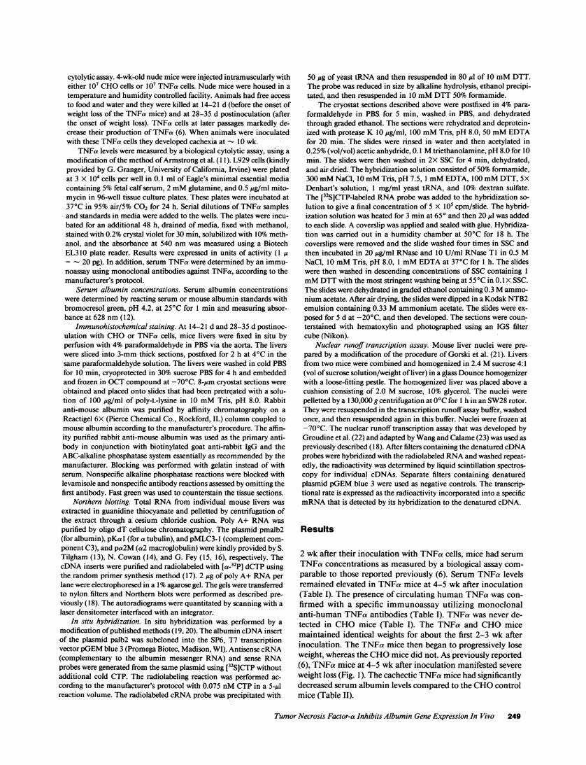
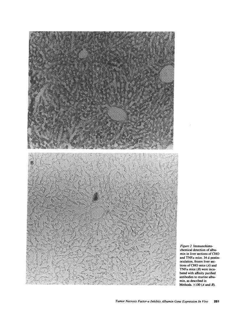
Livers from cachectic TNFa and CHOmice were perfusedin situ and used for immunohistochemical studies. In theCHOmice, albumin was detected in high abundance diffuselythrough the hepatic lobule. On the other hand, in the cachecticTNFa mice, the abundance of albumin was greatly depressed(Fig. 2). As controls, sections were incubated in the absence offirst antibody, which did not demonstrate any specific stain-ing, or with antimurine Ig as the first antibody, which did notdemonstrate any hepatocyte specific staining (data not shown).Northern blot analysis revealed a marked decrease in thesteady state levels of albumin mRNAbut not of f3-actinmRNAin the cachectic TNFa mice compared with the CHOmice (Fig. 3 A). The decrease in hepatic albumin protein andmRNAlevels in this model of cachexia is comparable to othermodels of weight loss reported previously by us (24) and others(25, 26).
If TNFa modulates albumin gene expression indepen-dently of its effects of malnutrition and weight loss, we wouldexpect TNFa to decrease albumin production before the onsetof weight loss. Before the onset of weight loss, TNFa mice hadsignificantly decreased serum albumin levels compared toage-matched CHOcontrol mice (Table II). In addition, im-munohistochemically detected albumin was decreased in thehepatocytes of TNFa mice before weight loss (data notshown).
In order to investigate the mechanism by which TNFainhibits albumin production, expression of the albumin genewas assessed by measuring steady state mRNAlevels, cellularlocalization, and transcriptional activity. Hepatic albuminmRNAcontent was measured in 10 precachetic TNFa miceand 10 age-matched CHOmice, and a representative Northern
/--lk>~~~~~~410 20
Days Post Inoculation
Figure 1. Change inweight of CHOandTNFa mice postinocu-
o CHO lation of CHOor TNFacells. CHOor TNFacells (10') were inocu-lated into age-matchedmale nude mice. Each
ThFCa point represents themean weight
30 ^0 change±SEM of at leastfour mice.
Table I. Serum Albumin Levels in CHOand TNFa Mice
Experimental conditions* Serum albumint
g/dl
Experiment I: Precachexic TNFa miceCHOmice, 2 wk (n = 5) 1.9±0.1TNFamice, 2 wk (n = 6) 1.7±0.1
Experiment II: Cachexic TNFa miceCHOmice, 4 wk (n = 4) 2.7±0.2TNFa mice, 4 wk (n = 5) 2.1±0.1TNFa mice, 10 wk (n = 4) 1.7±0.1
* Nude mice were inoculated with either CHOcells (CHO-mice) orTNFa cells (TNFa-mice) as described in Methods. The number ofweeks designates the time postinoculation of cells.
Determined as described in Methods. P < 0.05 for TNFa mice, 2wk, P < 0.01 for TNFa-mice, 4 wk and P < 0.001 for TNFa-mice,10 wk.
blot is shown in Fig. 3 B. The albumin mRNAlevels of theCHOmice were 100 ± 10.3 (mean ± SEMin relative densi-tometry units) and of the TNFa mice were 13.3 ± 3.8 (P< 0.005). Additional analysis of hepatic mRNArevealed nochanges in fl-actin, complement C3, or a-tubulin levels in theTNFa mice (Fig. 3 B).
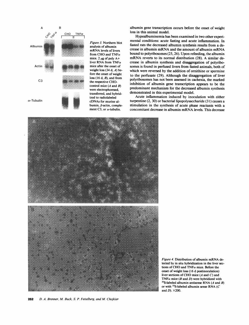
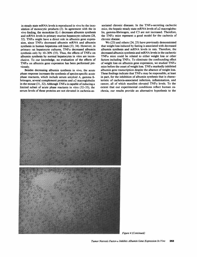
To assess the expression of the albumin gene throughoutthe hepatic lobules, we performed in situ hybridization using a35S-albumin antisense cRNAprobe. In the CHOmice, abun-dant albumin mRNAwas detected in the liver by in situ hy-bridization (Fig. 4 A). As previously reported (27), highestlevels of albumin mRNAwere found in the periportal zone(acinar zone 1) with diminished levels peripherally (acinarzone 3). In contrast, the TNFa mice before weight loss had amarked decrease in albumin mRNAlevels throughout the he-patic lobule, with loss of the gradient between acinar zones 1and 3 (Fig. 4 B). Minimal background in situ hybridizationwas observed when a 35S-labeled sense albumin RNAprobewas used as a control, therefore, validating the specificity of theantisense probe (Fig. 4, Cand D).
Decreased albumin mRNAlevels may reflect either de-creased albumin gene transcription or increased albuminmRNAdegradation. Transcription runoff assays were per-formed in order to measure albumin gene expression in he-patic nuclei obtained from TNFa and CHOmice 16 d postin-oculation. Albumin gene transcription was decreased by 90%in TNFa murine livers in a representative runoff assay (Fig. 5).This inhibition of albumin gene transcription apparently ac-counts for the decreased albumin mRNAin the TNFa murinelivers. The transcriptional rates of the f3-actin gene were un-changed (Fig. 5) suggesting a selective effect of TNFa on albu-min gene expression.
Discussion
In this study we use a novel model of cachexia to maintainchronically elevated serum levels of TNFa in mice. The resultsdemonstrate that elevated serum levels of TNFa lead to de-creased albumin gene transcription (Fig. 5), decreased steadystate levels of albumin mRNAthroughout the hepatic lobule(Figs. 3 and 4), decreased albumin production (Fig. 2), anddecreased serum albumin levels (Table II). The inhibition of
250 D. A. Brenner, M. Buck, S. P. Feitelberg, and M. Chojkier
30
25
20
15
10
5
0
! , -. .; ' '0 N , t ,
A C < 4X<.MS' n~f#"~'SA A
7&~~~~~~Yr #4 _> ,~rtzZ-- r.,t ; /-
r rv
tA~~~~ ~ ~ ~ ~ ~ ~ ~ ~~~~~~'A
............../,% '\ A'~~~~~~~~~~~~~~~~~~~~~~~~~~~~~~~~~~~~~~~~~~.-e~~~~~~~~~~~~~~~~r~~4
Figure 2. Immunohisto-chemical detection of albu-min in liver sections of CHOand TNFa mice. 34 d postin-oculation, frozen liver sec-tions of CHOmice (A) andTNFa mice (B) were incu-bated with affinity purifiedantibodies to murine albu-min, as described inMethods. X100 (A and B).
Tumor Necrosis Factor-a Inhibits Albumin Gene Expression In Vivo 251
A B
K0 !< d CHO TNFa0< 4v I 11 I
Albumin
Actin 4i@ t
C3
ct-Tubulin
Figure 3. Northern blotanalysis of albuminmRNAlevels of liversfrom CHOand TNFamice. 2 ;g of poly A+liver RNAfrom TNFamice after the onset ofweight loss (34 d, A) be-fore the onset of weightloss (16 d, B), and fromthe respective CHO-control mice (A and B)were electrophoresed,transferred, and hybrid-ized to radiolabeledcDNAs for murine al-bumin, fl-actin, comple-ment C3, or a-tubulin.
albumin gene transcription occurs before the onset of weightloss in this animal model.
Hypoalbuminemia has been examined in two other experi-mental conditions: acute fasting and acute inflammation. Infasted rats the decreased albumin synthesis results from a de-crease in albumin mRNAand the amount of albumin mRNAbound to polyribosomes (25, 26). Upon refeeding, the albuminmRNAreverts to its normal distribution (28). A similar de-crease in albumin synthesis and disaggregation of polyribo-somes is found in perfused livers from fasted animals, both ofwhich were reversed by the addition of ornithine or spermineto the perfusate (29). Although the disaggregation of liverpolyribosomes has not been assessed in cachexia, the markedinhibition of albumin gene transcription appears to be thepredominant mechanism for the decreased albumin synthesisdemonstrated in this experimental model.
Acute inflammation induced by inoculation with eitherturpentine (2, 30) or bacterial lipopolysaccharide (31) causes astimulation in the synthesis of acute phase reactants with aconcomitant decrease in albumin mRNAlevels. This decrease
Figure 4. Distribution of albumin mRNAde-tected by in situ hybridization in the liver sec-tions of CHOand TNFa mice. Before theonset of weight loss ( 16 d postinoculation)liver sections of CHOmice (A and C) andTNFa mice (B and D) were hybridized with"S-labeled albumin antisense RNA(A and B)or with IIS-labeled albumin sense RNA(Cand D). X200.
252 D. A. Brenner, M. Buck, S. P. Feitelberg, and M. Chojkier
in steady state mRNAlevels is reproduced in vivo by the inoc-ulation of monocytic products (2). In agreement with the invivo finding, the monokine IL-I decreases albumin synthesisand mRNAlevels in primary murine hepatocyte cultures (28,32). TNFa might have a direct role in albumin gene expres-sion, since TNFa decreased albumin mRNAand albuminsynthesis in human hepatoma cell lines (33, 34). However, inprimary rat hepatocyte cultures, TNFa decreased albuminsynthesis only by 10-30% (35). Thus, the effects of TNFa onalbumin synthesis by normal hepatocytes in vitro are incon-clusive. To our knowledge, no evaluation of the effects ofTNFa on albumin gene expression has been performed pre-viously.
Besides decreasing albumin synthesis in vivo, the acutephase response increases the synthesis of species-specific acutephase reactants, which include serum amyloid A, gamma fi-brinogen, several complement proteins and a2 macroglobulinin the mouse (31, 32). Although TNFa is capable of inducing alimited subset of acute phase reactants in vitro (32-35), theserum levels of these proteins are not elevated in cachexia-as-
sociated chronic diseases. In the TNFa-secreting cachecticmice, the hepatic steady state mRNAlevels of a2 macroglobu-lin, gamma-fibrinogen, and C3 are not increased. Therefore,the TNFa mice represent a good model for the cachexia ofchronic disease.
We(23) and others (24, 25) have previously demonstratedthat weight loss induced by fasting is associated with decreasedalbumin synthesis and mRNAlevels in rats. Therefore, thedecreased albumin synthesis and mRNAlevels in the cachecticTNFa mice could be related to either weight loss or otherfactors including TNFa. To eliminate the confounding effectof weight loss on albumin gene expression, we studied TNFamice before the onset of weight loss. TNFa markedly inhibitedalbumin gene transcription despite the absence of weight loss.These findings indicate that TNFa may be responsible, at leastin part, for the inhibition of albumin synthesis that is charac-teristic of cachexia-associated infection, inflammation, andcancer, all of which manifest elevated TNFa levels. To theextent that our experimental conditions reflect human ca-chexia, our results provide an alternative hypothesis to the
Figure 4 (Continued)
Tumor Necrosis Factor-a Inhibits Albumin Gene Expression In Vivo 253
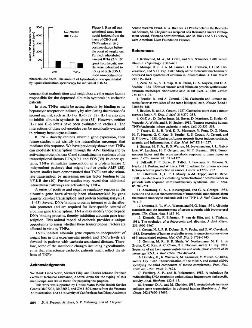
E 5000 Figure 5. Run-off tran-01
4--% r-I Albumin scriptional assay froma. 0. 4000_ actin nuclei isolated from the
dlivers of CHOandu 3000 TNFa mice at 16 d
postinoculation before00 2000 the onset of weight loss.
2' " Purified radiolabeled, 1000 nascent RNA(2 X 10⁷
* m. _ cpm) from hepatic nu-_ ] _ IIclei were hybridized to
CHO TNF a 0.5 ug of each cDNAinsert immobilized on
nitrocellulose filters. The amount of hybridization was quantitatedby liquid scintillation spectoscopy for individual cDNAs.
concept that malnutrition and weight loss are the major factorsresponsible for the depressed albumin synthesis in cachecticpatients.
In vivo, TNFa might be acting directly by binding to itshepatocyte receptor or indirectly by stimulating the release of asecond agonist, such as IL- 1 or IL-6 (37, 38). IL-1 is also ableto inhibit albumin synthesis in vitro (35). However, neitherIL- 1 nor IL-6 levels have been evaluated in cachexia. Theinteractions of these polypeptides can be specifically evaluatedin primary hepatocyte cultures.
If TNFa directly inhibits albumin gene expression, thenfuture studies must identify the intracellular messenger thatmediates this response. Wehave previously shown that TNFacan modulate transcription through the AP-I binding site byactivating protein kinase Cand stimulating the synthesis of thetranscriptional factors JUN/AP- 1 and FOS (39). In other sys-tems, TNFa stimulates transcription in a protein kinase Cindependent pathway that might involve cyclic AMP(38).Recent studies have demonstrated that TNFa can also stimu-late transcription by increasing nuclear factor binding to theNF-KB site (40). Further studies may reveal that additionalintracellular pathways are activated by TNFa.
A series of positive and negative regulatory regions in thealbumin gene have already been characterized by genetransfer, cell-free transcription, and protein binding assays (21,41-43). Several DNA-binding proteins interact with the albu-min promoter and are required for liver-specific control ofalbumin gene transcription. TNFa may modify these hepaticDNA-binding proteins, thereby inhibiting albumin gene tran-scription. This animal model of cachexia provides a uniqueopportunity to assess whether these transcriptional factors areaffected in vivo by TNFa.
TNFa inhibits albumin gene expression independent ofweight loss in this experimental model, and TNFa levels areelevated in patients with cachexia-associated diseases. There-fore, some of the metabolic changes including hypoalbumin-emia that characterize cachectic patients might reflect the ef-fects of TNFa.
Acknowlednments
Wethank Linda Veloz, Michael Filip, and Charles Johnson for theirexcellent technical assistance, Andrea Arata for the typing of thismanuscript, and Braun Brelin for preparing the graphics.
This work was supported by United States Public Health ServiceGrants DK07202, DK38652, and GM41804, grants from the VeteransAdministration, and a University of California at San Diego Academic
Senate research award. D. A. Brenner is a PewScholar in the Biomedi-cal Sciences, M. Chojkier is a recipient of a Research Career Develop-ment Award, Veterans Administration, and M. Buck and S. Feitelbergwere American Liver Foundation Fellows.
References
1. Rothschild, M. A., M. Oratz, and S. S. Schreiber. 1988. Serumalbumin. Hepatology. 8:385-401.
2. Mosage, H. J., J. A. M. Janssen, J. H. Franssen, J. C. M. Haf-kenscheid, and S. H. Yap. 1987. Study of the molecular mechanism ofdecreased liver synthesis of albumin in inflammation. J. Clin. Invest.79:1635-1641.
3. Zern, M. A., S. H. Yap, R. K. Strair, G. A. Kaysen, and D. A.Shafritz. 1984. Effects of chronic renal failure on protein synthesis andalbumin messenger ribonucleic acid in rat liver. J. Clin. Invest.73:1167-1174.
4. Beutler, B., and A. Cerami. 1986. Cachectin and tumour ne-crosis factor as two sides of the same biological coin. Nature (Lond.).320:584-588.
5. Beutler, B., and A. Cerami. 1987. Cachectin: more than a tumornecrosis factor. N. Engl. J. Med. 316:379-385.
6. Oliff, A., D. Defeo-Jones, M. Boyer, D. Martinez, D. Kiefer, G.Vuocolo, A. Wolfe, and S. H. Socher. 1987. Tumors secreting humanTNFa/cachectin induce cachexia in mice. Cell. 50:555-563.
7. Tracey, K. J., H. Wei, K. R. Manogue, Y. Fong, D. G. Hesse,H. T. Nguyen, G. C. Kuo, B. Beutler, R. S. Cotran, A. Cerami, andS. F. Lowry. 1988. Cachectin/tumor necrosis factor induces cachexia,anemia, and inflammation. J. Exp. Med. 167:1211-1227.
8. Starnes, H. F. Jr., R. S. Warren, M. Jeevanandam, J. L. Gabri-love, W. Larchian, H. F. Oettgen, and M. F. Brennan. 1988. Tumornecrosis factor and the acute metabolic response to tissue injury inman. J. Clin. Invest. 82:1321-1325.
9. Balkwill, F., F. Burke, D. Talbot, J. Tavernier, R. Osborne, S.Naylor, H. Durbin, and W. Fiers. 1987. Evidence for tumour necrosisfactor/cachectin production in cancer. Lancet. ii: 1229-1232.
10. Lahdevirta, J., C. P. J. Maury, A.-M. Teppo, and H. Repo.1988. Elevated levels of circulating cachectin/tumor necrosis factor inpatients with acquired immunodeficiency syndrome. Am. J. Med.85:289-291.
11. Armstrong, C. A., J. Klostergaard, and G. A. Granger. 1985.Isolation and initial characterization of tumoricidal monokine(s) fromthe human monocytic leukemia cell line THP- 1. J. NatL Cancer Inst.74:1-9.
12. Doumas, B. T., W. A. Watson, and H. G. Biggs. 1971. Albuminstandards and the measurement of serum albumin with bromocresolgreen. Clin. Chim. Acta. 31:87-96.
13. Kioussis, D., F. Eiferman, P. van de Rijn, and S. Tilgham.1981. The evolution of a fetoprotein and albumin. J. Bio. Chem.256:1960-1967.
14. Cowan, N. J., P. R. Dobner, E. V. Fuchs, and D. W. Cleveland.1983. Expression of human a-tubulin genes: interspecies conservationof 3' untranslated regions. Mol. Cell. Biol. 3:1738-1745.
15. Gehring, M. R., B. R. Shiels, W. Northemann, M. H. L. deBruijn, C.-C. Kan, A. C. Chain, D. J. Noonan, and G. H. Fey. 1987.Sequence of rat liver a2-macroglobulin and acute phase control of itsmessenger RNA. J. Biol. Chem. 262:446-454.
16. Domdey, H., K. Wiebauer, M. Kazmaier, V. Muller, K. Odink,and G. Fey. 1982. Characterization of the mRNAand cloned cDNAspecifying the third component of mouse complement. Proc. Natl.Acad. Sci. USA. 79:7619-7623.
17. Feinberg, A. P., and B. Volgenstein. 1983. A technique forradiolabelling DNArestriction endonuclease fragments to high specificactivity. Anal. Biochem. 132:6-13.
18. Brenner, D. A., and M. Chojkier. 1987. Acetaldehyde increasescollagen gene transcription in cultured human fibroblasts. J. Biol.Chem. 262: 17690-17695.
254 D. A. Brenner, M. Buck, S. P. Feitelberg, and M. Chojkier
19. Angerer, L. M., M. H. Stoler, and R. C. Angerer. 1987. In situhybridization with RNAprobes: an annotated recipe. In In situ hybrid-ization: Applications to neurobiology. K. L. Valentino, J. H. Ebenne,and J. D. Barchas, editors. Oxford University Press, New York.3:42-70.
20. Ingham, P. W., K. R. Howard, and D. Ish-Horowicz. 1985.Transcription pattern of the Drosophila segmentation gene hairy. Na-ture (Lond.). 318:439-445.
21. Gorski, K., M. Carneiro, and U. Schibler. 1986. Tissue-specificin vitro transcription from the mouse albumin promoter. Cell.47:767-776.
22. Groudine, M., M. Peretz, and H. Weintraub. 1981. Transcrip-tional regulation of hemoglobin switching in chicken embryos. Mol.Cell. Bio. 3:281-288.
23. Wang, X.-F., and K. Calame. 1985. The endogenous immuno-globulin heavy chain enhancer can activate tandem VH promotersseparated by a large distance. Cell. 43:659-665.
24. Chojkier, M., M. Flaherty, B. Peterkofsky, G. H. Majmudar,R. G. Spanheimer, and D. A. Brenner. 1988. Different mechanismsdecrease hepatic collagen and albumin production in fasted rats. Hepa-tology. 8:1040-1045.
25. Yap, S. H., R. K. Strair, and D. A. Shafritz. 1978. Effect of ashort term fast on the distribution of cytoplasmic albumin messengerribonucleic acid in rat liver. J. Biol. Chem. 253:4944-4950.
26. Shafritz, D. A., S. H. Yap, and R. K. Strair. 1979. Regulation ofalbumin synthesis by rat liver. Mo. Bio. Rep. 5:71-78.
27. Evarts, R. P., P. Nagy, E. Marsden, and S. S. Thorgeirsson.1987. In situ hybrization studies on expression of albumin and a-feto-protein during the early stages of neoplastic transformation in rat liver.Cancer Res. 47:5469-5475.
28. Yap, S. H., R. K. Strair, and S. A. Shafritz. 1978. Identificationof albumin mRNPsin the liver. Biochem. Biophys. Res. Commun.83:427-433.
29. Oratz, M., M. A. Rothschild, S. S. Schreiber, A. Burks, J.Mongelli, and B. Matarese. 1983. The role of urea cycle and poly-amines in albumin synthesis. Hepatology. 3:567-571.
30. Princen, J. M. G., W. Niewenhuizen, G. P. B. M. Mol-Backx,and S. H. Yap. 1981. Direct evidence of transcriptional control offibrinogen and albumin synthesis in rat liver during the acute phaseresponse. Biochem. Biophys. Res. Commun. 102:717-723.
31. Ciliberto, G., R. Arcone, E. F. Wagner, and U. Ruther. 1987.Inducible and tissue-specific expression of human C-reactive proteinin transgenic mice. EMBO(Eur. Mol. Biol. Organ.) J. 6:4017-4022.
32. Ramadori, G., J. D. Sipe, C. A. Dinarello, S. B. Mizel, andH. R. Colten. 1985. Pretranslational modulation of acute phase he-
patic protein synthesis by murine recombinant interleukin 1 (IL- 1) andpurified human IL-1. J. Exp. Med. 162:930-942.
33. Darlington, G. J., D. R. Wilson, and L. B. Lachman. 1986.Monocyte-conditioned medium, interleukin-1, and tumor necrosisfactor stimulate the acute phase response in human hepatoma cells invitro. J. Cell Biol. 103:787-793.
34. Perlmutter, D. H., C. A. Dinarello, P. I. Punsal, and H. R.Colten. 1986. Cachectin/tumor necrosis factor regulates hepaticacute-phase gene expression. J. Clin. Invest. 78:1349-1354.
35. Koj, A., A. Kurdowska, D. Magielska-Zero, H. Rokita, J. D.Sipe, J. M. Dayer, S. Demczuk, and J. Gauldie. 1987. Limited effectsof recombinant human and murine interleukin 1 and tumour necrosisfactor on production of acute phase proteins by cultured rat hepato-cytes. Biochem. Int. 14:553-560.
36. Andus, T., P. C. Heinrich, J. Bauer, T.-A. Tran-Thi, K. Decker,D. Mannel, and H. Northoff. 1987. Discrimination of hepatocyte-stimulating activity from human recombinant tumor necrosis factor a.Eur. J. Immunol. 17:1193-1197.
37. Kohase, M., L. T. May, I. Tamm, J. Vilcek, and P. B. Sehgal.1987. A cytokine network in human diploid fibroblasts: interactions offl-interferons, tumor necrosis factor, platelet-derived growth factor,interleukin-1. Mol. Cell. Bid. 7:273-280.
38. Zhang, Y., J.-X. Lin, and J. Vilcek. 1988. Synthesis of inter-leukin 6 (interferon-fl2/B cell stimulatory factor 2) in human fibro-blasts is triggered by an increase in intracellular cyclic AMP. J. Biol.Chem. 263:6177-6182.
39. Brenner, D. A., M. O'Hara, P. Angel, M. Chojkier, and M.Karin. 1989. Prolonged activation of jun and collagenase genes bytumour necrosis factor-a. Nature (Lond.). 337:661-663.
40. Duh, E. J., W. J. Maury, T. M. Folks, A. S. Fauci, and A. B.Rabson. 1989. Tumor necrosis factor a activates human immunodefi-ciency virus type I through induction of nuclear factor binding to theNF-KB sites in the long terminal repeat. Proc. Natl. Acad. Sci. USA.86:5974-5978.
41. Babiss, L. E., R. S. Herbst, A. L. Bennett, and J. E. Darnell, Jr.1987. Factors that interact with the rat albumin promoter are presentboth in hepatocytes and other cell types. Genes & Dev. 1:256-267.
42. Pinkert, C. A., D. M. Ornitz, R. L. Brinster, and R. D. Palmiter.1987. An albumin enhancer located 10 kb upstream functions alongwith its promoter to direct efficient, liver-specific expression in trans-genic mice. Genes & Dev. 1:268-276.
43. Lichtsteiner, S., J. Warin, and U. Schibler. 1987. The interplayof DNA-binding proteins on the promoter of the mouse albumin gene.Cell. 51:963-973.
Tumor Necrosis Factor-a Inhibits Albumin Gene Expression In Vivo 255