tumeurs bénignes du foie -...
TRANSCRIPT

Tumeurs bénignes du foie
• Angiomes• Hyperplasies nodulaires focales• Adénomes• Tumeurs rares• Pseudo-tumeurs (stéatoses focales,
perturbations hémodynamiques)


Application à la (ponction-) biopsie
• HNF– critères
• disparition de l’architecture hépatique normale• présence de cloisons fibreuses• vaisseaux dystrophiques• néoductules et inflammation• remaniements sinusoïdaux
– proposition d’un score histologique

Application à la (ponction-) biopsie
• HNF

• Adénome– disparition de l’architecture hépatique
normale– pas de cloison fibreuse– travées épaisses formées de «grands»
hépatocytes éosinophiles ou stéatosiques– nombreuses artérioles
Application à la (ponction-) biopsie

HNF: données nouvelles
• Physiopathologie– hypothèse vasculaire: données cliniques
• forme primitive: lésion malformative due à un hyperdébit artériel localisé
• tumeurs HNF-like: secondaires à des pathologies vasculaires
– hypothèse vasculaire: données moléculaires• anomalies d’expression des facteurs angiogéniques
(angiopoïétines 1 et 2)
– rapports avec les oestroprogestatifs

•4% of all new cancer cases in the world
•Third most common cause of cancer-related deathamong men and the sixth among women
•85-90% = Hepatocellular carcinoma
Primary liver cancers
Nordenstedt Dig Liver Dis 2010

Inheritedmetabolic diseases
Etiological Factors of Hepatocellular Carcinoma
HBV350 million
carriers
chronic hepatitis
HCV170 million
carriers
cirrhosis (90% of cases)
hepatocellular carcinoma
Aflatoxin B1Ethanol consumption
• ß-tyrosinemia• hemochromatosisa1-antitrysin deficiency
• glycogen storage disease• Wilson’s disease•PFIC2
othercarcinogens
Male genderDiabetesObesity
Metabolic syndrome

Hepatocellular carcinoma
IN WESTERN COUNTRIES
90% of HCC on cirrhotic livers
Chronic liver disease 10% of HCC on non cirrhotic livers
Cirrhosis = pre-cancerous state with an annual incidence of 2- 5% for HCC
Normal liver

Hepatocellular carcinoma: Gross aspect
Expansive uninodular Expansive multinodular
Mixed DiffuseSatellite nodules

Diffuse cirrhosis-like HCC
Jakate Am J Surg Pathol 2010
Biopsy +

Hepatocellular carcinomaClassical type

Histological types of Hepatocellular Carcinoma (WHO)
Architecture•Trabecular•Pseudoglandular•Compact•Squirrhous
Cytology (variants)•Pleomorphic•Clear cells•Oncocytic•Sarcomatoid
Other aspects•Bile•Glycogen•Steatosis•Nuclear inclusions •Hyaline cytoplasmic globules•Mallory’s bodies•Pale bodies

Hepatocellular Carcinoma: Architecture

Hepatocellular Carcinoma: Cytological aspects

Steatohepatitic hepatocellular carcinoma
Salomao Am J Surg Pathol 2010
22 steatohepatitic HCC (35.5%)/62 HCC on explant livers with HCV cirrhosis
SteatosisMallory bodies Inflammatory infiltratesPericellular or trabecular fibrosis
Risk factors for NALFD/NASH: 63.6%Steatosis/Steatohepatitis: 63.6% in the non neoplastic liver

HCC ON CIRRHOTIC LIVERSMOST OFTEN AN EASY DIAGNOSIS
BY IMAGING TECHNIQUES
No need for an histological proof

Diagnosis of HCC on cirrhotic livers:MOST OFTEN AN EASY DIAGNOSIS
Nodule on cirrhotic liver > 2cmDiagnosis of HCC by non invasive imaging and laboratorycriteria� Hypervascular arterial supply/portal wash-out� High serum AFP� Pathological confirmation is not mandatory. It could be
necessary if the imaging data are not typical
Nodule 1-2 cm
Nodule < 1cm
� One or two imaging techniques � If inconclusive : Biopsy
� Repeat US

Hemorrhagic risk
Tumor cell seeding on biopsy track (2.7%) –biological glue
Usefulness of an associated biopsy in non tumor liver +++
Higher opportunity of immunostaining
Ultra-sound guided core biopsy – 16-18G

Small Nodules resulting from screening on cirrhotic livers
« Macronodules » : detected by US screening
Macroregenerative nodules
Low grade dysplastic nodules
High grade dysplastic nodules
Small HCC ≤ 2 cm�Early HCC�Progressed HCC
International working party Hepatology 1995;22:983-993International Consensus Group for Hepatocellular Neoplasia Hepatology 2009

Macronodule

Hepatocellular carcinoma: Multistep carcinogenesis

MACROGENERATIVE NODULESHyperplastic - Polyclonal - Benign
DYSPLASTIC NODULESLow grade High grade Well differentiated HCC
Thickness of liver cell platesExpansive growingPseudo-glands/ bileNuclear densitySmall cell dysplasiaNuclear atypiaIron load resistanceReticulin lossUnpaired artery density (actin) Sinusoid capillarization CD34
Macronodules on cirrhotic liver
Stromal invasion

Risk of transformation of dysplastic nodule into HCC
� LGDN: 20%
� HGDN: 20 – 80%
Dysplastic nodules and Hepatocellular carcinoma
Nault Hepatology 2014

Macroregenerative nodules

Dysplastic nodule

Normal

CD34

Low grade High grade Well differentiated
HCCPlate thickness 1 to 2 cells 2 to 3 cells > 3 cellsPseudoglands 0 0 ou + 0 ou +Nodule in the nodule 0 ou + + ou 0 + ou 0Portal tractsStromal invasion
+0
+ ou 00
0+
Reticulin + +/- -Unpaired arteries quelques + ++Atypia mild +, focal +Mitosis (1à 5/ 10 HPF) - rares variableDysplasia Large cell Large or small cell -Clonality Mono or poly Mono or poly mono
Dysplastic Nodules

� Reticulin stainingDecreased framework in HCC
� ImmunostainingSMA (smooth muscle actin): unpaired arteriesCD34: sinusoid capillarization
Glypican3: Fetal oncoproteinHSP70: Heat shock proteinGlutamine synthetase: Glutamine synthesis
International Group recommendations:2 positive markers/3
Specificity 100% Sensitivity 60% for nodules of 0,5 to 2cm
Additional diagnostic tools
International Consensus Group for Hepatocellular Neoplasia Hepatology 2009

MRN HCC
G3
HSP
GS
Tremosini Gut 2012Conventional histology

Nodule > 2cm ÎImaging diagnosis (typical features on one imaging technique)If atypical features ÎGuided biopsy
Nodule < 1cm Î Repetition of imaging
Nodule 1cm to 2cm Î Imaging diagnosis (typical features on 2* imaging techniques)If not ÎGuided biopsy
MRN / Dysplastic nodule / HCC ?
Limits +++In the nodule?In the nodule within the nodule?Low sensitivity for the diagnosis of early HCC?
EASL–EORTC Clinical Practice Guidelines: Management of hepatocellular carcinoma 2012
Small Nodules resulting from screening on cirrhotic livers

DIAGNOSIS OF HCC ON NON CIRRHOTIC LIVER
Pathological diagnosis of HCC is recommended for all nodules occurring in non-cirrhotic livers.
EASL–EORTC 2012

Hepatocellular carcinoma on non cirrhotic liver
• 10 to 15% of HCC in Western countries• Large tumors

Paradis Hepatology 2009
Metabolic syndrome and HCC
� Non cirrhotic liver 65%
� Larger tumors� Well differentiated HCC

Differential diagnosis of hepatocellular tumor ( adenoma/ well differentiated HCC)
Fibrous variant of HCC: cholangiocarcinoma, metastasis?
Other rare differential diagnoses: metastasis of Neuroendocrine carcinoma, epithelioid angiomyolipoma
Hepatocellular carcinoma on non cirrhotic liver

Cellular densityThickness of liver platesPseudoglandular structuresReticulin staining ++Cytonuclear atypia, Mitoses CD34 ??b-cateninGlypican 3
Clinical background +++Evolution
Hepatocellular carcinoma/ Adenoma

HCC/ Adenoma

B-catenin mutated adenoma
bb-catenin
Glutamine synthetase
Risk of transformation++

Wang Arch Pathol Lab Med 2008
GLYPICAN 3

• Abondant fibrous stroma• Differential diagnosis Immunohistochemistry
Hepatocellular carcinoma with fibrous stromaSclerosing HCC???

CC KL1 + Vimentin +CK7, CK8, CK18,CK19 +CK20 - or + focalEMA +CEAp + apical or cytopl
Colorectal : CK7 -, CK20 +Pancreas/Stomach : CK7 +, CK 20+/-
Meta
Immunophenotype of liver tumors
HCC Hep Par 1 + 73%/ poorly ≠ 63%Glypican 3 76-79%/ poorly ≠ 89%AFP + 17% à 62%CEAp +canalicular 45% à 80%/ CD10CK8, CK18 +CK7*, CK19, CK20 -EMA -

CEAp
Immunophenotype of HCC
CK19Hep-Par1 Glypican 3

Epithelioid angiomyolipoma
Exclusive monophasic component : epithelioid cellsÕ perivascular cells PEC / PECOMA
HMB45

Fibrolamellar carcinoma

� Rare (<2% of HCC)� Yound adults and children� Non cirrhotic liver� Normal AFP level� Better prognosis?
Fibrolamellar carcinoma

Fibrolamellar carcinoma
HepPar1+, CK8 et 18 + / CK7 +, EMA+

PATHOLOGICAL PROGNOSTIC INDICATORS IN HCC
A résumer (et à regrouper resection/transplantation)

Identification of pathological parameters with predictive efficiency (survival or recurrence)
from resection specimens
•Uninodular > multinodular > massive tumor •Tumor size • Capsule• Satellite nodules• Macrovascular invasion ++• Necrosis• Curative resection
Gross parameters


•Edmonson grading*•Nuclear grade*•Mitotic index•Capsular invasion•Microvascular invasion*•Intratumoral inflammation•Activity and fibrosis in non tumoral liver
Histological parameters
Identification of pathological parameters with predictive efficiency (survival or recurrence)
from resection specimens

Nuclear grade
Am J Surg Pathol 2002; 26: 25-34
425 curative resections

Microvascular invasion

WHOWell differentiatedModerately differentiatedPoorly differentiated
EDMONSONGrade I: Benign appearance - no or slight atypia -Adenoma? Dysplastic nodule? – Associated with higher grades
Grade II: Well differentiated – Trabecular or pseudoacinar architecture - Moderate atypia - Bile +
Grade III: Compact architecture - Marked atypia - Syncytial cells
Grade IV: Poorly differentiated - Marked atypia - Small cell or sarcomatoid features
Histological grading

I II
III IV
Edmonson’s grading

Post-chemoembolization changes

Post-chemoembolization changes

-High-throughput molecular studies: cDNA arrays, CGH arrays, microRNAs, proteome analysis
-Molecular classifications of HCC :Molecular signature with predictive efficiency IHC markers
-Characterization of signalisation pathways leading to new targeted therapies
The close future

EpCAM
o Epithelial cell adhesion molecule CD326
o Tumor-associated calcium signal transducer 1 gene
o Liver: oAdult liver: bile ducts, ductulesoFetal liver: hepatoblasts
o HCC: 35% + for EpCAM
Schmelzer JEM 2007Yamashita Cancer Res 2008

Bipotent progenitor cell of adult liver
hepatocytes
Portal tractAdult stem cell
Progenitor cell of adult liver
Ductular reaction
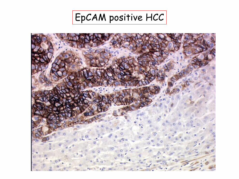
EpCAM positive HCC

Seok Hepatology 2012
The spectrum of liver cell tumors

Cholangiolocellular carcinoma30 cholangiolocellular carcinoma
Komuta Hepatology 2008
-Progenitor cells= target of liver carcinogenesis
CK19+
CLC = phenotypic homology with ductular reaction( = non tumoral HPCs)

Lineage of primary liver cancers
EMT
Komuta Hepatology 2008

Classical HCC
Progenitor-like HCC
Cholangiocarcinoma
Primary tumors on cirrhotic livers

Early HCCa/HCC of small size (≤ 2cm), well differentiated (G1), vaguely nodular
Progressed HCCa/HCC of small size (≤ 2cm), usually moderatelydifferentiated (G2), distinctly nodular type
HCC of not small size (>2cm) single or multiple
b/ HCC with stem/progenitor cell immunophenotype
c/ Mixed hepatobiliary carcinoma, classical type
d/Mixed hepatobiliary carcinomawith stem/progenitor cell phenotype and immunophenotype
PROPOSAL OF A NEW PATHOLOGICAL CLASSIFICATION
Roncalli Dig Liver Dis 2010

Akiba Am J Surg Pathol 2013
Classification relying upon the major component >50%
Combined Hepatocellular-CholangiocarcinomasClassification WHO 2010

Akiba Am J Surg Pathol 2013
� Two distinct components, hepatocellular carcinoma and cholangiocarcinoma
� Most often observed on chronic liver diseases (HCV, HBV, hemochromatosis)
HepPar1
CK19
Combined Hepatocellular-CholangiocarcinomasClassical type

Akiba Am J Surg Pathol 2013
� Nests of mature hepatocytes
� Peripheral clusters of small stem/progenitor cellsCK7+ CK19+ CD56+ c-kit+ EpCAM+
� Fibrous stroma
� Rare
Combined Hepatocellular-CholangiocarcinomaWith Stem cell features – typical type

Akiba Am J Surg Pathol 2013
� Cells intermediate betweenhepatocyte and cholangiocyte
� Strands, solids nests or trabeculae
� HepPar1+, AFP+, CK19+, c-kit+
� Minor components: HCC, CC,cholangiolocellular carcinoma
Combined Hepatocellular-CholangiocarcinomaWith Stem cell features – intermediate type

Akiba Am J Surg Pathol 2013
� Small anastomozing glands with fibrous stroma
� Cuboidal cells
� Coexpression of biliary and progenitor cell markers
� CK19, cKit, NCAM, EpCAM
� Minor components: HCC, CC
Combined Hepatocellular-CholangiocarcinomaWith Stem cell features – cholangiolocellular type

� Sorafenib
� Tivantinib: c-met receptor?
Targeted therapies in HCC?

Conclusion
Cirrhotic liver:Diagnosis of small tumors (1 -2 cm)?Prognostic factors: Tumor differentiation
Vascular invasionStem/progenitor cell markers
New prognosis markers from molecular studiesA continuous spectrum of liver cell tumors
Non cirrhotic liver:Biopsy for differential diagnosis? New prognosis markers from molecular studies
New therapies?