treatment of an iatrogenic subclavian artery ... · treatment of an iatrogenic subclavian artery...
TRANSCRIPT

237J Vasc Bras. 2013 Jul.-Set.; 12(3):237-242http://dx.doi.org/10.1590/jvb.2013.039
T H E R A P E U T I C C H A L L E N G E
Treatment of an iatrogenic subclavian artery pseudoaneurysm near vertebral artery branch:
endovascular approach or open surgery?
Pseudoaneurisma da artéria subclávia próximo à origem da artéria vertebral após punção inadvertida: tratamento endovascular ou cirurgia aberta?
Rodrigo Gibin Jaldin1, Matheus Bertanha1, Marcone Lima Sobreira1, Leandro Gobbo Braz2, Carlos Clayton Macedo de Freitas3, Winston Bonetti Yoshida4, Regina Moura4
INTRODUCTIONThe classical treatment of pseudoaneurysms is open
surgery for resection and end-to-end anastomosis, venous graft, suture, or bypass1. Surgeries to treat subclavian artery aneurysms have been attempted since 1818, first by Valentine Mott2, and then, in 1864, by Smyth, who was the first to achieve success. In 1924, Halsted described the difficulties of treating this type of lesion3. Open surgery involves considerable morbidity and mortality, particularly in the case of high-risk patients1 and urgent surgeries4.
Percutaneous endovascular treatments using covered stents or embolization, a minimally invasive method that does not require general anesthesia, has been one of the therapeutic alternatives in the last decade. Arterial lesions were first treated using an endovascular approach in 1915, by Carrel et al.4. The first animal studies with placement of endoluminal stents were published in 1969 by Dotter et al. In 1987, Nicholas Volodos5 performed the first endovascular correction of an aortic artery aneurysm in Kharkov, in the then Soviet Union, but this technique only became popular in 1991, when Parodi et al. published the first human study about the use of covered stents in the treatment of abdominal aortic aneurysms introduced percutaneously through the femoral artery6. After that, in 1992, they used covered stents for the treatment of an arteriovenous fistula7. In 1994,
Marin et al.8 conducted the first study about the use of covered stents to treat pseudoaneurysms.
Embolization was first used in 1930 in a study conducted by Brooks, who described a surgery for the embolization of a carotid-cavernous sinus fistula using muscle fragments. In 1968, Doppman used a percutaneous catheter and embolization to treat a case of intramedullary arteriovenous malformation9. Some years later, the transcatheter embolization technique was applied to the treatment of digestive hemorrhages10, urinary tract bleeding11, pelvic trauma12, arteriovenous fistulas and hemoptysis13. After the development of numerous embolization agents and a range of new angiographic resources, such as microcatheters, the percutaneous transcatheter embolization technique became an alternative therapeutic option and changed the course of these lesions.
Endovascular approaches are less invasive treatment options, and their use in the treatment of vascular trauma has become more frequent. In some situations, superselective transcatheter arterial embolization may be very useful because it controls bleeding and does not compromise essential functions. In this study, we discuss the advantages and disadvantages of the different therapeutic options to treat subclavian artery lesions.
1Universidade Estadual Paulista – UNESP, Discipline of Vascular and Endovascular Surgery, Botucatu, SP, Brazil.2Universidade Estadual Paulista – UNESP, Department of Anesthesiology, Botucatu, SP, Brazil.3Universidade Estadual Paulista – UNESP, Discipline of Neurosurgery and Interventional Neuroradiology, Botucatu, SP, Brazil.4Universidade Estadual Paulista – UNESP, Discipline of Vascular and Endovascular Surgery, Botucatu, SP, Brazil.Financial support: None.Conflicts of interest: No conflicts of interest declared concerning the publication of this article.Submitted on: 02.13.13. Accepted on: 05.03.13
Study carried out at Faculdade de Medicina de Botucatu-UNESP, Botucatu-SP, Brazil.
Subclavian pseudoaneurysm: endovascular approach or open surgery?
238 J Vasc Bras. 2013 Jul.-Set.; 12(3):237-242
• Endovascular approach using superselective em-bolization, a nonstandard indication, because itconsistsoftheocclusionoftheextravascularspacemaintainedbyflowcomingdirectlyfromthesub-clavianartery.
PART II - TREATMENTThe endovascular approach using superselective
embolization was chosen. Access was established by means of a retrograde puncture and catheterization of the right common femoral artery using an 11-cm 5F introducer sheath (St. Jude Medical®). To plan the intervention, arteriography of the aortic arc and of the supra-aortic trunks was performed using a pig-tail 5F catheter (Merit®), and the results showed extravasation of the contrast medium in the proximal segment of the right subclavian artery, feeding a pseudoaneurysm through a small fistula close to the vertebral artery branch (Figure 3). We decided to occlude the lesion using coils and fibrin sealant. For that purpose, a 260-cm long 0.035” hydrophilic guide wire (Aqualiner – Nipro®) was advanced, followed by a 5F headhunter catheter (Merit®) for selective catheterization of the brachiocephalic trunk and the right subclavian artery, to which the hydrophilic guide wire was anchored. An introducer with a larger caliber (8F, 90 cm; Flexor, Cook®) was introduced, and a JR 6F catheter was advanced over it. Inside it, an Excelsion microcatheter (Boston Scientific®) was advanced over a 205-cm long 0.014” micro guide wire (Transend, Boston, Scientific®) for ultra-superselective embolization of the rupture on the wall of the subclavian artery. The microcatheter placed in this area was used for the placement of detachable micro coils according to the following sequence: 12 mm × 20 cm Micrusphere Microcoil (Micrus®);
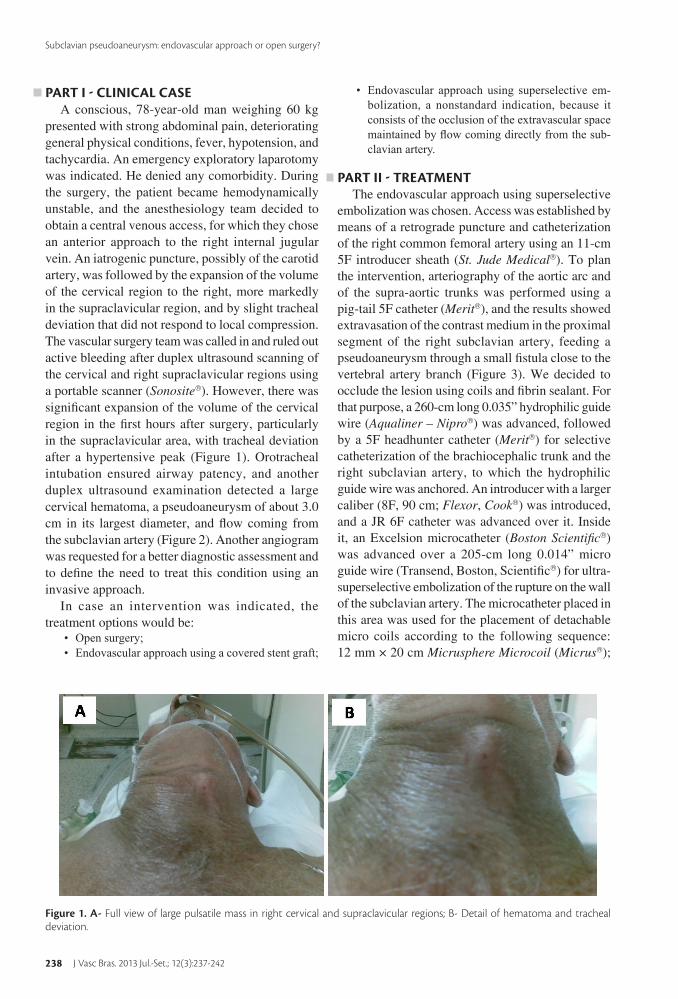
PART I - CLINICAL CASEA conscious, 78-year-old man weighing 60 kg
presented with strong abdominal pain, deteriorating general physical conditions, fever, hypotension, and tachycardia. An emergency exploratory laparotomy was indicated. He denied any comorbidity. During the surgery, the patient became hemodynamically unstable, and the anesthesiology team decided to obtain a central venous access, for which they chose an anterior approach to the right internal jugular vein. An iatrogenic puncture, possibly of the carotid artery, was followed by the expansion of the volume of the cervical region to the right, more markedly in the supraclavicular region, and by slight tracheal deviation that did not respond to local compression. The vascular surgery team was called in and ruled out active bleeding after duplex ultrasound scanning of the cervical and right supraclavicular regions using a portable scanner (Sonosite®). However, there was significant expansion of the volume of the cervical region in the first hours after surgery, particularly in the supraclavicular area, with tracheal deviation after a hypertensive peak (Figure 1). Orotracheal intubation ensured airway patency, and another duplex ultrasound examination detected a large cervical hematoma, a pseudoaneurysm of about 3.0 cm in its largest diameter, and flow coming from the subclavian artery (Figure 2). Another angiogram was requested for a better diagnostic assessment and to define the need to treat this condition using an invasive approach.
In case an intervention was indicated, the treatment options would be:
• Opensurgery;• Endovascularapproachusingacoveredstentgraft;
Figure 1. A- Full view of large pulsatile mass in right cervical and supraclavicular regions; B- Detail of hematoma and tracheal deviation.

Rodrigo Gibin Jaldin, Matheus Bertanha et al.
239J Vasc Bras. 2013 Jul.-Set.; 12(3):237-242
10 mm × 20 cm Micrusphere Microcoil (Micrus®); 10 mm × 30 cm Axium 3D Detachable Coil System (EV3®); 4 mm × 12 cm Axium 3D Detachable Coil System (EV3®); and 7 mm × 18 cm Microplex Coil system (Microvention - Terumo®). After their detachment, angiography showed that the lesion was closed and the vertebral artery opacification improved, which suggested that the pseudoaneurysm had been “stealing” flow from this artery (Figure 4).
Although the result achieved after coil deployment was satisfactory, we chose to complement the procedure using a surgical sealant, because there was no actual wall containing extravascular flow. Cyanoacrylate (Gluebran®-2, GEM®) was selected instead of fibrin sealant because we feared that the latter might adhere to the catheter before it produced the desired effect in the pseudoaneurysm lesion. The treatment was effective, hemodynamic stability was achieved, and hematoma volume was substantially reduced (Figure 5). Control duplex scanning was performed 24 h and 48 h after the procedure, and pseudoaneurysm occlusion was confirmed.
DISCUSSIONA central venous line may be greatly useful in
monitoring the cardiovascular functions of patients in critical condition and as a route to administer vasoactive drugs or solutions that would irritate peripheral veins, such as total parenteral nutrition14-16. With advances in intensive care practices, the number of such procedures has increased progressively, and numerous, more severe complications are seen today14,16. According to the literature, the most frequent complications are incorrect positioning, arterial puncture and pneumothorax, and they may often be diagnosed at the time of the procedure17. The main causes of complications are the inexperience of the operating physician or the possible anatomic variations14,16. Moreover, there is no direct visualization of structures, which increases the chances of errors17. The compression of hematomas and the control of mechanical ventilation parameters, in cases of superficial ventilation or apnea, as well as the use of ultrasound scanning to guide puncture, may minimize problems associated with central
Figure 3. A- Selective arteriogram of brachiocephalic branch shows contrast medium extravasation from lesion in right subcla-vian artery close to vertebral artery branch. B- Angiogram shows that subclavian artery lesion is close to vertebral artery branch.
Figure 2. Duplex scan shows large pseudoaneurysm and char-acteristic bidirectional flow.

Subclavian pseudoaneurysm: endovascular approach or open surgery?
240 J Vasc Bras. 2013 Jul.-Set.; 12(3):237-242
Figure 4. A- Final angiogram after pseudoaneurysm was corrected using superselective embolization and micro coils. B- Lesion details.
In the surgical treatment of subclavian artery pseudoaneurysms, proximal control may require the exposure of the retroclavicular segment of the subclavian artery, with or without thoracotomy, which poses risks to important structures, such as the left phrenic nerve to the left of the thoracic duct and the brachial plexus, in addition to hemorrhage during disection20. In this case in particular, the endovascular approach was, in the authors’ opinion, the best option, because it would be difficult to control the lesion during open surgery as the supraclavicular space was occupied by the pseudoaneurysm, which might complicate access. Moreover, the lesion was located in the subclavian artery between the first and second anatomic segments, and open surgery would require that the segment covered by the anterior scalene muscle be approached. The first treatment option was the placement of a covered stent21, but the proximity to the vertebral artery branch indicated risk of occlusion that might lead to cerebellar ischemia, which would further complicate the patient’s already serious clinical condition. The choice was, therefore, to proceed with superselective embolization to treat the lesion.
After the advent of micro catheters and micro guide wires, initially designed for neuroradiology interventions22,23, pseudoaneurysms can be treated selectively without compromising blood flow to adjacent regions. The precise deployment of micro coils is possible after their correct positioning is confirmed using arteriography. This technique minimizes the risk of coil migration and has better immediate results.24
venous accesses16. Pseudoaneurysms of large vessels or the subclavian artery are rare after central venous punctures and are estimated to occur in 0.05 to 2% of the cases. Complications may include the expansion and compression of respiratory and adjacent neurovascular structures, rupture, thrombosis and skin erosion and external bleeding18,19.
Figure 5- Significant reduction of cervical hematoma volume 48 h after procedure.
Rodrigo Gibin Jaldin, Matheus Bertanha et al.
241J Vasc Bras. 2013 Jul.-Set.; 12(3):237-242
13. Castaneda-Zuniga W, Epstein M, Zollikofer C, et al. Embolization of multiple pulmonary artery fistulas. Radiology. 1980;134:309-10. PMid:7352205.
14. Domino KB, Bowdle A, Posner KL, Spitellie PH, Lee LA, Cheney FW. Injuries and liability related to central vascular catheters. Anesthesiology. 2004;100:1411-8. PMid:15166560. http://dx.doi.org/10.1097/00000542-200406000-00013
15. Schwengel DA, McGready J, Berenholtz SM, Kozlowski LJ, Nichols DG, Yaster M. Peripherally inserted central catheters: a randomized, controlled prospective trial in pediatric surgical patients. Anesth Analg. 2004;99:1038-43. PMid:15385346. http://dx.doi.org/10.1213/01.ANE.0000132547.39180.88
16. Feller-Kopmann D. Ultrasound-guided internal jugular access: a proposed standartized approach and implications for training and practice. Chest. 2007;132:302-9. PMid:17625091. http://dx.doi.org/10.1378/chest.06-2711
17. Abood GJ, Davis KA, Esposito TJ, Luchette FA, Gamelli RL. Comparison of routine chest radiograph versus clinical judgement to determine adequate central line placement in critically ill patients. J Trauma Injury Infect Crit Care. 2007;63:50-6. PMid:17622868. http://dx.doi.org/10.1097/TA.0b013e31806bf1a3
18. Peces R, Navascues RA, Baltar J, Laures AS, Alvarez-Grande J. Pseudoaneurysm of the thyrocervical complicating percutaneous internal jugular-vein catheterization for hemodialysis. Nephrol Dial Transplant. 1998;13:1009-11. PMid:9568871. http://dx.doi.org/10.1093/ndt/13.4.1009
19. Sales e Silva EG, Moreira RW, Arcenio Neto E, et al. Tratamento endovascular de pseudo-aneurisma da artéria subclávia em criança hemofílica. J Vasc Bras. 2006;5(2):151-6.
20. Akgun S, Civelek A, Baltacioglu F, Ekici G. Successful endovascular repair of a subclavian artery pseudoaneurysm. Nephrol Dial Transplant. 1999;14:2219-21. PMid:10489237. http://dx.doi.org/10.1093/ndt/14.9.2219
21. Cox MW, Whittaker DR, Martinez C, Fox CJ, Feuerstein IM, Gillespie DL. Traumatic pseudoaneurysms of the head and neck: early endovascular intervention. J Vasc Surg. 2007;46(6):1227-33. PMid:18154999. http://dx.doi.org/10.1016/j.jvs.2007.08.021
22. Murayama Y, Vinuela F, Duckwiler GR, Gobin YP, Guglielmi G. Embolization of incidental cerebral aneurysms by using the Guglielmi detachable coil system. J Neurosurg. 1999;90:207-14. PMid:9950490. http://dx.doi.org/10.3171/jns.1999.90.2.0207
23. Cardozo MA, Lichtenfels E, Erling N Jr, Raupp E, Tarasconi DP. Tratamento endovascular de aneurisma da artéria renal por embolização com micromolas preservando o fluxo sangüíneo renal: relato de caso. J Vasc Br. 2007;6(2):167-170. http://dx.doi.org/10.1590/S1677-54492007000200012
24. Klein GE, Szolar DH, Breinl E, Raith J, Schreyer HH. Endovascular treatment of renal artery aneurysms with conventional non-detachable microcoils and Guglielmi detachable coils. Br J Urol. 1997;79:852-60. PMid:9202549. http://dx.doi.org/10.1046/j.1464-410X.1997.00157.x
In conclusion, embolization with micro coils may be one more alternative for a less invasive treatment of iatrogenic puncture lesions.
REFERENCES1. Ohki T, Veith FJ. Five-year experience with endovascular grafts
for the treatment of aneurysmal, occlusive and traumatic arterial lesions. Cardiovasc Surg. 1998;6(6):552-565. http://dx.doi.org/10.1016/S0967-2109(98)00073-8
2. Robert TB, Derek RK, Michael WG. Complicated right subclavian artery pseudoaneurysm after central veno puncture. Ann Thorac Surg. 1996;62:581-2. http://dx.doi.org/10.1016/0003-4975(96)00385-2
3. Matthew JD, Keith DC, Ronald PS, Dominic AD. Atherosclerotic aneurysm of the intrathoracic subclavian artery: a case report and review of the literature. J Vasc Surg. 1995;21:521-9. http://dx.doi.org/10.1016/S0741-5214(95)70297-0
4. Marin ML, Hollier L, Avrahami R, Parsons R. Varying Strategies For Endovascular Repair Of Abdominal And Iliac Artery Aneurysms. Surg Clin North Am. 1998;78(4):631-45. http://dx.doi.org/10.1016/S0039-6109(05)70338-8
5. Volodos NL, Karpovich IP Shekhanin VE, et al. A case of distant transfemoral endoprosthesis of the thoracic artery using a self-fixing synthetic prosthesis in traumatic aneurysm (article in Russian). Grudn Khlr. 1988;6:84-6.
6. Criado FJ. EVAR at 20: the unfolding of a revolutionary new technique that changed everything . J Endovasc Ther. 2010;17(6):789-96. PMid:21142491. http://dx.doi.org/10.1583/10-3291.1
7. Parodi JC, Schonholz L, Ferreira J . Endovascular Stent-Graft Treatment of Traumatic Arterial Lesions. Ann Vasc Surg. 1999;13:121-129. PMid:10072450. http://dx.doi.org/10.1007/s100169900230
8. Marin ML, Veith F, Pane’tta T, Cynamon J, et al. Transluminally Placed Endovascular Stented Graft Repair For Arterial Trauma . J Vasc Surg . 1994;20:466-73. http://dx .doi .org/10.1016/0741-5214(94)90147-3
9. Uflacker R, Mourão GS, Piske RL. Treating complications of subclavian vein puncture by embolization of the internal mamary artery. Cardiovasc Intervent Radiol. 1991;14:15-15. http://dx.doi.org/10.1007/BF02577708
10. Derauf BJ, Hunter DW, Sirr SA, et al. Peripheral embolization of diffuse hepatic arteriovenous malformations in a patient hereditary hemorragic telangiectasia. Cardiovasc Intervent Radiol. 1987;10:10-83. http://dx.doi.org/10.1007/BF02577971
11. Uflacker R, Paolini RM, Lima S. Management of traumatic hematuria by selective renal artery embolization. J of Urol. 1984;132:662-7. PMid:6471207.
12. Clark RA, Gallant TE, Alexander ES. Angiographic management of traumatic arteriovenous f istulas : cl inical results . Radiology. 1983;147:9-13. PMid:6828768.

Subclavian pseudoaneurysm: endovascular approach or open surgery?
242 J Vasc Bras. 2013 Jul.-Set.; 12(3):237-242
CorrespondenceRodrigo Gibin Jaldin
Hospital das Clínicas – Laboratório Vascular Distrito de Rubião Jr., s/n – UNESP Campus Botucatu
CEP 18618-780 - Botucatu (SP), Brazil Fone: (14) 3811-6305
E-mail: [email protected], [email protected]
Author informationRGJ is visiting professor, discipline of Vascular and Endovascular,
Universidade Estadual Paulista (UNESP). MB is assistant professor, discipline of Vascular and Endovascular
Surgery, Universidade Estadual Paulista (UNESP). MLS is professor, discipline of Vascular and Endovascular Surgery,
Universidade Estadual Paulista (UNESP). LGB is professor, Department of Anesthesiology, Universidade
Estadual Paulista (UNESP). CCMF is assistant professor, discipline of Neurosurgery and
Interventional Neuroradiology, Universidade Estadual Paulista (UNESP).
WBY is full professor, discipline of Vascular and Endovascular Surgery, Universidade Estadual Paulista (UNESP).
RM is professor, discipline of Vascular and Endovascular Surgery, Universidade Estadual Paulista (UNESP).
Author’s contributionsConception and design: RGJ, MLS
Analysis and interpretation: RGJ, MB, MLS, WBY, RM Data collection: RGJ, MB, MLS, CCMF, LGB, RM
Writing the article: RGJ, WBY, MLS Critical revision of the article: RGJ, MLS, RM, WBY
Final approval of the article*: RGJ, MB, MLS, CCMF, LGB, WBY, RM Statistical analysis: N/A
Overall responsibility: RGJ Obtained funding: None.
*All authors should have read and approved of the final version of the article submitted to J Vasc Bras.