transport and permeability properties of human caco-2 cells: an in vitro model of the intestinal...
TRANSCRIPT

Journal of Controlled Release, 11 (1990) 25-40 25 Elsevier Science Publishers B.V., Amsterdam - - Printed in The Netherlands
TRANSPORT AND PERMEABILITY PROPERTIES OF H U M A N CACO-2 CELLS: AN IN VITRO MODEL OF THE INTESTINAL EPITHELIAL CELL BARRIER*
G. Wilson*, I.F. Hassan, C.J. Dix t*, I. Williamson, R. Shah *tt, M. Mackay** Advanced Drug De/ivery Research, Ciba-Geigy Pharmaceudca/s, Wimblehurst Road, Horsham, West Sussex RH 12 4AB (Great Britain)
and P. Artursson Department of Pharmaceutics, Uppsala University, Box 580, S-751 23 Uppsa/a (Sweden)
Key words: Caco-2 cells; intestinal epithelial; cell line; cell model
When on nitrocellulose filters, in chambers, Caco-2 cells form a confluent monolayer with several properties characteristic of differentiated intestinal epithelial cells. The cell layers are morphol- ogically polar and have well-developed brush borders at their apical surface. The monolayers are functional in the carrier-mediated transport of taurocholic acid and in the receptor-mediated endocytic transport of cobalamin-intrinsic factor. These active transport pathways, characteristic of the distal ileum, are specific and unidirectional (apical to basolateral). The Caco-2 model dis- plays low permeability to the non-specific transepithelial passage of macromolecules. In addition, our results indicate that the model has potential for predicting in vivo absorption of low molecular weight drugs. The reproducibility and long-term viability of this human model, together with the ability to conveniently perform qualitative and quantitative transport studies across an intact epithelial layer, confer significant advantages over other in vitro models used for absorption studies.
INTRODUCTION
A number of in vitro methods have been used to study the absorption of drugs through the in- testinal epithelium [1]. The majority of these systems do not have the morphological and
*Paper presented at the Fourth International Symposium on Recent Advances in Drug Delivery Systems, Salt Lake City, UT, U.S.A., February 21-24, 1989. **To whom correspondence should be addressed. tPresent address: Smith Kline and French Laboratories, P.O. Box 1539, King of Prussia, PA 19406-0939, U.S.A. ttPresent address: Glaxo Group Research, Glaxo Labora- tories, Greenford Road, Greenford, Middlesex, Great Britain. rePresent address: Clinical Research Centre, Watford Road, Harrow, Middlesex, Great Britain.
functional properties of normal adult human epithelial layers. Moreover, they lack the via- bility and versatility required for quantitative measurements of transepithelial drug trans- port and for the examination of transport mechanisms. The development of human cell culture systems has been limited by the lack of retention of anatomical and biochemical fea- tures of differentiated cells in vivo. In particu- lar, the use of isolated intestinal epithelial cells has been slow to progress because these cells are difficult to culture and have limited viabil- ity [ 2 ]. The use of human tumour cell lines has also been restricted by their lack of differen- tiated properties in culture. Recently, attention has turned to human adenocarcinoma cell lines that reproducibly display a number of proper-
0168-3659/90/$03.50 © 1990 Elsevier Science Publishers B.V.
26
ties characteristic of differentiated intestinal epithelial cells [3-5 ]. These models are proving extremely useful for studies on a variety of in- testinal functions, e.g. cellular differentiation, and the transport of proteins in the biosyn- thetic and endocytic pathways. However, their application to drug transport processes has, un- til recently, received little attention. We there- fore set out to characterise the transport and permeability properties of human Caco-2 cells grown on filters, to determine whether such a system could be used to study the transepithe- lial transport of drugs, including peptides and proteins. Of the available human tumour cell lines, we chose the Caco-2 cell line as it ap- peared to have the most highly differentiated properties under standard culture conditions [6]. The Caco-2 model has been examined for the presence of carrier-mediated transport and a receptor-mediated endocytic pathway. The transepithelial passage of low molecular weight drugs and macromolecules has also been mea- sured. From these studies it is clear that the model has a number of morphological and func- tional properties that are similar to those of the normal human small intestine.
MATERIALS AND METHODS
Cell culture
The Caco-2 cell line, Fogh et al. [7], was ob- tained from Professor Colin Hopkins, Imperial College, University of London. Cells of passage numbers 80-110 were used. Caco-2 cells were routinely grown in a medium (maintenance medium) comprised of Dulbecco's modified Eagle's medium (DMEM), containing 20% (v/ v) foetal calf serum (FCS), 1% (w/v) non-es- sential amino acids, and 1% (w/v) glutamine, in an atmosphere of 90% air and 10% CO2. Rou- tine passaging of cell stocks was carried out in 75 cm 2 and 180 cm 2 flasks (Costar, Cambridge, MA, U.S.A.). For filter-cultures, cells were grown on nitrocellulose filters (uncoated pore
size 0.45 #m) in 30 mm diameter Millicell-HA chambers (Millipore, Bedford, MA, U.S.A.). These chambers were inserted into the wells of six well plates. For transferring from plastic dishes to filters, the cells were trypsinized with a solution containing 0.25% (w/v) trypsin (Sigma Chemicals, U.K., T8128) and 0.2% (w/ v) EDTA, and washed twice in maintenance medium before being plated at 2 × 10 ~ cells per millicell-HA chamber. Cells in 2 ml mainte- nance medium, containing penicillin (100 U / ml) /s t reptomycin ( 100/~g/ml ) were placed on the filters; 2 ml of the same medium was placed under the filter in the well of the culture plate. The filter-grown cultures were incubated at 37°C in a humidified atmosphere of 90% air, 10% CO2. After 48 h, the upper and lower cul- ture medium was changed daily. Cells were maintained on filters for up to 30 days.
Cell number
Filter-grown cells were cultured for varying times, fixed with 2.5% glutaraldehyde and stained with haemotoxylin. Filters were dehy- drated using an ascending series of ethanol with the final step in xylene. The filters were mounted in DPX mountant (BDH, U.K.) and cells counted using a light microscope (Leitz, Orthoplan) connected to an image analyser (Joyce-Loebl, Magiscan 2A).
Electrical resistance measurements
The permeability of cell monolayers was de- termined by electrical resistance measure- ments using the method of von Bonsdorff et al. [8]. Minimum Essential Medium (MEM) + 0.1% (w/v) bovine serum albumin (BSA), 5 ml and 3 ml to the apical and basolateral chamber, respectively, were added prior to measurement and allowed to equilibrate at 25 °C for 15 min. Salt bridges were connected to each chamber and a current of 100/ tA applied across the monolayer. The potential difference across the monolayer was measured and the electrical re- sistance ( ~ / c m 2) derived from Ohms Law.

Transmission electron microscopy (TEM)
Cells on plastic or on filters were washed in fresh maintenance medium and fixed with a so- lution containing 2.5% glutaraldehyde and 4% formaldehyde (freshly prepared from parafor- maldehyde) in 0.1 M cacodylate buffer, pH 7.3, for 1 h at 25 ° C. The cells were then rinsed in 0.1 M cacodylate buffer and fixed with 1% os- mium tetroxide in 0.1 M cacodylate buffer, for 1 h at 25°C. The cells were finally rinsed in 0.1 M cacodylate buffer. Filters were removed from the plastic surround, cut into quarters and placed in cacodylate buffer. Filters and cells grown on plastic were dehydrated through an ascending series of ethanol, infiltrated, embed- ded in Epon resin and cured in an oven at 60 ° C for 48 h. The filter squares were embedded in flat embedding moulds and the cells grown on plastic were embedded in situ. Cells were sec- tioned on a Reichert 0M4 microtome, stained with uranyl acetate and lead citrate, visualised and photographed on a Philips CM10 electron microscope. Materials used throughout were of an appropriate electron microscopy grade.
Scanning electron microscopy (SEM)
Cells were fixed and dehydrated using the procedures for TEM. At the final 100% ethanol wash, the samples were dried in an Edwards (Cambridge, U.K. ) critical point dryer. Filters were glued onto an SEM stub and a fine gold film evaporated onto the surface in an Edwards sputter coater. The cells were viewed in a Cam- bridge Stereoscan 200 electron microscope.
Horseradish peroxidase cytochemistry
Horseradish peroxidase (HRP), 10 mg/ml, was applied basolaterally to filter-grown cells and incubated for 1 h at 4°C, 20°C or 37°C. The cells were washed in fresh maintenance medium before adding an initial fixative con- taining 0.05% glutaraldehyde and 4% formal- dehyde in 0.1 M cacodylate buffer, pH 7.3, for
27
i h at 25 ° C. The cells were permeabilised in 5% (w/v) sucrose overnight and washed in 0.1 M cacodylate buffer. An incubation mixture (50 mg diaminobenzidine, 34/tl H202, in 50 ml of 0.1 M cacodylate buffer, pH 7.4) was added and incubated for 60 min at 25 °C in the dark. The cells were rinsed in 0.1 M cacodylate buffer. They were finally fixed in a solution containing 2.5% glutaraldehyde and 4% formaldehyde in 0.1 M cacodylate buffer, pH 7.3, for 1 h at 25 ° C and then incubated with 1% osmium tetroxide for 1 h at 25°C. The cells were dehydrated through an ascending series of ethanol, pro- cessed for TEM as previously described, sec- tioned on a Reichert OM4 microtome and were n o t stained with uranyl acetate or lead citrate.
Permeability to macromolecules
All experiments were performed using Caco- 2 cells cultured for 15 days in Millicell-HA chambers. [3H ] Inulin (New England Nuclear, 1.37 Ci/mmol), 0.5 #Ci, in 2 ml MEM, 0.1% (w/v) BSA was added to the apical surface and incubated at 37 ° C in an atmosphere of 95% rel- ative humidity. At various times, samples of the apical and basolateral fluid were removed, added to 5 ml scintillant (Lumagel, LKB) and counted using a Beckman LS1801 liquid scintillation spectrometer. Samples were removed for GPC analysis on a 7.5 × 30 cm TSK G3000PW col- umn (Tosoh, Japan) with a 7.5 × 7.5 PWH pre- column. The mobile phase was 0.1 M LiBr in H20) at a flow rate of 0.5 ml/min.
HRP activity was measured by the method of Gallati and Pracht [9]. The assay was adapted to allow use of a standard 96-well microtitre plate. Absorbance at 450 nm was determined by a Dynatech (Guernsey, Channel Islands) MR600 plate reader, and the concentrations determined from an HRP (Type II HRP, 100,000 units/527 mg solid: Sigma Chemicals, Poole, U.K. ) standard curve.

28
Taurocholic acid transport
Experiments were performed using Caco-2 cells cultured in Millicell-HA chambers as out- lined above. Cells were used at varying times after addition to the filters as indicated. [14C]Taurocholic acid (Amersham, U.K.; 56 mCi/mmol) , 0.5 ttCi in 2 ml media, was added to either the apical or basolateral fluid. Specific transport was determined from competition with an excess of taurocholic acid (1 mg/ml) . After 4 h at 37°C (95% relative humidity and 10% CO2) apical and basolateral samples were removed, 14 ml scintillant (Lumagel, LKB) added and counted using a Beckman LS1801 liquid scintillation spectrometer. Results were corrected to dpm by comparison with standard quench curves and are expressed as nmol [ 14C ] taurocholic acid transported per four hours across a filter of cells. A Lineweaver-Burk plot of the data was prepared.
Transepithelial transport of vitamin B1 2
Complexes between porcine intrinsic factor (IF: Sigma Chemicals, Poole, U.K ) and cyano- [57Co ] cobalamin (Amersham, U.K. ), for bind- ing and transport studies were prepared as de- scribed previously [10]. Cyano[~TCo]cob - alamin-intrinsic factor (16 pM) was added to either the apical or the basolateral medium, in the presence and absence of a fifty-fold molar excess of cobalamin-intrinsic factor. The cells were incubated at 37 °C for 24 h. At the end of the incubation period, cells were cooled to 4 ° C. The cells were washed twice with PBS, pH 7.4, and solubilised with 0.1 M NaOH. The solubi- lised cells and the medium from the upper and lower chambers were counted using an LKB multi-gamma spectrometer, to determine cell- associated and transcellularly transported cob- alamin, respectively.
Transepithelial transport of drugs
[3H]Propranolol (New England Nuclear) [3H]metoprolol and [3H]atenolol (obtained
from Dr K.J. Hoffman, H/issle AB, MSlndal, Sweden) had specific activities between 0.5 and 26.6 Ci/mmol. The compounds were mixed with unlabelled drugs to give a final concentration of 1 × 10 -7 M. The drugs were diluted in air- buffered DMEM containing 0.1% human serum albumin and 10 m M N-2-hydroxyethylpipera- zine-N-2-ethanesulphonic acid (HEPES) and were added to the apical filter chambers of Caco- 2 cells cultured on polycarbonate filters (Nu- cell, Nucleopore). Drug-free medium (2 ml) was added to the apical or basolateral chamber, re- spectively. Experiments were performed on cells which had been cultured for 15 days. The plates were incubated in air at 37°C and relative hu- midity of 95% for up to 150 min. At suitable time intervals, 50 #l samples were withdrawn from the apical and basolateral chambers, 5 ml scintillant added (Lumagel, LKB) and sam- ples counted in a Tricarb 1900 liquid scintilla- tion spectrometer (Packard Instruments) . Re- sults were corrected to dpm by comparison with standard quench curves. Rate constants (Kapp/ h) were obtained from linear regression analy- sis of the slopes of the initial appearance rates of drug in the basolateral fluid, before more than 10% of drug had been transported.
RESULTS AND DISCUSSION
Growth and morphology of epithelial layers
Human Caco-2 cells can be routinely grown as monolayers, on filters, in Millicell HA or Nu- cell chambers in a similar fashion to the system developed for Madin-Darby canine kidney (MDCK) cells [8]. In this system the apical (mucosal) and basolateral (serosal) culture fluids are separated allowing the transport of molecules from one culture fluid to the other to be determined. Cells were added to untreated

29
2 . 4
2 . 2
~ " 2 . 0 - ' o
x
L_ ~ 1 . 8
2 H
m 1 . 6 - (D
1 . 4
\ \
I I ] ] I J I 0 4 8 1 2 1 6 2 0 2 4
T i m e ( d a y s )
Fig. 1. The number of Caco-2 cells on nitrocellulose filters with time. Results ± SEM for 12 samples are shown.
filters at a high density in an attempt to achieve a functional monolayer as quickly as possible.
The growth pattern is shown in Fig. 1. De- crease in the cell number is probably due to un- attached cells sloughing off the filter. From about 15 days, the cell number remains rela- tively constant. Other laboratories have used collagen-coated polycarbonate filters as a permeable support [11-13], although coating the filters with collagen or other extracellular materials is probably not an absolute require- ment for the growth of a monolayer [12,13]. When low densities of cells are added to colla- gen-coated filters there is an increase in cell number until a confluent monolayer is formed [11]. The typical morphology of Caco-2 cells grown on nitrocellulose filters is shown in Figs. 2-4.
When grown on plastic (Fig. 2a), the cells show some morphological properties of a nor-
Fig. 2a. Transmission electron micrographs of day 15 Caco- 2 cells. Electron micrograph of a section through Caco-2 cells grown on plastic ( • ). Wide intracellular spaces were common and tight junctions (--,) were seen at the apical membrane. Magnification × 6327.
mal epithelium in that they form monolayers with tight junctions connecting cells at their apical surface. On the plastic substratum, there are large spaces between the basolateral sur- faces of adjacent cells probably due to the in- tercellular accumulation of fluid. At the apical surface of the cells, the microvilli are sparse and poorly developed. In contrast, Caco-2 cells grown on filters (Fig. 2b), show more closely opposed lateral surfaces and well developed ap- ical brush borders.
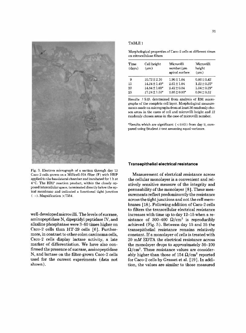
Histochemical localisation of HRP (Fig. 3) demonstrates that the tight junctions between the apical surfaces of each cell are impermeable

30
Fig. 2b. Transmission electron micrographs of day 15 Caco-2 cells. Electron micrograph of a section through Caco-2 cells grown on a Millicell-HA filter (F), showing extensive apical microvilli. Tight junctions ( --* ) were seen at the apical mem- brane. Magnification X 5836.
to the macromolecular probe. No penetration of HRP through the tight junctions was ob- served at either 4 o C, 20 °C or 37 ° C. Although these features are more characteristic of nor- mal small intestinal epithelial cells, the mor- phological properties of the Caco-2 cells are not identical to these cells. For example, we have observed that the cells accumulate glycogen, which is similar to previous reports [ 14 ].
Morphological measurements on the Caco-2 cell layers (Table 1) indicate that there is an increase in cell height and microvilli height up to day 15, and that these properties remain rel- atively constant up to day 23. However, micro- villi number per micrometer does not show a significant increase until day 23. The cell height found is significantly lower than the value of 25 ttm reported by Neutra and Padykula [ 15 ], for the normal human intestinal epithelium, and the value of 29.6 ttm at day 16 determined for Caco-2 cells grown on collagen-coated polycar- bonate fil ters[ 11 ].
We have consistently observed that con- fluent cell layers of Caco-2 cells are not homo- geneous with respect to certain morphological parameters. In particular, heterogeneity of mi- crovilli, investigated by SEM of the apical sur- face of the monolayer (Fig. 4), shows the pres- ence of dense microvilli, clusters of microvilli and a few cells having low numbers of micro- villi. The large deviations derived from micro- villi number per ttm of apical surface (Table 1 ) also reflect this heterogeneity. To determine the correlation between cellular heterogeneity and the functional properties of Caco-2 monolay- ers, an analysis of clones isolated from the par- ent culture is in progress. Distinct cell types, i.e. goblet cells, and colonic cells have been isolated from human HT-29 cultures [ 16,17 ]. Cells with the typical appearance of goblet cells have not been detected in the Caco-2 cell line.
It has been shown by a number of groups that Caco-2 cells have several brush border hydro- lase activities consistent with the presence of
31
T A B L E 1
Morphological propert ies of Caco-2 cells at different t imes on nitrocellulose filters
Time Cell height Microvilli Microvilli (days) (zm) number/~tm height
apical surface (]lm)
9 10.72 ± 2.10 1.96 ± 1.04 0.86 ± 0.42 15 14.24 ± 1.49 a 2.61 ± 1.04 1.22 ± 0.25 a 20 14.84 ± 1.69 a 2.42 ± 0.84 1.04 ± 0.29 a 23 17.24 ± 1.55 a 2.85 ± 0.93 a 0.94 ± 0.32
Results ± S.D. determined from analysis of E M micro- graphs of the complete cell layer. Morphological measure- ments made on micrographs from at least 36 randomly cho- sen areas in the cases of cell and microvilli height and 12 randomly chosen areas in the case of microvilli number.
aResults which are significant ( < 0.05) from day 9, com- pared using S tudent t- test assuming equal variance.
Fig. 3. Electron micrograph of a section through day 15 Caco-2 cells grown on a Mill±cell-HA filter (F) with H R P applied to the basolateral chamber and incubated for 1 h at 4°C. The H R P reaction product, wi th in the closely op- posed intercellular space, t e rmina ted directly below the ap- ical membrane and indicated a functional t ight junct ion ( --, ). Magnif icat ion × 7354.
well-developed microvilli. The levels of sucrase, aminopeptidase N, dipeptidyl peptidase IV, and alkaline phosphatase were 3-60 times higher on Caco-2 cells than HT-29 cells [6]. Further- more, in contrast to other colon carcinoma cells, Caco-2 cells display lactase activity, a late marker of differentiation. We have also con- firmed the presence of sucrase, aminopeptidase N, and lactase on the filter-grown Caco-2 cells used for the current experiments (data not shown).
Transepithelial electrical resistance
Measurement of electrical resistance across the cellular monolayer is a convenient and rel- atively sensitive measure of the integrity and permeability of the monolayer [8]. These mea- surements reflect predominantly the resistance across the tight junctions and not the cell mem- branes [ 18 ]. Following addition of Caco-2 cells to filters the transcellular electrical resistance increases with time up to day 12-15 when a re- sistance of 300-400 ~ / c m 2 is reproducibly achieved (Fig. 5). Between day 15 and 25 the transepithelial resistance remains relatively constant. If a monolayer of cells is treated with 20 mM EDTA the electrical resistance across the monolayer drops to approximately 50-100 Ft/cm 2. These resistance values are consider- ably higher than those of 154 ~ / c m 2 reported for Caco-2 cells by Grasset et al. [19]. In addi- tion, the values are similar to those measured

32
Fig. 4. Scanning electron micrographs of day 15 Caco-2 cells grown on Millicell-HA filters. (a) A low magnification photo- graph, showing microvilli heterogeneity: a dense microvilli covering (D) and a cell with a sparse covering of apical microvilli (B). Magnification X 6688. (b) A high magnification photograph, clearly showing the apical intercellular boundary ( ~ ) . Magnification × 22227.
across colonic tissue and are much higher than the transepithelial resistances across the small intestine [20]. However, methods used to de- rive resistance values from cell monolayers and excised tissue are different and therefore com- parisons should be considered with care.
Carrier-mediated transport of taurocholic acid
Bile acids are t ransported across the small intestinal mucosal membrane by either passive diffusion or by an active transport process [21 ].

5 0 0
4 0 0 - o
8 a~ 3 0 0
c (Q 4~ co .m to
o: 2 0 0 -
to u
u
a~ l O 0
w
Time (days)
Fig. 5. The transepithelial electrical resistance of Caco-2 cells on nitrocellulose filters with time. Results ± S.E.M. for 12 samples are shown.
In the rat, a specific transport protein is in- volved in Na+-dependent bile acid transport across the brush-border membrane [22]; the transport of bile acids across the basolateral membranes is thought to occur by anion ex- change [23 ]. Such an active transport process occurs from the lumen to the blood.
The transport of [14C ]taurocholic acid across monolayers of Caco-2 cells on filters is shown in Fig. 6. These results show that the specific transport of [14C ]taurocholate is unidirec- tional, from the apical to the basolateral side, and that the polarity and rate of transport are maintained by the monolayers for a period of greater than 20 days in culture. Greater than 80% of the transcellular transport can be inhib- ited by a one-hundred-fold molar excess of un- labelled taurocholate added with the radiola- belled bile acid to the apical culture fluid. In contrast, there is little inhibition of [14C]taurocholate transport when excess un-
33
120
,~ 1 0 0
o ~z 8 0 - o
2c 4J
,,,_ ,.~ 6 0
8~ [2 0
E 4 0 - ~s
o 2 0 ~
S O3
O-
I I I 1 I I I I I 8 1.0 1 2 1 4 t 6 :18 2 0 2 2 2 4
Time (Oays)
Fig. 6. The specific transport of [ 14C ] taurocholic acid across Caco-2 cells cultured on nitrocellulose filters. Specific transport of taurocholic acid (apical to basolateral, ( O ) ; basolateral to apical, ( O ) ). Result ± S.E.M. for 3 samples are shown.
labelled taurocholate is added to the basolateral culture fluid, demonstrating that in Caco-2 cells [ltC ]taurocholate is actively transported against a concentration gradient (Fig. 7).
The binding and uptake of [ 14C ] taurocholic acid is saturable with an apparent Km for the transcellular transport of [ 14C ] taurocholate of 42.5 + 2.8pM (Fig. 8). This apparent Km is con- sistent with the results of Barnard and Ghishan [ 24 ] who reported a Km for taurocholic acid up- take into human brush-border membrane ves- icles of 37 + 7/tM. The conformity of these re- sults may suggest that the apical membrane represents the rate-limiting step for the trans- port of taurocholic acid. However, further work is needed to verify this. To our knowledge, the Caco-2 system is the first human in vitro intes- tinal epithelial layer that has been shown to ac- tively transport bile acids from the apical to ba-

3 4
F. o ~ 5 ~ 0 r-~ (_ .~
o -p E g s #
(_
200]
1oo
CONTROL
D APICAL
Fig. 7. Competit ion of [ ~4C ] taurocholic acid transport (ap- ical to basolateral) across Caco-2 cells. The control was compared to a molar excess of unlabelled taurocholate on either the basolateral or apical side. Transport was mea- sured at day 15. Results _+ S.E.M. for 3 samples are shown.
solateral side. The transport of taurocholate across Caco-2 cells has also been reported by Hidalgo et al. [11 ]. It should thus prove useful for comparing the transport of bile acids and their derivatives.
Caco-2 monolayers will also specifically transport phenylalanine [25]. In the Millicell HA chamber system described here, the trans- port of leucine is specific, polar (from apical to basolateral), and saturable (Woodcock et al., manuscript in preparation).
P e r m e a b i l i t y t o m a c r o m o l e c u l e s
In order to investigate the receptor-mediated transport of macromolecules across the intes- tinal epithelial layer it is important to measure the overall permeability of cell monolayers to macromolecular probes which are not trans-
2 o
o
o c- o
f0 4 J
.,.~
o ~ l '-
( . . ~ .
n ° ~ o "0 ~ E C f -
f_ ~- 0
o
o_ i f )
0
4 . 0
s . o
2 . 0 • •
l . O
7 _0.1 . . . . . . . . ~, o.~, 0.~, 0.',
0 O"
0 50 ~00 ~50 200 250 300
T e u r o c h o l i c A c i d C o n c e n t r a t i o n (pM)
Fig. 8. Saturation of [ 14C ] taurocholic acid transport {api- cal to basolateral ) across Caco-2 cells. Increasing amounts of unlabelled taurocholate were added to a constant amount of [ 14C ]taurocholic acid giving final taurocholic acid con- centrations as indicated. Results +_ S.E.M. for 6 samples are shown. The curve was fitted to the data using the func- t ion y=a[x/(b+x) ]. The Km was derived from the Line- weaver-Burk plot shown ( inset) .
TABLE 2
Permeability of Caco-2 monolayers on filters to macromolecules
Macromolecule M.W. Transport rate a Transport amount (kDa) (fl cell-1 h -1) (% applied dose/h)
HRP 40 1.60+_ 0.54 0.0001 [3H ]inulin 5.2 370.5 +_45.5 0.03
"Macromolecule applied to apical fluid: appearance rate in basola- teral fluid determined. Enzymically active HRP measured; intact [3H ] inulin determined by GPC.

35
ported by specific carrier or receptor-mediated processes. This was done morphologically us- ing HRP and biochemically using HRP and ra- diolabelled inulin. These probes will not nor- mally penetrate tight junctions of an intact epithelium.
Results on the transepithelial passage of HRP and [3H]inulin across cells grown on filters for 15 days are shown in Table 2. Low concentra- tions of HRP (0.0001% of applied dose) and [3H]inulin (0.03% of applied dose) were de- tected in the basolateral culture fluid 1 h later. In the Caco-2 system the rate of transcellular transport of enzymically active HRP, from the apical to basolateral side, was 1.6 + 0.54 f l /cel l - ' h - ' . This rate is similar to the rate of 1.8 fl/ cell-1 h-1 reported for the transport of HRP across the MDCK system [8]. The use of probes such as [3H]inulin presented problems due to degradation of the molecule and subsequent re- lease of low molecular weight radiolabelled molecules. In such cases measurement of the percentage of intact macromolecule in the ba- solateral fluid is required. The results for [3H]inulin, shown in Table 2, were determined after GPC analysis of the basolateral fluid. Most of the radiolabelled material, (74%), ran as low molecular weight peaks corresponding to [3H ] glucose or [3H ] fructose and [3H ]water. It is likely that the sucrose moieties in [3H ] inulin are degraded to [3H ] glucose and [3H ] fructose by the brush border sucrase present on the Caco- 2 cells.
Each macromolecular probe has unique characteristics with respect to the stability of the molecule on the brush borders, and in the lysosome. Therefore, their main use is to com- pare the permeability of Caco-2 monolayers at different times, under different culture condi- tions and when exposed to extracellular com- ponents that might disrupt the monolayer. The passage of HRP and [3H ] inulin across cells on filters for 15 days or longer was consistently low, correlating with the full development of the morphological, electrical, and bile acid trans- port properties of the monolayer. The histo-
chemical localisation of HRP (results not shown) was qualitatively similar to that de- scribed for human [26] and rat tissues [27]. When added to apical or basolateral mem- branes at 20°C or 37°C the enzyme could be located in a variety of endocytic compartments. However, no penetration of tight junctions was detected. Therefore, with respect to the passage of HRP, both histochemical and biochemical analyses show that filter-grown Caco-2 cells are relatively impermeable to the transport of the molecule. Ellinger and Pavelka [27 ] have shown that a small amount of HRP added to the apical surface of the rat jejunum is extracellularly lo- cated on the basolateral membrane, presum- ably due to transcellular transport. This pro- cess did not occur when HRP was added to the basolateral membrane in their experiments.
Receptor-mediated endocytic transport
Caco-2 cells will specifically bind and endo- cytose cyano [57Co ] cobalamin-intrinsic factor [10,28 ]. This transport pathway is mediated by the internalisation of intrinsic factor by spe- cific receptors located in the intermicrovillous pits on the apical membranes of distal ileal ep- ithelial cells [29]. In initial studies performed on cells grown for 15 days on filters, the trans- port of cyano[57Co]cobalamin across Caco-2 monolayers could not be detected [10]. How- ever, when monolayers grown for longer times on filters (20 days onwards) were used, trans- cellular transport of cyano [57Co ] cobalamin was detected.
The results in Fig. 9 show that this transport only occurs in the apical to basolateral direc- tion. The transport is specific since it can be inhibited by 90% using a fifty-fold molar excess of unlabelled cobalamin-intrinsic factor added to the apical fluid, but not by cobalamin alone. It is likely that the transcellular transport of cyano[57Co]cobalamin across Caco-2 cells is determined by the polarity of intrinsic factor receptors and the polar secretion of a transcob- alamin II (TCII)-like protein. Receptors for

36
4J L o 4 o (/) E
4 J
• ~t . , 4
E ' * -
m ~ 20 i'o
~ r u
u ~
4 - ~
u ~ 0
- 2 0 A P I C A L B A S O L A T E R A L
B CELL-ASSOCIATED
TRANSPORT
Fig. 9. Uptake and t ranspor t of cyano[57Co]cobalamin across Caco-2 cells grown on Millicell-HA filters at day 30. Result show cell-associated cyano [~7Co ] cobalamin, apical and basolateral respectively, and t ranspor ted cy- ano [~VCo]cobalamin, apical to basolateral and basolateral to apical, respectively. Results -+ S.D. of 3 samples are shown.
T A B L E 3
IF-dependent cobalamin uptake and t ranspor t in Caco-2 cells grown for 28 days on filters (data taken from Dix et al. [30 ] )
Parameter fmol/106cells Molecules per cell
IF-receptors a 1.5_+ 0.15 900 _+90 [57Co]Cbl:
In ternal ised/24 h 42.2 _+ 4.2 2.5 _+ 0.25 _+ 104 Transpor ted /24 h 29.9 _+ 2.8 1.8 _+ 0.16 × 10⁴
TCII-l ike protein: Secreted/24 h 372.0 -+ 13.1 2.2 _+ 0.08 × 10 ~
Results are represented _+ S.D., n - - 3. aThese values were determined using cells grown on plastic.
intrinsic factor become concentrated on the ap- ical surface between day 14 and 28, and the cells synthesise and secrete TCII-like protein exclu-
sively into the basolateral fluid after 20 days [30]. Therefore, in Caco-2 monolayers the re- ceptor-mediated transcellular transport of cob- alamin becomes fully functional at a much later time than do the transport pathways for leucine or taurocholate.
A summary of the components of the path- way involved in the intrinsic factor-dependent transport of cobalamin is shown in Table 3. To our knowledge the Caco-2 cell system is the first human cell line that will internalise intrinsic factor and transcellularly transport cobalamin. The use of the cobalamin pathway has been suggested for the delivery of peptide and pro- tein drugs [31] and the Caco-2 model should be useful for examining this. Uncomplexed cy- ano [~7Co ] cobalamin will also pass across Caco- 2 cell layers by an intrinsic factor-independent pathway [ 28 ]. However, the mechanism of this pathway is not known.
Transport of drugs
A series of beta-blockers of known absorp- tion rates in vivo were used in initial studies to investigate the absorptive properties of the Caco-2 monolayer to low molecular weight drugs. For these studies, polycarbonate filters were preferred to nitrocellulose as the extent of binding of hydrophobic drugs to these filters was much lower (Artursson et al., unpublished results).
The absorption of the radiolabelled drugs was determined by measuring their disappearance from the apical fluid, and their appearance in the basolateral fluid (Fig. 10). The rate con- stants (Kap,/h) for drug appearance in the ba- solateral fluid are shown in Table 4, and were obtained under conditions that would minimise the influence of drug molecules diffusing back in the opposite direction. The correlations be- tween disappearance and appearance rate in the Caco-2 system, their lipophilicities and their overall extent of absorption in vivo are shown in Table 4. For the lipophilic drugs metoprolol and propranolol, the correlations are good.

120
b
x
c o
E OJ o
Z [ 9
g
100
80
60
40
20
..... ~ ......... i
m .i i~.:m • ~ .... D///
.... mfi~ "¸
m< , ......... , ......... , ......... , ......... •
I I I I I I I I I 0 2 0 4 0 6 0 8 0 1 0 0 : 1 2 0 1 4 0 ~ 6 0
Time (m~n)
Fig. 10. The apical to basolateral transport of beta-blockers across Caco-2 cells cultured on polycarbonate filters at day 15. The disappearance from the apical chamber ( ) and appearance in the basolateral chamber ( . . . . ) of [3H]atenolol ( A ) , [3H]metoprolol ( 0 ) and [3H]pro- pranolol ( . ) vs time is represented. Results _+ S.E.M. of 3 samples are shown.
TABLE 4
Transepithelial absorption of atenolol, metoprolol, and pro- pranolol, in vitro and in vivo
Drug Relative Oral dose Transport across lipid absorption Caco-2 cells solubility ~ (To)6 (Kapp/h)C
Atenolol 0.003 50 0.00168 Metoprolol 0.15 95 0.222 Propranolol 5.4 90 0.329
aDistribution coefficients n-octanol/buffer, pH7.0, 20°C; Woods and Robinson, 1981 [32]. 6Ruffalo et al. [33]; Mason et al. [34]. CAppearance rate in the basolateral fluid.
However, in the cell culture system, there is lit- tle uptake and transepithelial passage of aten- olol even though its extent of absorption in vivo is reported to be up to 50% of the applied dose
37
[32]. The reasons for this apparent discrep- ancy are not yet clear, but may be the result of the higher transmembrane resistance of the Caco-2 system than the small intestine. As the transmembrane resistance is a function of par- acellular permeability, the paracellular route, which is more important for the passive trans- port of hydrophilic drugs such as atenolol, would be expected to have a lower permeability in the Caco-2 model. The concentration of proprano- lol in the basolateral fluid is lower than ex- pected from the disappearance rate from the apical side. We believe that this is due to some accumulation of this drug inside the cells and on the filters.
Although studies on many other drugs and comparison with their percentage absorption in vivo will be required to determine how far one can extrapolate from the in vitro system to the whole animal, the current results indicate that the cell culture system provides a convenient system for measuring passive uptake and trans- cellular transport rates across an isolated hu- man cell barrier.
Using the culture conditions described, Caco- 2 cells on filters provide a highly reproducible system which is fully viable and functional for longer than 20 days. In contrast to other in vitro systems, e.g. isolated gut segments, intestinal rings, isolated cell suspensions, and membrane vesicles, the formation of an intact epithelial layer on filters, in chambers, allows the undi- rectional and quantitative transport of mole- cules across the cell layer to be measured. The long-term functionality of this system is a con- siderable advantage compared to the limited vi- ability of isolated tissues mounted in double- diffusion chambers. Although the detailed ef- fects of a number of culture variables on the growth and function of Caco-2 cells are not fully understood, data from a number of laboratories show that intact, functional monolayers can be achieved on different filters and under a variety of culture conditions.
The region of the normal human GI-tract represented by the Caco-2 model is still un-

38
clear. While the possession of brush-border hy- drolases and t ransport pathways for bile acids and cobalamin are properties of the distal ileum, the electrical properties are more indicative of a colonic epithelium. In addition, Caco-2 cells may represent a foetal phenotype, as some of the "differentiated" properties displayed by co- lon carcinoma cells are transiently expressed in the foetal colon [4-6].
As the parent Caco-2 cell line is morpholog- ically heterogeneous, the t ransport properties of the monolayer are probably the sum of the contributions of different cell types. Recent work in our laboratory in obtaining a number of clones, with different electrical and trans- port properties, from the parent Caco-2 cell population (Woodcock et al., manuscript in preparation) together with the isolation of dif- ferent cell types from HT-29 cells [ 16,17], sug- gests that it may be possible to develop more homogeneous layers representing cells from de- fined regions of the GI-tract. A potential diffi- culty will be to produce such monolayers with a range of different cell types present in the gut, such as mucus-producing goblet cells and M- cells.
In our current use of the Caco-2 system the apical and basolateral fluids are unstirred. However, the flexibility to mount filters in dou- ble diffusion chambers allows stirred systems to be used. Recent work in our laboratory has found that the carrier-mediated t ransport of amino acids occurs at the same rate in unstirred and stirred Caco-2 systems (Woodcock et al., manuscript in preparat ion).
Work in this and other laboratories has shown that Caco-2 cells possess functionally active t ransport pathways for folate [35 ], phenylala- nine [25], sugars [36] and bile acids [37], and the receptor-mediated system for the uptake of cobalamin-intrinsic factor [10,28]. The system described here should therefore provide a pow- erful in vitro tool for measuring the absorption of drugs and delivery systems which utilise these active t ransport systems. The use of an HT-29 system to determine the mechanism of trans-
port of cephalexin has recently been reported [38]. In addition, there is some evidence to sug- gest that the Caco-2 and other human cell sys- tems may also prove useful in predicting the in vivo absorbability of a range of drugs, including peptides [39,40] although many more exam- ples are required to test the extent of such cor- relations between absorption in the human cell systems and oral absorption in vivo.
ACKNOWLEDGMENTS
The authors gratefully acknowledge Sian Woodcock for maintaining the Caco-2 cell line and Dr K.J. Hoffman, H~ssle AB, MSlndal, Sweden for the gift of [3H]beta blockers. We are also grateful to Professor Ronald Bor- chardt, Kansas, for sending us a preprint of a manuscript describing their work on the Caco- 2 system [11]. We thank Professor Eric Tom- linson for his comments on the manuscript. This contribution is publication number 81 from the Advanced Drug Delivery Unit of Ciba- Geigy Pharmaceuticals.
REFERENCES
1 I. Osiescka, P.A. Porter, R.T. Borchardt, J.A. Fix and C.R. Gardner, In vitro drug absorption models. Brush border membrane vesicles, isolated mucosal cells and everted intestinal rings: Characterization and salicy- late accumulation, Pharm. Res., 2 (1985) 284-293.
2 M.P. Moyer, Culture of human gastrointestinal epi- thelial cells, Proc. Soc. Exp. Biol. Med., 174 (1983) 11-15.
3 M. Rousset, The human colon carcinoma lines HT-29 and Caco-2: Two in vitro models for the study of in- testinal differentiation, Biochimie, 68 (1986) 1035- 1040.
4 M. Pinto, M.D. Appay, P. Simon-Assmann, N. Dracopoli, J. Fogh and A. Zweibaum, Enterocytic differentiation of cultured human cancer cells by re- placement of glucose by galactose in the medium, Biol. Cell., 47 (1983) 232-330.
5 M. Neutra and D. Louvard, Differentiation of intes- tinal cells in vitro, in: Modern Cell Biology: Func- tional Epithelial Cells in Culture, Alan R. Liss, New York, NY, 1989, pp. 363-398.

6 A. Zweibaum, M. Laburthe, E. Grasset and D. Louvard, Use of cultured cell lines in studies of in- testinal cell differentiation and function, in: M. Field and R.A. Frizzell (Eds.), Handbook of Physiology, The Gastrointestinal System, IV, American Physiological Society, Oxford University Press, New York, NY, 1989, in press.
7 J. Fogh, W.C. Wright and J.D. Loveless, Absence of HeLa cell contamination in 169 cell lines derived from human tumors, J. Natl. Cancer. Inst., 58 (1977) 209- 214.
8 C.H. von Bonsdorff, S.D. Fuller and K. Simons, Api- cal and basolateral endocytosis in Madin-Darby can- ine kidney (MDCK) cells grown on nitrocellulose fil- ters, EMBO J., 4 (1985) 2781-2792.
9 V.H. Gallati and I. Pracht, Horseradish peroxidase: Kinetic studies and optimisation of the peroxidase ac- tivity determination with the substrate H202 and 3,3',5,5'-tetramethylbenzidine, J. Clin. Chem. Clin. Biochem., 23 (1985) 453-460.
10 C.J. Dix, H.Y. Obray, I.F. Hassan and G. Wilson, Vi- tamin B12 transport through polarised monolayers of a colon carcinoma cell line, Biochem. Soc. Trans., 15 (1987) 439-440.
11 I.J. Hidalgo, T.J. Raub and R.T. Borchardt, Charac- terisation of the human colon carcinoma cell line (Caco-2) as a model system for intestinal epithelial permeability, Gastroenterology, 96 (1989) 736-749.
12 A.R. Hilgers and P.S. Burton, Human colon carci- noma cell monolayers as a model for drug transport across the intestinal epithelium. I. Morphological and permeability characteristics, Pharm. Res., 5 (1988) PD 947.
13 R. Stratford, L. Bergin, J. Gimple, M. Kihfeld, C. van Pelt, S. White and A. Dantzig, Effect of base- ment membrane on properties of a human intestinal cell line (Caco-2), Pharm. Res., 5 (1988) PD 951.
14 M. Rousset, G. Chevalier, J.P. Rousset, E. Dussaulx and A. Zweibaum, Presence and cell-growth related variations of glycogen in human colorectal adenocar- cinoma cell lines in culture, Cancer Res., 39 (1979) 531-534.
15 M. Neutra and H. Padykula, in: L. Weiss (Ed.), His- tology: Cell and Tissue Biology, Elsevier, Amsterdam, 1983, pp. 658-660
16 C. Huet, C. Sahuquillo-Merino, E. Cordier and D. Louvard, Absorptive and mucus-secreting sub- clones isolated from a multipotent intestinal cell line (HT29) provide new models for cell polarity and ter- minal differentiation, J. Cell Biol., 105 (1987) 345- 358.
17 C. Augeron, J.J. Maoret, C.L. Laboisse and E. Grasset, Permanently differentiated cell clones established from the human colonic adenocarcinoma cell line HT-29:
39
Possible models for the study of ion transport and mu- cus production, in: F. A1varado and C.H. Van Os (Eds.), Ion-Gradient-Coupled Transport, Elsevier, Amsterdam, 1986, pp. 363-366.
18 J. Madara, Increases in guinea pig small intestinal transepithelial resistance induced by osmotic loads are accompanied by rapid alterations in absorptive-cell tight-junction structure, J. Cell Biol., 97 (1983) 125- 136.
19 E. Grassett, M. Pinto, E. Dussaulx, A. Zewibaum and J.F. Desjiux, Epithelial properties of a human colonic carcinoma cell line Caco-2: Electrical parameters, Amer. J. Physiol., 247 (1984) C260-267.
20 D.W. Powell, Intestinal water and electrolyte trans- port, in: L.R. Johnson (Ed.), Physiology of the Gas- trointestinal Tract, Raven Press, New York, NY, 1987, pp. 1267-1305.
21 F.A. Wilson, Intestinal transport of bile acids, Amer. J. Physiol., 241 (1981) G83-G92.
22 W. Kramer, G. Burckhardt, F.A. Wilson and G. Kurtz, Bile-salt binding polypeptides in brush-border mem- brane vesicles from rat small intestine revealed by photoaffinity labelling, J. Biol. Chem., 258 (1983) 3623-3627.
23 S.L. Weinberg, G. Burckhardt and F.A. Wilson, Tau- rocholate transport by rat intestinal basolateral mem- brane vesicles. Evidence for the presence of an anion exchange transport system, J. Clin. Invest., 78 (1986) 44-50.
24 J.A. Barnard and F.K. Ghishan, Taurocholate trans- port by human ileal brush border membrane vesicles, Gastroenterology, 93 (1987) 925-933.
25 I.J. Hidalgo and R.T. Borchardt, Amino-acid trans- port in a novel model system of the intestinal epithe- lium (Caco-2 cell), Pharm. Res., 5 (1988) PD 950.
26 J. Blok, A.A. Mulder-Stapel, L.A. Ginsel and W.T. Daems, Endocytosis in absortive cells of cultured hu- man small intestinal tissue: Horseradish peroxidase, lactoperoxidase, and ferritin as markers, Cell. Tissue Res., 216 (1981) 1-13.
27 A. Ellinger and M. Pavelka, Colchicine-induced tu- bular, vesicular and cisternal organelle aggregates in absorptive cells of the small intestine of the rat. II. Endocytosis studies, Biol. Cell., 58 (1986) 31-42.
28 R. Muthiah and B. Seetharam, [57Co ] Cyanocobalamin uptake by human colon adenocarcinoma cell line (Caco-2), J. Cell Biol., 105 (1987) 235a.
29 J.S. Levine, R.H. Allen, D.H. Alpers and B. Seetharam, Immunocytochemical localisation of intrinsic factor-cobalamin receptors in dog ilium: Dis- tribution of intracellular receptors during cell matur- ation, J. Cell. Biol., 98 (1984) 1110-1117.

40
30 C.J. Dix, I.F. Hassan, H.Y. Obray, R. Shah and G. Wilson, The transport of vitamin B12 through po- larised monolayers of Caco-2 cells, Gastroenterology ( 1989 ) in press.
31 G.J. Russell-Jones and H.J. Aizpurua, Vitamin B12: a novel carrier for orally presented antigens, Proc. Int. Symp. Controlled Release Bioact. Mater., 15 (1988) 142-143.
32 P.B. Woods and M.L. Robinson, An investigation of the comparative liposolubilities of beta-adrenoceptor blocking agents, J. Pharm. Pharmacol., 33 (1981) 172- 173.
33 R.L. Ruffalo, S.M. Garabedian-Ruffalo and B.N. Garrett, A rational therapeutic approach to the treatment of essential hypertension: Part I, Cardio- vasc. Rev. Rep., 7 (1986) 692-702.
34 W.D. Mason, N. Winer, G. Kochak, I. Cohen and R. Bell, Kinetics and absolute bioavailability of aten- olol, Clin. Pharm. Ther., 25 (1979)408-415.
35 M.L. Vincent, R.M. Russell and V. Sasak, Folic acid uptake characteristics of a human colon carcinoma cell line, Caco-2, Hum. Nutrit., Clin. Nutrit., 39C (1985) 355-360.
36 A. Blais, P. Bissonnette and A. Berteloot, Common characteristics for Na +-dependent sugar transport in Caco-2 cells and human fetal colon, J. Membrane Biol., 99 (1987) 113-125.
37 I.J. Hidalgo and R.T. Borchardt, Transport of tauro- cholic acid in an intestinal epithelial model system (Caco-2 cell), Pharm. Res., 5 (1988) PD 949.
38 A.H. Dantzig and L. Bergin, Carrier-mediated uptake of cephalexin in human intestinal cells, Biochem. Bio- phys. Res. Commun., 155 (1988) 1082-1087.
39 S. Buchmann, C. Reinke and H. Leuenberger, Devel- opment of an in vitro absorption model with a cell monolayer, Proc. Int. Symp. Controlled Release Bioact. Mater., 15 (1988) 246-247.
40 P.S. Burton, A.R. Hilgers and R.A. Conradi, Human colon carcinoma cell monolayers as a model for drug transport across the intestinal mucosa. II. Structure- absorptivity studies with a series of small peptides, Pharm. Res., 5 (1988) PD 948.