transport and epithelial secretion of the cardiac glycoside, digoxin, by human intestinal epithelial...
TRANSCRIPT

B 1996 Stockton Press All rights reserved 0007-1188/96 $12.00 X
Transport and epithelial secretion of the cardiac glycoside,digoxin, by human intestinal epithelial (Caco-2) cellsMegan E. Cavet, *Mike West & 'Nicholas L. Simmons
Gastrointestinal Drug Delivery Research Centre, Department of Physiological Sciences, University of Newcastle upon Tyne,Medical School, Newcastle upon Tyne, NE2 4HH and *Glaxo Wellcome Medicines Research Centre, Respiratory Diseases Unit,Gunnels Wood Road, Stevenage, Hertfordshire, SG1 2NY
1 Human intestinal epithelial Caco-2 cells have been used to investigate the transepithelial permeationof the cardiac glycoside, digoxin.2 Transepithelial basal to apical [3H]-digoxin flux exceeds apical to basal flux, a net secretion of [3H]-digoxin being observed. At 200 gM digoxin, net secretory flux (Jnet) was 10.8 +0.6 nmol cm2 h-'.Maximal secretory flux (Jmax) of vinblastine was 1.3 + 0.1 nmol cm-2 h-'. Cellular uptake of digoxin wasdifferent across apical and basal cell boundaries. It was greatest across the basal surface at 1 gM, whereasat 200 gM, apical uptake exceeded basal uptake.3 Net secretion of [3H]-digoxin was subject to inhibition by digitoxin and bufalin but was not inhibitedby ouabain, convallatoxin, and strophanthidin (all 100 gM). Inhibition was due to both a decrease in J-aand an increase in Jab. Uptake of [3H]-digoxin at the apical surface was increased by digitoxin andbufalin. All cardiac glycosides decreased [3H]-digoxin uptake at the basal cell surface (except for 100 gMdigitoxin).4 The competitive P-glycoprotein inhibitors, verapamil (100 pM), nifedipine (50 gM) and vinblastine(50 gM) all abolished net secretion of [3H]-digoxin due to both a decrease in Jb-a and an increase in Jab.Cellular accumulation of [3H]-digoxin was also increased across both the apical and basal cell surfaces. 1-
Chloro-2,4,-dinitrobenzene (10 gM), a substrate for glutathione-S-transferase and subsequent ATP-dependent glutathione-S-conjugate secretion, failed to inhibit net secretion of [3H]-digoxin. The increasein absorptive permeability Pa b ( = Ja b/Ca) and cellular [3H]-digoxin uptake upon P-glycoproteininhibition, showed that the intestinal epithelium was rendered effectively impermeable by ATP-dependent extrusion at the apical surface.5 A model for [3H]-digoxin secretion by the intestinal epithelium is likely to involve both diffusionaluptake and Na+-K+ pump-mediated endocytosis, followed by active extrusion at the apical membrane.
Keywords: Digoxin; cardiac glycoside; P-glycoprotein; intestine; epithelium; Caco-2 cells
Introduction
The intestinal permeation of cardiac glycosides such as digoxinand digitoxin has been thought to be solely determined by therelative polarity of the glycoside molecule; thus absorptioninversely parallels polarity with digitoxin> acetyldigoxin-> digoxin > lanatoside C> ouabain (Rietbrock & Woodcock,1989). Lauterbach (1981) has discussed evidence that showsoral absorption of cardiac glycosides is not simply described bysimple diffusive processes. A strict inverse relationship betweenpolarity and absorption rate does not hold for all glycosides.Indeed the absorption coefficient of digoxin decreased as afunction of time after continued oral dosage in the rat (Lau-terbach, 1981). Such discrepancies may be explained by theobservations of intestinal secretion of digoxin by Lauterbach(1981) in isolated preparations of intestinal mucosae main-tained in vitro. Similar observations of high transport ratesfrom blood to mucosal directions of transport of cardiac gly-cosides have been made for human isolated intestinal mucosae(Damm & Woermann, 1974).An important problem encountered in digitalis therapy is
that associated with a substance-specific elimination pathwaywhich is subject to physiological variability and changes indisease (Rietbrock & Woodcock, 1981). Thus maintenance ofstable plasma concentrations at constant dosage has beenproblematic. For digoxin and other renal-dependent glycosidesit has been assumed that the only pharmacokinetically sig-nificant route of elimination is renal. Intestinal secretory ca-
'Author for correspondence.
pacity will not only affect absorption kinetics, but will alsocontribute to whole body clearance of cardiac glycosides par-ticularly with renal insufficiency and in the elderly.
In human intestinal epithelial Caco-2 cell monolayers, wehave previously shown that vinblastine absorption displays anon-linear dependence upon luminal (apical) vinblastine con-centration, vinblastine absorption increasing markedly at highsubstrate concentrations (Hunter et al., 1993a). Such beha-viour is related to the presence of a substantial secretory ca-pacity for vinblastine (Hunter et al., 1993a,b). This secretion isinhibited by verapamil, consistent with the expression of theATP-dependent P-glycoprotein pump at the apical (brush-border) membrane (Hunter et al., 1993a,b). A monoclonalantibody (MRK16) not only directly localizes the protein tothe brush-border membrane of Caco-2 cells but also inhibitstransepithelial vinblastine secretion (Hunter et al., 1993b).Upon inhibition of P-glycoprotein by verapamil and dideoxy-forskolin, vinblastine absorption is increased (Hunter et al.,1993a).
P-glycoprotein acts as an energy-dependent efflux pump fora range of cytotoxic and hydrophobic compounds (Hunter etal., 1993a; Burton et al., 1993; Karlsson et al., 1993). Since P-glycoprotein is normally expressed at the apical surface of in-testinal epithelia (Cordon-Cardo et al., 1990) it is likely that P-glycoprotein may function as a natural mechanism in detox-ification and to limit intestinal permeation of natural toxiccompounds found as normal constituents of diet. It should benoted that anti-cancer cytotoxic agents such as vinblastinewere originally isolated from natural sources (plants, mi-crobes).
Brifish Joumal of Phamacology (1996) 118, 1389 1396
M.E. Cavet et a! Intestinal tansport of digoxin
The Caco-2 cell system has proved an appropriate modelsystem for intestinal epithelial permeability studies (Hidalgo etal., 1989; Artursson & Karlsson, 1991). This human intestinalepithelial cell line expresses functional transporters typical ofthe in vivo epithelium e.g. the H +-coupled dipeptide carriers atboth apical and basolateral membranes (Thwaites et al.,1993a,b) capable of selective absorption of cephalosporins(Dantzig & Bergin, 1990; Inui et al., 1992) and ACE inhibitorswhich show high oral bioavailability in vivo. The retention ofboth absorptive and secretory capacities (above) typical of theintact intestine in situ confirm the suitability of this humanmodel system to determine the mechanistic basis of unusualabsorption kinetics.
The purpose of the present study was twofold; first to in-vestigate the kinetics of transepithelial transport of the cardiacglycoside, digoxin, in order to identify the possible existence ofa specific secretory pathway in human intestinal cells andsecond to examine the contribution of P-glycoprotein (MDR1)in such a secretory pathway.
Methods
Cell culture
Caco-2 cells were obtained from Dr I. Hassan (Ciba-GeigyPharmaceuticals, Horsham, Sussex) and used between pas-
sage number 95-114. The cells were cultured in Dulbecco'sModified Eagle's Medium (DMEM) containing 4.5 g 1'
glucose and supplemented with 1% non-essential aminoacids, 2 mM L-glutamine, 10% (v/v) foetal calf serum andgentamycin (60 Mg ml-'). Cell monolayers were prepared byseeding at high density (4.4- 5.0 x 10⁵ cells cm-2) onto tissueculture inserts (Transwell polycarbonate filters (Costar)).Cell monolayers were maintained at 37°C in a humidifiedatmosphere of 5% CO2 in air. Cell confluence was estimatedby microscopy and determination of transepithelial electricalresistance (RT) using a WPI Evometer fitted with 'chopstick'electrodes (World Precision Instruments, Stevenage, Hert-fordshire), measured at 37°C in Krebs buffer (Hunter et al.,1991). Cell layers were typically used when the transepithe-lial resistance across the monolayer was between 200-300 QI.
Transepithelial transport experiments
Uptake and transport experiments with digoxin were per-formed 14-21 days after seeding and 18-24 h after feeding.Transepithelial flux measurements were performed as de-scribed previously (Thwaites et al., 1993a). Briefly, the cellmonolayers (24.5 mm in diameter) were washed by sequen-tial transfer through 4 beakers containing 500 ml of mod-ified Krebs buffer (all mmol 11): NaCl 137, KCl 5.4,CaCl2 2.8, MgSO4 1.0, NaH2PO4 0.3, KH2PO4 0.3, glucose10, HEPES/Tris 10 (pH 7.4, 37°C) and placed in 6-wellplates, each well containing 2 ml of modified Krebs buffer.Krebs buffer (2 ml; pH 7.4) was placed in the upper filtercup (apical solution) and the filters were incubated for10 mmn at 37°C. The experimental composition of the buffersin the apical and basal chambers were identical except wherestated otherwise. Radiolabelled [3H]-digoxin or [3H]-ouabainand [14C]-mannitol (0.1 MCi ml-') were added to either theapical or basolateral chamber and in each case an equivalentconcentration of unlabelled substrate was present in thecontralateral chamber. For experiments where the unlabelleddigoxin concentration was varied and where other drugswere present, equal concentrations were present in both theapical and basolateral bathing solutions. Fluxes in the ab-sorptive (apical-to-basal, Jab) and secretory (basal-to-apical,Jb-a) directions were determined for 1 h (after a 20 minpreincubation to establish a state of linear flux) on adjacentpaired cell monolayers and are expressed as nmol cm-2 h-'or pmol cm-2 h-1.
The passive (paracellular) route across the epithelium wasestimated by concurrent mannitol flux determinations. Man-nitol flux into the contralateral chamber was typically 2% atthe end of the incubation period. Flux values of > 5% led torejection of the monolayer and associated digoxin flux de-termination. At the end of the incubation period cell mono-layers were washed by sequential transfer through 4 beakerscontaining 500 ml volumes of Krebs buffer (pH 7.4) at 40C toremove any loosely-associated radiolabel, and removed fromthe insert. Cell monolayer-associated radiolabel was de-termined by scintillation counting. Cellular uptake of digoxinis expressed as pM or as a cell/medium (C/M) ratio. Cell heightwas determined by confocal microscopy and this value wasused in the determination of intracellular volume. Determi-nations of the transepithelial transport of vinblastine sulphate(10 nM- 100 yM vinblastine sulphate with [3H]-vinblastinesulphate as tracer) were made identically to digoxin.
Cellular uptake of 86Rb across apical and basolateralsurfaces
Cell monolayers were washed by sequential transfer through 4beakers containing 500 ml Krebs solution (pH 7.4) at 37°Cand placed in fresh 6-well plates containing 2 ml pre-warmedKrebs solution (pH 7.4) in both apical and basolateral com-partments. Uptake was initiated by replacing the apical orbasal bathing solution with an experimental solution con-taining 86Rb (1 MCi ml-'). After a 10 min incubation the apicalor basal bathing solution was rapidly aspirated and the cellmonolayer was washed by sequential transfer through 4 bea-kers containing 500 ml ice-cold Krebs solution (pH 7.4). Cell-associated 86Rb activity was determined after extraction indistilled water by scintillation spectrometry using the Cher-enkov effect (Aiton et al., 1982).
Estimation ofNa+-K+ pump density using [3H]-ouabainbinding
K+-sensitive ouabain uptake over a 15 min incubation wasdetermined at saturating ouabain concentrations (for pumpinhibition) as a measure of Na+-K+ pump density (Lamb etal., 1981). The difference in binding (i.e. K+-sensitive uptake)between a high affinity pump conformation (K+-free media)and a low affinity pump conformation (15 mM K+-media) at15 min gives an estimate of the specific binding to the Na+/K+ATPase (Lamb et al., 1981). Cell monolayers were washed bysequential transfer through 4 beakers containing 500 ml K+-free Krebs solution (K+ salts omitted) (pH 7.4) at 37°C andplaced in fresh 6-well plates containing 2 ml pre-warmedKrebs solution (pH 7.4) with 0.1 ,Ci ml- [3H]-ouabain ineither K+-free Krebs or 15 mM KCl-containing Krebs solution(the basolateral compartment). Alternatively radioactive soaksolutions were applied to the apical compartment with K+-freeKrebs solution in the basal compartment. After incubation for15 min, monolayers were washed 4 times by sequential transferthrough ice-cold K+-free Krebs solution. Cell-associatedradioactivity was determined as described for flux determina-tions.
Materials
[I4C]-mannitol (specific activity 50 Ci mmol- 1) and [3H]-di-goxin (specific activity 20 Ci mmol-') were from New EnglandNuclear (Stevenage, Hertfordshire). [3H]-ouabain (specific ac-tivity 50 Ci mmol-'), [3H]-vinblastine sulphate (specific activ-ity 16 Ci mmol-') and 86Rb (0.5-1O mCi mg-1 rubidium)were from Amersham (Little Chalfont, Buckinghamshire).Cardiac glycosides were from Sigma (Poole, Dorset). All car-diac glycosides were added to Krebs buffer from ethanolicstock solutions except ouabain which was added directly. Cellculture media, supplements and plastic were supplied by LifeTechnologies (Paisley, Scotland). All other chemicals werefrom Merck (Poole, Dorset).
1390

1391
!Cs 0.18 +0.01 cm h 1X -2 (n= 12). There is no asymmetry inthe measured mannitol fluxes, Pb-a not being statistically sig-
are expressed as mean + s.e.mean (n). Statistical ana- nificant from Pa-b (0.17 + 0.01 cm h'- x 10-2 (n = 12), P> 0.05,s performed using Student's unpaired t test or one-way NS.). Net secretory flux of digoxin (Jnet = Jb-a - Ja-b) was con-of variance (ANOVA) with a Dunnetts post test for centration-dependent, but failed to reach saturation even atcomparisons (Graph-Pad, Instat). Kinetic constants 200 guM digoxin, which was the highest concentration used due
;haelis-Menten kinetics were calculated by non-linear to solubility in the modified Krebs solution. Though absorp-on with the method of least squares (FigP, Biosoft, tive permeability at 200 gIM digoxin was very similar to thatdge, U.K.). observed at 1 gM (1.15 +0.17 cm h'- x 10-2 (n= 11)), that for
secretory permeability (P-a = Jb-a/Cb; where Cb is basal solutionconcentration) was reduced as digoxin concentration wasraised (7.38 +0.52 cm h'- x 10-2 (n= 12) (at 1 UM) versus5.41 +0.34 cm h-1 x 10⁻² (n= 11), P<0.05) suggesting the
pithelial secretion and cellular accumulation of existence of a saturable process. It should be noted that evenfgoxin with inclusion of 200,M digoxin in the incubation media,
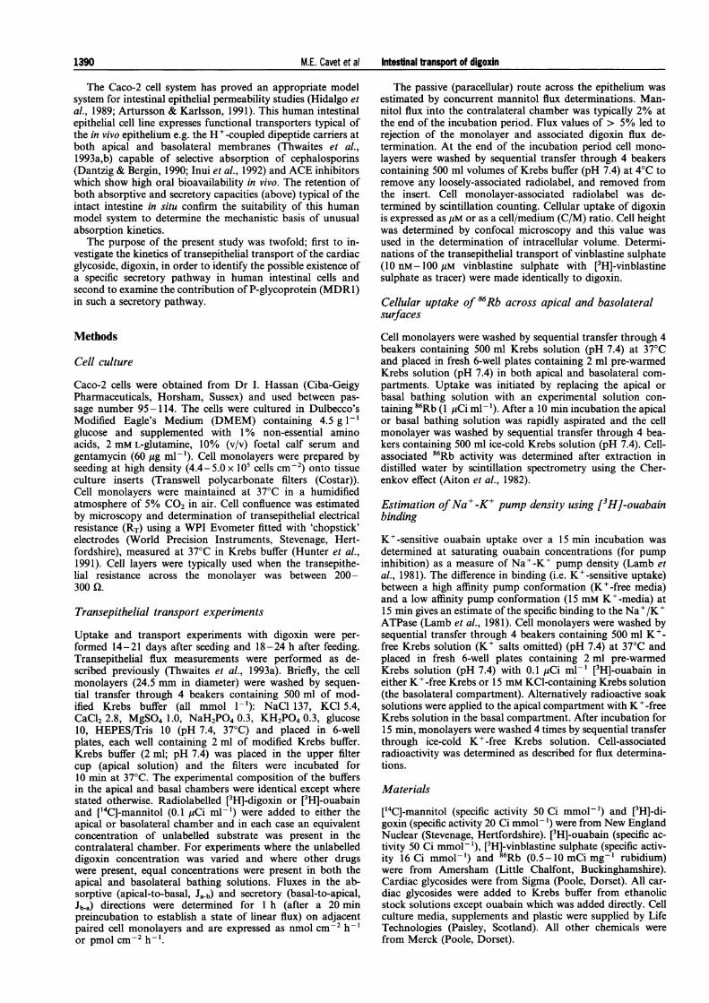
transepithelial mannitol permeability was not alteredla shows that in epithelial layers of human intestinal (Pa-b =0.16+0.01 cm h-' x 10-2 and Pb-a =0.16+0.01 cm h-sal to apical flux (Jb a) exceeds apical to basal flux (Jab) X 10-2 (n= 11), P >0.05, NS versus controls at 1 gM digoxin),-digoxin; that is Caco-2 epithelia are capable of net confirming that the high concentrations of cardiac glycoside don of digoxin from basal to apical surfaces. The ab- not adversely affect epithelial integrity over the time course ofpermeability of digoxin (Pab =Jab/Ca; where Ca is the experiment. Calculation of an apparent Km and Jmax for netsolution concentration) at 1 gM digoxin was digoxin secretion (Figure la) gave 0.65+0.18 mM and
.11 cm h' x 10-2 (n = 12). Concurrent measurement 46.4+ 10.4 nmol cm-2 h-1 respectively. This level of net se-absorptive permeability Pab for [14C]-mannitol gave cretion and that observed at the maximal concentration ex-
perimentally (200 gM digoxin) appears incompatible with themaximal secretory capacity observed for P-glycoprotein sub-strates such as vinblastine previously reported in Caco-2 in-
a testinal epithelia (1.3 nmol cm-2 h-', Hunter et al., 1993a,b).If vinblastine and digoxin secretion are mediated by an iden-tical pathway it would be expected that the maximal secretorycapacity would be identical. Accordingly, the maximal vin-blastine secretion and digoxin secretion were assessed in anidentical cell batch. For vinblastine secretion the measured Kmand Jmax were 13.8+2.8 gM and 1.3+0.1 nmol cm-2 h-'.(d.f. = 15). In contrast, the apparent Km and Jmax fordigoxin in the same cell batch were 0.26+0.11 mM and23.3+6.1 nmol cm-2 h-1.
Figure lb shows the concentration-dependence of apparent[3H]-digoxin uptake by Caco-2 cells from apical or basalbathing solutions. At low bathing solution digoxin con-centration e.g. 1 gM there was an asymmetry in that [3H]-di-goxin uptake at the basal surface exceeded that at the apical
0 50 100 150 200 surface; expressed as a cell to medium ratio (C/M) and as-
Digoxin concentration (gm) suming uniform distribution of the label within the cytosol, theb C/M ratio for apical [3H]-digoxin was 0.63 + 0.01, whereas for
basal [3H]-digoxin it was 1.57+0.13 (n= 12), P<0.01. Incomparison, that for [14C]-mannitol was 0.26 + 0.03 and0.17+0.01 at the apical and basolateral membranes respec-tively after the prolonged 1 h incubation for flux determina-tions. As digoxin concentration is raised to 200 gM these ratioschange to 1.26+0.28 and 0.58+0.01 respectively. Binding ofdigoxin to the filter was 0.034 + 0.009% (n =3) and0.036 + 0.006% (n = 3) of total radioactivity added in the apicaland basal compartments respectively (at 1 gM digoxin). Thiscompares with 0.123+0.006% (n=6) and 0.402+0.035%(n = 6) of total radioactive digoxin into the cells from the apicaland basolateral compartments respectively (at 1 gM digoxin).
rJ 1 t f fffAsfilter binding was small in comparison to accumulation__ I E_ 83______________ S within the cells, showed no asymmetry and may not be re-
1 10 50 100 150 200 presentative of the level of filter binding which occurs whenDigoxin concentration (gM) cells are present, cellular accumulation data were not modified
for filter binding.Figure 1 Concentration-dependence of secretion and cellularaccumulation of digoxin by human intestinal Caco-2 cell monolayers.(a) Transepithelial [3H]-digoxin fluxes (0.1 yCi/ml-, 3.6nM) were
determined in adjacent paired monolayers for apical to basal flux (Ja-
b) (l) and basal to apical flux (J, a) (A). Net secretory flux was = Jb-a-Ja-b(*)- Increasing concentrations of unlabelled digoxin were
present in both apical and basal bathing fluid compartments. All
data mean+s.e., n=11-12 epithelia per point. (b) Cellularaccumulation of digoxin after 1 h incubation with increasing digoxinconcentrations at the apical (open columns) and basal (hatchedcolumns) membrane surfaces. [3H]-digoxin present in either apical or
basal bathing solution. All data mean+ s.e., n = 11-12 epithelia.
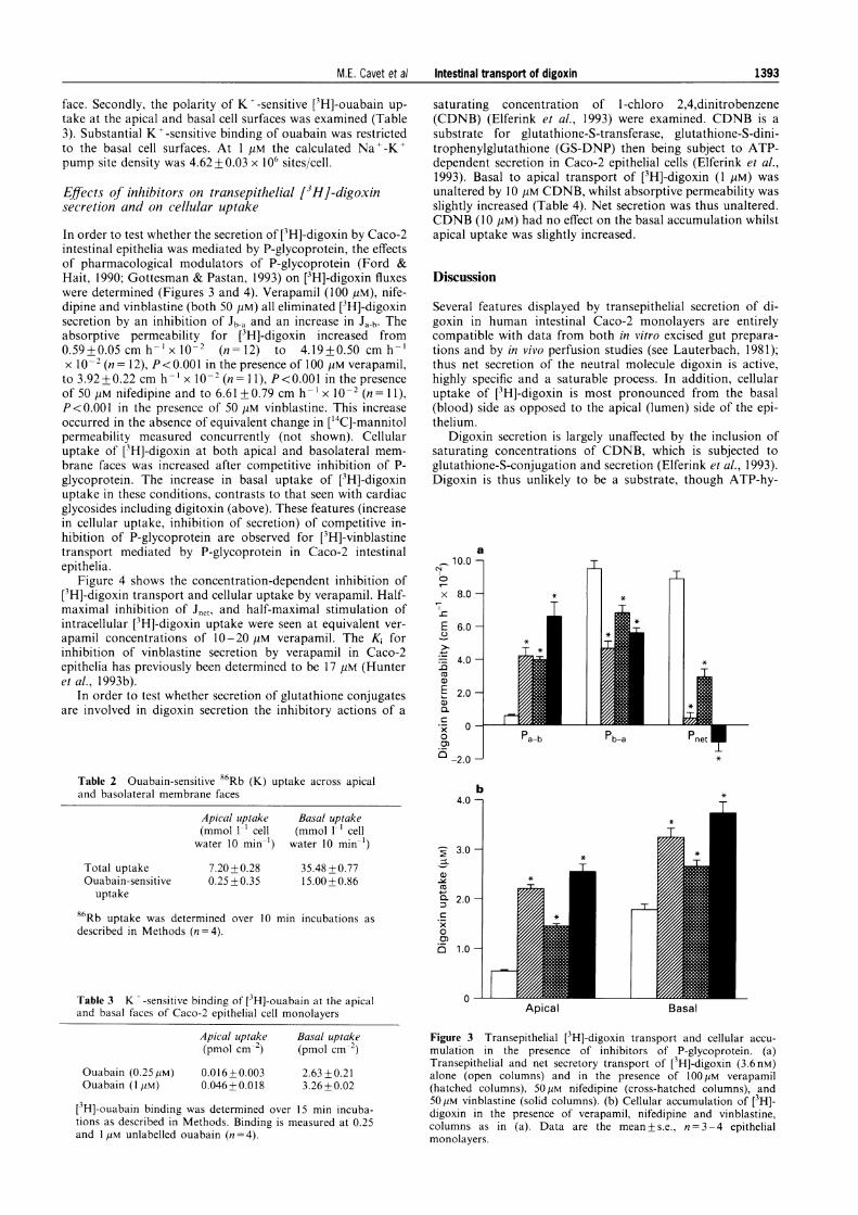
Effect of various cardiac glycosides on transepithelial[ H]-digoxin secretion and cellular accumulation
Figure 2 and Table 1 demonstrate a partial structure-activity
relationship for the ability of various cardiac glycosides to
affect transepithelial [3H]-digoxin secretion. Ouabain, stro-
phanthidin and convallatoxin (all at 20 pM and 100 gM) failed
to inhibit digoxin secretion. In contrast digitoxin (at 20 gM and
100 gM) and bufalin (100 gM) were effective in inhibition of net
[3H]-digoxin secretion. This inhibitory action by digitoxin was
Statisti
Resultslysis wa:analysismultiplefor MicregressivCambri(
Results
Transej[3H]-di,Figurecells badof [3H]-secretio:sorptiveapical0.87 +0of the
20
' 15E
EC 10x
x 50CD
iCu
Cu
0..C
0C)
1391M.E. Cavet et al Intestinal transport of digoxin

M.E. Cavet et al Intestinal transport of digoxin
due to both a reduction in Jb-a and an increase in absorptiveflux (Ja-b) (Figure 2a). The absorptive permeability for [3H]-digoxin increased from 0.47 +0.06 cm h-1 x 10⁻² (n=4) to3.51+0.72 cm h-'x 10-2 (n=3), P<0.01 in the presence of
a-, 10.0-C4
0
x 8.0-
E 6.0-+-
._ 4.0.0a)E 2.0-CL
00)0 -2.0
b2.5 -1
2.0-2
:I
az,v, 1.5-
x 1.0-0CD 5
0.5 -
o0
Vnet
Apical Basal
Figure 2 Transepithelial [3H]-digoxin transport and cellular accu-mulation in the presence of ouabain and digitoxin. (a) Transepithelialand net secretory transport of [3H]-digoxin (3.6 nM) alone (opencolumns) and in the presence of 20yM ouabain (upward slopinghatched columns,/), 100yM ouabain (cross-hatched columns), 20MMdigitoxin (downward sloping hatched columns,\), and 100MMdigitoxin (solid columns). (b) Cellular accumulation of [3H]-digoxinin the presence of ouabain and digitoxin as (a). Data are themean+s.e., n=3-4 epithelial monolayers per data point.
100 gM digitoxin. This increase occurred in the absence ofchange in ['4C]-mannitol permeability measured concurrently(control Pab for mannitol=0.23+0.01 cm h-1 x 10⁻² (n=4),plus 100 jiM digitoxin Pab for mannitol=0.24+ 0.19cm h-' x 10-2 (n=3) P>0.05, NS), confirming that epithelialintegrity was not affected in these conditions. The ability ofdigitoxigenin, the aglycone of digitoxin, to inhibit [3H]-digoxinsecretion was also investigated. At 20 gM and 100 gM digi-toxigenin failed to inhibit secretory [3H]-digoxin permeability(Pnet in controls was 6.80+0.53 cm h-' x 10-2, plus digitox-igenin 5.92+0.15 cm h- x 10-2 and 6.70+0.27 cm h-lx 10-2 respectively, all n= 4, NS versus control data).
Since digoxin itself fails to compete effectively with [jH]-digoxin for secretion, an absence of inhibition as with ouabain(Figure 2a) may reflect a low-affinity interaction with the se-cretory transport. Accordingly, transepithelial [3H]-ouabainfluxes were measured to confirm that no secretion of this car-diac glycoside was present. At 7 nM ouabain Pab was just0.23+0.06 cm h-' x 10-' (n=4), whilst Pba was 0.15+0.03cm h'l x I0O- (n = 4). These permeabilities were not altered byinclusion of 100 jyM ouabain, digoxin or digitoxin. Thus thereis no evidence of transepithelial secretion, the absolute per-meability of this molecule is markedly lower than either di-goxin or mannitol and the transepithelial transport isconsistent with an entirely passive mechanism.
Figure 2b and Table 1 show that cardiac glycosides haveopposite actions on [3H]-digoxin uptake at the apical and ba-solateral membrane faces. Digitoxin at 20 gM and 100 gM andbufalin at 100 pM markedly increased uptake across the apicalmembrane; ouabain, strophanthidin and convallatoxin (at100 jyM) were without effect. In contrast ouabain, stro-phanthidin and convallatoxin all depressed [3H]-digoxin up-take at the basolateral membrane. At the basolateralmembrane face, digitoxin (100 gM) had no effect on uptakeand bufalin (100 pM) depressed [3H]-digoxin uptake comparedto control levels, but gave an increase in cellular uptake at theapical membrane in a concentration-dependent manner incomparison to those cardiac glycosides that did not inhibitdigoxin transepithelial secretion.
It is likely that the ability of cardiac glycosides to reduce[3H]-digoxin uptake at the basolateral membrane face is relatedto their ability to bind to and inhibit the Na+-K' pump.Accordingly, the polarity of expression of the Na'-K'ATPase was investigated by determining the location of oua-bain-sensitive 86Rb(K) uptake (Table 2). 86Rb(K) uptakeacross the basolateral membrane was 5 fold higher than thatacross the apical membrane. A ouabain-sensitive componentof 86Rb(K) uptake was observed only at the basolateral sur-
Table 1 Effect of cardiac glycosides on the transport and cellular uptake of digoxin (1 gM) (n = 3-11)
Relativepotency
(digitoxigenin = 1) Pne, (cm h-' x 102)
Control
Digoxin(100 MM)
Strophanthidin(20 MM)
Strophanthidin(100 UM)
Convallatoxin(20 MM)
Convallatoxin(100 AM)
Bufalin(20 gM))
Bufalin(100 MM)
0.3 to 6.3
0.1 to 0.5
1.3 to 3.8
10.7
7.70+0.31(100%)
7.33 +0.43(95.2%)
8.11 +0.65(105%)
6.86+0.52(89.1%)
8.77 + 0.38(114%)
7.88 + 0.69(102%)
7.22+0.47(93.8%)
2.19+0.07(28.4%)*
Apicaluptake (MM)0.46+0.01(100%)
0.63 +0.10(137%)
0.36+0.03(78.3%)
0.38+0.00(82.6%)
0.31+0.00(67.4%)
0.35 +0.02(76.1%)
0.53 + 0.04(115%)
0.90 + 0.03(196%)*
Basaluptake (MM)
1.59 +0.09(100%)
0.41 +0.02(25.8%)*0.44+0.04(27.7%)*0.49 +0.02(30.8%)*0.48+0.01(30.2%)*0.45 + 0.03(28.3%)*0.65 + 0.03(40.9%)*1.01 +0.04(63.5%)*
Relative potencies of cardiac glycosides for inotropic activity from Thomas et al. (1990). * Significantly different from control values:P < 0.05.
1392
M.E. Cavet et al Intestinal transport of digoxin
face. Secondly, the polarity of Kt -sensitive [3H]-ouabain up-take at the apical and basal cell surfaces was examined (Table3). Substantial K -sensitive binding of ouabain was restrictedto the basal cell surfaces. At 1 AM the calculated Na'-K'pump site density was 4.62 +0.03 x 10⁶ sites/cell.
Effects of inhibitors on transepithelial [3H]-digoxinsecretion and on cellular uptake
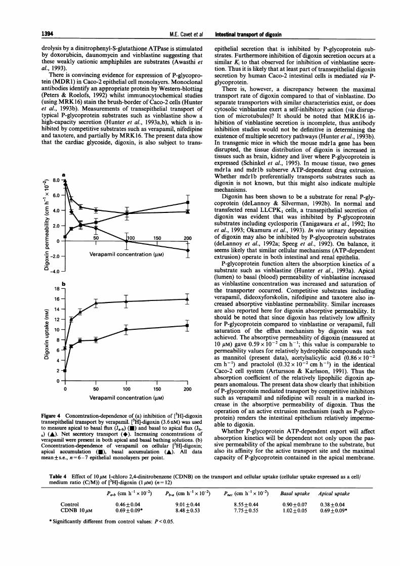
In order to test whether the secretion of ['H]-digoxin by Caco-2intestinal epithelia was mediated by P-glycoprotein, the effectsof pharmacological modulators of P-glycoprotein (Ford &Hait, 1990; Gottesman & Pastan, 1993) on [3H]-digoxin fluxeswere determined (Figures 3 and 4). Verapamil (100 pM), nife-dipine and vinblastine (both 50 pM) all eliminated [3H]-digoxinsecretion by an inhibition of Jb-a and an increase in Ja-b. Theabsorptive permeability for [3H]-digoxin increased from0.59+0.05 cm h-' x o0-2 (n= 12) to 4.19+0.50 cm h-'x 10-2 (n= 12), P<0.001 in the presence of 100 pM verapamil,to 3.92 +0.22 cm h'- x 10-2 (n= 11), P<0.00I in the presenceof 50 pM nifedipine and to 6.61+0.79 cm h-' x 10-2 (n= 11),P<0.001 in the presence of 50,lM vinblastine. This increaseoccurred in the absence of equivalent change in ['4C]-mannitolpermeability measured concurrently (not shown). Cellularuptake of [3H]-digoxin at both apical and basolateral mem-brane faces was increased after competitive inhibition of P-glycoprotein. The increase in basal uptake of [3H]-digoxinuptake in these conditions, contrasts to that seen with cardiacglycosides including digitoxin (above). These features (increasein cellular uptake, inhibition of secretion) of competitive in-hibition of P-glycoprotein are observed for [3H]-vinblastinetransport mediated by P-glycoprotein in Caco-2 intestinalepithelia.
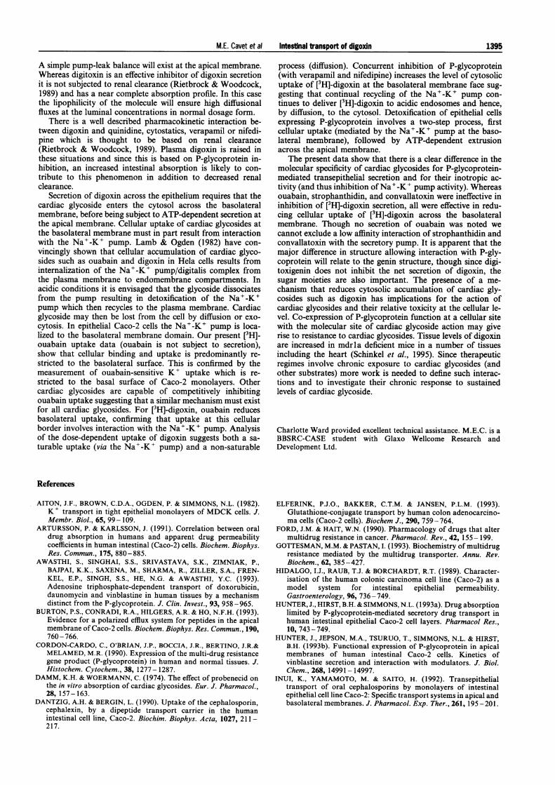
Figure 4 shows the concentration-dependent inhibition of[3H]-digoxin transport and cellular uptake by verapamil. Half-maximal inhibition of Jnet, and half-maximal stimulation ofintracellular ['H]-digoxin uptake were seen at equivalent ver-apamil concentrations of 10-20 gM verapamil. The Ki forinhibition of vinblastine secretion by verapamil in Caco-2epithelia has previously been determined to be 17 JM (Hunteret al., 1993b).
In order to test whether secretion of glutathione conjugatesare involved in digoxin secretion the inhibitory actions of a
Table 2 Ouabain-sensitive 86Rb (K) uptake across apicaland basolateral membrane faces
Apical uptake(mmol 1-' cell
water 10 min-')
Total uptakeOuabain-sensitive
uptake
Basal uptake(mmol 1' cell
water 10 min-')
7.20+0.28 35.48+0.770.25+0.35 15.00+0.86
86Rb uptake was determined over 10 min incubations asdescribed in Methods (n=4).
Table 3 K -'-sensitive binding of [3H]-ouabain at the apicaland basal faces of Caco-2 epithelial cell monolayers
Apical uptake Basal uptake(pmol cm 2) (pmol cm 2)
Ouabain (0.25IM)Ouabain (I Jam)
0.016+0.003 2.63+0.210.046+0.018 3.26+0.02
[3H]-ouabain binding was determined over 15 mmn incuba-
tions as described in Methods. Binding is measured at 0.25and 1 [M unlabelled ouabain (n = 4).
saturating concentration of l-chloro 2,4,dinitrobenzene(CDNB) (Elferink et al., 1993) were examined. CDNB is asubstrate for glutathione-S-transferase, glutathione-S-dini-trophenylglutathione (GS-DNP) then being subject to ATP-dependent secretion in Caco-2 epithelial cells (Elferink et al.,1993). Basal to apical transport of ['H]-digoxin (1 tiM) wasunaltered by 10 JM CDNB, whilst absorptive permeability wasslightly increased (Table 4). Net secretion was thus unaltered.CDNB (10 pM) had no effect on the basal accumulation whilstapical uptake was slightly increased.
Discussion
Several features displayed by transepithelial secretion of di-goxin in human intestinal Caco-2 monolayers are entirelycompatible with data from both in vitro excised gut prepara-tions and by in vivo perfusion studies (see Lauterbach, 1981);thus net secretion of the neutral molecule digoxin is active,highly specific and a saturable process. In addition, cellularuptake of [3H]-digoxin is most pronounced from the basal(blood) side as opposed to the apical (lumen) side of the epi-thelium.
Digoxin secretion is largely unaffected by the inclusion ofsaturating concentrations of CDNB, which is subjected toglutathione-S-conjugation and secretion (Elferink et al., 1993).Digoxin is thus unlikely to be a substrate, though ATP-hy-
a10.0 a P
2-2.0 *
x 8.0
E 6.0
4.0
CL
.CR Qo Pa-b Pb-aP
-.
b4.0 7
3.0-
a)
0 2.0-C:.x0
o 1.0-
0-
-r
T
rApical Basal
Figure 3 Transepithelial [3H]-digoxin transport and cellular accu-mulation in the presence of inhibitors of P-glycoprotein. (a)Transepithelial and net secretory transport of [3H]-digoxin (3.6 nM)alone (open columns) and in the presence of 100 ,UM verapamil(hatched columns), 50[M nifedipine (cross-hatched columns), and50[pM vinblastine (solid columns). (b) Cellular accumulation of [3H]-digoxin in the presence of verapamil, nifedipine and vinblastine,columns as in (a). Data are the mean + s.e., n = 3 - 4 epithelialmonolayers.
1393
t- .u
M.E. Cavet et at Intestinal transport of digoxin
drolysisby doxcthese weal., 1993
Theretein (MIantibodi(Peters z(using Net al., 1typical Ihigh-cafhibited land tax(that the
v- 8.00
X 6.0
T-
._
2.00.2o
Cx-2.0.00)'R-4.0
Figure 4transepitito measui
a) (A). IverapamilConcentrapical amean+ s.
by a dinitrophenyl-S-glutathione ATPase is stimulated epithelial secretion that is inhibited by P-glycoprotein sub-)rubicin, daunomycin and vinblastine suggesting that strates. Furthermore inhibition of digoxin secretion occurs at aDakly cationic amphiphiles are substrates (Awasthi et similar Ki to that observed for inhibition of vinblastine secre-3). tion. Thus it is likely that at least part of transepithelial digoxinD is convincing evidence for expression of P-glycopro- secretion by human Caco-2 intestinal cells is mediated via P-DR1) in Caco-2 epithelial cell monolayers. Monoclonal glycoprotein.ies identify an appropriate protein by Western-blotting There is, however, a discrepancy between the maximal& Roelofs, 1992) whilst immunocytochemical studies transport rate of digoxin compared to that of vinblastine. Do4RK16) stain the brush-border of Caco-2 cells (Hunter separate transporters with similar characteristics exist, or does.993b). Measurements of transepithelial transport of cytosolic vinblastine exert a self-inhibitory action (via disrup-P-glycoprotein substrates such as vinblastine show a tion of microtubules)? It should be noted that MRK16 in-)acity secretion (Hunter et al., 1993a,b), which is in- hibition of vinblastine secretion is incomplete, thus antibodyby competitive substrates such as verapamil, nifedipine inhibition studies would not be definitive in determining the)tere, and partially by MRK16. The present data show existence of multiple secretory pathways (Hunter et al., 1993b).cardiac glycoside, digoxin, is also subject to trans- In transgenic mice in which the mouse mdrla gene has been
disrupted, the tissue distribution of digoxin is increased intissues such as brain, kidney and liver where P-glycoprotein isexpressed (Schinkel et al., 1995). In mouse tissue, two genesmdrla and mdrlb subserve ATP-dependent drug extrusion.
a Whether mdrlb preferentially transports substrates such asdigoxin is not known, but this might also indicate multiplemechanisms.
Digoxin has been shown to be a substrate for renal P-gly-coprotein (deLannoy & Silverman, 1992b). In normal andtransfected renal LLCPKI cells, a transepithelial secretion ofdigoxin was evident that was inhibited by P-glycoproteinsubstrates including cyclosporin (Tanigawara et al., 1992; Itoet al., 1993; Okamura et al., 1993). In vivo urinary deposition
I r 50 100 150 200 of digoxin may also be inhibited by P-glycoprotein substrates(deLannoy et al., 1992a; Speeg et al., 1992). On balance, it
i concntraion gm)seems likely that similar cellular mechanisms (ATP-dependent_ Verapamil concentration (SM) lextrusion) operate in both intestinal and renal epithelia.P-glycoprotein function alters the absorption kinetics of a
substrate such as vinblastine (Hunter et al., 1993a). Apical(lumen) to basal (blood) permeability of vinblastine increased
b as vinblastine concentration was increased and saturation ofthe transporter occurred. Competitive substrates includingverapamil, dideoxyforskolin, nifedipine and taxotere also in-creased absorptive vinblastine permeability. Similar increasesare also reported here for digoxin absorptive permeability. Itshould be noted that since digoxin has relatively low affinityfor P-glycoprotein compared to vinblastine or verapamil, fullsaturation of the efflux mechanism by digoxin was notachieved. The absorptive permeability of digoxin (measured at10 gM) gave 0.59 x 10 2 cm h-'; this value is comparable topermeability values for relatively hydrophilic compounds suchas mannitol (present data), acetylsalicylic acid (0.86 x 10⁻²cm h-') and practolol (0.32 x 10⁻²cm h-') in the identicalCaco-2 cell system (Artursson & Karlsson, 1991). Thus the
____________________________________ |absorption coefficient of the relatively lipophilic digoxin ap-pears anomalous. The present data show clearly that inhibition
0 50 100 150 200 of P-glycoprotein mediated transport by competitive inhibitorsVerapamil concentration (gM) such as verapamil and nifedipine will result in a marked in-
crease in the absorptive permeability of digoxin. Thus theCo[3H]-digoxin operation of an active extrusion mechanism (such as P-glyco-Concentration-dependence of (a) inhibition of [3Hl-digoxin protein) renders the intestinal epithelium relatively imperme-
ielial transport by verapamil. [3H]-digoxin (3.6nM) was used able to digxnre apical to basal flux (Ja-b) (E) and basal to apical flux (J1- aWheto P-goxln.Net secretory transport (*). Increasing concentrations of Whether P-glycoprotein ATP-dependent export will affectil were present in both apical and basal bathing solutions. (b) absorption kinetics will be dependent not only upon the pas-ation-dependence of verapamil on cellular [3Hl-digoxin; sive permeability of the apical membrane to the substrate, butLccumulation (U), basal accumulation (A). All data also its affinity for the active transport site and the maximal.e., n = 6-7 epithelial monolayers per point, capacity of P-glycoprotein contained in the apical membrane.
Table 4 Effect of 10gM I-chloro 2,4-dinitrobenzene (CDNB) on the transport and cellular uptake (cellular uptake expressed as a cell/medium ratio (C/M)) of [3H]-digoxin (1 AM) (n = 12)
Pa-b (cm h-' x 10-2) Pba (cm h-' x 10-2) Pt (cm h-' X 10-2) Basal uptake Apical uptake
8.55+0.447.75 +0.55
0.90+0.07 0.38 +0.041.02+0.05 0.69 +0.09*
*Significantly different from control values: P < 0.05.
ice
0)a)Q1XR0o0_
18
16
14
12
10
8
6
4
2
0
ControlCDNB IOuAM
0.46+0.040.69+ 0.09*
9.01+0.448.48 +0.53
1394
I
M.E. Cavet et al Intestinal transport of digoxin 1395
A simple pump-leak balance will exist at the apical membrane.Whereas digitoxin is an effective inhibitor of digoxin secretionit is not subjected to renal clearance (Rietbrock & Woodcock,1989) and has a near complete absorption profile. In this casethe lipophilicity of the molecule will ensure high diffusionalfluxes at the luminal concentrations in normal dosage form.
There is a well described pharmacokinetic interaction be-tween digoxin and quinidine, cytostatics, verapamil or nifedi-pine which is thought to be based on renal clearance(Rietbrock & Woodcock, 1989). Plasma digoxin is raised inthese situations and since this is based on P-glycoprotein in-hibition, an increased intestinal absorption is likely to con-tribute to this phenomenon in addition to decreased renalclearance.
Secretion of digoxin across the epithelium requires that thecardiac glycoside enters the cytosol across the basolateralmembrane, before being subject to ATP-dependent secretion atthe apical membrane. Cellular uptake of cardiac glycosides atthe basolateral membrane must in part result from interactionwith the Na+-K+ pump. Lamb & Ogden (1982) have con-vincingly shown that cellular accumulation of cardiac glyco-sides such as ouabain and digoxin in Hela cells results frominternalization of the Na'-K+ pump/digitalis complex fromthe plasma membrane to endomembrane compartments. Inacidic conditions it is envisaged that the glycoside dissociatesfrom the pump resulting in detoxification of the Na'-K+pump which then recycles to the plasma membrane. Cardiacglycoside may then be lost from the cell by diffusion or exo-cytosis. In epithelial Caco-2 cells the Na+-K+ pump is loca-lized to the basolateral membrane domain. Our present [3H]-ouabain uptake data (ouabain is not subject to secretion),show that cellular binding and uptake is predominantly re-stricted to the basolateral surface. This is confirmed by themeasurement of ouabain-sensitive K+ uptake which is re-stricted to the basal surface of Caco-2 monolayers. Othercardiac glycosides are capable of competitively inhibitingouabain uptake suggesting that a similar mechanism must existfor all cardiac glycosides. For [3H]-digoxin, ouabain reducesbasolateral uptake, confirming that uptake at this cellularborder involves interaction with the Na+-K+ pump. Analysisof the dose-dependent uptake of digoxin suggests both a sa-turable uptake (via the Na+-K+ pump) and a non-saturable
process (diffusion). Concurrent inhibition of P-glycoprotein(with verapamil and nifedipine) increases the level of cytosolicuptake of [3H]-digoxin at the basolateral membrane face sug-gesting that continual recycling of the Na'-K+ pump con-tinues to deliver [3H]-digoxin to acidic endosomes and hence,by diffusion, to the cytosol. Detoxification of epithelial cellsexpressing P-glycoprotein involves a two-step process, firstcellular uptake (mediated by the Na'-K' pump at the baso-lateral membrane), followed by ATP-dependent extrusionacross the apical membrane.
The present data show that there is a clear difference in themolecular specificity of cardiac glycosides for P-glycoprotein-mediated transepithelial secretion and for their inotropic ac-tivity (and thus inhibition ofNa'-K' pump activity). Whereasouabain, strophanthidin, and convallatoxin were ineffective ininhibition of [3H]-digoxin secretion, all were effective in redu-cing cellular uptake of [3H]-digoxin across the basolateralmembrane. Though no secretion of ouabain was noted wecannot exclude a low affinity interaction of strophanthidin andconvallatoxin with the secretory pump. It is apparent that themajor difference in structure allowing interaction with P-gly-coprotein will relate to the genin structure, though since digi-toxigenin does not inhibit the net secretion of digoxin, thesugar moieties are also important. The presence of a me-chanism that reduces cytosolic accumulation of cardiac gly-cosides such as digoxin has implications for the action ofcardiac glycosides and their relative toxicity at the cellular le-vel. Co-expression of P-glycoprotein function at a cellular sitewith the molecular site of cardiac glycoside action may giverise to resistance to cardiac glycosides. Tissue levels of digoxinare increased in mdrla deficient mice in a number of tissuesincluding the heart (Schinkel et al., 1995). Since therapeuticregimes involve chronic exposure to cardiac glycosides (andother substrates) more work is needed to define such interac-tions and to investigate their chronic response to sustainedlevels of cardiac glycoside.
Charlotte Ward provided excellent technical assistance. M.E.C. is aBBSRC-CASE student with Glaxo Wellcome Research andDevelopment Ltd.
References
AITON, J.F., BROWN, C.D.A., OGDEN, P. & SIMMONS, N.L. (1982).K+ transport in tight epithelial monolayers of MDCK cells. J.Membr. Biol., 65, 99-109.
ARTURSSON, P. & KARLSSON, J. (1991). Correlation between oraldrug absorption in humans and apparent drug permeabilitycoefficients in human intestinal (Caco-2) cells. Biochem. Biophys.Res. Commun., 175, 880-885.
AWASTHI, S., SINGHAI, S.S., SRIVASTAVA, S.K., ZIMNIAK, P.,BAJPAI, K.K., SAXENA, M., SHARMA, R., ZILLER, S.A., FREN-KEL, E.P., SINGH, S.S., HE, N.G. & AWASTHI, Y.C. (1993).Adenosine triphosphate-dependent transport of doxorubicin,daunomycin and vinblastine in human tissues by a mechanismdistinct from the P-glycoprotein. J. Clin. Invest., 93, 958 -965.
BURTON, P.S., CONRADI, R.A., HILGERS, A.R. & HO, N.F.H. (1993).Evidence for a polarized efflux system for peptides in the apicalmembrane of Caco-2 cells. Biochem. Biophys. Res. Commun., 190,760-766.
CORDON-CARDO, C., O'BRIAN, J.P., BOCCIA, J.R., BERTINO, J.R.&MELAMED, M.R. (1990). Expression of the multi-drug resistancegene product (P-glycoprotein) in human and normal tissues. J.Histochem. Cytochem., 38, 1277-1287.
DAMM, K.H. & WOERMANN, C. (1974). The effect of probenecid onthe in vitro absorption of cardiac glycosides. Eur. J. Pharmacol.,28, 157-163.
DANTZIG, A.H. & BERGIN, L. (1990). Uptake of the cephalosporin,cephalexin, by a dipeptide transport carrier in the humanintestinal cell line, Caco-2. Biochim. Biophys. Acta, 1027, 211 -217.
ELFERINK, P.J.O., BAKKER, C.T.M. & JANSEN, P.L.M. (1993).Glutathione-conjugate transport by human colon adenocarcino-ma cells (Caco-2 cells). Biochem J., 290, 759-764.
FORD, J.M. & HAIT, W.N. (1990). Pharmacology of drugs that altermultidrug resistance in cancer. Pharmacol. Rev., 42, 155-199.
GOTTESMAN, M.M. & PASTAN, I. (1993). Biochemistry of multidrugresistance mediated by the multidrug transporter. Annu. Rev.Biochem., 62, 385-427.
HIDALGO, I.J., RAUB, T.J. & BORCHARDT, R.T. (1989). Character-isation of the human colonic carcinoma cell line (Caco-2) as amodel system for intestinal epithelial permeability.Gastroenterology, 96, 736-749.
HUNTER, J., HIRST, B.H. & SIMMONS, N.L. (1993a). Drug absorptionlimited by P-glycoprotein-mediated secretory drug transport inhuman intestinal epithelial Caco-2 cell layers. Pharmacol Res.,10, 743-749.
HUNTER, J., JEPSON, M.A., TSURUO, T., SIMMONS, N.L. & HIRST,B.H. (1993b). Functional expression of P-glycoprotein in apicalmembranes of human intestinal Caco-2 cells. Kinetics ofvinblastine secretion and interaction with modulators. J. Biol.Chem., 268, 14991-14997.
INUI, K., YAMAMOTO, M. & SAITO, H. (1992). Transepithelialtransport of oral cephalosporins by monolayers of intestinalepithelial cell line Caco-2: Specific transport systems in apical andbasolateral membranes. J. Pharmacol. Exp. Ther., 261, 195 - 201.
1396 M.E. Cavet et al Intestinal transport of digoxin
ITO, S., KOREN, G., HARPER, P.A. & SILVERMAN, M. (1993).Energy-dependent transport of digoxin across renal tubular cellmonolayers (LLCPK1). Can. J. Physiol. Pharmacol., 71, 40-47.
KARLSSON, J., KUO, S., ZIEMNIAK, J. & ARTURSSON, P. (1993).Transport of celiprolol across human intestinal epithelial (Caco-2) cells: mediation of secretion by multiple transporters includingP-glycoprotein. Br. J. Pharmacol., 110, 1009- 1016.
LAMB, J.F. & OGDEN, P. (1982). Internalisation of ouabain andreplacement of sodium pumps in the plasma membranes of HeLacells following block with cardiac glycosides. Q. J. Exp. Physiol.,67, 105-119.
LAMB, J.F., OGDEN, P. & SIMMONS, N.L. (1981). Autoradiographiclocalisation of [3H]-ouabain bound to cultured epithelial cellmonolayers of MDCK cells. Biochim. Biophys. Acta, 644, 333-340.
DELANNOY, I.A.M., KOREN, G., KLEIN, J., CHARUK, J. & SILVER-MAN, M. (1992a). Cyclosporin and quinidine inhibition of renaldigoxin excretion; evidence for luminal secretion of digoxin. Am.J. Physiol., 263, F613-F622.
DELANNOY, I.A.M. & SILVERMAN, M. (1992b). The MDR1 geneproduct P-glycoprotein mediates the transport of the cardiacglycoside, digoxin. Biochem. Biophys. Res. Commun., 30, 551-557.
LAUTERBACH, F. (1981). Intestinal absorption and secretion ofcardiac glycosides. In Cardiac Glycosides, Part II. ed. Greeff, K.pp. 105-139. Berlin, Heidelberg, New York: Springer-Verlag.
OKAMURA, N., HIRAI, M., TANIGAWARA, Y., TANAKA, K.,YASUHARA, M., UEDA, K., KOMANO, T. & HORI, R. (1993).Digoxin-cyclosporin A interaction: Modulation of the multidrugtransporter P-glycoprotein in the Kidney. J. Pharmacol. Exp.Ther., 266, 1614-1625.
PETERS, W.H.M. & ROELOFS, H.M.J. (1992). Biochemical character-isation of resistance to mitoxanthrone and adriamycin in Caco-2human adenocarcinoma cells; a possible role for glutathione-S-transferases. Cancer Res., 52, 1886- 1890.
RIETBROCK, N. & WOODCOCK, B.G. (1989). Handbook of Renal-Independent Cardiac Glycosides; Pharmacology and ClinicalPharmacology. pp. 71-94. Chichester, UK: Ellis Horwood Ltd.
SCHINKEL, A.H., WAGENAAR, E., VAN DEEMTER, L., MOL,C.A.A.M. & BORST, P. (1995). Absence of the mdrla P-glycoprotein in mice affects tissue distribution and pharmacoki-netics of dexamethasone, digoxin and cyclosporin A. J. Clin.Invest., 96, 1698-1705.
SPEEG, K.V., MALDONADA, A.L., LIACI, J. & MUIRHEAD, D. (1992).Effect of cyclosporine on colchicine secretion by the kidneymultidrug transporter studied in vivo. J. Pharmacol. Exp. Ther.,261, 50-55.
TANIGAWARA, Y., OKAMURA, N., HIRAI, M., YASUHARA, M.,UEDA, K., KIOKA, N., KOMANO, T. & HORI, R. (1992). Transportof digoxin by human P-glycoprotein expressed in a porcinekidney epithelial cell-line (LLCPK1). J. Pharmacol. Exp. Ther.,263, 840-845.
THOMAS, R., GRAY, P. & ANDREWS, J. (1990). Digitalis: its mode ofaction, receptor and structure-activity relationships. Adv. DrugRes., 19, 313 - 562.
THWAITES, D.T., BROWN, C.D.A., HIRST, B.H. & SIMMONS, N.L.(1993a). Transepithelial glycylsarcosine transport in intestinalCaco-2 cells mediated by expression of H + -coupled carriers atboth apical and basal membranes. J. Biol. Chem., 268, 7640-7642.
THWAITES, D.T., BROWN, C.D.A., HIRST, B.H. & SIMMONS, N.L.(1993b). H + -coupled dipeptide (glycylsarcosine) transport acrossapical and basal borders of human intestinal Caco-2 cellmonolayers display distinctive characteristics. Biochim. Biophys.Acta, 1151, 237-245.
(Received January 15, 1996revised March 12, 1996
accepted March 29, 1996)