translational health research
TRANSCRIPT

Translational Health Research
Cervical cancer

http://www.thelancet.com/

YESTERDAYIn 1940s, cervical cancer was a major cause of death among women of childbearing age in the United States.
TODAYCervical cancer – once one of the most common cancers affecting U.S. women – now ranks 14th in frequency. Because precancerous lesions found by Pap smears can be treated and cured before they develop into cancer, and because cervical cancer is often detected before it becomes advanced, the incidence and death rates for this disease are relatively low.
HOW?Better visualization:- Introduction Papanicolaou (Pap) smear test in which a sample of cervical cells is examined under a microscope to detect cellular abnormalities. The incidence of invasive cervical cancer declined dramatically. Between 1955 and 1992, U.S. cervical cancer incidence and death rates declined by more than 60%. and automationAutomation:- Introduction of ThinPrep®, FocalPoint® (FDA approved) systems which screens and suggest potential cases to cytopathologists
http://report.nih.gov/NIHfactsheets/ViewFactSheet.aspx?csid=76http://report.nih.gov/NIHfactsheets/ViewFactSheet.aspx?csid=76

‘’Cancer Facts and Figures 2012’’ America cancer society

Why cant I just mimic the existing technology?

Experinced CytopathologistsCOSTAccuracy (False neg.)Massive screening Automated imagingComputational FacilityExtension to rural areasBusiness ModelAwareness..
OUR CONCERN


Monolayer Preparation
9
Collection of cervical smears from transformation zone and endo cervix by Ayer’s spatula and endo-cervical brush respectively into polysol solution.
Centrifugation at 2000-4000 rpm for 5 mins, supernatant discarded and re-suspension of pallet in fresh polysol solution
Preparation of cervical monolayer smear using cyto-centrifuge with appropriate funnel and filter cards.
Fixation of the smear with 95% ethanol for 10 mins.
Pap staining of the monolayer cervical smear using hematoxylin (stains nucleus blue), orange G (stains cytoplasm of superficial cells pink) and eosine-azure (stains cytoplasm of intermediate cells blue to green)

Feature Identification & Significance
No. Nuclear Feature Significance
1 Brightness of Nucleus Most Significant
2 Area of Nucleus Significant
3 Irregularity of the Nuclear Membrane
Significant
4 Nucleus Roundness Significant
5 Size of Chromatin Granule
Significant
Table shows the importance of feature significance in 40 X magnification
12
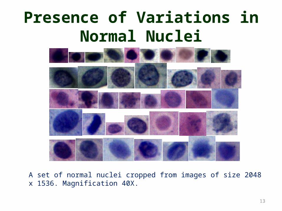
Presence of Variations in Normal Nuclei
13
A set of normal nuclei cropped from images of size 2048 x 1536. Magnification 40X.

Presence of Variations in Abnormal Nuclei
14
A set of abnormal nuclei. Image size 404 x 270. Courtesy by International Agency for Research on Cancer cervical cells image database. Magnification 40X

Ambiguity in Nuclear Features
15
a b c da.Malignant Intermediate Cell Nucleus. b.Normal Parabasal Cell Nucleusc.Malignant Superficial Cell Nucleus. d.Normal Parabasal Cell Nucleus

Feature Identificatio
n
Feature Identificatio
n
Classification
Classification
Implementating out of box Features and Classification
Feature Quantificatio
n
Feature Quantificatio
n
16

Nucleus Localization & Segmentation
A three phase technique is adopted for segmentation of nucleus.
17

18
Color Imbalance• As we are intend to implement color
image processing , there should be no disruptions in color cue
• Sources of descriptiona) Light source (temperature dependent)b) Digital camera (biasing of any one
channel of R,G,B)
CIE standard Color gamut provided and limitation of screen display

19
White Balancing Cont.
Pap stained image of a Normal smear under microscope at 40x
Pap stained image of a Abnormal smear under microscope at 40x
White Balancing Step
White Balanced Images

20
Image Enhancement
Top two images are white balanced. Bottom two are enhanced with CLAHE

21
Haematoxyline Probability Plane

Segmentaion performance
BilateralFilteredimage
22
InitialMask
ColorDeconvoutput
Original image
https://www.dropbox.com/sh/p2kt9e181mdx9eh/NfHrVzhrTC
L Planeof LabColorspace
Saturation +
Intensity
Level set Method
Morphological filtering

23
Results of Active Contour(Level Sets)

24Segmented results

25Segmented results

Target Features of the nucleus for computer assisted diagnosis
1. Area2. Perimeter3. ConvexArea4. Filled area5. Eccentricity6. MajorAxisLength7. MinorAxisLength8. EquivDiameter9. MinorAxisLength10. Standard deviation of Red color values on
nucleus11. Mean value of Red color value on nucleus12. Standard deviation of Green color values on
nucleus13. Mean value of Green color value on nucleus14. Standard deviation of Blue color values on
nucleus
1. Mean value of Blue color value on nucleus
2. Entropy of red color values of nucleus3. Entropy of Green color values of
nucleus4. Entropy of Blue color values of
nucleus5. contrast of gray values of nucleus6. correlation of gray values of nucleus7. energy homogeneity of gray values of
nucleus8. standard deviation of Laws mask
response9. mean of Laws mask response 10. standard deviation of LBP(local
binary pattern) response11. mean of LBP response
26

SVM based classification for 25 nuclear features
28