transformative concepts for drug design: target wrapping
TRANSCRIPT


Transformative Concepts for Drug Design:Target Wrapping

“This page left intentionally blank.”
Ariel Fernández
Transformative Conceptsfor Drug Design:Target Wrapping
123
Prof. Dr. Ariel FernándezKarl F. Hasselmann Chair in EngineeringRice UniversityDepartment of BioengineeringE200K George R. Brown HallHouston TX 77005USA
ISBN 978-3-642-11791-6 e-ISBN 978-3-642-11792-3DOI 10.1007/978-3-642-11792-3Springer Heidelberg Dordrecht London New York
Library of Congress Control Number: 2010922998
© Springer-Verlag Berlin Heidelberg 2010This work is subject to copyright. All rights are reserved, whether the whole or part of the material isconcerned, specifically the rights of translation, reprinting, reuse of illustrations, recitation, broadcasting,reproduction on microfilm or in any other way, and storage in data banks. Duplication of this publicationor parts thereof is permitted only under the provisions of the German Copyright Law of September 9,1965, in its current version, and permission for use must always be obtained from Springer. Violationsare liable to prosecution under the German Copyright Law.The use of general descriptive names, registered names, trademarks, etc. in this publication does notimply, even in the absence of a specific statement, that such names are exempt from the relevant protectivelaws and regulations and therefore free for general use.
Cover design: WMXDesign GmbH, Heidelberg
Printed on acid-free paper
Springer is part of Springer Science+Business Media (www.springer.com)
Preface
To a man with a hammer everything looks like a nail.Mark Twain
Notwithstanding the enticing promises of the post-genomic era, the pharmaceuticalworld appears to be in a state of disarray. Drug discovery seems riskier and moreuncertain than ever as projects get routinely terminated in mid-stage clinical trials,as the dearth of new targets becomes apparent, and as successful therapeutic agentsare often recalled whenever an idiosyncratic side effect is detected. Exploiting thehuge output of genomic data to make more efficacious and safer drugs has provento be much more difficult than anticipated. More than ever, the lead in the phar-maceutical industry depends on the ability to harness innovative research, and thistype of innovation can only come from one source: fundamental knowledge. Thisbook has a place in this scenario, as it introduces fundamental discoveries in basicbiomolecular research that hold potential to become transformative and broaden thetechnological base of the pharmaceutical industry.
The book takes a fresh and fundamental look at the problem of how to design aneffective drug with controlled specificity. Within the pharmaceutical industry, it isof course superfluous to recall that the principal bottleneck in developing new drugsis the clinical uncertainty stemming from the lack of control of specificity. Chemistsknow how to increase affinity, but when they do this, the affinity of the drug tostructurally similar molecules also increases, target discrimination becomes verydifficult, and adverse side-effects due to unwanted binding are usually sufficientlysevere to render the drug unusable.
The secret of how nature manages to design molecules with extraordinarily highand specific affinities lies in cooperativity. In medicine, we are nearly always work-ing in aqueous media and therefore cooperativity needs to be looked at in the specificcontext of aqueous systems.
Recognizing that these concepts are unfamiliar to most practitioners, the firstpart of this book (Chaps. 1, 2, 3, 4, 5, and 6) explains these matters very carefullystarting from a fairly elementary physico-chemical level. The second part of thebook (Chaps. 7, 8, 9, 10, 11, 12, 13, and 14) is devoted to practical applications. Weare aiming at nothing less than a paradigm shift in drug design.
Thus, cooperativity emerges as a molecular design principle in Chaps. 7, 8, 9, 10,11, 12, 13, and 14, but this incarnation is only possible after the concept is exploredfrom architectural, biophysical, bioinformatics and evolutionary perspectives in thepreparatory Chaps. 1, 2, 3, 4, 5, and 6.
v

vi Preface
This book is above all addressed to scientists working at the cutting edge ofresearch in the pharmaceutical industry, but the material is at the same time fullyaccessible to senior undergraduates or graduate students interested in fundamentalconcepts on drug discovery. It essentially covers my lectures on systems biol-ogy and molecular design, an elective undergraduate and graduate level course forbioengineering majors at Rice University.
It has been a pleasure to work with the talented staff at Springer. I am especiallygrateful to Marion Hertel (executive editor), and to Cornelia Kinsky, Beate Siek andSam Roobesh for their helpful cooperation and enduring patience.
Houston, USA Ariel Fernández

Contents
1 Protein Cooperativity and Wrapping: Two Themes in theTransformative Platform of Molecular Targeted Therapy . . . . . 11.1 Many-Body Problems for the Drug Designer . . . . . . . . . . 11.2 Cooperative Protein Interactions: The Need for the
Wrapping Concept . . . . . . . . . . . . . . . . . . . . . . . 31.3 Poorly Wrapped Hydrogen Bonds are Promoters
of Protein Associations . . . . . . . . . . . . . . . . . . . . . 61.4 Wrapping Defects Are Sticky . . . . . . . . . . . . . . . . . . 91.5 Cooperative Drug–Target Associations: A Window
into Molecular Engineering Possibilities . . . . . . . . . . . . 12References . . . . . . . . . . . . . . . . . . . . . . . . . . . . . . . 14
2 Wrapping Defects and the Architecture of Soluble Proteins . . . . 172.1 How Do Soluble Proteins Compensate
for Their Wrapping Defects? . . . . . . . . . . . . . . . . . . 172.2 Thermodynamic Support for the Dehydron/Disulfide
Balance Equation . . . . . . . . . . . . . . . . . . . . . . . . 222.3 Evolutionary Support for the Balance Equation . . . . . . . . 242.4 Wrapping Translates into Protein Architecture . . . . . . . . . 24References . . . . . . . . . . . . . . . . . . . . . . . . . . . . . . . 26
3 Folding Cooperativity and the Wrapping of IntermediateStates of Soluble Natural Proteins . . . . . . . . . . . . . . . . . . 273.1 Many-Body Picture of Protein Folding:
Cooperativity and Wrapping . . . . . . . . . . . . . . . . . . 273.2 Hydrogen Bond Wrapping Requires Cooperative Folding . . . 303.3 Generating Cooperative Folding Trajectories . . . . . . . . . . 323.4 Wrapping Patterns Along Folding Trajectories . . . . . . . . . 373.5 Nanoscale Solvation Theory of Folding Cooperativity:
Dynamic Benchmarks and Constant of Motion . . . . . . . . . 413.6 Dehydronic Field Along the Folding Pathway
and the Commitment to Fold . . . . . . . . . . . . . . . . . . 45References . . . . . . . . . . . . . . . . . . . . . . . . . . . . . . . 46
vii
viii Contents
4 Wrapping Deficiencies and De-wetting Patterns in SolubleProteins: A Blueprint for Drug Design . . . . . . . . . . . . . . . 494.1 Hydration Defects in Soluble Proteins . . . . . . . . . . . . . 494.2 Wrapping as a Marker of Local De-wetting Propensity . . . . 504.3 Dehydrons Are Loosely Hydrated . . . . . . . . . . . . . . . 524.4 Displacing Loose Hydrating Molecules:
A Blueprint for the Drug Designer . . . . . . . . . . . . . . . 56References . . . . . . . . . . . . . . . . . . . . . . . . . . . . . . . 57
5 Under-Wrapped Proteins in the Order-Disorder Twilight:Unraveling the Molecular Etiology of Aberrant Aggregation . . . 595.1 Dehydron Clusters and Disordered Regions . . . . . . . . . . 595.2 Discrete Solvent Effects Around Dehydrons . . . . . . . . . . 615.3 Dielectric Modulation of Interfacial Water Around Dehydrons 655.4 A Study Case: Dielectric Quenching in the p53
DNA-Binding Domain . . . . . . . . . . . . . . . . . . . . . 675.5 Proteins with Dehydron Clusters . . . . . . . . . . . . . . . . 695.6 Misfolding and Aggregation: Consequences of a
Massive Violation of Architectural Constraints . . . . . . . . . 72References . . . . . . . . . . . . . . . . . . . . . . . . . . . . . . . 77
6 Evolution of Protein Wrapping and Implications for theDrug Designer . . . . . . . . . . . . . . . . . . . . . . . . . . . . . 796.1 An Evolutionary Context for the Drug Designer . . . . . . . . 796.2 Wrapping Across Species: Hallmarks of Nonadaptive
Traits in the Comparison of Orthologous Proteins . . . . . . . 806.3 Wrapping and Natural Selection . . . . . . . . . . . . . . . . 836.4 How Do Humans Cope with Inefficient Selection? . . . . . . . 84
6.4.1 Regulatory Patterns for Paralog Proteins . . . . . . . . 856.4.2 Wrapping Deficiency Causes Dosage
Imbalance and Regulation Dissimilarity . . . . . . . . 876.5 Human Capacitance to Dosage Imbalances
in the Concentrations of Under-Wrapped Proteins . . . . . . . 936.6 Why Should the Drug Designer Be Mindful
of Molecular Evolution? . . . . . . . . . . . . . . . . . . . . 94References . . . . . . . . . . . . . . . . . . . . . . . . . . . . . . . 95
7 Wrapping as a Selectivity Filter for Molecular TargetedTherapy: Preliminary Evidence . . . . . . . . . . . . . . . . . . . 977.1 The Specificity Problem in Drug Design . . . . . . . . . . . . 977.2 Ligands as Wrappers of Proteins in PDB Complexes:
Bioinformatics Evidence . . . . . . . . . . . . . . . . . . . . 1037.3 Poor Dehydron Wrappers Make Poor Drugs . . . . . . . . . . 1057.4 Wrapping as a Selectivity Filter . . . . . . . . . . . . . . . . . 1067.5 Wrapping as a Selectivity Filter: An Exercise in Drug Design . 1077.6 Wrapping-Based Selectivity Switch . . . . . . . . . . . . . . 113References . . . . . . . . . . . . . . . . . . . . . . . . . . . . . . . 113
Contents ix
8 Re-engineering an Anticancer Drug to Make It Safer:Modifying Imatinib to Curb Its Side Effects . . . . . . . . . . . . 1178.1 Rational Control of Specificity: Toward a Safer Imatinib . . . 1178.2 Unique De-wetting Hot Spots in the Target Protein
Provide a Blueprint for Drug Design . . . . . . . . . . . . . . 1198.3 In Silico Assays of the Water-Displacing Efficacy
of a Wrapping Drug . . . . . . . . . . . . . . . . . . . . . . . 1258.4 High-Throughput Screening: Test-Tube Validation
of the Engineered Specificity . . . . . . . . . . . . . . . . . . 1258.5 In Vitro Assays: Selectively Modulating Imatinib Impact . . . 1278.6 In Vitro Assay of the Selective Anticancer Activity
of the Wrapping Design . . . . . . . . . . . . . . . . . . . . . 1318.7 Enhanced Safety of the Wrapping Redesign in Animal
Models of Gastrointestinal Stromal Tumor . . . . . . . . . . . 1348.8 Controlled Specificity Engineered Through Rational
Design: Concluding Remarks . . . . . . . . . . . . . . . . . . 139References . . . . . . . . . . . . . . . . . . . . . . . . . . . . . . . 139
9 Wrapping Patterns as Universal Markers for Specificityin the Therapeutic Interference with Signaling Pathways . . . . . 1419.1 The Need for a Universal Selectivity Filter for
Rationally Designed Kinase Inhibitors . . . . . . . . . . . . . 1419.2 Computational Tool Box for Comparative Analysis
of Molecular Attributes Across the Human Kinome . . . . . . 1439.2.1 Wrapping Inferences on Proteins with
Unreported Structure . . . . . . . . . . . . . . . . . . 1439.2.2 Alignment of Targetable Molecular Features
Across the Human Kinome . . . . . . . . . . . . . . . 1449.3 Is Wrapping Pharmacologically Relevant?
A Bioinformatics Analysis . . . . . . . . . . . . . . . . . . . 1449.4 A Target Library for the Human Kinome:
Broadening the Technological Basis of Drug Discovery . . . . 1529.5 Useful Annotations of a Library of
Specificity-Promoting Target Features . . . . . . . . . . . . . 1539.6 The Dehydron Library as a Technological Resource . . . . . . 159References . . . . . . . . . . . . . . . . . . . . . . . . . . . . . . . 160
10 Fulfilling a Therapeutic Imperative in Cancer Treatment:Control of Multi-target Drug Impact . . . . . . . . . . . . . . . . 16310.1 Is There Really a Case for Promiscuous Drugs
in Anticancer Therapy? . . . . . . . . . . . . . . . . . . . . . 16310.2 Cleaning Dirty Drugs with Selectivity Filters:
Basic Insights . . . . . . . . . . . . . . . . . . . . . . . . . . 16510.3 Cleaning Dirty Drugs by Exploiting the Wrapping
Filter: Proof of Concept . . . . . . . . . . . . . . . . . . . . . 166
x Contents
10.4 Cleaning Staurosporine Through a WrappingModification: A Stringent Test . . . . . . . . . . . . . . . . . 173
10.5 Systems Biology Insights into Wrapping-DirectedDesign of Multi-target Kinase Inhibitors . . . . . . . . . . . . 177
10.6 Controlling the Cross-Reactivity of Sunitinib toEnhance Therapeutic Efficacy and Reduce Side Effects . . . . 179
10.7 Is a Paradigm Shift in Drug Discovery Imminent? . . . . . . . 183References . . . . . . . . . . . . . . . . . . . . . . . . . . . . . . . 184
11 Inducing Folding By Crating the Target . . . . . . . . . . . . . . . 18711.1 Induced Folding: The Bête Noire of Drug Design . . . . . . . 18711.2 Wrapping the Target: A Tractable Case of Induced
Folding . . . . . . . . . . . . . . . . . . . . . . . . . . . . . 18811.3 Kinase Inhibitors Designed to Crate Floppy Regions . . . . . . 19011.4 Steering Induced Folding with High Specificity:
The Emergence of the Crating Design Concept . . . . . . . . 195References . . . . . . . . . . . . . . . . . . . . . . . . . . . . . . . 195
12 Wrapper Drugs as Therapeutic Editors of Side Effects . . . . . . 19712.1 The Editor Concept . . . . . . . . . . . . . . . . . . . . . . . 19712.2 Editing Drugs to Curb Side Effects . . . . . . . . . . . . . . . 19812.3 Designing a Therapeutic Editor Using the Wrapping
Selectivity Filter . . . . . . . . . . . . . . . . . . . . . . . . . 20312.4 Therapeutic Editing: Toward a Proof of Principle . . . . . . . 20512.5 Future Perspectives for the Editing Therapy . . . . . . . . . . 208References . . . . . . . . . . . . . . . . . . . . . . . . . . . . . . . 209
13 Wrapper Drugs for Personalized Medicine . . . . . . . . . . . . . 21113.1 Wrapping as a Biomarker in Personalized Drug Therapy . . . 21113.2 Targeting Oncogenic Mutations with Wrapper Drugs . . . . . 21413.3 Closing Remarks . . . . . . . . . . . . . . . . . . . . . . . . 215References . . . . . . . . . . . . . . . . . . . . . . . . . . . . . . . 215
14 Last Frontier and Back to the Drawing Board:Protein–Water Interfacial Tension in Drug Design . . . . . . . . . 21714.1 Interfacial Tension Between Protein and Water:
A Missing Chapter in Drug Design . . . . . . . . . . . . . . . 21714.2 Disrupting Protein–Protein Interfaces with Small Molecules . 222References . . . . . . . . . . . . . . . . . . . . . . . . . . . . . . . 223
Epilogue . . . . . . . . . . . . . . . . . . . . . . . . . . . . . . . . . . . 225
Index . . . . . . . . . . . . . . . . . . . . . . . . . . . . . . . . . . . . . 227
Chapter 1Protein Cooperativity and Wrapping:Two Themes in the TransformativePlatform of Molecular Targeted Therapy
In spite of the enticing promises of the post-genomic era, the pharmaceutical worldappears to be in a state of disarray. Projects get routinely terminated in mid-stageclinical trials, the scarcity of new targets is ever so apparent, and successful thera-peutic agents are often recalled as idiosyncratic side effects are detected in patientsubpopulations. The vast and seemingly endemic problems of the pharmaceuti-cal industry are not confined to the scientific realm but the latter has much to dowith the current stagnation. Properly harvesting and ultimately exploiting the out-put of genomic forays to make more efficacious and safer drugs has proven to bemuch more difficult than originally thought. In spite of the huge output of inte-grative post-genomic studies, drug discovery and development remain essentiallya serendipitous endeavor where high-throughput screening and toxicological stud-ies are favored over rational molecular design. Thus, more than ever, the lead inthe pharmaceutical industry depends pivotally on our ability to harness innova-tive high-risk research. This chapter and ultimately this book may have a place inthis scenario, as we introduce fundamental discoveries in basic biomolecular-levelresearch that hold potential to become transformative and broaden the technologicalbase of the pharmaceutical industry.
This chapter sets the tone for the entire book as it delineates the molecular basisof cooperativity, a crucial biomolecular concept largely overlooked in drug design.Cooperativity is shown to be tightly related to a particular molecular attribute of tar-get proteins known as “wrapping.” This structure-based feature and its exploitationin the contexts of drug safety, specificity, and personalized therapy will become theleitmotiv of the book.
1.1 Many-Body Problems for the Drug Designer
As we examine the current state of molecular targeted therapy, the first thing thatstrikes our attention is the fact that the design of therapeutic drugs is hardly everrational. While the biophysical principles governing the affinity of a drug for a tar-get biomolecule are believed to be understood, the control of specificity, the safety,and the idiosyncratic efficacy of the therapeutic agents remain very opaque sub-jects. They are typically dealt with through painstaking trial and error and at an
1A. Fernández, Transformative Concepts for Drug Design: Target Wrapping,DOI 10.1007/978-3-642-11792-3_1, C© Springer-Verlag Berlin Heidelberg 2010

2 1 Protein Cooperativity and Wrapping
enormous cost because our a priori understanding of the therapeutic context issketchy at best. Thus, clinical uncertainty and unpredictable adverse effects oftenhamper or impede drug development and this situation is unlikely to change unlessa higher level of conceptual innovation is effectively incorporated in the discoverypipeline.
We shall narrow down our treatment of these vast problems to small-moleculedrugs purposely engineered to target human proteins and thereby inhibit their bio-logical function. Aiming at a paradigm shift in the field, we advocate a translationaltop-down approach that takes us back to the very fundamentals of protein associ-ations as we introduce a foundational platform for a next generation of safer andmore effective drugs.
In molecular therapy, we often deal with water-soluble proteins that are targetedby man-made ligands; therefore the efficacy and target specificity of a moleculardesign depends pivotally on our understanding of protein–ligand associations. Inthis regard, there is a crucial property that seems to have been missed altogether inrational drug design: cooperativity. We somewhat narrowly define this property asthe concurrent participation of different regions of the biomolecule to promote andsustain intramolecular or intermolecular interactions. In plain terms, “cooperativityis the nonadditive contribution to protein interactions,” a peculiar property oftenillustrated by the phrase “the whole is more than the sum of the parts.” Cooperativityis thus an attribute of natural proteins that plays a decisive role in determininghow the peptide chain folds into its native 3D structure and form associations orcomplexes with other molecules.
In our context of interest, the nonadditive nature of protein associations impliesthat the rational drug designer faces a many-body problem: the interactions betweenthe protein target and the drug/ligand involve more than groups matched up in apairwise fashion at the target–ligand interface. Because protein–ligand interactionstake place in an aqueous medium, this many-body problem is a very special one.As we shall advocate throughout this book, matching groups with complementarybiochemical properties across the target/ligand interface is only one aspect of whatrational design is about and by no means the decisive one, as the evidence attests.The next generation of molecular designs must take into account modes of associ-ation or binding above and beyond pairwise intermolecular interactions involvinggroups in the ligand and their purported matched groups in the target.
Be as it may, the current design paradigm is unlikely to change anytime soonunless a clear case can be made for cooperativity, and the right computational toolsare brought to fruition to operationally incorporate this concept in drug design.Cooperativity will broaden the technological base of drug design only if it is intro-duced in a manner that is suitable to address the manifold practical problems thatthe pharmaceutical industry is currently addressing. Thus, as we deal with cooper-ativity, perhaps the first core question that needs to be dealt with is: What sort ofmany-body problem is the drug designer facing and how can this knowledge playadvantageously to address the major therapeutic imperatives of today and tomor-row? The answer to this pressing question will unravel as we labor through the pagesof this book and will be hinted at in this chapter.
1.2 Cooperative Protein Interactions: The Need for the Wrapping Concept 3
1.2 Cooperative Protein Interactions:The Need for the Wrapping Concept
Protein structure in solution is assumed to arise and be sustained by forces thatare essentially electrostatic [1–4]. Even the hydrophobic attraction between twononpolar groups, an entropic effect arising from the minimization of unfavorableinterfaces with water, includes a major electrostatic contribution as it increases theextent of hydrogen bonding among surrounding water molecules [3]. The forces thatdrive protein folding and protein associations are actually modulated by an impor-tant factor often neglected: the shaping of the solvent microenvironment wherein theforces become operational [2]. Since the shaping of the microenvironment arounda pairwise intramolecular interaction requires the participation of other regions ofthe molecule, we may state that cooperativity is inherent to the folding of a proteinchain [4–9].
To illustrate the importance of cooperativity, we may recall that an electrostaticinteraction occurring in bulk water is 78 times weaker than the same interaction in ananhydrous medium [2, 10]. Thus, the stability and strength of pairwise interactionsbetween different parts of the peptide chain is determined not only by the atomicgroups directly engaged in the interaction but also by the groups involved in shap-ing their microenvironment by promoting the expulsion of surrounding water [10].The latter contributors are just as important, as they determine either the persistenceor the ephemeral nature of the interactions and, ultimately, the integrity of the pro-tein structure [2]. In fact, low-permittivity microenvironments around the backbonehydrogen bonds of a self-interacting polypeptide chain are essential to promote andsustain its structure and have been the focus of much attention as we attempt tounderpin the physical basis of cooperativity [2, 4, 11].
The backbone of a protein or peptide chain is highly polar, comprising an amideand carbonyl group per residue. This chemical feature introduces constraints onthe nature of the hydrophobic collapse [9] and on the chain composition of fold-able proteins, i.e., those capable of sustaining such a collapse [12, 13]. Thus, thehydrophobic collapse entails the dehydration of backbone amides and carbonylsand such a process would be thermodynamically unfavorable unless amides andcarbonyls engage in hydrogen bonding with each other [9]. Only a hydrophobiccollapse that ensures the formation and protection of backbone hydrogen bonds islikely to be conducive to sustainable folding [11].
The hydration of amides and carbonyls competes with the formation of theintramolecular hydrogen bonds. Thus, the structural integrity of proteins is compro-mised by a “deficiently wrapped” backbone [11, 14]. Wrapping refers to a cluster ofnonpolar groups around a pre-formed Coulombic interaction [13]. The need for pro-tection of intramolecular hydrogen bonds from water attack is an important factor indetermining the chain composition of a foldable protein, that is, of a chain capableof sustaining a soluble structure and folding expeditiously and reproducibly [11].
As noted above, the strength and stability of backbone hydrogen bonds clearlydepend on the microenvironment where they occur: The proximity of nonpolargroups to a hydrogen bond enhances the electrostatic interaction by de-screening
4 1 Protein Cooperativity and Wrapping
the partial charges or lowering the local environment permittivity [10, 12]. Thesenonpolar groups also stabilize the hydrogen bond by destabilizing the nonbondedstate, i.e., by hindering the hydration of the polar groups in the nonbonded state [12,13]. Thus, to guarantee the integrity of soluble protein structure, most intramolec-ular hydrogen bonds must be surrounded or “wrapped” by nonpolar groups fairlythoroughly as to become significantly dehydrated [11–14].
To make the wrapping concept more precise, we need a definition that enables adirect assessment of the extent of hydrogen bond protection from structure coordi-nates. This parameter, denoted ρ, is given by the number of side chain carbonaceousnonpolar groups (CHn, n = 0, 1, 2, 3) contained within a desolvation domainthat represents the hydrogen bond microenvironment. This domain is defined as thereunion of two intersecting spheres of fixed radius (∼thickness of three water layers)centered at the α-carbons of the residues paired by the hydrogen bond. In structuresof PDB-reported soluble proteins, backbone hydrogen bonds are protected on aver-age by ρ = 26.6 ± 7.5 side chain nonpolar groups for a desolvation sphere ofradius r = 6 Å. The desolvation domain adopted for a hydrogen bond is a residue-based feature, incorporating a descriptor of the local environment of each of thepaired residues [13]. It fully subsumes the local environment of the hydrogen bonditself since the heavy atoms N and O are invariably within 6 Å of the α-carbons ofthe paired residues and hence fully contained in the intersection of the desolvationspheres.
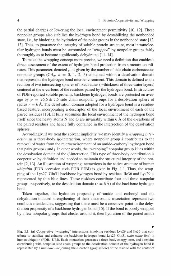
Accordingly, if we treat the solvent implicitly, we may identify a wrapping inter-action as a three-body ijk-interaction, where nonpolar group k contributes to theremoval of water from the microenvironment of an amide–carbonyl hydrogen bondthat pairs groups i and j. In other words, the “wrapping” nonpolar group k lies withinthe desolvation domain of the ij-interaction. This type of three-body contribution iscooperative by definition and needed to maintain the structural integrity of the pro-tein [2, 13]. An illustration of wrapping interactions in the native structure of humanubiquitin (PDB accession code PDB.1UBI) is given in Fig. 1.1. Thus, the wrap-ping of the Lys27–Gln31 backbone hydrogen bond by residues Ile36 and Lys29 isrepresented by thin blue lines. These residues contribute four and three nonpolargroups, respectively, to the desolvation domain (r = 6 Å) of the backbone hydrogenbond.
Taken together, the hydration propensity of amide and carbonyl and thedehydration-induced strengthening of their electrostatic association represent twoconflictive tendencies, suggesting that there must be a crossover point in the dehy-dration propensity of a backbone hydrogen bond [15]. If the bond is poorly wrappedby a few nonpolar groups that cluster around it, then hydration of the paired amide
�Fig. 1.1 (a) Cooperative “wrapping” interactions involving residues Lys29 and Ile36 that con-tribute to stabilize and enhance the backbone hydrogen bond Lys27–Gln31 (thin white line) inhuman ubiquitin (PDB.1UBI). Each interaction generates a three-body energy term, and a residuecontributing with nonpolar side chain groups to the desolvation domain of the hydrogen bond isrepresented by a thin blue line joining the α-carbon (gray sphere) of the residue with the center of

1.2 Cooperative Protein Interactions: The Need for the Wrapping Concept 5
a
b
Fig. 1.1 (continued) the amide–carbonyl hydrogen bond. Conventional colors are used for atomrepresentation and the protein backbone is represented schematically, except for the two residuespaired by the backbone hydrogen bond that are displayed in full backbone detail. Only the sidechains of the wrapping residues 29Lys and 36Ile are shown. (b) Location of the residues in aribbon rendering the native structure of human ubiquitin
6 1 Protein Cooperativity and Wrapping
and carbonyl is favored and prevails, but as the hydrogen bond becomes betterwrapped, the surrounding water loses too many hydrogen bonding partnerships andthus may be favorably removed [13]. This observation is essential to rationalizethe cooperative two-state nature of the folding of single-domain proteins [16–18],as shown in Chap. 3: we may say that the state of hydration of a protein hydrogenbond is in a statistical sense a local reflection of the degree of progress of the foldingprocess.
1.3 Poorly Wrapped Hydrogen Bonds are Promotersof Protein Associations
The structural integrity of a soluble protein is contingent on its ability to excludewater from its amide–carbonyl hydrogen bonds [11, 12]. Thus, water-exposedintramolecular hydrogen bonds, the so-called dehydrons, constitute structural weak-nesses taking the particular form of wrapping deficiencies [12, 19, 20]. On theother hand, these defects favor the removal of surrounding water as a means tostrengthen and stabilize the underlying electrostatic interaction [13, 20, 21], andthus are predictably implicated in protein associations [21], aberrant aggregation[22], and macromolecular recognition [23, 24]. By exogenously contributing to thewrapping of pre-formed hydrogen bonds, these associations in effect remove thewrapping defects, thereby stabilizing the structure.
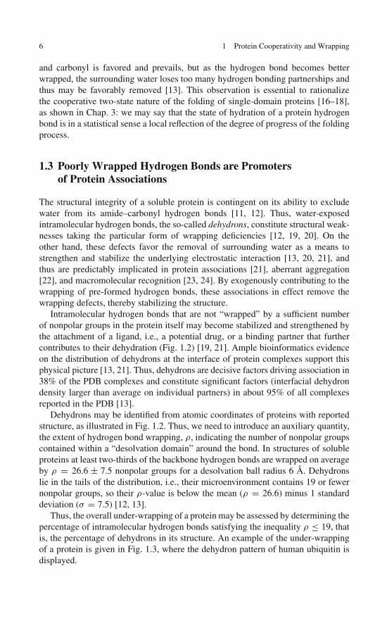
Intramolecular hydrogen bonds that are not “wrapped” by a sufficient numberof nonpolar groups in the protein itself may become stabilized and strengthened bythe attachment of a ligand, i.e., a potential drug, or a binding partner that furthercontributes to their dehydration (Fig. 1.2) [19, 21]. Ample bioinformatics evidenceon the distribution of dehydrons at the interface of protein complexes support thisphysical picture [13, 21]. Thus, dehydrons are decisive factors driving association in38% of the PDB complexes and constitute significant factors (interfacial dehydrondensity larger than average on individual partners) in about 95% of all complexesreported in the PDB [13].
Dehydrons may be identified from atomic coordinates of proteins with reportedstructure, as illustrated in Fig. 1.2. Thus, we need to introduce an auxiliary quantity,the extent of hydrogen bond wrapping, ρ, indicating the number of nonpolar groupscontained within a “desolvation domain” around the bond. In structures of solubleproteins at least two-thirds of the backbone hydrogen bonds are wrapped on averageby ρ = 26.6 ± 7.5 nonpolar groups for a desolvation ball radius 6 Å. Dehydronslie in the tails of the distribution, i.e., their microenvironment contains 19 or fewernonpolar groups, so their ρ-value is below the mean (ρ = 26.6) minus 1 standarddeviation (σ = 7.5) [12, 13].
Thus, the overall under-wrapping of a protein may be assessed by determining thepercentage of intramolecular hydrogen bonds satisfying the inequality ρ ≤ 19, thatis, the percentage of dehydrons in its structure. An example of the under-wrappingof a protein is given in Fig. 1.3, where the dehydron pattern of human ubiquitin isdisplayed.

1.3 Poorly Wrapped Hydrogen Bonds are Promoters of Protein Associations 7
Fig. 1.2 (a) Dehydron in a soluble protein. The dehydron (ρ = 18), marked in green, pairs twobackbone groups (amide and carbonyl, conventional colors for atoms). The microenvironmentis indicated by two intersecting gray spheres centered at the α-carbons of the paired residues.Wrapping side chain groups are shown in light blue and only side chains contributing (fully or par-tially) to the dehydration of the hydrogen bond are indicated. (b) The drug depicted in the figureacts as an exogenous wrapper of the hydrogen bond (gray bond, ρ = 21) turning the dehydron intoa well-protected bond (the three atoms marked with ∗ complete the desolvation of the dehydron)
Fig. 1.3 Illustration of the under-wrapping of protein structure. Dehydron pattern of human ubiq-uitin (ribbon display in Fig. 1.1b). Dehydrons are indicated as green segments joining the α-carbonsof the paired units, well-wrapped hydrogen bonds (ρ > 19) are shown in light gray, and the proteinbackbone is conventionally shown as blue virtual bonds joining the α-carbons of consecutive aminoacid units. The displayed structure has 33 backbone hydrogen bonds, of which 11 are dehydrons.Thus, the extent of under-wrapping for this protein is 33%

8 1 Protein Cooperativity and Wrapping
a
c
b
d
Fig. 1.4 (a) Intermolecular wrapping in the human HIV-1 protease dimer (PDB.1A30) as a meansof protecting the enzyme structure from water attack. Dehydrons are indicated as green seg-ments joining the α-carbons of the paired units, well-wrapped hydrogen bonds are shown in lightgray, and the protein backbone is conventionally shown as virtual bonds joining the α-carbons of
1.4 Wrapping Defects Are Sticky 9
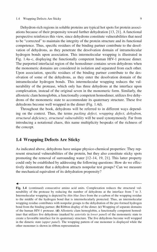
Dehydron-rich regions in soluble proteins are typical hot spots for protein associ-ations because of their propensity toward further dehydration [13, 21]. A functionalperspective reinforces this view, since dehydrons constitute vulnerabilities that needto be “corrected” to maintain the integrity of the protein structure and its functionalcompetence. Thus, specific residues of the binding partner contribute to the desol-vation of dehydrons, as they penetrate the desolvation domain of intramolecularhydrogen bonds upon association. This intermolecular wrapping is illustrated inFig. 1.4a–c, displaying the functionally competent human HIV-1 protease dimer.The purported interfacial region of the homodimer contains seven dehydrons whenthe monomeric domains are considered in isolation and separated from each other.Upon association, specific residues of the binding partner contribute to the des-olvation of some of the dehydrons, as they enter the desolvation domain of theintramolecular hydrogen bonds. This intermolecular wrapping reduces the vul-nerability of the protease, which only has three dehydrons at the interface uponcomplexation, instead of the original seven in the monomeric form. Similarly, theallosteric clam hemoglobin, a functionally competent homodimer, utilizes five dehy-drons of the monomeric state to accommodate its quaternary structure. These fivedehydrons become well wrapped in the dimer (Fig. 1.4d).
Throughout the book, dehydrons will be referred to in different ways depend-ing on the context. Thus, the terms packing defect, wrapping defect, dehydron,structural deficiency, structural vulnerability will be used synonymously. Far fromintroducing a notational chaos, this name multiplicity bespeaks of the richness ofthe concept.
1.4 Wrapping Defects Are Sticky
As indicated above, dehydrons have unique physico-chemical properties: They rep-resent structural vulnerabilities of the protein, but they also constitute sticky spotspromoting the removal of surrounding water [12–14, 19, 21]. This latter propertycould only be established by addressing the following questions: How do we effec-tively demonstrate that a dehydron attracts nonpolar test groups? Can we measurethe mechanical equivalent of its dehydration propensity?
�Fig. 1.4 (continued) consecutive amino acid units. Complexation reduces the structural vul-nerability of the protease by reducing the number of dehydrons at the interface from 7 to 3.Intermolecular wrapping is depicted by thin blue lines from the α-carbon of the wrapping residueto the middle of the hydrogen bond that is intermolecularly protected. Thus, an intermolecularwrapping residue contributes with nonpolar groups to the dehydration of the pre-formed hydrogenbond from the binding partner. (b) Ribbon display of the dimer. (c) Wrapping of separate domainsof the human HIV-1 protease. (d) Allosteric clam hemoglobin, a functionally competent homod-imer that utilizes five dehydrons (marked by asterisks in lower panel) of the monomeric state tocreate a favorable interface for its quaternary structure. The five dehydrons become well wrappedin the dimeric state (upper panel). The wrapping pattern of one monomer is displayed while theother monomer is shown in ribbon representation

10 1 Protein Cooperativity and Wrapping
Fig. 1.5 (a) High-precision total reflection setup to measure the adsorption uptake of proteins ontoa hydrophobic surface under controlled hydrodynamic conditions [19]. The adsorption uptake isproportional to the photon loss due to local alterations in the refractive index of a Langmuir–Blodgett (LB) layer that constitutes the wrapping medium for the protein. (b) Orthogonalitybetween dehydronic field exerted on the test hydrophobe (h) along coordinate R and the Coulombfield exerted along coordinate r between two spherical charges q, q′
Reported experimental work addressed these questions by measuring the adsorp-tion of proteins with wrapping defects and equivalent surface hydrophobicity (areaof solvent-exposed nonpolar surface) onto a “wrapping” layer [19]. This wrappingmedium consists of a Langmuir–Blodgett phospholipid film coating a waveguide, asshown in Fig. 1.5a. These high-precision experiments made use of evanescent-fieldspectroscopic interrogation of the wrapping medium enabling a direct measurementof the protein adsorption uptake. This observable is determined by detecting localchanges in refractive index of the phase within which total reflection of the incident

1.4 Wrapping Defects Are Sticky 11
light occurred [19]. Thus, a beam from a He–Ne laser travels through a waveguideat an incidence angle suitable for total reflection within the medium. The adsorbedmolecules alter the refractive properties of the hydrophobic layer and consequentlyalter the critical angle for total reflection. Thus, protein adsorption is commensuratewith photon loss resulting from the extent of local refraction or “evanescent field.”Hence adsorption uptake can be determined by the loss of photons due to refractivephoton leakage from the total reflection pattern.
For proteins with comparable surface hydrophobicity, the adsorption uptakecorrelates strongly with the extent of protein under-wrapping [19]. As an ade-quate control, only proteins with the same extent of surface hydrophobicity orsolvent-exposed nonpolar area were included in the comparative analysis. Hence,the attractive drag exerted by dehydrons on test hydrophobes became accessible.The net gain in Coulomb energy associated with wrapping a dehydron has beenexperimentally determined to be ∼ 4 kJ/mol [19]. The adhesive force exerted by adehydron on a hydrophobe at 6 Å distance is ∼7.8 pN, a magnitude comparable tothe hydrophobic attraction between two nonpolar moieties that frame unfavorableinterfaces with water.
This study was motivated by the earlier observations that dehydrons play a pivotalrole in driving protein associations, as such associations contribute intermolecularlyto the wrapping of pre-formed structure [12,14], as discussed in the previous section.In consistency with current terminology, the force stemming from the dehydrationpropensity of the partially wrapped hydrogen bond is hithertofore termed dehy-dronic. The dehydronic force arises as a nonpolar group approaches a dehydron witha net effect of immobilizing and ultimately removing surrounding water molecules.This displacement lowers the polarizability of the microenvironment which, in turn,de-shields the paired charges [12, 19]. Thus, a net attractive force is exerted by thedehydron on a nonpolar group and this force represents the mechanical equivalentof the dehydration propensity of an unburied pre-formed hydrogen bond. Since thewater molecules solvating an amide and carbonyl paired by a dehydron are nec-essarily depleted of some hydrogen bonding partners, the work required for theirultimate removal from the bond surroundings is minimal [12, 22]. The dehydronicfield, denoted �(R), is necessarily orthogonal to the Coulomb field generated bythe polar (amide–carbonyl) pair and may be described within a quasi-continuoustreatment of the solvent by the equation
�(R) = −∇R [4πε(R)]−1 qq′/r, (1.1)
where R represents the position vector of the hydrophobe or nonpolar group withrespect to the center of mass of the hydrogen-bonded polar pair, ∇R is the gra-dient taken with respect to this vector, r is the distance between the charges ofmagnitude q and q′ paired by the hydrogen bond (Fig. 1.5b), and the local per-mittivity coefficient ε = ε(R) subsumes the polarizability of the microenvironment,which is generically dependent on the position of the test hydrophobe [2, 14]. Anappropriate expression for ε(R) valid at nanoscales is unavailable at present [25],because of the discreteness of the dielectric medium and the need to include indi-vidual solvent dipole correlations [26]. Although a mean-field dielectric description
12 1 Protein Cooperativity and Wrapping
is unsatisfactory, it is still possible to assert that �(R) is an attractive force since adecrease in ‖R‖ entails a decrease in local polarization which, in turn, enhances theCoulomb attraction.
Building on this analysis, we may quantify the net hydrophobicity η of a hydro-gen bond by taking into account the surface flux of the dehydronic field generatedby the hydrogen bond. This field is given by ω−1�(R) (ω = volume of testhydrophobe). Thus, in accord with Gauss theorem we obtain
η =∫ ∫
�
�(R).d σ (R), (1.2)
where � is the closed surface of the dehydration domain of the hydrogen bond (cf.Fig. 1.2) and dσ (R) is the differential surface area vector at position R.
1.5 Cooperative Drug–Target Associations: A Windowinto Molecular Engineering Possibilities
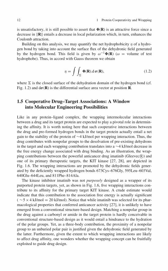
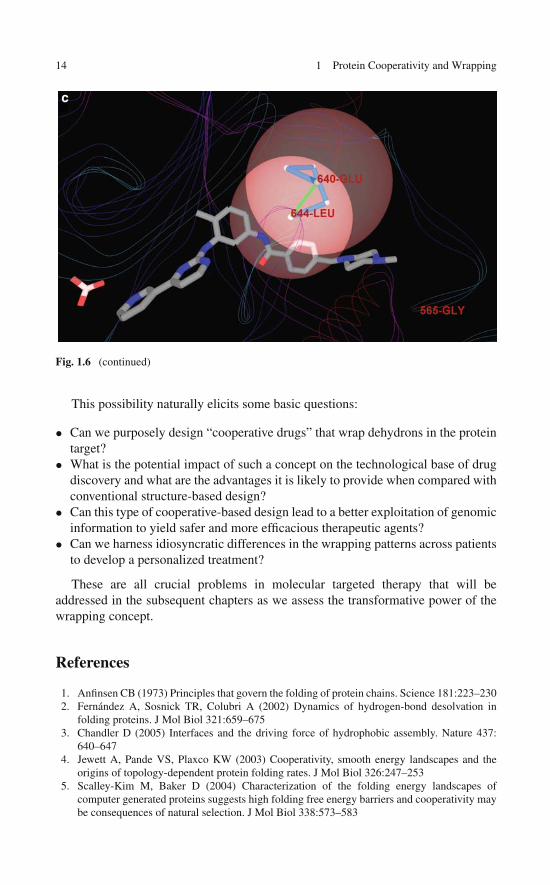
Like in any protein–ligand complex, the wrapping intermolecular interactionsbetween a drug and its target protein are expected to play a pivotal role in determin-ing the affinity. It is worth noting here that such cooperative interactions betweenthe drug and pre-formed hydrogen bonds in the target protein actually entail a netgain to the stability of the protein of ∼4 kJ/mol per wrapping interaction. Thus, thedrug contributes with nonpolar groups to the desolvation of pre-existing dehydronsin the target and each wrapping contribution translates into a ∼4 kJ/mol decrease inthe free energy change associated with drug binding. As an illustration, the wrap-ping contributions between the powerful anticancer drug imatinib (Gleevec R©) andone of its primary therapeutic targets, the KIT kinase [27, 28], are depicted inFig. 1.6. The wrapping interactions are promoted by the dehydronic fields gener-ated by the deficiently wrapped hydrogen bonds 673Cys–676Gly, 595Leu–603Val,640Glu–644Leu, and 811Phe–814Ala.
The kinase inhibitor imatinib was not purposely designed as a wrapper of itspurported protein targets, yet, as shown in Fig. 1.6, five wrapping interactions con-tribute to its affinity for the primary target KIT kinase. A crude estimate wouldindicate that this contribution to the association free energy is actually significant( ∼5 × 4 kJ/mol = 20 kJ/mol). Notice that while imatinib was selected for its phar-macological properties that conferred anticancer activity [27], it is unlikely to haveemerged from a conventional structure-based design. Matching a nonpolar group inthe drug against a carbonyl or amide in the target protein is hardly conceivable inconventional structure-based design as it would entail a hindrance to the hydrationof the polar groups. Yet, as a three-body contribution, the proximity of a nonpolargroup to an unburied polar pair is justified given the dehydronic field generated bythe latter. Furthermore, given the extent to which wrapping interactions are likelyto affect drug affinity, one wonders whether the wrapping concept can be fruitfullyexploited to guide drug design.

1.5 Drug – Target Associations 13
a
b
Fig. 1.6 (a) Dehydrons in the KIT kinase wrapped intermolecularly by the kinase inhibitor ima-tinib in the crystallized drug/target complex (PDB.1T46). The drug nonpolar groups contributing tothe wrapping upon association are marked by circles. The dehydrons (green) wrapped by the druginvolve residue pairs 673Cys–676Gly, 595Leu–603Val, 640Glu–644Leu, and 811Phe–814Ala.(b) Simplified tube rendering of the protein backbone provided as visual aid. (c) Detail of inter-molecular wrapping interaction between imatinib and the KIT kinase. The drug penetrates thedesolvation domain (intersecting pink spheres) of KIT dehydron 640Glu–644Leu upon binding,contributing with two nonpolar groups to the desolvation of the pre-formed hydrogen bond
14 1 Protein Cooperativity and Wrapping
c
Fig. 1.6 (continued)
This possibility naturally elicits some basic questions:
• Can we purposely design “cooperative drugs” that wrap dehydrons in the proteintarget?
• What is the potential impact of such a concept on the technological base of drugdiscovery and what are the advantages it is likely to provide when compared withconventional structure-based design?
• Can this type of cooperative-based design lead to a better exploitation of genomicinformation to yield safer and more efficacious therapeutic agents?
• Can we harness idiosyncratic differences in the wrapping patterns across patientsto develop a personalized treatment?
These are all crucial problems in molecular targeted therapy that will beaddressed in the subsequent chapters as we assess the transformative power of thewrapping concept.
References
1. Anfinsen CB (1973) Principles that govern the folding of protein chains. Science 181:223–2302. Fernández A, Sosnick TR, Colubri A (2002) Dynamics of hydrogen-bond desolvation in
folding proteins. J Mol Biol 321:659–6753. Chandler D (2005) Interfaces and the driving force of hydrophobic assembly. Nature 437:
640–6474. Jewett A, Pande VS, Plaxco KW (2003) Cooperativity, smooth energy landscapes and the
origins of topology-dependent protein folding rates. J Mol Biol 326:247–2535. Scalley-Kim M, Baker D (2004) Characterization of the folding energy landscapes of
computer generated proteins suggests high folding free energy barriers and cooperativity maybe consequences of natural selection. J Mol Biol 338:573–583

References 15
6. Fernández A, Colubri A, Berry RS (2002) Three-body correlations in protein folding: Theorigin of cooperativity. Physica A 307:235–259
7. Fernández A, Kostov K, Berry RS (1999) From residue matching patterns to protein foldingtopographies: General model and bovine pancreatic trypsin inhibitor. Proc Natl Acad Sci USA96:12991–12996
8. Fernández A, Colubri A, Berry RS (2000) Topology to Geometry in protein folding: Beta-lactoglobulin. Proc Natl Acad Sci USA 97:14062–14066
9. Fernández A, Kardos J, Goto J (2003) Protein folding: Could hydrophobic collapse be coupledwith hydrogen-bond formation? FEBS Lett 536:187–192
10. Fernández A (2001) Conformation-dependent environments in folding proteins. J Chem Phys114:2489–2502
11. Fernández A, Kardos J, Scott R, Goto Y, Berry RS (2003) Structural defects and the diagnosisof amyloidogenic propensity. Proc Natl Acad Sci USA 100:6446–6451
12. Fernández A (2004) Keeping dry and crossing membranes. Nat Biotechnol 22:1081–1084
13. Pietrosemoli N, Crespo A, Fernández A (2007) Dehydration propensity of order-disorderintermediate regions in soluble proteins. J Proteome Res 6:3519–3526
14. Fernández A, Scott R (2003) Dehydron: A structure-encoded signal for protein interactions.Biophys J 85:1914–1928
15. Avbelj F, Baldwin RL (2003) Role of backbone solvation and electrostatics in generatingpreferred peptide backbone conformations: distributions of phi. Proc Natl Acad Sci USA100:5742–5747
16. Krantz BA, Moran LB, Kentsis A, Sosnick TR (2000) D/H amide kinetic isotope effects revealwhen hydrogen bonds form during protein folding. Nat Struct Biol 7:62–71
17. Fersht A (2000) Transition-state structure as a unifying basis in protein-folding mechanisms:Contact order, chain topology, stability, and the extended nucleus mechanism. Proc Natl AcadSci USA 97:1525–1929
18. Plaxco KW, Simmons KT, Baker D (1998) Contact order, transition state placement and therefolding rates of single domain proteins. J Mol Biol 277:985–994
19. Fernández A, Scott LR (2003). Adherence of packing defects in soluble proteins. Phys RevLett 91:018102
20. Fernández A, Zhang X, Chen J (2008) Folding and wrapping soluble proteins: Exploring themolecular basis of cooperativity and aggregation. Prog Nucleic Acids Res Transl Sci 83:57–87
21. Fernández A, Scheraga HA (2003) Insufficiently dehydrated hydrogen bonds as determinantsof protein interactions. Proc Natl Acad Sci USA 100:113–118
22. Fernández A, Berry RS (2003) Proteins with H-bond packing defects are highly interac-tive with lipid bilayers: Implications for amyloidogenesis. Proc Natl Acad Sci USA 100:2391–2396
23. Deremble C, Lavery R (2005) Macromolecular recognition. Curr Opin Struct Biol 15:171–175
24. Ma B, Elkayam T, Wolfson H, Nussinov R (2003) Protein-protein interactions: Structurallyconserved residues distinguish between binding sites and exposed protein surfaces. Proc NatlAcad Sci USA 100:5772–5777
25. Fernández A (2003) What caliber pore is like a pipe? Nanotubes as modulators of iongradients. J Chem Phys 119:5315–5319
26. Despa F, Fernández A, Berry RS (2004) Dielectric modulation of biological water. Phys RevLett 93:228104
27. Demetri G (2002) Efficacy and safety of imatinib mesyalte in advanced gastrointestinalstromal tumors. N Engl J Med 347:472–480
28. Fernández A, Sanguino A, Peng Z et al (2007) An anticancer C-kit kinase inhibitor isreengineered to make it more active and less cardiotoxic. J Clin Invest 117:4044–4054

“This page left intentionally blank.”
Chapter 2Wrapping Defects and the Architectureof Soluble Proteins
Wrapping defects in soluble proteins represent local weaknesses of the nativestructures and have received little attention, especially by the drug design com-munity. The protein structure may be inherently weak at sites where hydration ofthe backbone is locally hampered by formation of an intramolecular hydrogen bondwhich in turn is not stabilized through complete burial within a hydrophobic envi-ronment. This chapter explores the architectural implications stemming from theexistence of these vulnerabilities. Thus, the unburied backbone hydrogen bondsor dehydrons are shown to be compensated by disulfide bridges that are neededto maintain the structural integrity in extracellular environments. Examination ofall reported soluble structures reveals that the number of disulfide bonds correlatestightly with the number of dehydrons in a 1:5 ratio. The results have implicationsfor biomolecular design as they introduce universal constraints in the architectureof water-soluble proteins.
2.1 How Do Soluble Proteins Compensatefor Their Wrapping Defects?
Backbone hydration, prevalent in the unfolded state of a polypeptide chain, is oftenhindered in a soluble folded state as backbone amides and carbonyls are pairedthrough hydrogen bonds [1, 2]. Yet, the thermodynamic cost of dehydration is notalways compensated, especially if the backbone hydrogen bond is not completelysequestered from solvent. In soluble proteins, such bonds may be readily identi-fied from the structural coordinates by determining the number of nonpolar groupswithin the bond microenvironment [3–5]. These unburied backbone hydrogen bondsconstitute structural deficiencies and represent markers for protein associations[5]. In turn, these associations are required to maintain the structural integrityof the protein through intermolecular protection of the pre-formed hydrogenbonds [5].
A thorough examination of the protein data bank (PDB) singles out toxin pep-tides with picomolar affinity for the Kv1.3 potassium channel [6], such as HsTX1(PDB.1QUZ), as members of the protein family with the highest extent of structural
17A. Fernández, Transformative Concepts for Drug Design: Target Wrapping,DOI 10.1007/978-3-642-11792-3_2, C© Springer-Verlag Berlin Heidelberg 2010

18 2 Wrapping Defects and the Architecture of Soluble Proteins
deficiency. The unburied hydrogen bonds in such proteins can make up to 100% ofthe backbone hydrogen bonds. This observation immediately suggests a reason forthe extremely high target affinity of neurotoxins: according to Chap. 1, we expect ahuge dehydronic field for such biomolecules.
A separate analysis reveals that such proteins contain an inordinately large num-ber of disulfide bonds, with an average of 11 when normalized to 100 amino acids.These observations prompt us to investigate the relation between structural defi-ciency and disulfide bonds in search for a balance equation that reflects a statisticalcorrelation between structural strengths and vulnerabilities of soluble proteins andpolypeptides. The balance equation unraveled in this chapter is likely to impact thedesign of soluble proteins and enable a better control of their functional modulationin relation to environmental redox conditions.
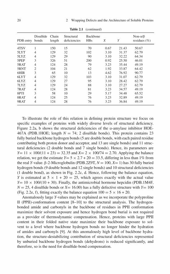
A comprehensive wrapping analysis of the PDB yields the structural deficienciesof an exhaustive nonredundant set of 2,989 monomeric uncomplexed soluble pro-teins or peptides with disulfide links and 8,975 proteins without disulfide links [7].These data are compiled as illustrated in Table 2.1 for some selected PDB entries.Peptide chains were excluded from the analysis if their structural integrity requiredprosthetic groups or cation coordination. In order to compare protein or peptidechains of different lengths, two normalized parameters were used to characterize aprotein structure: Y = number of structural deficiencies per 100 backbone hydrogenbonds and X = number of disulfide bonds per 100 amino acids. Proteins were binnedaccording to their X-value in integer groups with n = 0, 1,. . .,18, where proteins withno disulfide bonds (X= 0) belong to group n = 0 and proteins with X in the rangen < X ≤ n + 1 belonged to group n + 1. The mean Y-value and standard deviationwere computed for each n-group and the results are shown in Fig. 2.1a. A tight X–Ylinear correlation (R2= 0.96) results and is further corroborated by the raw X–Y-trendline generated by linear regression on all (X, Y) data points (Fig. 2.1b). Takentogether, the results from Fig. 2.1 unambiguously reveal a simple balance relationY = 5X + 20. This statistical relation introduces a 1:5 ratio to buttress vulnerableproteins and allows for a 20%-baseline in structural deficiency.
Table 2.1 Structural parameters for few selected monomeric uncomplexed soluble proteins withdisulfide bridges lacking prosthetic groups and scaffolding cation coordination
PDB entryDisulfidebonds
Chainlength
Structuraldeficiencies
BackboneHBs X Y
Non-α/βresidues (%)
2PNE 2 81 37 37 2.47 100.00 100.001M4F 4 25 8 8 16.00 100.00 68.001EZG 8 81 33 48 9.87 68.75 77.781HOE 2 74 11 34 2.70 32.35 54.052Z9T 1 100 10 40 1.00 25.00 50.00135L 4 129 35 109 3.10 32.11 63.57153L 2 185 38 185 1.08 20.54 48.65154L 2 185 39 194 1.08 20.10 48.65172L 1 164 47 188 0.61 25.00 30.491A2J 1 189 57 196 0.53 29.08 39.151A39 9 402 69 250 2.24 27.60 59.70

2.1 How Do Soluble Proteins Compensate for Their Wrapping Defects? 19
Table 2.1 (continued)
PDB entryDisulfidebonds
Chainlength
Structuraldeficiencies
BackboneHBs X Y
Non-α/βresidues (%)
1A3P 2 45 9 18 4.44 50.00 82.221A43 1 87 22 67 1.15 32.84 49.431A67 2 108 20 62 1.85 32.26 48.151A7M 3 180 37 155 1.67 23.87 41.671AC5 3 483 91 370 0.62 24.59 51.761ACJ 3 537 77 404 0.56 19.06 50.091ACW 3 29 18 18 10.34 100.00 41.381ACX 2 108 20 44 1.85 45.45 56.481ADX 3 40 5 7 7.50 71.43 100.001ADZ 3 71 11 35 4.23 31.43 76.061AE5 4 225 33 123 1.78 26.83 62.221AEC 3 218 33 155 1.38 21.29 55.961AFH 4 93 39 74 4.30 52.70 46.241AG2 1 103 15 80 0.97 18.75 43.691AGG 4 48 11 14 8.33 78.57 87.501AGI 3 125 18 82 2.40 21.95 48.801AGY 2 200 43 158 1.00 27.22 51.501AH1 2 129 15 51 1.55 29.41 60.471AHK 3 129 13 43 2.33 30.23 80.621AHL 3 49 6 14 6.12 42.86 87.763DHM 1 100 10 50 1.00 20.00 52.003DIH 7 122 35 97 5.74 36.08 52.463EGP 1 108 8 37 0.93 21.62 58.333EHS 3 476 93 359 0.63 25.91 40.343EMY 1 329 44 198 0.30 22.22 49.853ENG 7 213 34 119 3.29 28.57 66.673EO5 1 171 47 125 0.58 37.60 48.543EOW 2 221 21 75 0.90 28.00 70.143ETP 2 187 22 89 1.07 24.72 51.873EXD 4 129 35 113 3.10 30.97 63.573EZM 2 101 31 57 1.98 54.39 42.573GF1 3 70 25 53 4.29 47.17 77.143LYM 4 129 36 109 3.10 33.03 63.573LZ2 4 129 32 91 3.10 35.16 64.343MAN 1 302 58 252 0.33 23.02 50.993PTE 1 349 70 297 0.29 23.57 50.433RAT 4 124 28 81 3.23 34.57 49.193RSD 4 124 25 78 3.23 32.05 49.193SEB 1 238 27 153 0.42 17.65 50.003SSI 2 113 26 69 1.77 37.68 59.293TGF 3 50 13 34 6.00 38.24 68.003TGL 3 269 42 221 1.12 19.00 49.814AIT 2 74 13 35 2.70 37.14 54.054APE 1 330 49 195 0.30 25.13 47.884CMS 3 323 53 203 0.93 26.11 42.724ENG 7 210 34 119 3.33 28.57 67.144RAT 4 124 29 82 3.23 35.37 49.194TGL 3 269 36 204 1.12 17.65 50.19
20 2 Wrapping Defects and the Architecture of Soluble Proteins
Table 2.1 (continued)
PDB entryDisulfidebonds
Chainlength
Structuraldeficiencies
BackboneHBs X Y
Non-α/βresidues (%)
4TSV 1 150 15 70 0.67 21.43 50.675LYT 4 129 32 102 3.10 31.37 62.795LYZ 4 129 29 90 3.10 32.22 64.345PEP 3 326 51 200 0.92 25.50 46.015RAT 4 124 28 79 3.23 35.44 49.195RNT 2 104 21 62 1.92 33.87 64.426HIR 3 65 10 13 4.62 76.92 90.776LYT 4 129 32 103 3.10 31.07 62.796LYZ 4 129 27 95 3.10 28.42 62.797LYZ 4 129 24 88 3.10 27.27 62.797RAT 4 124 28 81 3.23 34.57 49.198PTI 3 58 10 29 5.17 34.48 65.528RAT 4 124 25 76 3.23 32.89 49.199RAT 4 124 28 76 3.23 36.84 49.19
To illustrate the role of this relation in defining protein structure we focus onspecific examples of proteins with widely diverse levels of structural deficiency.Figure 2.2a, b shows the structural deficiencies of the α-amylase inhibitor HOE-467A (PDB.1HOE; length N = 74; 2 disulfide bonds). This protein contains 23fully buried backbone hydrogen bonds (5 are double bonds, with each paired residuecontributing both proton donor and acceptor, and 13 are single bonds) and 11 struc-tural deficiencies (2 double bonds and 7 single bonds). Hence, its parameters areY= 11 × 100/(11 + 23) = 32.35 and X= 2 × 100/74 = 2.70. Applying the balancerelation, we get the estimate Y= 5 × 2.7 + 20 = 33.5, differing in less than 1% fromthe real Y-value. β-2-Microglobulin (PDB.2Z9T, N = 100, X= 1) has 30 fully buriedhydrogen bonds (9 double bonds and 12 single bonds) and 10 structural deficiencies(1 double bond), as shown in Fig. 2.2c, d. Hence, following the balance equation,Y is estimated at 5 × 1 + 20 = 25, which agrees exactly with the actual valueY= 10 × 100/(10 + 30). Finally, the antimicrobial hormone hepcidin (PDB.1M4F,N = 25, 4 disulfide bonds or X= 16.00) has a fully defective structure with Y= 100(Fig. 2.2e, f), fitting exactly the balance equation 100 = 5 × 16 + 20.
Anomalously large Y-values may be explained as we incorporate the polyprolineII (PPII)-conformation content [8–10] to the structural analysis. The hydrogen-bonded amide and carbonyls in the backbone of residues in PPII conformationmaximize their solvent exposure and hence hydrogen bond burial is not requiredas a provider of thermodynamic compensation. Hence, proteins with large PPIIcontent in their folded native state maximize their backbone exposure to sol-vent to a level where backbone hydrogen bonds no longer hinder the hydrationof amides and carbonyls [9]. At this anomalously high level of backbone hydra-tion, the structure-destabilizing contribution of structural deficiencies representedby unburied backbone hydrogen bonds (dehydrons) is reduced significantly, andtherefore, so is the need for disulfide-bond compensation.

2.1 How Do Soluble Proteins Compensate for Their Wrapping Defects? 21
Fig. 2.1 Number of structural deficiencies (unburied backbone hydrogen bonds, dehydrons) nor-malized to 100 backbone hydrogen bonds (Y) plotted against number of disulfide bonds normalizedto 100 amino acids (X) for PDB-reported soluble proteins. a Mean Y-value (square) and standarddeviation (error bar) for proteins grouped according to their number of disulfide bonds. Proteinswere binned according to their X-value in integer groups with n = 0, 1,. . .,18, where proteins withno disulfide bonds (X= 0) belong to group n = 0 and proteins with n < X ≤ n + 1 belong to group n+ 1. b All (X, Y)-data points from the nonredundant exhaustive set of PDB entries for uncomplexedsoluble proteins

22 2 Wrapping Defects and the Architecture of Soluble Proteins
Fig. 2.2 Structural deficiencies in soluble proteins. The protein backbone is shown as virtualbonds (blue) joining consecutive α-carbons in the peptide chain. Light-gray segments joiningα-carbons represent completely buried backbone hydrogen bonds and green segments representstructural deficiencies (unburied backbone hydrogen bonds). A tube/ribbon representation is addedfor visual aid. Cysteines involved in disulfide bonds are identified by side chain display. Structuraldeficiencies (a, c, e) and tube/ribbon representation (b, d, f), respectively, of α-amylase inhibitorHOE-467A (PDB.1HOE) (a, b), β-2 microglobulin (PDB.2Z9T) (c, d), and antimicrobial hormonehepcidin (PDB.1M4F) (e, f)
2.2 Thermodynamic Support for the Dehydron/DisulfideBalance Equation
The dehydron/disulfide balance relation clearly identifies proteins with excess(Y > 5X + 20) or lack (Y < 5X + 20) of structural deficiencies, with the former likelyto be more favorably denatured than the latter under equivalent redox and denatura-tion conditions. To test this prediction, thermodynamic data on thermal denaturation(Table 2.2) were obtained for an exhaustive set of proteins for which structural infor-mation was also available [7]. Thus, the thermal denaturation free energy change,G, under reducing conditions and comparable temperatures [11], was obtainedfor monomeric uncomplexed PDB-reported proteins with disulfide bonds and lack-ing prosthetic groups or ion coordination. A significant anticorrelation was found

2.2 Thermodynamic Support for the Dehydron/Disulfide Balance Equation 23
Table 2.2 Thermodynamic and structural parameters of soluble proteins. Thermal denaturationfree energy change, G, under reducing conditions and comparable temperatures for an exhaustiveset of monomeric uncomplexed proteins with disulfide bonds and without prosthetic groups or ioncoordination [11]. Deviations from the balance relation are measured by Y–(5X + 20) and shownto anticorrelate tightly (R2 = 0.72, Fig. 2.3) with the denaturation free energies
PDB entry Y–(5X+ 20) G(kcal/mol) T (C) pH Reference
1BSQ −9.83 11.10 40.00 7.00 Int J Biol Macromol 38, 9–17(2006)
1RTB −5.00 10.10 25.00 8.40 Biophys Chem 127, 51–63 (2007)4LYZ −3.30 9.02 26.85 7.00 Biopolymers 85, 264–273 (2007)1CX1 −3.26 5.38 24.85 7.09 Biochemistry 37, 3529–3537
(1998)1QG5 1.13 8.80 40.00 7.00 Int J Biol Macromol 38, 9–17
(2006)2AIT 2.39 6.70 25.00 5.00 J Mol Biol 223, 769–779 (1992)3SSI 8.82 4.07 20.00 7.00 J Mol Biol 249, 625–635 (1995)1HIC 25.58 5.02 25.00 7.00 Eur J Biochem 202, 67–73 (1991)1PMC 38.33 1.10 20.00 3.00 Nat Struct Biol 3, 45–53 (1996)
Fig. 2.3 Anticorrelation between denaturation free energy (G) and excess structural defectswith respect to the balance relation, measured by Y–(5X + 20). The exhaustive set of monomericuncomplexed proteins with disulfide links and the respective denaturation conditions are given inTable 2.2. The coefficient R2= 0.72 for the linear fit was obtained by linear regression
24 2 Wrapping Defects and the Architecture of Soluble Proteins
(R2= 0.72, Fig. 2.3) between the deviation from the balance equation, mea-sured as Y–(5X + 20), and the thermal denaturation free energy (G). This tightanticorrelation provides a thermodynamic validation of the balance equation.
The 5:1 rule may be justified on thermodynamic grounds. Thus, Doig andWilliams [12] addressed the inconsistencies in Flory′s treatment of the entropiccontribution to protein denaturation, calculating G for denaturation for a cross-linked protein versus its non-cross-linked counterpart. At physiological temperatureof 300 K, they estimated G ≈ 4.4 kcal/mol. This value is essentially independentof protein length and loop size and best represents the insensitivity of experimentalvalues to loop size-dependent configurational entropies [13].
This 4.4 kcal/mol constant agrees reasonably well with the free energy contribu-tion associated with the native-state destabilization brought about by five structuraldeficiencies. If we take into account that –0.93 kcal/mol is the free energy changeassociated with complete dehydration of an unburied backbone hydrogen bond[4, 14], we may estimate the net destabilization effect promoted by five structuraldeficiencies at 0.93 kcal/mol × 5 = 4.65 kcal/mol. This value is in close proxim-ity to the Doig–Williams constant. The thermodynamic agreement supports the 5:1golden ratio for protein buttressing arising from structural analysis.
2.3 Evolutionary Support for the Balance Equation
Since the evolutionary axis is germane to any biological analysis, the followingquestion naturally arises: Is the architectural constraint defined by the dehy-dron/disulfide balance equation respected by evolution? Bioinformatics evidence onorthologous proteins (homologs across species) supports the tenet of evolutionaryconservation. Thus, we may compare the structural deficiency and normalized num-ber of disulfide bonds across 1105 homolog pairs that differ in at least one disulfidebond (one homolog may have no disulfide bond). The changes in X and Y (X, Y)were obtained for homolog pairs identified by their respective PDB accessions andthe deviation from ideality was measured as = Y– 5X. For all homolog pairswe obtained /Y < 11%, with Y associated with either homolog. Table 2.3 illustratesthe tightest evolutionary conservation of the architectural constraint across homologpairs with nontrivial buttressing differences.
2.4 Wrapping Translates into Protein Architecture
This chapter introduced a basic design principle that can be rationalized throughan analogy. Just like defiance of gravity in building engineering requires buttress-ing to preserve the integrity of the building, protein design allowing for backbonehydration (the force counteracting structural cohesion) requires disulfide bridges tomaintain the structural integrity of the protein. In this regard, this work unravels twoconstants that define a fundamental architectural constraint in soluble proteins: Aftersuitable normalization, a single disulfide bond stabilizes five structural deficiencies

2.4 Wrapping Translates into Protein Architecture 25
Table 2.3 Evolutionary conservation of the balance relation Y = 5X + 20.
SCOP family aSequenceidentity Y–5X X Y Homolog 1 Homolog 2
Fibronectin type II module 63.125 –0.1 0.21 0.95 1QO6 1E8Bβ-Glycanases 40 –0.1 0.31 1.45 1FH8 1I1Xβ-Glycanases 42.13836478 –0.1 0.31 1.45 1E0V 1B30C-Type lysozyme 57.69230769 –0.09 0.79 3.86 1KXY 1LHMC-Type lysozyme 58.46153846 –0.09 0.79 3.86 1JIS 1LHMRibonuclease A-like 30.23255814 –0.09 0.79 3.86 1RUV 1B1ERibonuclease A-like 31.00775194 –0.09 0.79 3.86 1RUV 1H52Acetylcholinesterase-like 30.82437276 –0.07 0.18 0.83 2CKM 1K4YC-Type lysozyme 67.69230769 –0.07 0.79 3.88 1LMP 2BQMC-Type lysozyme 56.92307692 –0.07 0.79 3.88 1LSM 2BQKEukaryotic proteases 31.47410359 –0.07 0.29 1.38 1EX3 1DSTSnake venom toxins 32.89473684 –0.07 0.4 1.93 1TXA 1ERAPepsin-like 40 –0.06 0.31 1.49 1PSN 1FQ6Ribonuclease A-like 31.74603175 –0.06 0.73 3.59 1RTB 1K5BC-Type lysozyme 59.23076923 –0.05 0.79 3.9 1IR7 1HNLC-Type lysozyme 59.23076923 –0.05 0.79 3.9 1XEK 1HNLC-Type lysozyme 59.23076923 –0.05 0.79 3.9 1UIH 1HNLRibonuclease A-like 31.00775194 –0.05 0.79 3.9 4RAT 1K5AC-Type lysozyme 58.46153846 –0.05 0.79 3.9 2HS7 2BQKPlant proteinase inhibitors 47.74774775 –0.04 0.34 1.66 1TIH 1FYBPapain-like 70.21943574 –0.03 0.84 4.17 1ITO 2PBHβ-Glycanases 36.36363636 –0.03 0.31 1.52 2XYL 1B3XC-Type lysozyme 84.72222222 –0.03 1.02 5.07 3LZ2 1LSGRibonuclease A-like 31.00775194 –0.03 0.79 3.92 1RHB 1K5A
(a) SCOP: structural classification of proteins (Murzin AG, Brenner SE, Hubbard T, Chothia C(1995) SCOP: a structural classification of proteins database for the investigation of sequencesand structures. J Mol Biol 247:536–540)
and every soluble protein has a 20%-baseline level of structural deficiency. Theseconstants define statistically a design principle.
The baseline structural deficiency Y = 20 represents the maximum of a tight dis-tribution (standard deviation σ = 2.25) of Y-values for the structural deficiency ofsoluble proteins with no disulfide bridges. This baseline Y-value implies that solubleproteins are not perfectly packed and maintain at least 20% of unburied backbonehydrogen bonds. Since such structural deficiencies locally promote backbone hydra-tion, they belong to an intermediate region between order and disorder and hencerepresent markers of structural flexibility. Thus, because of its universality, theY = 20 constant may be interpreted as the baseline flexibility needed for proteinfunction.
In this chapter, dehydrons were characterized as structural deficiencies. Thesedeficiencies are of a special kind: They are promoters of backbone hydration [4]and hence destabilizers of the native structure. On the other hand, disulfide bondspre-formed in the denatured state reduce the structure-destabilizing conformationalentropy cost associated with the folding process [12], hence stabilizing the native

26 2 Wrapping Defects and the Architecture of Soluble Proteins
structure. Thus, it should be generally expected that the two major and oppositecontributors to native structure destabilization would be correlated, as revealed bythe balance equation described in this chapter.
This relation is hence likely to assist the molecular engineering of soluble pro-teins. Furthermore, since disulfide bridges can be formed or dismantled in accordwith redox environmental conditions, the relation presented is likely to enable thetype of design fine-tuning that may be required for an environmental modulation ofthe protein function.
As the dehydron/disulfide organizational principle is established and shown tohold for soluble proteins, we cannot fail to notice that it also introduces a new setof problems stemming from a basic question What is in physical terms the fate ofa soluble protein whose structure significantly violates the architectural constraintsdefined by the balance equation? This issue will be explored in Chap. 5.
References
1. Baldwin RL (2003) In search of the energetic role of peptide hydrogen bonds. J Biol Chem278:17581–17588
2. Powers ET, Deechongkit S, Kelly JW (2006) Backbone-backbone H-bonds make context-dependent contributions to protein folding kinetics and thermodynamics: Lessons from amide-to-ester mutations. In: Peptide Solvation and H-bonds, Baldwin RL, Baker D, eds, Adv ProteinChem 72:40–79, Elsevier Academic Press, San Diego, California
3. Fernández A, Berry RS (2002) Extent of Hydrogen-bond protection in folded proteints: Aconstraint on packing architectures. Biophys J 83:2475–2481
4. Pietrosemoli N, Crespo A, Fernández A (2007) Dehydration propensity of order-disorderintermediate regions in soluble proteins. J Proteome Res 6:3519–3526
5. Fernández A, Scheraga HA (2003) Insufficiently dehydrated hydrogen bonds as determinantsfor protein interactions. Proc Nat Acad Sci USA 100:113–118
6. MacKinnon R, Reinhart PH, White MN (1988) Charybdotoxin block of Shaker K+ channelssuggests that different types of K+channels share common features. Neuron 1:997–1001
7. Fernández A, Berry RS (2002) Extent of Hydrogen-bond protection in folded proteins: Aconstraint on packing architectures. Biophys J 83:2475–2481
8. Pentelute BL, Gates ZP, Tereshko V et al (2008) X-ray structure of snow flea antifreeze pro-tein determined by racemic crystallization of synthetic protein enantiomers. J Am Chem Soc130:9695–9701
9. Adzhubei AA, Sternberg MJE (1993) Left-handed polyproline II helices commonly occur inglobular proteins. J Mol Biol 229:472–493
10. Shi Z, Woody RW, Kallenbach NR (2002) Is polyproline II a major backbone conformationin unfolded proteins? In: Unfolded Proteins, Rose G, ed, Adv Protein Chem 62:163–240
11. Kumar MD (2006) ProTherm and ProNIT: thermodynamic databases for proteins and protein-nucleic acid interactions. Nucleic Acids Res 34:D204–D206
12. Doig AJ, Williams DH (1991) Is the hydrophobic effect stabilizing or destabilizing in proteins:The contribution of disulphide bonds to protein stability. J Mol Biol 217:389–398
13. Betz SF (1993) Disulfide bonds and the stability of globular proteins. Protein Sci 2:1551–1558
14. Fernández A, Scott R (2003) Adherence of packing defects in soluble proteins. Phys Rev Lett91:018102

Chapter 3Folding Cooperativity and the Wrappingof Intermediate States of SolubleNatural Proteins
This chapter focuses on the molecular basis of cooperativity as a means to under-stand the folding of soluble natural proteins. We explore the concept of proteinwrapping, its intimate relation to cooperativity, and its bearing on the expediencyof the folding process for natural proteins. As previously described, wrapping refersto the environmental modulation or protection of intramolecular electrostatic inter-actions through an exclusion of surrounding water that takes place as the chainfolds onto itself. Thus, a special many-body picture of the folding process is shownto emerge where the folding chain not only interacts with itself but also shapesthe microenvironments that stabilize or destabilize the interactions. This picturereflects a competition between chain folding and backbone hydration leading to theprevalence of backbone hydrogen bonds for natural foldable proteins. A constant ofmotion governing the folding process emerges from the analysis.
3.1 Many-Body Picture of Protein Folding:Cooperativity and Wrapping
The physical underpinnings to the protein folding process remain elusive or, rather,difficult to cast in a useful form that enables structure prediction [1–10]. Thus, thepossibility of inferring the folding pathway of a soluble protein solely from physicalprinciples continues to elude major research efforts.
A major difficulty arises as we attempt to tackle this problem: as a peptide chainfolds onto itself, it also shapes the microenvironments of the intramolecular interac-tions, and hence the strength and stability of such interactions need to be rescaledaccording to the extent to which they become “wrapped” or surrounded by otherparts of the chain. Thus, interactions between different parts of the peptide chain notonly entail the units directly engaged in the interaction but also the units involvedin shaping their microenvironment, and the latter are just as important as they deter-mine either the persistence or the ephemeral nature of such interactions. This factmakes the folding problem essentially a many-body problem and points to the heartof cooperativity, a pivotal attribute of the folding process [4, 6]. Furthermore, ithighlights the intimate link between cooperativity and wrapping: intramolecular
27A. Fernández, Transformative Concepts for Drug Design: Target Wrapping,DOI 10.1007/978-3-642-11792-3_3, C© Springer-Verlag Berlin Heidelberg 2010
28 3 Folding Cooperativity and the Wrapping of Intermediate States
hydrogen bonds prevail only if properly wrapped and this requires a cooperativeprocess.
To further explore the molecular basis of cooperativity, we need to examine thefolding process from a physico-chemical perspective: With an amide and carbonylgroup per residue, the backbone of the protein chain is highly polar and this molec-ular property imposes severe constraints on the nature of the hydrophobic collapseand on the chain composition of proteins capable of sustaining such a collapse [2,9, 11]. Thus, the hydrophobic collapse entails the dehydration of backbone amidesand carbonyls and such a process would be thermodynamically disfavored if it werenot for the possibility of amides and carbonyls to engage in hydrogen bonding witheach other. Hence, not every hydrophobic collapse qualifies as being conducive tofolding the protein chain: Only a collapse that ensures the formation and protectionof backbone hydrogen bonds is likely to ensure an expedient folding of the chain [2].On the other hand, polar group hydration competes with intramolecular hydrogenbonds, compromising the structural integrity of proteins with a deficiently wrappedbackbone [12]. Thus, the need for formation and protection of intramolecular hydro-gen bonds from water attack imposes constraints on the chain composition of anefficient folder capable of sustaining a reproducible and expedient collapse.
In accord with this picture, it has been postulated that as water-soluble proteinsfold, the hydrogen bond pairing of backbone amides and carbonyls is concurrentwith the hydrophobic collapse of the chain [11, 13]. This fact has been rationalizedtaking into account that the thermodynamic cost associated with the dehydration ofunpaired polar groups is relatively high and that the hydrophobic collapse hindersthe backbone hydration by shielding it from water. On the other hand, the strengthand stability of hydrogen bonds clearly depend on the microenvironment where theyoccur: The proximity of nonpolar groups to a hydrogen bond enhances the electro-static interaction by de-screening the partial charges and stabilizes it by hinderingthe hydration of the polar groups in the nonbonded state. Thus, to guarantee theintegrity of soluble protein structure and the expediency of the folding process, mostintramolecular hydrogen bonds must be surrounded or “wrapped” by nonpolargroups fairly thoroughly so as to become significantly dehydrated at all times dur-ing the folding process. This observation has implications at an ensemble-averagelevel accessible to experimentalists [13, 14]. As shown below, it may help under-stand the fact that single domain proteins are likely to be two-state folders, with asingle kinetic barrier dominating the folding process at the ensemble-average level[13, 15].
The hydration propensity of amide and carbonyl and the dehydration-inducedstrengthening of their electrostatic association represent two conflictive tendencies,suggesting that there must be a crossover point in the dehydration propensity of abackbone hydrogen bond. If the bond is poorly wrapped by a few nonpolar groupsthat cluster around it, then hydration of the paired amide and carbonyl is likely to befavorable, but as the hydrogen bond becomes better wrapped intramolecularly, thesurrounding water loses too many hydrogen bonding partnerships and thus furtherremoval of surrounding water is promoted. Thus, the dehydronic field per hydrogenbond must significantly increase as hydrogen bonds get sufficiently dehydrated and

3.1 Many-Body Picture of Protein Folding: Cooperativity and Wrapping 29
this enhancement promotes further compaction of the chain and commits its fold.This switch-over behavior from backbone hydration to dehydration of backbonehydrogen bonds translated into an enhanced dehydronic field reflects the commit-ment to fold into a compact structure in which most backbone hydrogen bonds willbe thoroughly dehydrated. This general picture is schematized in Fig. 3.1. The ratio-nalization of the two-state folding kinetics of single domain proteins is thus basedon the cooperative nature of wrapping interactions.
The crossover point in hydrogen bond dehydration propensity may be regardedas representing a local characterization of the folding transition state if we adoptthe backbone hydrogen bond dehydration as a generic folding coordinate. Once thefolding process has progressed beyond the crossover point, further dehydration ofthe backbone is favored in consonance with the downhill nature of the folding pro-cess beyond the transition state [15]. Thus, a transition state conformation commitsthe chain to fold partly because the partially wrapped hydrogen bonds trigger theirfurther desolvation, in turn fostering further chain compaction (Fig. 3.1). This com-paction is essential to augment the number of nonpolar groups within the hydrogenbond microenvironments, thus protecting the bonds from water attack.
Fig. 3.1 The cross-over behavior from backbone hydration to dehydration of backbone hydrogenbonds triggered by hydrophobic collapse translates at the ensemble folding-kinetics level into acommitment of the chain to fold as the folding reaction progresses beyond the transition state

30 3 Folding Cooperativity and the Wrapping of Intermediate States
However compelling, the scenario described above requires a rigorous justifica-tion that entails an understanding of the crossover behavior at a local level. Thisunderstanding prompts us to focus on the solvent environment of individual hydro-gen bonds during the conformational exploration performed by the protein chain asthe hydrophobic collapse takes place [12, 16, 17].
3.2 Hydrogen Bond Wrapping Requires Cooperative Folding
The dynamics of hydrogen bond wrapping concurrent with the folding processcannot be probed directly at this time, much like the conformational explorationdefining the folding process still remains inaccessible to experimentalists [13–15].To make progress in our understanding of the wrapping dynamics, we need to cap-ture this process through ab initio folding algorithms independently benchmarkedand validated against PDB-reported structures and experimental data on the foldingkinetics [2, 13, 15].
To properly describe folding cooperativity, we exploit a program that algorith-mically reproduces folding dynamics by stochastically and coarsely representingtransitions in the conformation space of individual residues and effectively incor-porating wrapping three-body interactions. To access realistic folding timescalesbeyond the microsecond range, the program builds on an implicit solvent modelof cooperativity [6] and exploits a coarse-grained representation of the torsionalRamachandran (�, �)-state of each residue [10]. The implicit treatment of thesolvent dictates that the program assesses at each step the microenvironments ofintramolecular hydrogen bonds that form as byproducts of the chain attempt atachieving a sustainable compaction.
In order to reduce computation time and access relevant timescales, the microen-vironmental dielectric within which intramolecular hydrogen bonds form is com-puted in a coarse manner at each step of the simulations. The strength and stabilityof intramolecular hydrogen bonds are thus rescaled with each computational step inconsonance with the degree of dryness or dehydration of the bond determined by theoverall folding state of the chain. Thus, the extent of bond dehydration defines itsnet hydrophobicity (see (1.2)), which in turn promotes further wrapping begettingfurther compaction of the chain.
The ab initio folding algorithm reproduces the essential dynamical features ofcooperativity while possessing predictive potential in regard to both endpoint struc-ture and folding pathway [2]. Thus, the algorithm appears to reproduce a basicfeature of the folding kinetics of single domain proteins: its purported two-statenessobserved at an ensemble-average level of experimentation [13]. This dynamic fea-ture is likely to arise as an overall consequence of the crossover behavior inhydrogen bond dehydration propensity, although a “hard proof” of this statementhas not been produced to this day.
In spite of the difficulties inherent to a direct probing, the folding of a solubleprotein is believed to be accompanied by a progressive structuring, immobiliza-tion, and eventual exclusion of water surrounding backbone hydrogen bonds. This

3.2 Hydrogen Bond Wrapping Requires Cooperative Folding 31
process turns hydrogen bonds into major determinants of the folding pathway andcompensates for the thermodynamic penalties associated with desolvation of thebackbone polar groups [18]. Thus, the need to wrap hydrogen bonds as a means toensure their integrity has been shown to determine a constraint and hence definea constant of motion in the long-time limit behavior of coarse-grained ab initiosimulations. In this regard, an examination of one of the longest available (1 μs)all-atom simulations with explicit solvent [19] reveals that the extent of wrappingis a constant of motion for the folding trajectory [2]. It is well documented thatsuch stabilization is best achieved by clustering five residues with nonpolar sidechain groups around the backbone hydrogen bonds, an arrangement that yields anoptimal stabilization of the intramolecular hydrogen bond with a minimal confor-mational entropy cost [2]. The results hereby reported thus support and clarify theview that hydrophobic surface burial should be commensurate with hydrogen bondformation [13, 20] and enable us to introduce a basic wrapping motif inherent tothe folding dynamics of soluble proteins. Ultimately, the crossover behavior out-lined in Fig. 3.1 is reproduced, lending ample support to the two-state picture thatemerged from ensemble-averaged kinetic studies and to the observed expediency ofthe folding process.
The connection between hydrophobic interactions, water structuring, and thestrength of hydrogen bonds was first delineated by Scheraga and co-workers [21].Thus, the inaccessibility of hydrogen bonds to solvent takes place as the proteinstrategically places hydrophobes around its backbone polar groups. This processinduces hydrogen bond formation as a means to compensate for the unfavorableburial of the backbone polar groups. In this regard, natural questions arise andwill be addressed through the examination of wrapping dynamics along the foldingprocess:
(a) Does the wrapping or protection of backbone hydrogen bonds promote anexpedient folding and if so, how?
(b) How can we identify the conformation or conformational ensemble that com-mits the chain to fold expeditiously into its native structure?
(c) Can we establish whether hydrophobic collapse precedes or is concurrent withsecondary structure formation?
We approach these problems by establishing a relationship between the modula-tion of electrostatic shielding and the wrapping of the protein conformation alonggenerated folding trajectories. This link is delineated based on statistical informationon hydrophobic clustering around native backbone hydrogen bonds, informationthat leads us to establish a pervasive wrapping motif in native structures [22].
The analysis is carried out exploiting both a coarse-grained ab initio folding algo-rithm based on an implicit solvent model [18] and a long (1 μs) all-atom moleculardynamics simulation [19] of the type pioneered by the Karplus [23], Levitt [24],and Brooks [25] groups. Both algorithmic approaches reveal a nearly constant aver-age extent of hydrogen bond wrapping along the folding trajectories with relativelysmall dispersion, suggesting the existence of a constant of motion.
32 3 Folding Cooperativity and the Wrapping of Intermediate States
These results provide evidence supporting a dynamic picture of folding inwhich surface burial is commensurate with hydrogen bond formation or, phraseddifferently, hydrophobic collapse is concurrent with the formation of secondarystructure [9, 13, 26]. Furthermore, the results add a new element to the picture:hydrophobic collapse occurs productively in so far as hydrophobes can also exert aprotective role [26], wrapping the backbone hydrogen bonds, and they do so to thesame extent throughout the folding pathway, irrespective of the number of hydrogenbonds present at a given time. This last assertion enables us to postulate a constantof motion underlying the folding process.
3.3 Generating Cooperative Folding Trajectories
To validate the previous scenario, the dynamics of backbone desolvation are studiedusing the so-called folding machine (FM), an ab initio wrapping-based algorithmdesigned to generate low-resolution folding pathways [2, 7, 8, 18], and contrastedagainst wrapping information extracted from all-atom explicit solvent simulations[19] as well as experimental kinetics data [13].
The trustworthiness of the FM-generated folding pathways is guaranteed by anindependent corroboration of the predictive value of the FM. This algorithm hasbeen successful at predicting crucial dynamic features of complex folders that donot follow the two-state kinetic picture, such as β-lactoglobulin [8]. The native-likeand nonnative conformations occurring along the folding pathway in this presumednonhierarchical folder as well as the productive role of nonnative interactions inpreventing misfolding or incorrect structure condensation were predicted throughthe FM and subsequently validated experimentally [27]. This corroboration addedsignificant leverage to the wrapping-based FM algorithm, making it a powerful toolto study folding cooperativity.
Rather than encompassing all the structural detail for each step, the FM focuseson the time evolution of backbone torsional constraints imposed by steric clasheswith side chains and rescales the chain’s intramolecular potential according tothe wrapping microenvironments around pairwise dielectric-dependent interactions.Thus, each pairwise interaction of the peptide chain, whether hydrophobic or elec-trostatic, is effectively enhanced or weakened according to the extent of burial ofthe paired groups. The net decrease of free energy associated with the backbonehydrogen bond desolvation is typically only in the range 0.5–1.2 kcal/mol, due tothe opposing increase in the solvation free energy of the polar amide and carbonylgroups. The implicit treatment of the solvent requires that we introduce three-bodycorrelations to characterize the wrapping of pre-formed intramolecular interactions,and accordingly, rescale the internal energy terms with each iteration; that is, everytime the pattern of three-body correlations around an interaction has changed. Boththe reduction in structural resolution and the implicit solvent treatment are essen-tial to make realistic folding timescales (>1 μs) accessible to the FM computations.Thus, the FM ab initio approach is geared to generate folding pathways with a coarsestructural resolution needed to make folding timescales computationally accessible.

3.3 Generating Cooperative Folding Trajectories 33
The FM algorithm utilizes no a priori information on target folds (unlike the so-called Go models which use the native fold as input for the simulator [28–30]),nor does it incorporate any energetic biases. The model focuses on the torsional�, � constraints that are applied to backbone torsional state due to the steric hin-drances imposed by the side chains. Each residue is assigned to a region, or basin,in the Ramachandran map, and changes in configuration occur by hopping to a newbasin.
By dealing with the evolution of constraints (i.e., Ramachandran basins) ratherthan the backbone torsional coordinates themselves, the dynamics are judiciouslysimplified [31]. The algorithm consists of a stochastic simulation of the coarselyresolved dynamics, simplified to the level of time-evolving Ramachandran basinassignments. An operational premise is that steric restrictions imposed by the sidechains on the backbone may be subsumed into the basin-hopping dynamics. Theside chain constraints define regions in the Ramachandran map that can be exploredin order to obtain an optimized pattern of nonbonded interactions.
The basin location of each residue coarsely defines the topology of the proteinconformation. This string of basin locations, termed the local topology matrix orLTM(t), reflects the inherent geometrical constraints of a real polypeptide chain. Theprecise coordinates of the chain (i.e., the physical realization of an LTM) are definedby explicit �, � angles determined by an optimization process that is turned onevery 10 hopping steps. To maintain structural continuity during a folding trajectory,the explicit dihedral angles are retained for each residue from one time step to thenext until that residue Ramachandran basin is scheduled to change.
To make torsional moves in 3D space, translating the “modulo-basin topology,”a conformation is generated with a set of explicit �, � angles compatible withthe basin string. This explicit realization is used to identify the extent of struc-tural involvement of each residue. As expected, the more structurally involved theresidue, the less likely it may be engaged in a basin hopping, and the algorithmicrules do in fact reflect this fact. The degree of structural involvement is quantifiedenergetically with a semiempirical potential. This potential is used to determinewhich residues change their Ramachandran basin in the next step. Upon a basintransition, the new structure is energetically minimized by changing �, � angleswithin the chosen basins.
The basin-hopping probability is dependent on the extent of structural engage-ment of the residue, which is defined by the energetic cost associated with the virtualmove of changing basin, higher the more structurally engaged the residue is. On theother hand, the probability of hopping to a target basin (given that a hopping moveis scheduled to occur) depends on the target basin lake area or its microcanonicalentropy. To fit experimental folding measurements [13, 15, 32], a free residue isassigned a basin-hopping rate fixed at 109 Hz. The basic tenets governing inter-basin hopping in the FM algorithm are as follows: (a) interbasin hopping is slowerthan intrabasin exploration. This “adiabatic tenet” warrants a subordination of thebackbone (�, �) search to the LTM evolution or “modulo-basin dynamics”; (b)side chain torsional exploration occurs on a faster timescale than backbone LTMdynamics. The last premise introduces a second adiabatic approximation, justifying

34 3 Folding Cooperativity and the Wrapping of Intermediate States
the averaging of side chain torsional motions in the stages of folding that precedea final side chain fine-tuning on the native backbone fold. This simplification isadequate to represent early stages of compaction and hydrogen bond wrapping.
The FM captures the molecular basis of folding cooperativity by introducingan effective enhancement of dielectric-dependent two-body interactions accord-ing to the extent of wrapping of the interaction (see Figs. 1.1 and 1.2). Thecooperative effect translates as a rescaling of the zero-order (in-bulk) pairwise con-tributions depending on the number of wrapping side chain groups.The weakeningof hydrophobic attractions depending on the extent of hydrophobic burial of thepaired nonpolar groups is treated in a similar manner.
Three alternative and interchangeable representations of the folding state ofa chain are simultaneously captured by the FM, as depicted in Fig. 3.2a–c: themodulo-basin Ramachandran torsional state (LTM), the 3D rendering, and thewrapping pattern. Figure 3.2 shows the endpoint conformation of a representa-tive simulation for the thermophilic variant of protein G (PDB.1GB4) performed at313 K, pH 7, and consisting of 106 steps [2]. The endpoint was largely reproducedin 66 of 91 runs and has RMSD ∼3.0 Å from PDB entry 1GB4.
The wrapping model subsumed into the FM algorithm reflects the fact thathydrogen bonds are extremely context-sensitive. The algorithm, however, treats thesolvent implicitly. This simplification requires that we introduce three-body cor-relations involving the wrapping residues (Fig. 3.2) as an operational means toincorporate rescalings of the intramolecular potential according to the microenvi-ronmental modulations that take place during the course of folding. Explicitly, thesecorrelations rescale the “zeroth-order” pairwise interactions by determining theirextent of desolvation. For consistency, the wrapping of a hydrogen bond is alsointroduced in a coarse-grained manner in this analysis. Thus, in contrast with thedetailed wrapping assessment (cf. Fig. 1.2), here the wrapping parameter, denotedρcoar, indicates the number of residues contributing with nonpolar side chain groupsto the dehydration of the hydrogen bond. Each residue contributing to the dehydra-tion of a hydrogen bond determines a three-body correlation (cf. Fig. 1.1). Noticethat we have introduced a notational distinction absent in Chap. 1: a three-body cor-relation denotes something coarser than a three-body interaction. The latter refersto a wrapping group while the former refers to residues contributing with wrappinggroups (often more than one) to shield a hydrogen bond. The overall number ofthree-body correlations, denoted C3 = C3(t), is invariably smaller than the numberof three-body interactions I3 = I3(t), and both are roughly proportional for mostnatural proteins (I3 ≈ 2.5C3).
The wrapping effect may also be cast in thermodynamic terms: due totheir destabilizing effect on the nonbonded state, the hydrophobes surround-ing a dielectric-dependent interactive pair become enhancers of the interaction.As folding progresses, the effective hydrophobic energy contribution becomesprogressively transferred into an effective energy of backbone desolvation in whichthe amide–carbonyl hydrogen bonds become determinants of protein structure andcompensate for the rise in self-energy of the paired groups.

3.3 Generating Cooperative Folding Trajectories 35
Fig. 3.2 Three representations of the conformational state of thermophilic variant of protein G(PDB code: 1GB4) obtained after 106 FM iterations. (a) LTM or backbone torsional state repre-sented “modulo Ramachandran basin.” Each basin is indicated by the quadrant in the (�, �)-toruswhere it occurs. Thus, blue stands for the extended residue conformations including the β-strandstates, red corresponds to a manifold of conformations containing the right-handed α-helix, greendenotes the basin containing the left-handed helical conformation, while gray represents the basinin the lower right quadrant which is only accessible to Gly. (b) Three-dimensional ribbon rep-resentation of the endpoint chain conformation. (c) Wrapping state of the chain in the endpointconformation. The chain backbone is shown as virtual bonds joining α-carbons depicted in pink,hydrogen bonds are shown as gray segments joining α-carbons, and three-body “wrapping” corre-lations (cf. Fig. 1.1) are shown as thin blue lines joining the α-carbon of the wrapping residue withthe center of the wrapped hydrogen bond. A wrapping residue is defined as a contributor of non-polar groups to the hydrogen-bond microenvironment. Thus, the FM keeps track of the pairwiseinteractions as well as of the evolving microenvironments of such interactions determined by theevolving chain conformation. Reprinted from [35], with permission from Elsevier
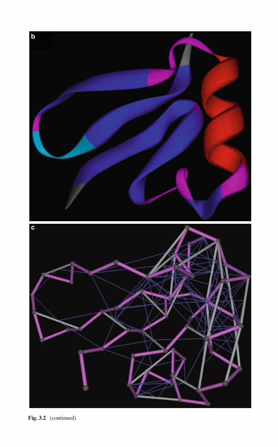
b
c
Fig. 3.2 (continued)
3.4 Wrapping Patterns Along Folding Trajectories 37
To take into account this effect, we incorporate a phenomenological approach towrapping electrostatics [10, 33] as a means of incorporating changes in permittivityassociated with changes in the conformation-dependent environments that affectpairwise interactions. This procedure entails a rescaling of the intramolecular poten-tial terms as folding progresses, an operation requiring keeping track of vicinalhydrophobe positions with each FM iteration (Fig. 3.2c).
3.4 Wrapping Patterns Along Folding Trajectories
To take into account the effect of wrapping on dielectric-dependent pairwiseinteractions and assess its role in defining cooperativity, we have adopted a semiem-pirical procedure to algorithmically keep track of the conformation-dependentmicroenvironments. Thus, the in-bulk potential energy contributions are regardedas zero-order terms, while cooperative effects arise due to the wrapping offavorable interactions brought about by hydrophobic third-body participation(cf. Fig. 3.2c).
A basic question is addressed through this analysis: What is the dynamic rele-vance of optimal wrapping in regard to the protein’s commitment to fold? To tacklethis question, we analyze a representative simulation for the thermophilic variant ofprotein G. This simulation performed at 313 K, pH 7, consists of 106 steps and wasessentially reproduced in 66 of 91 FM runs. All the runs generated a stationary foldwithin RMSD ∼4 Å from the native structure (PDB entry 1GB44) and a dramaticdecrease in potential energy around 0.6 ms (Fig. 3.3).
In accord with experimental tenets, direct examination of the time-dependentbehavior of contact order suggests a nucleation process, whereby a sustainablelarge-scale organization is achieved only at 8×10⁻⁴s after a relatively lengthy trial-and-error process (0–0.63 ms), followed by a critical regime (0.63–0.8 ms). Directinspection of Fig. 3.4 reveals that the transition from local to large-scale organiza-tion is actually defined by a sudden burst in the number of three-body correlationsstarting at 6.0 × 10⁻⁴ − 7 × 10⁻⁴ s.
The nucleation picture revealed by Figs. 3.3 and 3.4 has been further confirmedby examination of total internal energy of the peptide chain and solvent-exposedarea plots. The energy experiences a sudden decrease in the 6.3×10⁻⁴−8.0×10⁻⁴ sregion concurrent with a dramatic decrease in the solvent-exposed area. The pointat which the protein is actually committed to fold (Fig. 3.5) can be inferredby performing runs with different starting conformations extracted from the6.3×10−4 −8.0×10⁻⁴s time window. This commitment arises when a sustainablenumber of three-body correlations (native or nonnative!) equal to or larger than thefinal almost stationary number is reached (Fig. 3.4). In the case of protein G, theburst time window is 6.3 × 10⁻⁴ − 8.0 × 10⁻⁴ s and a sustainable population ofthree-body correlations is maintained in the region 7.0 (±0.2) × 10−4 s.
A similar FM computation was carried out for ubiquitin [32]. The exposed sur-
face area at the transition state is estimated to be 7200 Å2, while the random coil
conformation exposes approximately 10,800 Å2. Thus, we find that the transition

38 3 Folding Cooperativity and the Wrapping of Intermediate States
Fig. 3.3 Internal energy and number of hopping residues along a reproducible representative FMtrajectory for the thermophilic variant of protein G. A hopping residue is defined as tagged fora Ramachandran basin transition in the coarse-grained stochastic process that underlies the FMtorsional dynamics. Reprinted from [35], with permission from Elsevier
state buries 3,600 Å2, approximately 60% of the total area buried in the native fold,
in good agreement with the experimental results [13].The dominant folding pathway for the variant of protein G is coarsely displayed
in Fig. 3.4, where the abscissas denote residue numbers and the ordinates, the timeaxis. The Ramachandran basin assignment for each residue is given as a function oftime and the topology of the entire chain is designated by a row in the histogram.The blue color (basin 1) designates the large basin containing the β strand and PP2conformations, the red color (basin 2) designates the basin containing the right-handed helix conformation, green (basin 3) designates the basin containing the left-handed helix coordinates, and gray (basin 4) corresponds to the fourth basin presentonly in glycine. The figure clearly reveals the emergence of a stable large-scaleorganization that prevails after a critical period located at the 6.3 × 10⁻⁴ − 8.0 ×10⁻⁴ s interval.
We have also reported the extent of protection of backbone hydrogen bonds alongthe folding pathway. Figure 3.6 shows that the average extent of hydrogen bondprotection, ρcoar(t), converges to the value ρcoar = 5 in the long-time limit that startsright after the trial-and-error period; that is, at the sharp burst in C3 = C3(t). Thisregime is associated with the region C3 > 60. The stationary native-like population

3.4 Wrapping Patterns Along Folding Trajectories 39
Fig. 3.4 Three views of the wrapping/folding dynamics for the thermophilic variant of protein Gobtained from a representative expeditious FM trajectory. The left panel represents the time evo-lution of the local topology matrix (LTM) with the abscissas denoting residue numbers and theordinates, the time axis. The Ramachandran basin assignment for each residue is given as a func-tion of time and the topology of the entire chain is designated by a row in the histogram. Colorconvention is consistent with Fig. 3.2b. The middle panel represents the different roles exerted byresidues along the chain at different times. Thus, a pink residue-time entry indicates a residue exert-ing a protecting or wrapping role at the particular time, a gray entry indicates a residue engagedin pairwise interaction which is being protected or wrapped by other hydrophobes, and a blackentry indicates a dual role as protector or wrapper and also engagement in a hydrogen bond whichis being exogenously protected. The right panel indicates the total number of three-body cor-relations representing the wrapping dynamics concurrent with the expeditious folding process.Notice that the burst phase in three-body correlations coincides with the region of transition froma trial-and-error phase to a sustainable structure. Reprinted from [35], with permission fromElsevier
of 15( ± 1) backbone hydrogen bonds are protected by 75( ± 5) three-body correla-tions. We see that the value ρcoar = 5 becomes an approximate constant of motionin the critical region and beyond, that is for C3 > 60. A similar result holds forubiquitin [2]: The native-like stationary population of 28( ± 3) backbone hydrogenbonds is now wrapped by 140( ± 5) three-body correlations.
The wrapping results from Figs. 3.4 and 3.6 are more specific and informativethan earlier attempts at establishing whether buried surface area is commensuratewith hydrogen bond formation [13, 32]. It is difficult to infer from such studieswhether hydrophobic collapse triggers hydrogen bond formation or whether thelatter directs the former. However, Figs. 3.4 and 3.6 reveal that the productive
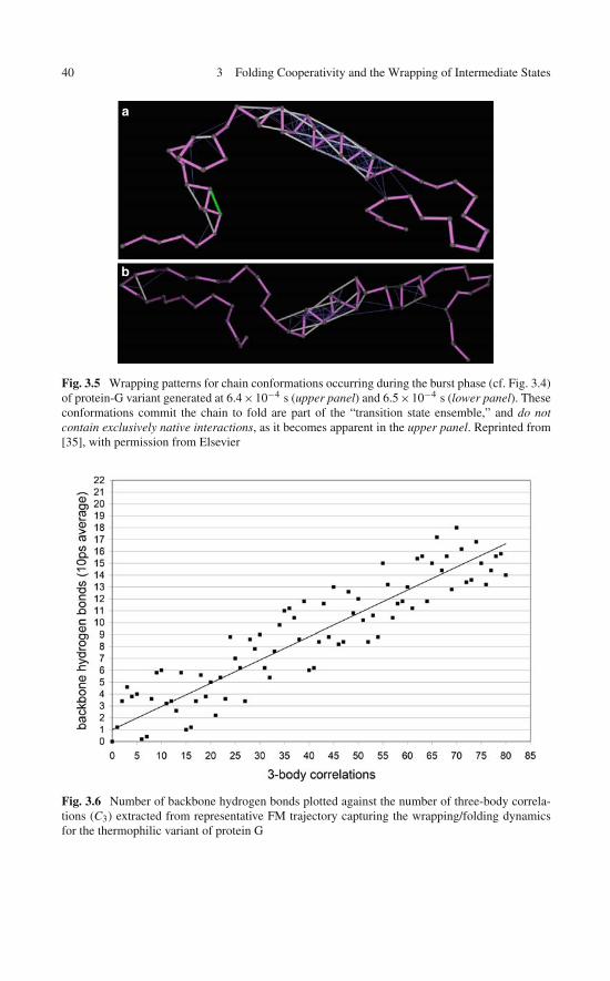
40 3 Folding Cooperativity and the Wrapping of Intermediate States
a
b
Fig. 3.5 Wrapping patterns for chain conformations occurring during the burst phase (cf. Fig. 3.4)of protein-G variant generated at 6.4×10⁻⁴ s (upper panel) and 6.5×10⁻⁴ s (lower panel). Theseconformations commit the chain to fold are part of the “transition state ensemble,” and do notcontain exclusively native interactions, as it becomes apparent in the upper panel. Reprinted from[35], with permission from Elsevier
Fig. 3.6 Number of backbone hydrogen bonds plotted against the number of three-body correla-tions (C3) extracted from representative FM trajectory capturing the wrapping/folding dynamicsfor the thermophilic variant of protein G
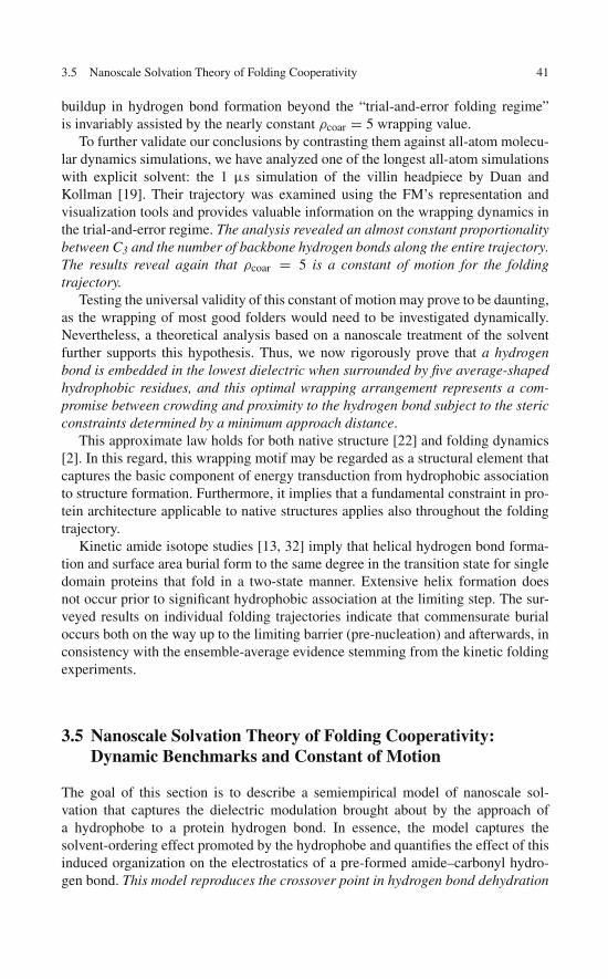
3.5 Nanoscale Solvation Theory of Folding Cooperativity 41
buildup in hydrogen bond formation beyond the “trial-and-error folding regime”is invariably assisted by the nearly constant ρcoar = 5 wrapping value.
To further validate our conclusions by contrasting them against all-atom molecu-lar dynamics simulations, we have analyzed one of the longest all-atom simulationswith explicit solvent: the 1 μs simulation of the villin headpiece by Duan andKollman [19]. Their trajectory was examined using the FM’s representation andvisualization tools and provides valuable information on the wrapping dynamics inthe trial-and-error regime. The analysis revealed an almost constant proportionalitybetween C3 and the number of backbone hydrogen bonds along the entire trajectory.The results reveal again that ρcoar = 5 is a constant of motion for the foldingtrajectory.
Testing the universal validity of this constant of motion may prove to be daunting,as the wrapping of most good folders would need to be investigated dynamically.Nevertheless, a theoretical analysis based on a nanoscale treatment of the solventfurther supports this hypothesis. Thus, we now rigorously prove that a hydrogenbond is embedded in the lowest dielectric when surrounded by five average-shapedhydrophobic residues, and this optimal wrapping arrangement represents a com-promise between crowding and proximity to the hydrogen bond subject to the stericconstraints determined by a minimum approach distance.
This approximate law holds for both native structure [22] and folding dynamics[2]. In this regard, this wrapping motif may be regarded as a structural element thatcaptures the basic component of energy transduction from hydrophobic associationto structure formation. Furthermore, it implies that a fundamental constraint in pro-tein architecture applicable to native structures applies also throughout the foldingtrajectory.
Kinetic amide isotope studies [13, 32] imply that helical hydrogen bond forma-tion and surface area burial form to the same degree in the transition state for singledomain proteins that fold in a two-state manner. Extensive helix formation doesnot occur prior to significant hydrophobic association at the limiting step. The sur-veyed results on individual folding trajectories indicate that commensurate burialoccurs both on the way up to the limiting barrier (pre-nucleation) and afterwards, inconsistency with the ensemble-average evidence stemming from the kinetic foldingexperiments.
3.5 Nanoscale Solvation Theory of Folding Cooperativity:Dynamic Benchmarks and Constant of Motion
The goal of this section is to describe a semiempirical model of nanoscale sol-vation that captures the dielectric modulation brought about by the approach ofa hydrophobe to a protein hydrogen bond. In essence, the model captures thesolvent-ordering effect promoted by the hydrophobe and quantifies the effect of thisinduced organization on the electrostatics of a pre-formed amide–carbonyl hydro-gen bond. This model reproduces the crossover point in hydrogen bond dehydration
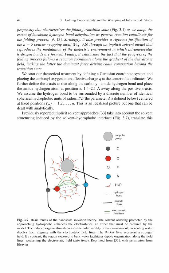
42 3 Folding Cooperativity and the Wrapping of Intermediate States
propensity that characterizes the folding transition state (Fig. 3.1) as we adopt theextent of backbone hydrogen bond dehydration as generic reaction coordinate forthe folding process [9, 13]. Strikingly, it also provides a rigorous justification ofthe n = 5 coarse-wrapping motif (Fig. 3.6) through an implicit solvent model thatreproduces the modulation of the dielectric environment in which intramolecularhydrogen bonds are formed. Finally, it establishes the fact that the progress of thefolding process follows a reaction coordinate along the gradient of the dehydronicfield, making the latter the dominant force driving chain compaction beyond thetransition state.
We start our theoretical treatment by defining a Cartesian coordinate system andplacing the carbonyl oxygen atom effective charge q at the center of coordinates. Wefurther define the x-axis as that along the carbonyl–amide hydrogen bond and placethe amide hydrogen atom at position r, 1.4–2.1 Å away along the positive x-axis.We assume the hydrogen bond to be surrounded by a discrete number of identicalspherical hydrophobic units of radius d/2 (the parameter d is defined below) centeredat fixed positions rj, j = 1,2, . . . , n. This is an idealized picture but one that can bedealt with analytically.
Previously reported implicit solvent approaches [33] take into account the solventstructuring induced by the solvent–hydrophobe interface (Fig. 3.7), translate this
Fig. 3.7 Basic tenets of the nanoscale solvation theory. The solvent ordering promoted by theapproaching hydrophobe enhances the electrostatics, an effect that must be captured by themodel. The induced organization decreases the polarizability of the environment, preventing waterdipoles from aligning with the electrostatic field lines. The thicker lines represent a strongerfield. By contrast, the region exposed to bulk water facilitates dipole organization along the fieldlines, weakening the electrostatic field (thin lines). Reprinted from [35], with permission fromElsevier

3.5 Nanoscale Solvation Theory of Folding Cooperativity 43
effect into a distance-dependent permittivity, ε (r), and quantify the effect on theCoulomb screening. A more heuristic, practical, and phenomenological approachis rooted in two pivotal components: (a) perturbation of the diffraction structure ofbulk water as hydrophobes are incorporated at fixed positions and (b) recovery oftheir solvent-structuring effect by inverse Fourier transforming the previous resultgiven in frequency space.
To propagate the solvent-structuring effect induced by the presence of thehydrophobic spheres, we replace the position-dependent dielectric by an integralkernel convoluted with the electric field at position r to represent the correlationswith the field at neighboring positions r′. This prompts us to replace the classicalPoisson equation by the rigorous relation (see Chap. 14):
div
[∫K
(r,r′,
{rj
})E
(r′) dr′
]= 4πqδ(r), (3.1)
where the kernel K(r,r′ {rj
})is parametrically dependent on the fixed hydrophobe
positions. In the absence of vicinal hydrophobic units, the correlations decay asexp
(− ∥∥r − r′∥∥ /ξ)
(ξ = characteristic correlation length). In the limit ξ → 0, weget K
(r, r′) ∼ δ
(r′ − r
), yielding the standard Poisson equation.
The correlation kernel reflects the relationship between diffraction and dielectric.For bulk water, we get K
(r, r′) = K
(r − r′) by inverse transforming its frequency
k-vector representation:
K(r − r′) =
∫exp
[ik · (
r − r′)]L (k) dk. (3.2)
In (3.2), L (k) = [(εw − ε0) /
(1 + εw ‖k‖2 ξ2/ε0
) + ε0]
, ξ ≈ 5 Å denotesthe characteristic length, and εw, ε0 are the permittivities of water and vacuum,respectively. To obtain the correlation kernel with n hydrophobic units, we need toincorporate their solvent-structuring effect:
K(r,r′ {rj
}) =[∫
exp[ik · (
r − r′)] L (k) dk]
×⎡⎣1 +
∑j=1,...,n
j(r,r′)
⎤⎦
. (3.3)
On purely phenomenological grounds we get the following approximation [2, 33,34]: j
(r,r′) ∼ exp
[− (∥∥r − rj∥∥ + ∥∥r′ − rj
∥∥)/�
], for
∥∥r − rj∥∥ and
∥∥r′ − rj∥∥ >
d/2 with characteristic length � subsuming the range of the water-structuring effect.This parameter is typically fixed at 1.8 Å, the effective thickness of a single waterlayer (cf. Fig. 3.7).

44 3 Folding Cooperativity and the Wrapping of Intermediate States
We now solve (3.1) and (3.3) by Fourier transformation, obtaining the elec-tric field E (r) by inverse Fourier transformation of the solution to (3.1) ink-representation:
∫E (r) dr = − (4πq)
∫exp (ik · r) ‖k‖−2 [
K(k,
{kj
})]−1dk . (3.4)
Direct residue evaluation at the first-order poles k = ±i (ε0/εw)1/2 ξ−1
(k = ‖k‖) and k = kj ± i�−1 yields the electric field E (r) by retaining only thereal part in the residue calculation:
E (r) =(
q/r2) [(
ε−10 − ε−1
w
)�
({rj
})(1 + r/ξ) exp (−r/ξ) + ε−1
w
], (3.5)
where
�({
rj}) = ∏
j=1,...,n
[1 + exp
(− ∥∥rj∥∥ /�
)]
× [(1 + exp ( − ∥∥r − rj
∥∥ /�)] (3.6)
and r = ‖r‖. Equations (3.5) and (3.6) describe the net effect of the wrappinghydrophobic arrangement on the electric field. In accord with (3.5) and (3.6), thewrapping effect can be captured by replacing the permittivity constant εw for bulkwater by an effective permittivity ε = ε
(r,
{rj
})defined as
ε =[(
ε−10 − ε−1
w
)�
({rj
})(1 + r/ξ) exp (−r/ξ) + ε−1
w
]−1. (3.7)
This quantity tends to the bulk limit for long interaction distances (r/ξ → ∞).We now turn to the problem of finding the optimal wrapping arrangement and
contrasting our implicit solvent result with the phenomenological result emergingfrom the simulations (cf. Figs. 3.4 and 3.6). Since ε−1
0 >> ε−1w , finding the wrap-
ping cluster with the lowest dielectric in its interior is tantamount to finding thearrangement
{rj
}that maximizes the function �
({rj
}), in accord with (3.7). We
thus find the optimal arrangement{rj
}of hydrophobes that yields the maximum
value �∗ for �({
rj})
. First, we compute the maximum �∗ (n) of �({
rj})
for eachfixed n subject to the constraint of preserving a minimum distance d between anytwo hydrophobes. The d value is taken to be 5 Å in accord with typical minimaldistances between α-carbon atoms in tertiary structure [22]. Our results are quali-tatively invariant in the range 4.5 Å ≤ d ≤ 6 Å. Using the Lagrange multipliersmethod to minimize the effective permittivity, we find that the optimal arrangementis invariably obtained by fixing n−2 hydrophobes at distance d from each other andequidistantly from the O and H atoms, and placing the remaining two along thex-axis at distance (� + η)(1 + n−2) (to first approximation) away from the C andN atoms, with η = C − O distance in the carbonyl group. This gives for n = 4(tetrahedron): �∗(4) = 3.419; for n = 5 (trigonal bipyramid): �∗(5) = 4.144; for
3.6 Dehydronic Field Along the Folding Pathway and the Commitment to Fold 45
n = 6 (square bipyramid), �∗(6) = 3.952; and for n = 7 (pentagonal bipyramid),�∗(7) = 3.421. Similar calculations for all n allow us to establish the followingorder relations:
�∗(3) < �∗(4) < �∗(5) > �∗(6) > �∗(7) > . . . (3.8)
Thus, �∗(n) has a single maximum at n = 5. This maximum is expected onthe basis of the two conflictive tendencies in the stabilization of a hydrogen bond:(1) bringing close to the hydrogen bond as many hydrophobes as possible and (2)bringing them as close to the hydrogen bond as possible. However, both demandsstart becoming mutually incompatible due to the steric hindrances implicit in theLennard–Jones repulsive terms.
In full agreement with the result described in Fig. 3.6 (ρcoar = 5 is a constantof motion along the folding trajectory), a hydrogen bond is embedded in the low-est dielectric when surrounded by five wrapping residues, the optimal compromisebetween crowding and proximity to the hydrogen bond. Thus, we have benchmarkedthe solvation theory against a rigorous dynamic result.
3.6 Dehydronic Field Along the Folding Pathwayand the Commitment to Fold
Equations (3.5), (3.6), and (3.7) enable us to compute the dehydronic field, that is,the mechanical equivalent of the dehydration propensity of hydrogen bonds madealong the folding process. This computation requires the evaluation of the gra-dient � (R) = −∇R [4πε (R)]−1 qq′/r of the electrostatic energy with respectto the position vector R of the test hydrophobe (in the simulations we adoptedmethane as test hydrophobe). This analysis is motivated by the need to supportthe two-state kinetic picture outlined in Fig. 3.1. To normalize for the number ofhydrogen bonds formed at any given time, we computed the dehydronic field per
hydrogen bond as ϑ (t) =⟨∥∥∥� (R)‖R‖=4Å
∥∥∥⟩t, where < >t denotes the average
over the hydrogen bonds formed by the chain at time t. The results along the fold-ing trajectory described in Figs. 3.2, 3.3, 3.4, 3.5, 3.6, and 3.7 are presented inFig. 3.8.
Contrasting Fig. 3.8 with Figs. 3.3 and 3.4 clearly validates the two-state foldingscenario depicted in Fig. 3.1: The dehydronic field starts as a very minor contributorduring the hydrophobic collapse of the chain. Yet, once hydrogen bonds are formedto compensate for the backbone burial and they become partially wrapped (transi-tion state ensemble), the dehydronic force becomes the driving force behind chaincompaction. Thus, the progress of the folding process follows a reaction coordi-nate dictated by the gradient of the dehydronic field, making the latter the dominantfactor that commits the chain to fold.
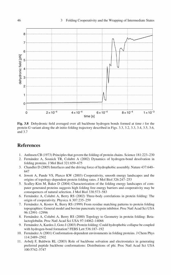
46 3 Folding Cooperativity and the Wrapping of Intermediate States
8
7
6
5
4
3
2
dehy
dron
ic fi
eld
[pN
]
1
0
0 2 × 10⁻⁴ 4 × 10⁻⁴ 6 × 10⁻⁴
time [s]8 × 10⁻⁴ 1 × 10⁻³
Fig. 3.8 Dehydronic field averaged over all backbone hydrogen bonds formed at time t for theprotein G variant along the ab initio folding trajectory described in Figs. 3.3, 3.2, 3.3, 3.4, 3.5, 3.6,and 3.7
References
1. Anfinsen CB (1973) Principles that govern the folding of protein chains. Science 181:223–2302. Fernández A, Sosnick TR, Colubri A (2002) Dynamics of hydrogen-bond desolvation in
folding proteins. J Mol Biol 321:659–6753. Chandler D (2005) Interfaces and the driving force of hydrophobic assembly. Nature 437:640–
6474. Jewett A, Pande VS, Plaxco KW (2003) Cooperativity, smooth energy landscapes and the
origins of topology-dependent protein folding rates. J Mol Biol 326:247–2535. Scalley-Kim M, Baker D (2004) Characterization of the folding energy landscapes of com-
puter generated proteins suggests high folding free energy barriers and cooperativity may beconsequences of natural selection. J Mol Biol 338:573–583
6. Fernández A, Colubri A, Berry RS (2002) Three-body correlations in protein folding: Theorigin of cooperativity. Physica A 307:235–259
7. Fernández A, Kostov K, Berry RS (1999) From residue matching patterns to protein foldingtopographies: General model and bovine pancreatic trypsin inhibitor. Proc Natl Acad Sci USA96:12991–12996
8. Fernández A, Colubri A, Berry RS (2000) Topology to Geometry in protein folding: Beta-lactoglobulin. Proc Natl Acad Sci USA 97:14062–14066
9. Fernández A, Kardos J, Goto J (2003) Protein folding: Could hydrophobic collapse be coupledwith hydrogen-bond formation? FEBS Lett 536:187–192
10. Fernández A (2001) Conformation-dependent environments in folding proteins. J Chem Phys114:2489–2502
11. Avbelj F, Baldwin RL (2003) Role of backbone solvation and electrostatics in generatingpreferred peptide backbone conformations: Distributions of phi. Proc Natl Acad Sci USA100:5742–5747
References 47
12. Fernández A (2004) Keeping dry and crossing membranes. Nat Biotechnol 22:1081–108413. Krantz BA, Moran LB, Kentsis A, Sosnick TR (2000) D/H amide kinetic isotope effects reveal
when hydrogen bonds form during protein folding. Nat Struct Biol 7:62–7114. Plaxco KW, Simmons KT, Baker D (1998) Contact order, transition state placement and the
refolding rates of single domain proteins. J Mol Biol 277:985–99415. Fersht A (2000) Transition-state structure as a unifying basis in protein-folding mechanisms:
Contact order, chain topology, stability, and the extended nucleus mechanism. Proc Natl AcadSci USA 97:1525–1929
16. Fernández A, Scott LR (2003) Adherence of packing defects in soluble proteins. Phys RevLett 91:018102
17. Fernández A (2003) What caliber pore is like a pipe? Nanotubes as modulators of iongradients. J Chem Phys 119:5315–5319
18. Fernández A, Shen M, Colubri A, Sosnick TR, Freed KF (2003) Large-scale context in proteinfolding: Villin headpiece. Biochemistry 42:664–671
19. Duan Y, Kollman PA (1998) Pathways to a protein folding intermediate observed in a 1-microsecond simulation in aqueous solution. Science 282:740–744
20. Baldwin RL (2002) Making a network of hydrophobic clusters. Science 295:1657–165821. Nemethy G, Steinberg IZ, Scheraga HA (1963) The influence of water structure and
hydrophobic contacts on the strength of side-chain hydrogen bonds in proteins. Biopolymers1:43–69
22. Fernández A, Berry RS (2002) Extent of hydrogen-bond protection in folded proteins: Aconstraint on packing architectures. Biophys J 83:2475–2481
23. Novotny J, Bruccoleri R, Karplus M (1984) Analysis of incorrectly folded protein models.Implications for structure predictions. J Mol Biol 177:787–818
24. Daggett V, Levitt M (1992) A model of the molten globule state from molecular dynamicssimulations. Proc Natl Acad Sci USA 89:5142–5146
25. Brooks CL, Case D (1993) Simulations of peptide conformational dynamics and thermody-namics. Chem Rev 93:2487–2502
26. Fernández A, Rogale K (2004) Sequence-space selection of cooperative model proteins. JPhys A: Math & Gen 37:197–202
27. Kuwata K, Shastry R, Cheng H, Hoshino M, Batt CA, Goto Y, Roder H (2001) Structural andkinetic characterization of early folding events in beta-lactoglobulin. Nat Struct Biol 8:151–155
28. Nymeyer H, Garcia AE, Onuchic JN (1998) Folding funnels and frustration in off-latticeminimalist protein landscapes. Proc Natl Acad Sci 95:5921–5928
29. Onuchic JN, Luthey-Schulten Z, Wolynes PG (1997) Theory of protein folding: The energylandscape perspective. Annu Rev Phys Chem 48:545–600
30. Chan HS, Dill KA (1997) From Levinthal to pathways to funnels. Nat Struct Biol 4:10–1931. Fernández A, Colubri A, Berry RS (2001) Topologies to geometries in protein folding:
Hierarchical and nonhierarchical scenarios. J Chem Phys 114:5871–588832. Shi Z, Krantz BA, Kallenbach N, Sosnick TR (2002) Contribution of hydrogen bonding to
protein stability estimated from isotope effects. Biochemistry 41:2120–212933. Pietrosemoli N, Crespo A, Fernández A (2007) Dehydration propensity of order-disorder
intermediate regions in soluble proteins. J Proteome Res 6:3519–352634. Despa F, Fernández A, Berry RS (2004) Dielectric modulation of biological water. Phys Rev
Lett 93:22810435. Fernandez A, Zhang X, Chen J (2008) Folding and wrapping soluble proteins: Exploring the
molecular basis of cooperativity and aggregation. Prog Mol Biol Transl Sci 83:53–88

“This page left intentionally blank.”
Chapter 4Wrapping Deficiencies and De-wettingPatterns in Soluble Proteins: A Blueprintfor Drug Design
This chapter unravels a link between wrapping deficiencies and defects in the hydra-tion shell of soluble proteins. The hydration shell of a soluble protein is not uniform:its tightness, marked by the mobility of interfacial water, is site-dependent andmodulates the propensity for protein associations. The most pronounced interfa-cial de-wetting propensity is promoted by dehydrons in the surface of the solubleproteins. Their dehydration is thermodynamically favored and hence may serve asa blueprint for drug design. The result has crucial implications for the designer asdrugs may be engineered to expel the labile parts of the target hydration shell uponassociation. Hence, subtle differences in the location of hydration defects in proteinswithin the same family may be exploited to enhance drug specificity, as demon-strated in Chap. 8. The subject of interfacial water is again taken up in Chap. 14 thatdeals with the disruption of protein-protein interfaces with competing drugs.
4.1 Hydration Defects in Soluble Proteins
Solvation is not uniform on the surface of soluble proteins, rather it is marked byvastly different mobilities of interfacial water molecules [1–5]. This uneven distri-bution of mobilities of the hydrating molecules affects protein folding dynamics andassociations. The hydration shell gets tighter or less mobile at interfacial regionswhere the hydrogen bond network of hydrating molecules is minimally disruptedand becomes looser in concave or flat nonpolar regions of the protein surface [3–5].Thus, dehydration patterns have been associated with surface topography [5] andwith fold topology [6], both determinants of protein associations. However, thepronounced de-wetting propensity of interfacial regions remains difficult to assessin general, as evidenced by the difficulty in predicting binding sites [7], in inferringpropensities for aberrant aggregation [8], and in rationally designing ligands,notwithstanding some level of success in ligand docking [9]. This chapter exploresde-wetting patterns determined by unburied or under-wrapped backbone hydrogenbonds, the dehydrons [10, 11] described in the preceding chapters. Since their levelsof hydration may be modulated by protein associations, dehydrons are implicatedin macromolecular recognition [8–11]. More precisely, dehydrons may become
49A. Fernández, Transformative Concepts for Drug Design: Target Wrapping,DOI 10.1007/978-3-642-11792-3_4, C© Springer-Verlag Berlin Heidelberg 2010

50 4 Wrapping Deficiencies and De-wetting Patterns in Soluble Proteins
stabilized and electrostatically strengthened by the attachment of a ligand or bindingpartner that further contributes to their dehydration. As indicated in Chap. 1, thenet gain in Coulomb energy associated with wrapping or protecting a dehydron hasbeen experimentally determined to be ∼4 kJ/mol [12]. The adhesive force exertedby a dehydron on a hydrophobic group at 6 Å distance is ∼7.8 pN, a magnitudecomparable to the hydrophobic attraction between two nonpolar moieties that frameunfavorable interfaces with water [12]. Furthermore, the fact that dehydrons pro-mote protein associations (Chap. 1) strongly suggests that they may signal sites witha loose hydration shell, that is, sites where the work needed to remove interfacialwater is minimal and amply compensated by the enhancement of the Coulombicenergy.
As shown in Chap. 8 and again in Chap. 14, this result will prove to be ofparamount relevance for the rational drug designer as it introduces a blueprint toguide ligand engineering: the de-wetting propensities in the target protein may besculpted into the ligand so that water is expelled from the interface upon associationwherever the work needed to do so is minimal and the dehydronic force is maximal.In other words, the drug/ligand may be engineered to tightly fit against the hot spotsof labile interfacial water.
4.2 Wrapping as a Marker of Local De-wetting Propensity
In this section we introduce a descriptor of hydration tightness or de-wetting propen-sity in order to assess the functional role of dehydrons. Tightness is defined bythe extent of mobility of hydrating molecules. Thus, the mean residence time ofhydrating molecules within a domain around each residue on the protein surfaceserves as an adequate indicator. As an illustration, we describe in detail the interfa-cial water mobility for the autonomously folded SH3 domain (PDB.1SRL) [13],an all-beta protein involved in the regulation of cell signaling. We also analyzeubiquitin (PDB.1UBI) [14], a quintessential α/β fold, and finally explore the rela-tionship between extreme dehydration propensity and amyloidogenic aggregation[9].
Our results lead us to identify dehydrons as the structural feature causativeof the most dramatic decrease in residence times (highest mobility) of hydratingmolecules. Dehydrons promote local de-wetting because the Coulomb energy of theintramolecular hydrogen bonds becomes magnified upon water removal, and the netgain in stability upon dehydration offsets the work required to remove surroundingwater [10, 15, 16].
In order to assess the local mobility of water molecules in the hydration shell,the local mean residence time, <τ>i, of hydrating molecules around residue i isdefined with respect to a microenvironment in the form of a spherical domain D(i)of 6 Å radius (∼width of three water layers [12]) centered at the α-carbon of residuei (Fig. 4.1a). The computations are performed for a range of radii (see below). Theresidence time is obtained as follows:

a
b
Fig. 4.1 (a) Basic scheme for the computation of de-wetting hot spots in soluble proteins.(b) Mean residence times of water molecules solvating the folded protein SH3 domain. Residuenumbering follows PDB file 1SRL. The one-letter amino acid code was adopted for clarity. Themean residence times are computed using (4.1) and extracted from MD simulations of the hydrationdynamics. The error bars represent variances and the thick rectangle represents the range of res-idence times for bulk water within a domain of the same dimensions of the one used to examinethe protein surface. The range for bulk water serves as benchmark to single out hot spots of weakhydration. Three residues (Ala12, Ile56, and Val61) are fully buried in the folded structure andthus their hydration shells contain no water molecules. Figure 4.1b reprinted with permission fromAmerican Institute of Physics [20]. Copyright 2007

52 4 Wrapping Deficiencies and De-wetting Patterns in Soluble Proteins
〈τ 〉i = ∫τ fi(τ )dτ
/(∫fi(τ )dτ
);
τ∫0
fi(τ ′)dτ ′ = Pi(0) − Pi(τ ) ;
Pi(τ ) = �−1�∫0
⎡⎣ ∑
v(t)∈U(i,t)w(t+τ )∈U(i,t+τ )
δ (v(t), w(t + τ ))
⎤⎦ dt ;
(4.1)
where fi (τ ) dτ/∫
fi(τ )dτ is the expected fraction of water molecules that exit D(i)within time interval [τ , τ + dτ ]; Pi(τ ) is the expected number of water moleculesremaining in D(i) at time τ (Pi(0) − Pi(τ ) = number of molecules that exited D(i)in the time period [0, τ ]); v(t), w(t + τ ) denote indexes labeling water moleculescontained in D(i) at times t and t + τ , respectively; U (i, t), U (i, t + τ) denotethe collection of indexes of water molecules contained in D(i) at times t andt + τ , respectively; δ is the Kronecker symbol (δ(v(t), w(t + τ )) = 1 if v(t) =w(t + τ ) and 0, otherwise); and the integration over variable t is carried out overthe interval of sampled times (t = 0 to t = � = 10 ns) after 5 ns of priorequilibration (the sampling is considered exhaustive since < τ ><< � for allresidues).
The mean residence times are obtained from classical trajectories generated bymolecular dynamics (MD) simulations starting from the PDB structure embeddedin a pre-equilibrated cell of explicitly represented water molecules and counteri-ons [17, 18]. Computations are performed by integration of Newton’s equations ofmotion with time step 2 fs using the GROMACS program [19] in an NPT ensemblewith box size 8 × 8 × 8 nm3 and periodic boundary conditions maintained at 300 Kand 1 atm. The box size is calibrated so that the solvation shell extended at least12 Å from the protein surface at all times. Specific details on the generation of theseclassical trajectories may be found in [20].
4.3 Dehydrons Are Loosely Hydrated
We start by providing a detailed analysis of the de-wetting propensities in the SH3domain (N = 55, PDB.1SRL) and their correlation with dehydrons in the struc-ture. The mean residence times of water molecules at the protein–water interfacehas an overall average value of 44 ps, nearly twice the residence time for bulkwater (∼21 ps, Fig. 4.1b) [21]. The hydration shell reveals some standard features.Thus, short residence times correspond to de-wetting propensity associated withhydrophobic hydration at cavities with relatively large curvature radius ( ∼ 6 Å)[22], like the hydrophobic pocket containing Trp43 (Figs. 4.1b and 4.2).
By contrast, exposed hydrophobic residues like Val35 may be effectively“clathrated” or accommodated within a water cavity that introduces a minimal per-turbation of the tetrahedral hydrogen bonding network of water. In fact, clathrationactually tightens the hydration shell (Figs. 4.1b and 4.2).

4.3 Dehydrons Are Loosely Hydrated 53
Fig. 4.2 PDB structure of SH3 domain indicating the packing defects in the protein. The backboneis displayed by blue segments representing virtual bonds joining α-carbons, well-wrapped hydro-gen bonds are shown as white segments joining α-carbons of paired residues, and dehydrons areshown in green. Some residue labels and side chains are displayed to illustrate hydration patterns.Reprinted with permission from American Institute of Physics [20]. Copyright 2007
Side chains with small exposed cations (Lys27 and Lys28) have the longest res-idence times since they are tightly hydrated as are polar residues in cavities (likeSer58), since their hydrogen bonding to water is protected. By contrast, exposedresidues with delocalized positive charges like Arg19 have looser hydration [23].
The residues with the shortest residence times (<τ>< 24 ps) and also with thelargest variances (< (τ−<τ > )2 >1/2 > 6 ps) are Gln33, His46, Ser47, and Gly51,indicating a highly fluctuating environment and a high de-wetting propensity. Asshown below, these residues are paired by the only backbone amide–carbonyl hydro-gen bonds which are incompletely wrapped or shielded from water by nonpolargroups of the protein chain. In accord with their shortest water residence times(Fig. 4.1b), dehydrons become favorably dehydrated, a process that decreases theoverall polarizability of their microenvironments. This dehydration strengthens theelectrostatic contribution and stabilizes the hydrogen bond by destabilizing the non-bonding or unbound state to the point of overcoming the work needed to removesurrounding water [8]. These packing defects and the decrease in medium polariz-ability or dielectric modulation they promote differ from packing-density variations

54 4 Wrapping Deficiencies and De-wetting Patterns in Soluble Proteins
in nonpolar interiors [24]: They relate to the exposure of the polar backbone due toincomplete wrapping by nonpolar side chains.
To explain the local weaknesses in the hydration shell of the protein (Fig. 4.1b),the de-wetting propensity of dehydrons is quantified and compared with that of well-wrapped hydrogen bonds. As shown in Chaps. 1 and 3, the de-wetting propensityhas a mechanical equivalent [12, 25]: The attractive force exerted on a nonpolar testgroup (i.e., methane, CH4) by a pre-formed hydrogen bond that may be strengthenedupon removal of surrounding water. As the nonpolar group approaches a dehydron,it displaces water thus decreasing the environmental polarity, thereby enhancing andstabilizing the bond. The wrapping microenvironment of a hydrogen bond may bedescribed by the set of position vectors
{Rj
}j=1,...,K = {
Rj(t)}
j=1,...,K , relative tothe hydrogen atom (origin of coordinates), of K nonpolar groups from the proteinchain within the two spheres of radius 6 Å centered at the α-carbons of the pairedresidues. All time-dependent coordinates are taken from MD trajectories.
To compute the de-wetting of intramolecular hydrogen bonds, we take intoaccount the modulation of local permittivity determined by a change in the “envi-ronmental coordinates”:
{Rj
}j=1,...,K → {
Rj, R}
j=1,...,K , where R is the positionvector of the test methane that approaches the bond orthogonally to the Coulombicfield. The energy change E associated with the change in wrapping is then givenby [12, 25]
E(t) = −(qq′/4πr)[ε−1 ({Rj, R}j=1,...,K
) − ε−1 ({Rj}j=1,...,K)]
, (4.2)
where q, q′ are the effective charges at the hydrogen and oxygen atoms of theamide–carbonyl hydrogen bond, r is the hydrogen bond length (r = r(t) = ‖r(t)‖,with r = position vector of oxygen), and the reciprocal permittivity ε−1, quanti-fying the Coulomb screening due to wrapping by nonpolar groups, is genericallygiven by [25]
ε−1({Rn}) = (ε−1o − ε−1
w )�({Rn})[(1 + r/ξ ) exp ( − r/ξ )] + ε−1w , (4.3)
where, as in Chap. 3, ε0, εw denote, respectively, the permittivity of vacuum andbulk water, �({Rn}) = �n=1,...
[1 + exp (−‖Rn‖ /�)
] [1 + exp (‖r − Rn‖ /�)
],
where ξ , fixed at 5 Å, is the characteristic length for water dipole reorientationinfluence, and � = 1.8Å is the characteristic length for water structuring around anonpolar group [25].
Applying (4.2) and (4.3) to the MD trajectories, we quantify the enhancement inthe Coulombic contribution to the hydrogen bond resulting from the decrease in per-mittivity as the test hydrophobe approaches the protein surface (Fig. 4.3). Thus, thede-wetting field �(R) at position R of the test hydrophobe generated by a hydrogenbond wrapped by K nonpolar groups is given by
�(R) = (qq′/4πr)∇R(ε−1({Rj, R}j=1,...,K)). (4.4)

4.3 Dehydrons Are Loosely Hydrated 55
Fig. 4.3 De-wetting field exerted by residues paired by hydrogen bonds in the SH3 domain. Thedehydration propensity of a hydrogen bond, ‖�(R)‖, is determined at ‖R‖ = 6 Å and is assignedto the two residues paired by the hydrogen bonds. The data displayed show a time average and vari-ance over the interval of sampled times (t = 0 to t = � = 10 ns) after 5 ns of prior equilibration.Reprinted with permission from American Institute of Physics [20]. Copyright 2007
The time averages of the magnitude and variance of the de-wetting field werecomputed for each residue in the protein (Fig. 4.3).
Comparison of Figs. 4.1b and 4.3 prompt the following conclusions: (a) the tight-ness of the hydration shell of a soluble protein is not uniform, yielding an unevendistribution of mobilities for hydrating molecules; (b) dehydrons generate the mostpronounced loosening of the hydration shell; and (c) dehydrons are endowed withdehydration propensities.
A similar analysis was conducted for other single-domain soluble proteins, unre-lated evolutionarily or topologically to the study case presented and the sameconclusions regarding the de-wetting propensity of dehydrons were shown to hold[20]. For example, we focused on the highly conserved ubiquitin (N = 76, 11 dehy-drons, PDB.1UBI). In this case dehydrons also generate the most intense de-wettingfield in the protein (Fig. 4.4), in accord with their role as structural markers for pro-tein associations. The de-wetting hot spots account for 9 out of the 11 dehydronsidentified for this structure, and only partially account for the remaining two. Thetwo dehydrons which do not fully promote de-wetting are Glu24–Asp52 and Pro19–Ser57. The larger residence times in these cases may be attributed to the pronouncedhydration demands of the two charged side chains in the former case and the spe-cial steric hindrance promoted by Pro19, which precludes water approach to thebackbone in the latter.

56 4 Wrapping Deficiencies and De-wetting Patterns in Soluble Proteins
Fig. 4.4 (a) Mean residence times of water molecules solvating the folded protein ubiquitin.Residue numbering follows the file PDB.1UBI. b PDB structure of ubiquitin highlighting thedehydrons (displayed in green). Reprinted with permission from American Institute of Physics[20]. Copyright 2007
4.4 Displacing Loose Hydrating Molecules:A Blueprint for the Drug Designer
In conclusion, the identification of structural determinants of protein/water interfa-cial behavior is vital to understand protein associations and to design drugs/ligandswith better affinity. The findings reported in this chapter represent a constructivestep in this direction, since we have singled out structural singularities in solubleproteins – the dehydrons – that behave as de-wetting hot spots. The identificationof these sites with defective and loose hydration opens up an engineering avenueto understand and manipulate protein/ligand associations. Thus, by sculpting on thedrug the de-wetting hot spots of the target, we take advantage of the minimal workassociated with removal of dehydron-solvating water molecules, amply compen-sated by the resulting enhancement of the electrostatic interaction (see Chap. 3). Inthis way, we may enhance the affinity for the protein target by adopting the patternof hydration defects in the target as a blueprint for molecular engineering (Fig. 4.5).This concept is brought to fruition in the reengineering of the powerful anticancer

References 57
Fig. 4.5 The pattern of defects in the hydration shell of a target protein is a blueprint for drugdesign. Interfacial water molecules solvating dehydrons have short residence time and hence areeasily displaced upon association with a purposely designed ligand. Their loss of hydrogen bondingpartnerships results from their partial confinement. Fast interfacial water molecules are repre-sented as thick black angular lines, while bulk-like water molecules are indicated as gray angularlines. Hydrogen bonds are shown as thin black lines, while dehydrons are indicated as dottedlines
drug imatinib (Gleevec), as described in Chap. 8. Furthermore, it heralds the adventof a new generation of drugs based on an assessment of the architecture of theprotein fold in relation to the solvating environment, as shown in Chap. 14. Theimplications of this concept for drug specificity will be explored in Chap. 8, wheredifferences in de-wetting patterns across purported targets enable us to engineer aselective therapeutic impact.
References
1. Otting G, Liepinsh E, Wüthrich K (1991) Protein hydration in aqueous solution. Science254:974–980
2. Qiu W, Kao Y, Zhang L et al (2006) Protein surface hydration mapped by site-specificmutations. Proc Natl Acad Sci USA 103:13979–13984
3. Makarov V, Andrews BV, Smith PE, Pettitt BM (2000) Residence times of water moleculesin the hydration sites of myoglobin. Biophys J 79:2966–2974
4. Chandler D (2005) Interfaces and the driving force of hydrophobic assembly. Nature 437:640–647
5. Cheng Y, Rossky PJ (1998) Surface topography dependence of biomolecular hydrophobichydration. Nature 392: 696–699
58 4 Wrapping Deficiencies and De-wetting Patterns in Soluble Proteins
6. Liu P, Huang X, Zhou R, Berne BJ (2005) Observation of a dewetting transition in the collapseof the melitin tetramer. Nature 437:159–162
7. Fernández A, Scheraga HA (2003) Insufficiently dehydrated hydrogen bonds as determinantsof protein interactions. Proc Natl Acad Sci USA 100:113–118
8. Fernández A, Kardos J, Scott R, Goto Y, Berry RS (2003) Structural defects and the diagnosisof amyloidogenic propensity. Proc Natl Acad Sci USA 100:6446–6451
9. Hetenyi C, van der Spoel D (2006) Blind docking of drug-sized compounds to proteins withup to a thousand residues. FEBS Lett 580:1447–1450
10. Fernández A, Berry RS (2004) Molecular dimension explored in evolution to promoteproteomic complexity. Proc Natl Acad Sci USA 101:13460–13465
11. Fernández A, Scott RL (2003) Dehydron: A structurally encoded signal for protein interaction.Biophys J 85:1914–1928
12. Fernández A, Scott LR (2003) Adherence of packing defects in soluble proteins. Phys RevLett 91:018102
13. Yu H, Rosen MK, Schreiber SL (1993) 1H and 15 N assignments and secondary structure ofthe Src SH3 domain. FEBS Lett 324:87–92
14. Vijay-Kumar S, Bugg CE, Cook WJ (1987) Structure of ubiquitin refined at 1.8 angstromresolution. J Mol Biol 194:531–544
15. van der Spoel D, van Maaren P, Larsson P, Timneanu N (2006) Thermodynamics of hydrogenbonding in hydrophilic and hydrophobic media. J Phys Chem B 110:4393–4398
16. Sheu S, Yang D, Selzle H, Schlag EW (2003) Energetics of hydrogen bonds in peptides. ProcNatl Acad Sci USA 100:12683–12687
17. Rizzo RC, Jorgensen WL (1999) OPLS All-atom model for amines: Resolution of the aminehydration problem. J Am Chem Soc 121:4827–4836
18. Jorgensen WL, Chandrasekhar J, Madura J, Impey RW, Klein ML (1983) Comparison ofsimple potential functions for simulating liquid water. J Chem Phys 79:926–935
19. Lindahl E, Hess B, Van der Spoel D (2001) GROMACS 3.0: A package for molecularsimulation and trajectory analysis. J Mol Model 7:302–317
20. Fernández A, Chen J, Crespo A (2007) Solvent-exposed backbone loosens the hydration shellof soluble folded proteins. J Chem Phys 126:245103
21. Denisov V, Halle B (1995) Protein hydration dynamics in aqueous solution. A comparison ofbovine pancreatic trypsin inhibitor and ubiquitin by oxygen-17 spin relaxation dispersion. JMol Biol 245:682–697
22. Lum K, Chandler D, Weeks JD (1999) Hydrophobicity at small and large length scales. J PhysChem B 103:4570–4577
23. Mason PE, Neilson GW, Dempsey CE, Barnes AC, Cruickshank JM (2003) The hydrationstructure of guanidinium and thiocyanate ions: Implications for protein stability. Proc NatlAcad Sci USA 100:4557–4561
24. Kocher JP, Prevost M, Wodak S, Lee B (1996) Properties of the protein matrix revealed by thefree energy of cavity formation. Structure 4:1517–1529
25. Fernández A, Sosnick TR, Colubri A (2002) Dynamics of hydrogen-bond desolvation infolding proteins. J Mol Biol 321:659–675

Chapter 5Under-Wrapped Proteins in the Order–DisorderTwilight: Unraveling the Molecular Etiologyof Aberrant Aggregation
Soluble folded proteins maintain their structural integrity by properly shielding theirbackbone amides and carbonyls from full hydration. Thus, a poorly wrapped back-bone hydrogen bond or dehydron constitutes an identifiable structural deficiency. Inthis chapter we describe the physical properties of proteins that possess large clus-ters of dehydrons in their soluble structure. We show that these clusters representunique structural singularities belonging to an order–disorder twilight and generatea sharp local quenching of the dielectric permittivity of the surrounding medium.The functional roles of these singularities are explored across natural proteins andrelated to their physical properties, with special emphasis on the molecular etiol-ogy of amyloidogenic aggregation. Our analysis required a description of interfacialwater that cannot be properly captured by conventional continuous models, wheresolvent degrees of freedom are typically averaged out.
5.1 Dehydron Clusters and Disordered Regions
As noted in the preceding chapters, the structural integrity of a soluble protein iscontingent on its capacity to exclude water from its amide–carbonyl hydrogen bonds[1, 2]. Thus, under-wrapped intramolecular hydrogen bonds or dehydrons consti-tute structural singularities representing packing defects that have been extensivelycharacterized as implicated in protein associations and macromolecular recognition[2–7, 8–13]. The strength and stability of dehydrons may be modulated by an exter-nal agent. More precisely, intramolecular hydrogen bonds which are not “wrapped”by a sufficient number of nonpolar groups may become stabilized and strengthenedby the attachment of a ligand or binding partner that further contributes to theirdehydration.
In this chapter we identify functional indicators of PDB-reported soluble pro-teins with clusters of dehydrons. Such regions, rich in structural vulnerabilities, maybe characterized as belonging to a “twilight zone” between order and native disor-der [4]. This characterization is already suggested by a strong correlation betweenwrapping of intramolecular hydrogen bonds (ρ) and propensity for structural disor-der (fd), as shown in Fig. 5.1. The correlation reveals that the inability to exclude
59A. Fernández, Transformative Concepts for Drug Design: Target Wrapping,DOI 10.1007/978-3-642-11792-3_5, C© Springer-Verlag Berlin Heidelberg 2010

60 5 Under-Wrapped Proteins in the Order–Disorder Twilight
Fig. 5.1 Correlation between the disorder score of a residue and the extent of wrapping (ρ) ofthe backbone hydrogen bond engaging that particular residue (if any). The disorder score on eachindividual residue was obtained for 2,806 nonredundant nonhomologous PDB domains. Residueshave been independently grouped in 45 bins of 400 residues each, according to the extent of wrap-ping (7 ≤ ρ ≤ 52). The average score has been determined for each bin (square) and the errorbars represent the dispersion of disorder scores within each bin. The strong correlation betweenthe disorder score and the extent of wrapping and the dispersions obtained imply that dehydronscan be safely inferred in regions where the disorder score is fd > 0.35. The red rectangle repre-sents the region of order–disorder twilight where the existence of dehydrons may be inferred fromdisorder score with absolute certainty. No hydrogen bond in monomeric domains reported in PDBwas found to possess less than seven wrappers, implying a threshold for structural sustainabil-ity in soluble proteins. Reprinted with permission from [19] copyright 2007 American ChemicalSociety
water intramolecularly from pre-formed hydrogen bonds is causative of the loss ofstructural integrity.
The disorder propensity may be accurately quantified by a sequence-based scoregenerated by the program PONDR-VLXT [14–16], a predictor of native disorderthat takes into account residue attributes such as hydrophilicity, aromaticity, andtheir distribution within the window interrogated. The disorder score (0 ≤ fd ≤ 1)is assigned to each residue within a sliding window, representing the predictedpropensity of the residue to be in a disordered region (fd = 1, certainty of disor-der; fd = 0, certainty of order). Only 6% of 1,100 nonhomologous PDB proteinsgave false-positive predictions of disorder in sequence windows of 40 amino acids.The strong correlation (over 2,806 nonredundant nonhomologous PDB domains,Fig. 5.1) between disorder score of a residue and extent of wrapping of the hydro-gen bond engaging the residue (if any) implies that clusters of dehydrons correspondto extended regions of structural vulnerability within a structured domain. Hence,the characterization of dehydron clusters as belonging to an order–disorder twilightzone appears to be warranted.

5.2 Discrete Solvent Effects Around Dehydrons 61
Both order and native disorder are well-characterized structural attributes ofprotein chains [16]. However, the highly vulnerable regions in a soluble folddescribed in this chapter belong to the novel category of “tamed disorder” becausethey can acquire and maintain a structured state only upon association. Neitherorder nor disorder is an adequate category to describe such dehydron-rich proteinregions.
In this chapter we explore the functional role of regions with the largest dehydronclusters. A cluster is defined as the maximal set of dehydrons with intersecting des-olvation domains or overlapping microenvironments. These regions actually belongto an order–disorder twilight and are shown to be strong dielectric modulators, i.e.,quenchers of the local dielectric permittivity, thus providing a local enhancement ofCoulomb fields nearby. This property arises since clusters of packing defects pro-mote local dehydration of the protein interface promoted by a significant reductionin hydrogen bonding partnerships of solvating water molecules.
These discrete effects relating to local water structuring around packing defectscannot be captured properly by existing continuous models of the interfacial electro-statics [17]. This is mainly because such models are based on mean-force potentialapproximations to solvent interactions, where solvent degrees of freedom are aver-aged out, and this is clearly an inappropriate ansatz to deal with cavities of the sizeof solvent molecules themselves.
5.2 Discrete Solvent Effects Around Dehydrons
The dehydrons in a soluble protein (ρ ≤ 19, for desolvation radius 6 Å) are par-tially exposed to solvent. As shown in the previous chapters, these bonds promotethe removal of hydrating molecules as a means to enhance the underlying polar pairelectrostatics. Furthermore, the resulting bond stabilization overcomes the amountof work needed to remove solvating water molecules [3, 5]. To describe this dehy-dration propensity, we first compute the extent of constraint of interfacial watermolecules. This parameter is identified with the thermal average of , the averagenumber of hydrogen bond partnerships involving water molecules within the des-olvation domain of each residue along the chain (0 ≤ ≤ 4, Fig. 5.2). As anillustration we focus on the DNA-binding domain of antitumor gene p53 [18]. Thisdomain was selected because it contains three of the largest dehydron clusters tobe found in PDB (Fig. 5.3). The functional significance of these clusters relatesto their interplay with the electrostatics of DNA recognition by this transcriptionfactor [19].
Figure 5.2 shows the thermal average, <>, of the average number of hydrogenbond partnerships involving water molecules within the desolvation domain for eachresidue. Three dehydration hot spots are apparent, comprising residues 171–181,236–246, and 270–289, respectively. The location of these hot spots corresponds tothe three major dehydron clusters shown in Fig. 5.3.
The thermal average, <>, of the number of hydrogen bond partnerships involv-ing water molecules hydrating the p53 DNA-binding domain was obtained from
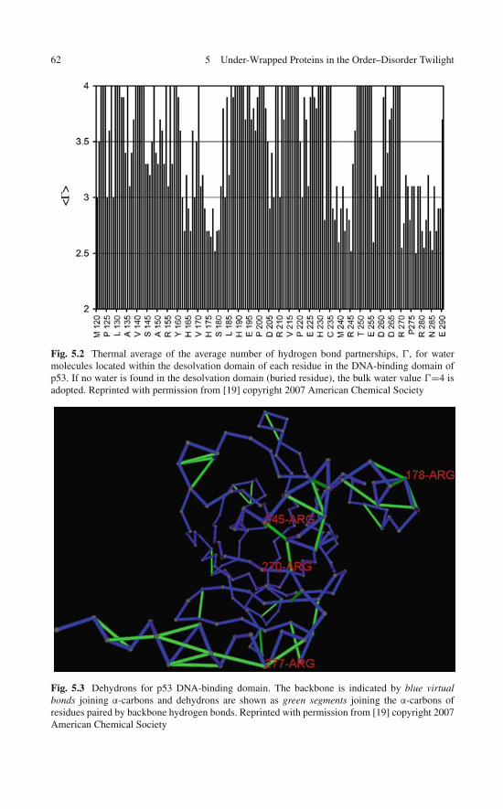
62 5 Under-Wrapped Proteins in the Order–Disorder Twilight
Fig. 5.2 Thermal average of the average number of hydrogen bond partnerships, , for watermolecules located within the desolvation domain of each residue in the DNA-binding domain ofp53. If no water is found in the desolvation domain (buried residue), the bulk water value =4 isadopted. Reprinted with permission from [19] copyright 2007 American Chemical Society
Fig. 5.3 Dehydrons for p53 DNA-binding domain. The backbone is indicated by blue virtualbonds joining α-carbons and dehydrons are shown as green segments joining the α-carbons ofresidues paired by backbone hydrogen bonds. Reprinted with permission from [19] copyright 2007American Chemical Society
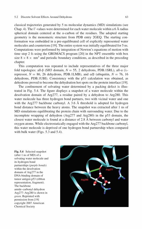
5.2 Discrete Solvent Effects Around Dehydrons 63
classical trajectories generated by 5 ns molecular dynamics (MD) simulations (seeChap. 4). The -values were determined for each water molecule within a 6 Å radiusspherical domain centered at the α-carbon of the residues. The adopted startinggeometry is the monomeric structure from PDB entry 2GEQ. The starting con-formation was embedded in a pre-equilibrated cell of explicitly represented watermolecules and counterions [19]. The entire system was initially equilibrated for 5 ns.Computations were performed by integration of Newton’s equations of motion withtime step 2 fs using the GROMACS program [20] in the NPT ensemble with boxsize 8 × 8 × nm3 and periodic boundary conditions, as described in the precedingchapter.
This computation was repeated to include representatives of the three majorfold topologies: all-β (SH3 domain, N = 55, 2 dehydrons, PDB.1SRL), all-α (λ-repressor, N = 86, 26 dehydrons, PDB.1LMB), and α/β (ubiquitin, N = 76, 16dehydrons, PDB.1UBI). Consistency with the p53 calculation was obtained, asdehydrons proved to become the dehydration hot spots on the protein interface [19].
The confinement of solvating water determined by a packing defect is illus-trated in Fig. 5.4. The figure displays a snapshot of a water molecule within thedesolvation domain of Arg277, a residue paired by a dehydron to Arg280. Thiswater molecule has three hydrogen bond partners, two with vicinal water and onewith the Arg277 backbone carbonyl. A 3.6 Å threshold is adopted for hydrogenbond distance between the heavy atoms. The snapshot was extracted after 1 ns ofMD simulations equilibrating the protein chain with surrounding water. Due to theincomplete wrapping of dehydron (Arg277 and Arg280) in the p53 domain, theclosest water molecule is found at a distance of 2.8 Å between carbonyl and wateroxygen atoms. While electrostatically engaged with the Arg277 backbone carbonyl,this water molecule is deprived of one hydrogen bond partnership when comparedwith bulk water (Figs. 5.3 and 5.4).
Fig. 5.4 Selected snapshot(after 1 ns of MD) of asolvating water molecule andits hydrogen bondpartnerships (purple bonds)within the desolvationdomain of Arg277 in theDNA-binding domain oftumor antigen p53 (ribbonrepresentation, fragment).The backboneamide–carbonyl dehydronArg277–Arg280 is shown ingreen. Reprinted withpermission from [19]copyright 2007 AmericanChemical Society
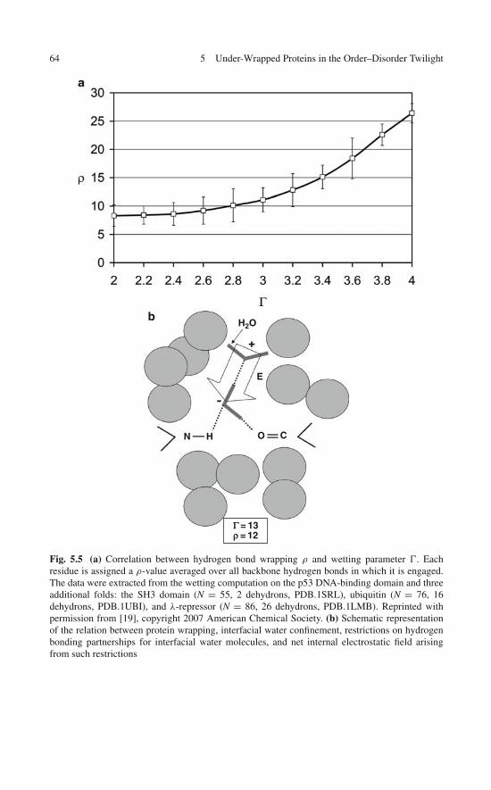
64 5 Under-Wrapped Proteins in the Order–Disorder Twilight
bH2O
E
N H O C
Γ = 13ρ = 12
Fig. 5.5 (a) Correlation between hydrogen bond wrapping ρ and wetting parameter . Eachresidue is assigned a ρ-value averaged over all backbone hydrogen bonds in which it is engaged.The data were extracted from the wetting computation on the p53 DNA-binding domain and threeadditional folds: the SH3 domain (N = 55, 2 dehydrons, PDB.1SRL), ubiquitin (N = 76, 16dehydrons, PDB.1UBI), and λ-repressor (N = 86, 26 dehydrons, PDB.1LMB). Reprinted withpermission from [19], copyright 2007 American Chemical Society. (b) Schematic representationof the relation between protein wrapping, interfacial water confinement, restrictions on hydrogenbonding partnerships for interfacial water molecules, and net internal electrostatic field arisingfrom such restrictions
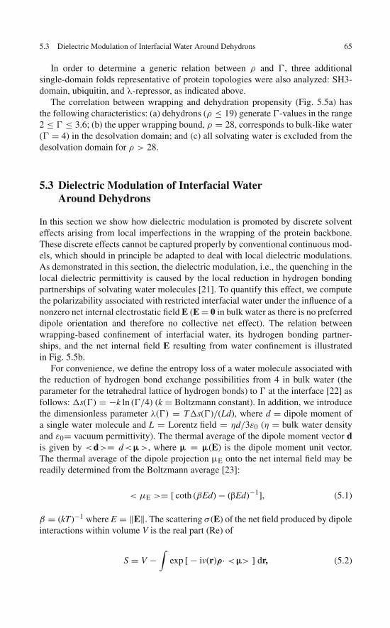
5.3 Dielectric Modulation of Interfacial Water Around Dehydrons 65
In order to determine a generic relation between ρ and , three additionalsingle-domain folds representative of protein topologies were also analyzed: SH3-domain, ubiquitin, and λ-repressor, as indicated above.
The correlation between wrapping and dehydration propensity (Fig. 5.5a) hasthe following characteristics: (a) dehydrons (ρ ≤ 19) generate -values in the range2 ≤ ≤ 3.6; (b) the upper wrapping bound, ρ = 28, corresponds to bulk-like water(G = 4) in the desolvation domain; and (c) all solvating water is excluded from thedesolvation domain for ρ > 28.
5.3 Dielectric Modulation of Interfacial WaterAround Dehydrons
In this section we show how dielectric modulation is promoted by discrete solventeffects arising from local imperfections in the wrapping of the protein backbone.These discrete effects cannot be captured properly by conventional continuous mod-els, which should in principle be adapted to deal with local dielectric modulations.As demonstrated in this section, the dielectric modulation, i.e., the quenching in thelocal dielectric permittivity is caused by the local reduction in hydrogen bondingpartnerships of solvating water molecules [21]. To quantify this effect, we computethe polarizability associated with restricted interfacial water under the influence of anonzero net internal electrostatic field E (E = 0 in bulk water as there is no preferreddipole orientation and therefore no collective net effect). The relation betweenwrapping-based confinement of interfacial water, its hydrogen bonding partner-ships, and the net internal field E resulting from water confinement is illustratedin Fig. 5.5b.
For convenience, we define the entropy loss of a water molecule associated withthe reduction of hydrogen bond exchange possibilities from 4 in bulk water (theparameter for the tetrahedral lattice of hydrogen bonds) to at the interface [22] asfollows: s() = −k ln (/4) (k = Boltzmann constant). In addition, we introducethe dimensionless parameter λ() = Ts()/(Ld), where d = dipole moment ofa single water molecule and L = Lorentz field = ηd/3ε0 (η = bulk water densityand ε0= vacuum permittivity). The thermal average of the dipole moment vector dis given by <d>= d<μ>, where μ = μ(E) is the dipole moment unit vector.The thermal average of the dipole projection μE onto the net internal field may bereadily determined from the Boltzmann average [23]:
< μE >= [ coth (βEd) − (βEd)−1], (5.1)
β = (kT)−1 where E = ‖E‖. The scattering σ (E) of the net field produced by dipoleinteractions within volume V is the real part (Re) of
S = V −∫
exp [ − iv(r)ρ· <μ> ] dr, (5.2)
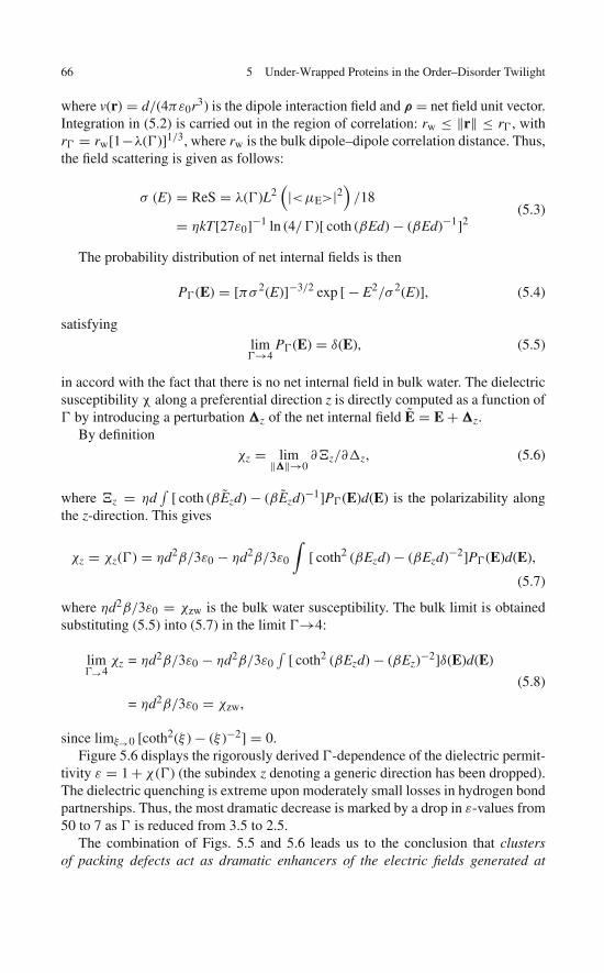
66 5 Under-Wrapped Proteins in the Order–Disorder Twilight
where v(r) = d/(4πε0r3) is the dipole interaction field and ρ = net field unit vector.Integration in (5.2) is carried out in the region of correlation: rw ≤ ‖r‖ ≤ r , withr = rw[1−λ()]1/3, where rw is the bulk dipole–dipole correlation distance. Thus,the field scattering is given as follows:
σ (E) = ReS = λ()L2(|<μE>|2
)/18
= ηkT[27ε0]−1 ln (4/)[ coth (βEd) − (βEd)−1]2(5.3)
The probability distribution of net internal fields is then
P(E) = [πσ 2(E)]−3/2 exp [ − E2/σ 2(E)], (5.4)
satisfying
lim→4
P(E) = δ(E), (5.5)
in accord with the fact that there is no net internal field in bulk water. The dielectricsusceptibility χ along a preferential direction z is directly computed as a function of by introducing a perturbation �z of the net internal field E = E + �z.
By definition
χz = lim‖�‖→0∂�z/∂z, (5.6)
where �z = ηd∫
[ coth (βEzd) − (βEzd)−1]P(E)d(E) is the polarizability alongthe z-direction. This gives
χz = χz() = ηd2β/3ε0 − ηd2β/3ε0
∫[ coth2 (βEzd) − (βEzd)−2]P(E)d(E),
(5.7)
where ηd2β/3ε0 = χzw is the bulk water susceptibility. The bulk limit is obtainedsubstituting (5.5) into (5.7) in the limit →4:
lim→4
χz = ηd2β/3ε0 − ηd2β/3ε0∫
[ coth2 (βEzd) − (βEz)−2]δ(E)d(E)
= ηd2β/3ε0 = χzw,
(5.8)
since limξ→0 [coth2(ξ ) − (ξ )−2] = 0.Figure 5.6 displays the rigorously derived -dependence of the dielectric permit-
tivity ε = 1 + χ () (the subindex z denoting a generic direction has been dropped).The dielectric quenching is extreme upon moderately small losses in hydrogen bondpartnerships. Thus, the most dramatic decrease is marked by a drop in ε-values from50 to 7 as is reduced from 3.5 to 2.5.
The combination of Figs. 5.5 and 5.6 leads us to the conclusion that clustersof packing defects act as dramatic enhancers of the electric fields generated at
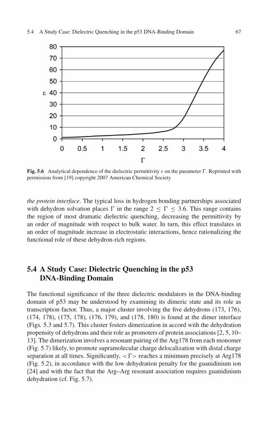
5.4 A Study Case: Dielectric Quenching in the p53 DNA-Binding Domain 67
Fig. 5.6 Analytical dependence of the dielectric permittivity ε on the parameter . Reprinted withpermission from [19] copyright 2007 American Chemical Society
the protein interface. The typical loss in hydrogen bonding partnerships associatedwith dehydron solvation places in the range 2 ≤ ≤ 3.6. This range containsthe region of most dramatic dielectric quenching, decreasing the permittivity byan order of magnitude with respect to bulk water. In turn, this effect translates inan order of magnitude increase in electrostatic interactions, hence rationalizing thefunctional role of these dehydron-rich regions.
5.4 A Study Case: Dielectric Quenching in the p53DNA-Binding Domain
The functional significance of the three dielectric modulators in the DNA-bindingdomain of p53 may be understood by examining its dimeric state and its role astranscription factor. Thus, a major cluster involving the five dehydrons (173, 176),(174, 178), (175, 178), (176, 179), and (178, 180) is found at the dimer interface(Figs. 5.3 and 5.7). This cluster fosters dimerization in accord with the dehydrationpropensity of dehydrons and their role as promoters of protein associations [2, 5, 10–13]. The dimerization involves a resonant pairing of the Arg178 from each monomer(Fig. 5.7) likely, to promote supramolecular charge delocalization with distal chargeseparation at all times. Significantly, <> reaches a minimum precisely at Arg178(Fig. 5.2), in accordance with the low dehydration penalty for the guanidinium ion[24] and with the fact that the Arg–Arg resonant association requires guanidiniumdehydration (cf. Fig. 5.7).

68 5 Under-Wrapped Proteins in the Order–Disorder Twilight
Fig. 5.7 Backbone/dehydron representation of the dimer interface for the DNA-binding domainof p53 (PDB.2GEQ). The side chains of the Arg178 of each monomer involved in a reso-nance pair are shown. Reprinted with permission from [19] copyright 2007 American ChemicalSociety
Besides Arg 178, there are three additional minima in < >, corresponding toresidues Arg245, Arg270, and Arg277 (Fig. 5.2). The latter three arginines playa pivotal role in DNA recognition [18], as discussed below. Residue Arg245 isengaged in the dielectric quenching region 236–246 and is part of the dehydron clus-ter involving pairs (236, 239), (237, 245), (237, 271), (239, 242), (239, 244), (240,242) (Fig. 5.3). Residues 270 and 277 lie within the dielectric quenching region270–289 and are part of the dehydron cluster involving pairs (237, 271), (274, 277),(277, 281), (280, 284), (281, 285), (282, 285), (285, 288) (Figs. 5.3 and 5.8). Thelatter cluster is one of the largest to be found in a PDB-reported soluble protein, asdiscussed below.
Direct examination of the protein–DNA complex (PDB.2GEQ) reveals thatthe three residues directly implicated in DNA recognition are precisely Arg245,Arg270, and Arg277 (Fig. 5.8). Residue Arg277 acts as intrabase intercalator, whilethe other two interact with the negatively charged backbone phosphates (Fig. 5.8).The electrostatics of protein–DNA recognition is not merely the result of match-ing charges along the geometrically compatible interfaces, otherwise ion pairswould prevail in water. Rather, electrostatic recognition of the DNA polyelectrolyterequires a device to promote dehydration at the protein–nucleic acid interface.The large dehydron clusters surrounding the three arginines directly implicatedin the protein–DNA association (Figs. 5.3 and 5.8) provide such an expedient, asthey quench the local dielectric (Fig. 5.2), thus enhancing the electrostatic recog-nition. Thus, the fact that the three arginines involved in DNA recognition arealso dehydration hot spots satisfies a functional imperative for the transcriptionfactor.

5.5 Proteins with Dehydron Clusters 69
Fig. 5.8 Protein–DNA complex of the DNA-binding domain of p53 (PDB.2GEQ). Side chains ofthe key residues directly implicated in DNA recognition, Arg245, Arg270, and Arg277 are shown.The pyridine base recognized by Arg277 is shown in yellow, while the individual DNA strands areshown in lilac and light magenta. Reprinted with permission from [19] copyright 2007 AmericanChemical Society
5.5 Proteins with Dehydron Clusters
A curated PDB-derived database was constructed free of redundancy for single-domain proteins with contour length N > 50. The database is comprised of 2,982entries with <50% pairwise identity in aligned sequences. Proteins were groupedaccording to n, the size of the dehydron cluster in the structure. The groups intersectto a considerable extent [19].
Each n-group has been lumped into five nondisjoint functional categories:biosynthesis, enzymology, cell signaling, cytoskeleton, and cancer. The contribu-tion of each category to each n-group is quantified through suitable normalization,taking into account the relative abundance of each category in the curated database.Thus, the number of PDB domains within an n-cluster in a functional category isdivided by the total number of PDB domains in the category. After normalization,percentages indicating representativity from each functional category are computedfor each n-group (Fig. 5.9).
The distribution of dehydron clusters becomes a marker of the functional cat-egory. Thus, biosynthesis peaks at 2, enzymology at 3, cell signaling at 6, whilecytoskeleton and cancer are monotonically increasing with dehydron cluster size.Intriguingly, the dominance of the cancer category becomes apparent for the poorestpacked proteins. The dearth of domains for n ≥ 7 facilitates individual identifica-tion and assessment. Thus, there are only five domains with n = 7: calmodulin(cell signaling, Fig. 5.10) [25], actin (cytoskeleton) [26], p53 DNA-binding domain(cancer, Figs. 5.3 and 5.7) [18], BRCT, the terminal repeat domain of breast cancergene BRAC1 [27], and the cellular prion protein (Fig. 5.11) [28]. The group with
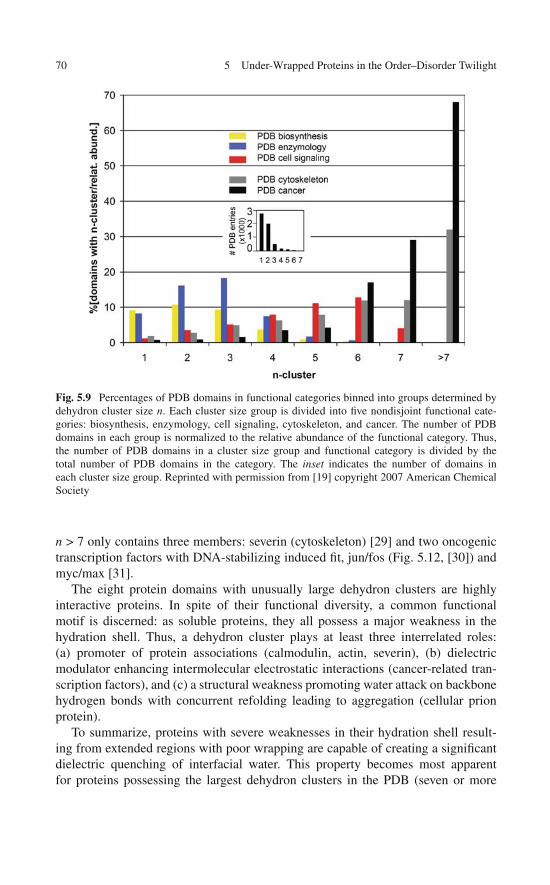
70 5 Under-Wrapped Proteins in the Order–Disorder Twilight
Fig. 5.9 Percentages of PDB domains in functional categories binned into groups determined bydehydron cluster size n. Each cluster size group is divided into five nondisjoint functional cate-gories: biosynthesis, enzymology, cell signaling, cytoskeleton, and cancer. The number of PDBdomains in each group is normalized to the relative abundance of the functional category. Thus,the number of PDB domains in a cluster size group and functional category is divided by thetotal number of PDB domains in the category. The inset indicates the number of domains ineach cluster size group. Reprinted with permission from [19] copyright 2007 American ChemicalSociety
n > 7 only contains three members: severin (cytoskeleton) [29] and two oncogenictranscription factors with DNA-stabilizing induced fit, jun/fos (Fig. 5.12, [30]) andmyc/max [31].
The eight protein domains with unusually large dehydron clusters are highlyinteractive proteins. In spite of their functional diversity, a common functionalmotif is discerned: as soluble proteins, they all possess a major weakness in thehydration shell. Thus, a dehydron cluster plays at least three interrelated roles:(a) promoter of protein associations (calmodulin, actin, severin), (b) dielectricmodulator enhancing intermolecular electrostatic interactions (cancer-related tran-scription factors), and (c) a structural weakness promoting water attack on backbonehydrogen bonds with concurrent refolding leading to aggregation (cellular prionprotein).
To summarize, proteins with severe weaknesses in their hydration shell result-ing from extended regions with poor wrapping are capable of creating a significantdielectric quenching of interfacial water. This property becomes most apparentfor proteins possessing the largest dehydron clusters in the PDB (seven or more

5.5 Proteins with Dehydron Clusters 71
Fig. 5.10 Dehydron pattern of calmodulin (PDB. 1CDM) complexed with the calmodulin-bindingdomain of calmodulin-dependent protein kinase II. The backbone is indicated by blue virtual bondsjoining α-carbons and dehydrons are shown in green. The intermolecular wrapping of calmodulinhydrogen bonds by the ligand side chains is indicated by thin blue lines. The extremities of theselines are the β-carbon of the residue contributing to the desolvation shell of the hydrogen bondand the midpoint of the hydrogen bond that is being wrapped intermolecularly. Reprinted withpermission from [19] copyright 2007 American Chemical Society
Fig. 5.11 Dehydron patternof the cellular prion protein(PDB.1QM0). Reprinted withpermission from [19]copyright 2007 AmericanChemical Society
dehydrons). We found that there are eight such proteins reported in PDB, possessingstructural regions in the order–disorder twilight. These regions belong to cancer-related (oncogenic or antitumor) proteins and also to highly interactive proteins andto a cellular prion that promotes misfolding and aberrant aggregation.
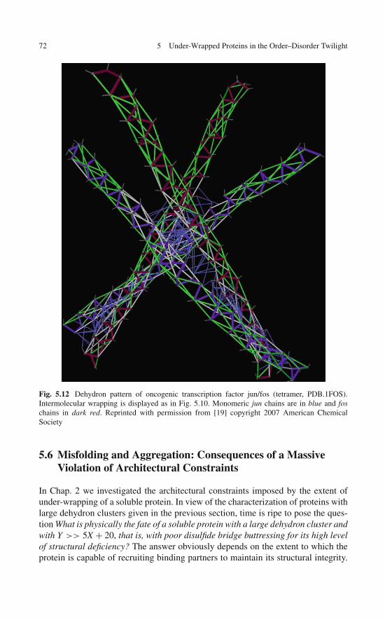
72 5 Under-Wrapped Proteins in the Order–Disorder Twilight
Fig. 5.12 Dehydron pattern of oncogenic transcription factor jun/fos (tetramer, PDB.1FOS).Intermolecular wrapping is displayed as in Fig. 5.10. Monomeric jun chains are in blue and foschains in dark red. Reprinted with permission from [19] copyright 2007 American ChemicalSociety
5.6 Misfolding and Aggregation: Consequences of a MassiveViolation of Architectural Constraints
In Chap. 2 we investigated the architectural constraints imposed by the extent ofunder-wrapping of a soluble protein. In view of the characterization of proteins withlarge dehydron clusters given in the previous section, time is ripe to pose the ques-tion What is physically the fate of a soluble protein with a large dehydron cluster andwith Y >> 5X + 20, that is, with poor disulfide bridge buttressing for its high levelof structural deficiency? The answer obviously depends on the extent to which theprotein is capable of recruiting binding partners to maintain its structural integrity.
5.6 Consequences of a Massive Violation of Architectural Constraints 73
As noted in Chap. 2, the worse wrapped proteins are potassium channel neurotox-ins but their buttressing is actually adequate for an extracellular protein with such alevel of structural deficiency.
On the other hand, the cellular form of the human prion (PDB.1QM0) is verypoorly buttressed for its extensive under-wrapping, belongs to the reducing cytosolicspace, contains spots of large dehydron density, and makes no obligatory com-plex. We know that prions misfold and aggregate aberrantly into highly organizedamyloidogenic fibrils. Is this then a generic behavior of proteins sharing the samemolecular attributes? This question prompts us to investigate the amyloid phasemore closely.
There is significant evidence supporting the view that amyloidogenic aggrega-tion is a generic phase of peptide chains [32–34]. The term generic phase refersto a 3D organization dominated by main chain interactions which is essentiallyinsensitive to the information encoded in the primary sequence. Such intermolecularassociations appear to be dominated by a basic structural motif: the cross-β structure[35], an intermolecular sheet-pleated pattern ubiquitous in fibrillogenic aggregation.This assertion remains conjectural, as no crystal of the fiber for natural prions hasbeen obtained. By contrast, the folded state of the peptide chain is relatively wellunderstood: natural soluble proteins tend to adopt single-molecule conformations ofmarginal stability.
While the folding process and its final stable outcome are crucially dependent onthe amino acid composition of the chain, the amyloid state appears to be fairly insen-sitive to this information: At first sight, amyloidogenic aggregation does not seem torequire an “aggregation code,” as some recent claims would have it [32–34]. But fur-ther analysis reveals that it must place severe constraints on the primary sequence,as some proteins tend to be prone to aggregate under physiological conditions [8],while others require extreme conditions to do so or simply do not aggregate repro-ducibly [32, 36]. In addition, negative design features of the folded state purposelyencoded in the protein primary sequence may avert aggregation [37]. Thus, it isnot entirely correct to characterize the aberrant aggregation as a “polymer physicsphase,” shared by polypeptides with arbitrary, suboptimal, or random sequence,in contrast with the folded state, determined unambiguously by the primarysequence.
Proteins are subject to selection pressure and adapt to become good folders, i.e.,expeditious structure seekers with a reproducible and stable soluble structure. Thisoptimization is needed to prevent the functionally competent fold from reverting toa primeval amyloid phase. On the other hand, certain sequences are better optimizedto escape aggregation than others even under conditions known to sustain the nativefold [8].
While amyloidogenic aggregation has been shown to be always plausible pro-vided sufficiently stringent denaturation conditions are applied [32–36], a markedamyloidogenic propensity has been detected on a number of proteins even underphysiological or near-physiological conditions, particularly if the monomeric fold-ing domain is deprived of its natural interacting partners [8]. Such findings implythat not all soluble structures have been optimized to the same degree in order

74 5 Under-Wrapped Proteins in the Order–Disorder Twilight
to avert aggregation, and that the more reliant the structure is on binding part-nerships or complexations, the more vulnerable it becomes in regard to revertingto the primeval phase. Thus, an over-expression of a folding domain with highcomplexation requirements in vivo, or the modification of its binding partners asa result of genetic accident, or any factor that distorts its natural interactive contextis likely to bolster a transition to an amyloidogenic state.
These observations lead us to the following question: What type of deficiency inthe native fold constitutes a signal for aberrant aggregation? A recent assessmentof the wrapping of soluble structure might prove critical to address this problem.For highly under-wrapped proteins (∼50% dehydrons or more), densities higherthan four dehydrons per 1,000 Å2 on the protein surface become inducers of proteinaggregation [8].
Thus, the condition of “keeping the structure dry in water” becomes a require-ment to preserve the structural integrity of soluble proteins and imposes a severebuilding constraint (and thereby an evolutionary pressure) on such proteins. It isexpected that the optimization of the structures resulting from this type of evolu-tionary constraint would be uneven over a range of soluble proteins, resulting inmarked differences in aggregation propensity.
This wrapping analysis clarifies the physical picture put forth by Dobson [32–34], in which amyloidogenic propensity depends crucially on the fact that mainchain interactions become dominant in detriment of the amino acid sequence thatencodes the folded state. Precisely, main chain interactions may dominate as themain chain of the folded state is not properly protected from water attack.
It is instructive to compare this statement with the local analysis of Avbelj andBaldwin [38] in the sense that backbone solvation is a determinant of β-sheetpropensity. Thus, an over-exposed backbone hydrogen bond in the native fold isan indicator of a failure in folding cooperativity, as it reveals an inability to removewater from an interactive polar pair by means of a many-body correlation, and at thesame time, it is a signal enabling the diagnosis of amyloidogenic propensity. Thus,the wrapping concept enables us to discern why some soluble proteins may havebeen better optimized to avoid amyloidogenic aggregation than others.
Direct inspection of the pattern of desolvation of the main chain clearly revealsthat the cellular fold of the human prion [39] (Fig. 5.11) is too vulnerable to waterattack and at the same time too sticky to avert aggregation. Clearly, its sequencehas not been optimized to “keep the backbone hydrogen bonds dry” in the foldedstate. In fact, their extent of exposure of backbone hydrogen bonds is the highestamong soluble proteins in the entire PDB, with the sole exception of some toxinswhose stable fold is held together by a profusion of disulfide bridges, as shown inChap. 2.
It is suggestive that an inability to protect the main chain is precisely the type ofdeficiency that best correlates with a propensity to reverse to a primeval aggrega-tion phase determined by main chain interactions. The actual mechanism by whichsuch defects induce or nucleate the transition is still opaque, although the inherentadhesiveness of packing defects obviously plays a role.
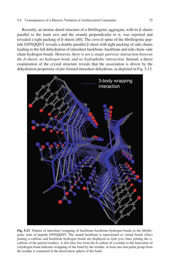
5.6 Consequences of a Massive Violation of Architectural Constraints 75
Recently, an atomic-detail structure of a fibrillogenic aggregate, with its β-sheetsparallel to the main axis and the strands perpendicular to it, was reported andrevealed a tight packing of β-sheets [40]. The cross-β spine of the fibrillogenic pep-tide GNNQQNY reveals a double-parallel β-sheet with tight packing of side chainsleading to the full dehydration of intrasheet backbone–backbone and side chain–sidechain hydrogen bonds. However, there is not a single pairwise interaction betweenthe β-sheets, no hydrogen bond, and no hydrophobic interaction. Instead, a directexamination of the crystal structure reveals that the association is driven by thedehydration propensity of pre-formed intrasheet dehydrons, as depicted in Fig. 5.13.
Fig. 5.13 Pattern of intersheet wrapping of backbone–backbone hydrogen bonds in the fibrillo-genic state of peptide GNNQQNY. The strand backbone is represented as virtual bonds (blue)joining α-carbons and backbone hydrogen bonds are displayed as light gray lines joining the α-carbons of the paired residues. A thin blue line from the β-carbon of a residue to the baricenter ofa hydrogen bond indicates wrapping of the bond by the residue: at least one non-polar group fromthe residue is contained in the desolvation sphere of the bond

76 5 Under-Wrapped Proteins in the Order–Disorder Twilight
This discussion naturally leads us to some evolutionary considerations. Aparadigmatic discovery in biology revealed that folds are conserved across species toperform specific functions. However, the wrapping of such folds is clearly not con-served [4]. This fact suggests how complex physiologies may be achieved withoutdramatically expanding genome size, a standing problem in biology. Considerablenetwork complexity may be achieved by actually fostering a higher level of com-plexation or binding partnership, as promoted by a more precarious wrappingof the isolated protein domains. According to our previous analysis, such com-plex design also entails an inherent danger: the reversal of highly under-wrappedfolding domains to an amyloidogenic phase even under physiological conditions.Amyloidosis is thus likely to be a consequence of high complexity in proteomicconnectivity, as dictated by the structural fragility of highly interactive proteins.
Prion-like aggregation has been widely recognized as a causative of pathogenicdysfunction [39], but recent work [41] has revealed that there may be also epigeneticconsequences to prion-like aggregation. These novel insights lead to a significantextension of the prion hypothesis. Furthermore, while a connection between proteinunder-wrapping and propensity for aberrant amyloidogenic aggregation has beenunraveled, no misfolding inference can be made for cases when the extent of under-wrapping is so severe that no soluble structure can be maintained by the monomericchain. This case appears to be of biological interest in light of recent research [41]that focused on nonpathogenic yeast prions. These prions are capable of promotingphenotypic polymorphism through a transmittable conformational change that theyregard as having epigenetic consequences, thus defining a protein-based elementof inheritance. The yeast prions studied, especially PSI in yeast gene Sup35, areunlikely to sustain a monomeric structure.
From the wrapping perspective, this is so because the prion sequences containlarge windows (>30 residues) containing mostly under-wrapping residues (espe-cially G, N, Q, S, A, P). In turn, these residues are under-wrappers because theycontain few nonpolar side chain groups, thereby exposing the backbone to thesolvent, while some (i.e., S, N, Q) even prevent other nonpolar groups from clus-tering around the backbone hydrogen bonds. The other under-wrapping residue,D, is unlikely to be found in such prions as its charge promotes hydration, thusintroducing a negative design element for aggregation.
These observations are in accordance with the structural characterization byKrishnan and Lindquist [41], which places yeast prions in the twilight between orderand disorder, in consonance with the wrapping-based characterization of the priondescribed in this chapter.
Thus, the epigenetic consequences associated with misfolding of severely under-wrapped proteins incapable of sustaining monomeric structure should be mandatorysubjects of any research agenda built on the premises of this chapter. The focus ofsuch endeavors are proteins endowed with large windows (>30) of under-wrappingresidues. When combined with tyrosine (probably needed for stacking), such motifsare likely promoters of self-aggregation leading to pathology [39] or epigeneticprion-based phenotype polymorphism [41]. Thus, future research efforts should bedirected at identifying highly under-wrapped human genes containing the sequence

References 77
motifs indicated and assessing the epigenetic consequences of their transformationinto sequestered aggregates. A preliminary scanning of ca. 16,000 human proteins inSwiss-Prot reveals 13 genes with the severely under-wrapped motif. Among them,RNF12, AF9, MAML2 are implicated in cancer promotion, while seven are involvedin transcription regulation, hence with likely epigenetic consequences associatedwith a conformational switching into sequestered aggregate states.
References
1. Fernández A, Sosnick TR, Colubri A (2002) Dynamics of hydrogen-bond desolvation infolding proteins. J Mol Biol 321: 659–675
2. Fernández A, Scheraga HA (2003) Insufficiently dehydrated hydrogen bonds as determinantsof protein interactions. Proc Natl Acad Sci USA 100:113–118
3. Fernández A, Scott LR (2003) Adherence of packing defects in soluble proteins. Phys RevLett 91:018102
4. Fernández A, Berry RS (2004) Molecular dimension explored in evolution to promoteproteomic complexity. Proc Natl Acad Sci USA 101:13460–13465
5. Fernández A, Scott RL (2003) Dehydron: A structurally encoded signal for protein interaction.Biophys J 85:1914–1928
6. Fernández A (2004) Keeping dry and crossing membranes. Nat Biotechnol 22:1081–10847. Fernández A, Scott RL, Berry RS (2006) Packing defects as selectivity switches for drug-
based protein inhibitors. Proc Natl Acad Sci USA 103:323–3288. Fernández A, Kardos J, Scott R, Goto Y, Berry RS (2003) Structural defects and the diagnosis
of amyloidogenic propensity. Proc Natl Acad Sci USA 100:6446–64519. Fernández A, Berry RS (2003) Proteins with hydrogen-bond packing defects are highly
interactive with lipid bilayers: Implications for amyloidogenesis. Proc Natl Acad Sci USA100:2391–2396
10. Deremble C, Lavery R (2005) Macromolecular recognition. Curr Opin Struct Biol 15:171–17511. Ma B, Elkayam T, Wolfson H, Nussinov R (2003) Protein-protein interactions: Structurally
conserved residues distinguish between binding sites and exposed protein surfaces. Proc NatlAcad Sci USA 100:5772–5777
12. Ma B, Pan Y, Gunasekaran K, Venkataraghavan RB, Levine AJ, Nussinov R (2005)Comparison of the protein-protein interfaces in the p53-DNA crystal structures: Towardselucidation of the biological interface. Proc Natl Acad Sci USA 102:3988–3993
13. Rajamani D, Thiel S, Vajda S, Camacho CJ (2004) Anchor residues in protein-proteininteractions. Proc Natl Acad Sci USA 101:11287–11292
14. Dunker AK, Cortese MS, Romero P, Iakoucheva LM, Uversky VN (2005) Flexible nets: Theroles of intrinsic disorder in protein interaction networks. FEBS J 272:5129–5148
15. Iakoucheva LM, Dunker AK (2003) Order, disorder, and flexibility: Prediction from proteinsequence. Structure (London) 11:1316–1317
16. Dunker AK, Obradovic Z (2001) The protein trinity-linking function and disorder. NatBiotechnol 19: 805–806
17. Wade RC, Gabdoulline RR, Lüdemann SK, Lounnas V (1998) Electrostatic steering andionic tethering in enzyme-ligand binding: Insights from simulations. Proc Natl Acad Sci USA95:5942–5949
18. Ho WC, Fitzgerald MX, Marmorstein R (2006) Structure of the p53 core domain dimer boundto DNA. J Biol Chem 281:20494–20502
19. Pietrosemoli N, Crespo A, Fernández A (2007) Dehydration propensity of order-disorderintermediate regions in soluble proteins. J Prot Res 6:3519–3526
20. Lindahl E, Hess B, Van der Spoel D: GROMACS 3.0: A package for molecular simulationsand trajectory analysis. J Mol Model 2001, 7: 302–317
78 5 Under-Wrapped Proteins in the Order–Disorder Twilight
21. Fernández A (2003) What caliber pore is like a pipe? Nanotubes as modulators of ionicgradients. J Chem Phys 119:5315–5319
22. Ben-Naim A (1980) Hydrophobic Interactions. Plenum Press, New York23. Debye P (1929) Polar Molecules. Dover, New York24. Mason PE, Neilson GW, Dempsey CE, Barnes AC, Cruickshank, JM (2003) The hydration
structure of guanidinium and thiocyanate ions: Implications for protein stability in aqueoussolution. Proc Natl Acad Sci USA 100:4557–4561
25. Kabsch W, Mannherz HG, Suck D, Pai EF, Holmes KC (1990) Atomic structure of the actin:DNase I complex. Nature 347:37–44
26. Meador WE, Means AR, Quiocho FA (1993) Modulation of calmodulin plasticity inmolecular recognition on the basis of x-ray structures. Science 262:1718–1721
27. Williams RS, Green R, Glover JN (2001) Crystal structure of the BRCT repeat region fromthe breast cancer-associated protein BRCA1. Nat Struct Biol 8:838–842
28. Zahn R, Liu A, Luhrs T et al (2000) NMR solution structure of the human prion protein. ProcNatl Acad Sci USA 97:145–150
29. Schnuchel A, Wiltscheck R, Eichinger L, Schleicher M, Holak, TA (1995) Structure of severindomain 2 in solution. J Mol Biol 247:21–27
30. Glover JN, Harrison SC (1995) Crystal structure of the heterodimeric bZIP transcription factorc-Fos-c-Jun bound to DNA. Nature 373:257–261
31. Lavigne P, Crump MP, Gagne SM et al (1998) Insights into the mechanism of heterodimer-ization from the 1H-NMR solution structure of the c-Myc-Max heterodimeric leucine zipper.J Mol Biol 281:165–181
32. Dobson CM (1999) Protein misfolding, evolution and disease. Trends Biochem Sci24:329–332
33. Dobson CM (2001) The structural basis of protein folding and its links with human disease.Philos Trans R Soc Lond B 356:133–145
34. Fändrich M, Dobson CM (2002) The behavior of polyamino acids reveals an inverse sidechain effect in amyloid structure formation. EMBO J 21:5682–5690
35. Sunde M, Blake CCF (1998) From the globular to the fibrous state: protein structure andstructural conversion in amyloid formation. Q Rev Biophys 31:1–39
36. Dobson CM (2002) Protein misfolding diseases: Getting out of shape. Nature 418:729–73037. Richardson JS, Richardson DC (2002) Natural β-sheet proteins use negative design to avoid
edge-to-edge aggregation. Proc Natl Acad Sci USA 99:2754–275938. Avbelj F, Baldwin RL (2003) Role of backbone solvation and electrostatics in generating
preferred peptide backbone conformations: Distributions of phi. Proc Natl Acad Sci USA100:5742–5747
39. Prusiner SB (1998) Prions. Proc Natl Acad Sci USA 95:13363–1338340. Nelson R, Sawaya M, Balbirnie M et al (2005) Structure of the cross-beta spine of amyloid-
like fibrils. Nature 435:773–77841. Krishnan R, Lindquist SL (2005) Structural insights into a yeast prion illuminate nucleation
and strain diversity. Nature 435:765–772

Chapter 6Evolution of Protein Wrappingand Implications for the Drug Designer
Proteins with common ancestry (homologs) typically share a common fold. Thisstructural similarity introduces major problems for rational drug design since amajor goal in the field is the control of specificity. Yet, as shown in this chapter,while the topology of the native structure is extremely similar across homologs,their wrapping patterns and expression regulation tend to be different, offering anovel opportunity to funnel the impact of a drug on clinically relevant targets.The evolutionary root of these dissimilarities across homologous proteins is care-fully dissected in this chapter both across species and within the human species.Furthermore, as hinted in this and demonstrated in subsequent chapters, the wrap-ping variations across homologs have profound consequences for drug design as weaim at engineering target-specific and species-specific therapeutic agents and buildinsightful animal models for disease or malignancy.
6.1 An Evolutionary Context for the Drug Designer
From a structural biology perspective, one of the most striking observations regard-ing protein evolution is the uncanny similarity of the 3D structure of proteinswith common ancestry. This similarity across homologs is often quantified by anRMSD <1.5Å for the aligned backbone atoms of the proteins and holds even atlow levels of sequence identity (∼30%) that are nevertheless sufficient to establishhomology.
From the perspective of a drug designer, this structural similarity acrosshomologs of the protein target is particularly troublesome. Thus, major goals indrug design are related or entangled with this evolutionary aspect. They are as fol-lows: (a) the control of drug specificity; (b) the engineering of species-selectivetherapeutic impact in the treatment of infectious diseases; and (c) the building ofinsightful animal models to properly assess drug efficacy. In all three contexts, theexistence of homologs of the protein target within the species (paralogs) or acrossspecies (orthologs) can introduce undesired cross-reactivities due to their high levelof structural similarity with the target. These cross-reactivities, in turn, can result
79A. Fernández, Transformative Concepts for Drug Design: Target Wrapping,DOI 10.1007/978-3-642-11792-3_6, C© Springer-Verlag Berlin Heidelberg 2010
80 6 Evolution of Protein Wrapping and Implications for the Drug Designer
in adverse and even health-threatening side effects or can render inconsequential orirrelevant the animal models for a particular disease or malignancy.
The fold may be highly conserved across homologs, but as shown in this chap-ter, the wrapping is not conserved and neither is the regulation of expression ofthe homologs. As described below, we should distinguish orthologs from paralogswhen assessing the evolutionary origins of these dissimilarities. Thus, in the case oforthologs, the variations arise mostly from differences in the level of efficiency ofnatural selection across different species. On the other hand, in paralogs the dissim-ilarity is rooted in the imperative to curb dosage imbalances that would arise if twocopies of the same gene would be retained undifferentiated. As shown in the sub-sequent chapters, these subtle variations of proteins that essentially share the same3D-fold have paramount consequences to engineer drugs that can fulfill the threegoals described above.
The picture that emerges from this chapter is essentially that wrapping consti-tutes a biological dimension utilized by evolution to promote complexity and atthe same time that wrapping differences within a fold offer escape routes to thefitness consequences introduced by paralog retention. Both evolutionary aspectswill be harnessed in the subsequent chapters as we herald the new generation ofwrapping-based drugs.
6.2 Wrapping Across Species: Hallmarks of NonadaptiveTraits in the Comparison of Orthologous Proteins
Proteins with common ancestry tend to share the same fold or at least the samestructural topology [1, 2]. Thus, essentially the same molecular machinery is opera-tive in organisms that diverged from each other billions of years ago. For example,the metabolic enzymatic function “dehydrofolate reductase” (DHFR) is exerted bya molecular machinery that has hardly changed in structure as we compare speciesin different kingdoms of life, like archea (i.e., Haloferax volcanii), bacteria (i.e.,Escherichia coli), and higher eukaryotes (i.e., Homo sapiens). When closely exam-ined, the wrapping of the DHFR has gotten worse (richer in dehydrons) in specieswith small population size, that is, those where natural selection became less effi-cient [3] (Fig. 6.1a). This trend is apparent even when examining highly conservedprotein domains, such as ubiquitin: There is a progressive enrichment in dehydronsas the species population size decreases (Fig. 6.1b).
For operational reasons, the under-wrapping or packing deficiency of a proteinmay be defined as ν = % dehydrons in the set of backbone hydrogen bonds. Aswe focus on any functionally competent fold, the anecdotal examples we harvestedsuggest a clear trend: ν(archea) < ν(bacteria) < ν(unicellular eukaryotes) < · · · <ν(higher eukaryotes). This trend probably follows from the fact that mildly delete-rious mutations have a better chance to prevail and get fixed in species with smaller

6.2 Wrapping Across Species 81
b
Fig. 6.1 (continued)
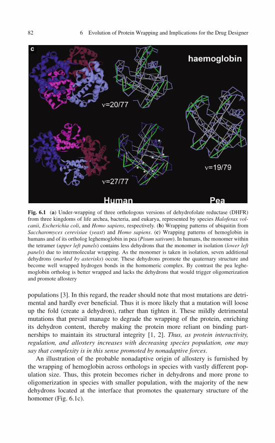
82 6 Evolution of Protein Wrapping and Implications for the Drug Designer
Fig. 6.1 (a) Under-wrapping of three orthologous versions of dehydrofolate reductase (DHFR)from three kingdoms of life archea, bacteria, and eukarya, represented by species Haloferax vol-canii, Escherichia coli, and Homo sapiens, respectively. (b) Wrapping patterns of ubiquitin fromSaccharomyces cerevisiae (yeast) and Homo sapiens. (c) Wrapping patterns of hemoglobin inhumans and of its ortholog leghemoglobin in pea (Pisum sativum). In humans, the monomer withinthe tetramer (upper left panels) contains less dehydrons that the monomer in isolation (lower leftpanels) due to intermolecular wrapping. As the monomer is taken in isolation, seven additionaldehydrons (marked by asterisks) occur. These dehydrons promote the quaternary structure andbecome well wrapped hydrogen bonds in the homomeric complex. By contrast the pea leghe-moglobin ortholog is better wrapped and lacks the dehydrons that would trigger oligomerizationand promote allostery
populations [3]. In this regard, the reader should note that most mutations are detri-mental and hardly ever beneficial. Thus it is more likely that a mutation will looseup the fold (create a dehydron), rather than tighten it. These mildly detrimentalmutations that prevail manage to degrade the wrapping of the protein, enrichingits dehydron content, thereby making the protein more reliant on binding part-nerships to maintain its structural integrity [1, 2]. Thus, as protein interactivity,regulation, and allostery increases with decreasing species population, one maysay that complexity is in this sense promoted by nonadaptive forces.
An illustration of the probable nonadaptive origin of allostery is furnished bythe wrapping of hemoglobin across orthologs in species with vastly different pop-ulation size. Thus, this protein becomes richer in dehydrons and more prone tooligomerization in species with smaller population, with the majority of the newdehydrons located at the interface that promotes the quaternary structure of thehomomer (Fig. 6.1c).
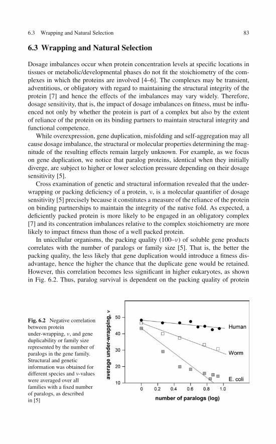
6.3 Wrapping and Natural Selection 83
6.3 Wrapping and Natural Selection
Dosage imbalances occur when protein concentration levels at specific locations intissues or metabolic/developmental phases do not fit the stoichiometry of the com-plexes in which the proteins are involved [4–6]. The complexes may be transient,adventitious, or obligatory with regard to maintaining the structural integrity of theprotein [7] and hence the effects of the imbalances may vary widely. Therefore,dosage sensitivity, that is, the impact of dosage imbalances on fitness, must be influ-enced not only by whether the protein is part of a complex but also by the extentof reliance of the protein on its binding partners to maintain structural integrity andfunctional competence.
While overexpression, gene duplication, misfolding and self-aggregation may allcause dosage imbalance, the structural or molecular properties determining the mag-nitude of the resulting effects remain largely unknown. For example, as we focuson gene duplication, we notice that paralog proteins, identical when they initiallydiverge, are subject to higher or lower selection pressure depending on their dosagesensitivity [5].
Cross examination of genetic and structural information revealed that the under-wrapping or packing deficiency of a protein, ν, is a molecular quantifier of dosagesensitivity [5] precisely because it constitutes a measure of the reliance of the proteinon binding partnerships to maintain the integrity of the native fold. As expected, adeficiently packed protein is more likely to be engaged in an obligatory complex[7] and its concentration imbalances relative to the complex stoichiometry are morelikely to impact fitness than those of a well packed protein.
In unicellular organisms, the packing quality (100–ν) of soluble gene productscorrelates with the number of paralogs or family size [5]. That is, the better thepacking quality, the less likely that gene duplication would introduce a fitness dis-advantage, hence the higher the chance that the duplicate gene would be retained.However, this correlation becomes less significant in higher eukaryotes, as shownin Fig. 6.2. Thus, paralog survival is dependent on the packing quality of protein
Fig. 6.2 Negative correlationbetween proteinunder-wrapping, ν, and geneduplicability or family sizerepresented by the number ofparalogs in the gene family.Structural and geneticinformation was obtained fordifferent species and ν-valueswere averaged over allfamilies with a fixed numberof paralogs, as describedin [5]

84 6 Evolution of Protein Wrapping and Implications for the Drug Designer
structure with p < 10−16 in Escherichia coli and p > 6.7 × 10⁻³ in humans [5].This contrast between simple and complex organisms is hard to interpret due to widedifferences at the proteome level. However, alternative measures point to a similartrend. For example, the average difference in packing deficiency between singletonsand duplicate genes is 18% in E. coli, 6% in worm (Caenorhabditis elegans), and∼2% in humans (Homo sapiens).
In humans, this insensitivity to dosage imbalance may be attributed in part toselection inefficiency arising from smaller population size [3], implying that theselection pressure exerted on paralogs of deficiently packed proteins has simplynot become operative. Alternatively, the higher complexity of expression regula-tion in higher eukaryotes may introduce a tolerance to dosage imbalance not foundin unicellular organisms. This chapter explores this latter possibility, focusing onevolution-related dosage imbalances and the mechanisms that humans possess tocope with the troubling aspects of selection inefficiency.
6.4 How Do Humans Cope with Inefficient Selection?
If selection is indeed inefficient in humans and this inefficiency enabled duplicategenes to stay around awaiting further specialization, significant dosage imbal-ances must have occurred during human evolution. How did we cope with suchimbalances, which are likely to be significant when the gene products are veryunder-wrapped? The key to this problem lies in the ability of humans to regulatethe expression of both gene copies dissimilarly through a plausible process thatinvolves random deleterious mutations.
To address this problem, we first assess the selection pressure on gene duplicatesexerted as paralogs are co-expressed at the mRNA (messenger RNA) level and henceare likely to compete for their interactive partners. Then we relate packing deficiency(ν) with differences in post-transcriptional regulation patterns within families. Thus,we investigate how differences in miRNA (microRNA) target patterns [8], tellingapart paralogs through different patterns of translational repression, impinge on theselection pressure on duplicate genes by mitigating dosage imbalances. In humansthese patterns are significantly dissimilar across paralogs of poorly packed proteinswhile nearly coincident across paralogs of well packed proteins [17], thus under-scoring a means to buffer dosage imbalance effects arising from gene duplication.
This miRNA-based capacitance is not expected to be nearly as significant inspecies with larger effective population size due to the higher efficiency of evo-lutionary forces in such organisms when compared with humans [3]. Thus, theselection pressure affecting the retention of gene duplicates is likely to be moreefficient in these organisms promoting adaptation through functional innovation orpurifying selection.
The next two sections are devoted to provide further evidence in support of theclaim that protein wrapping quality is a determinant of dosage sensitivity whileupholding the view that resilience to dosage imbalance is achieved in humans bydiversifying miRNA regulatory patterns across paralogs.

6.4 How Do Humans Cope with Inefficient Selection? 85
6.4.1 Regulatory Patterns for Paralog Proteins
As indicated in Chap. 1, soluble proteins may further protect their backbone hydro-gen bonds and improve packing quality through binding partnerships by increasingthe number of nonpolar groups in their hydrogen bond microenvironments [7, 9].Hence, the extent of intermolecular protection determines whether the complex isobligatory, ephemeral or adventitious, and thus packing quality may be recognizedas an important factor in determining dosage sensitivity [5].
Gene duplication introduces dosage imbalance and the resulting selection pres-sure on paralogs [6, 10, 11] appears to depend on the packing deficiency of theparental gene [5]. This trend is clear in E. coli and S. cerevisiae, but not so appar-ent in higher eukaryotes. This observation suggests that expression dissimilaritiesat the mRNA level and at post-transcriptional levels may be exploited to separateparalogs and avoid competition for the binding partners of the parental gene. Thus,to study human capacitance to dosage imbalance arising from gene duplication, weexamined families with paralog co-expression at the mRNA level [12] and assessedpost-transcriptional miRNA regulation patterns in relation to the packing quality ofthe proteins in the family.
To assess the role of miRNA regulation in the human capacitance to dosageimbalance, we selected human genes from an exhaustive set of 583 non-singletonfamilies for which genetic [13], evolutionary [14], structural [5], expression [12],and post-transcriptional [15, 16] data are available for at least two paralogs(Table 6.1). We obtained human gene information from the Ensembl GenomeDatabase (NCBI36). Using the Ensembl gene family annotation [13], 22,357 humangenes were grouped into 12,394 families [17]. Gene expression data were obtainedfrom Novartis Gene Expression Atlas [12] in the form of expression levels acrossa panel of 73 normal human tissues. We then mapped the putative miRNA tar-get patterns for each classified gene. Putative conserved target sites in the 3′-UTR(untranslated region) of each gene for 156 conserved microRNA families wereidentified using TargetScanS (Version 5.1). Thus, to determine co-expression andco-regulation patterns across paralogs, each gene i is represented by two vectors:
(1) A normalized mRNA expression vector �i/ ‖Φi‖, where the vector Φ i has 73entries indicating mRNA expression levels in 73 normal tissues [12] and ‖Φi‖is the norm of the vector.
(2) A normalized miRNA vector �i/ ‖Ψi‖ of 156 entries representing the pat-tern of miRNA-related repression efficacy on gene i, with ‖�i‖=vector norm.This representation is in accord with the identified target sites for the 156conserved miRNA families (broadly conserved, intermediately conserved, andmammalian specific) in 17,444 human genes [8, 15]. The nth entry in � i is�i (n) = 1 − 2s(i,n), where s(i, n) ≤ 0 is the context score of conservedmiRNA-binding site n in the 3’UTR of gene i [18]. Thus, �i(n) = 1 indi-cates full repressive efficacy of the nth miRNA conserved site on gene i(s(i, n) = −∞), while �i(n) = 0 (or s(i, n) = 0) indicates absolute lackof repressive regulatory power. The context score is known to correlate tightly

86 6 Evolution of Protein Wrapping and Implications for the Drug Designer
Table 6.1 PDB representation of selected human gene families of different sizes. Human geneinformation is from Ensembl Genome Database (NCBI36)
Ensembl ID Family size Swiss prot ID Gene name PDB ID
>ENSF00000000393 7 Q05066 SRY 1HRY_A>ENSF00000000393 7 P48431 SOX2 1O4X_B>ENSF00000000784 4 P08048 ZFY 1KLR_A>ENSF00000002256 4 Q8IUE1 TGIF2LX 2DMN_A>ENSF00000000640 6 Q9HC56 PCDH9 2EE0_A>ENSF00000000504 6 P38919 EIF4A3 2HXY_A>ENSF00000000661 16 Q01105 SET 2E50_A>ENSF00000001155 23 Q15414 RBMY1B 2FY1_A>ENSF00000001155 23 Q14011 CIRBP 1X5S_A>ENSF00000000702 5 P17612 PRKACA 2GU8_A>ENSF00000000530 19 P13501 CCL5 1B3A_A>ENSF00000000530 19 O00175 CCL24 1EIG_A>ENSF00000001027 4 Q92565 RAPGEF5 1WGY_A>ENSF00000000301 14 Q05923 DUSP2 1M3G_A>ENSF00000000301 14 Q16690 DUSP5 2G6Z_A>ENSF00000001036 2 O00763 ACACB 2DN8_A>ENSF00000002731 2 P61923 COPZ1 2HF6_A>ENSF00000000164 18 Q9Y3D6 CLDN15 1NZN_A>ENSF00000000212 11 Q9UHX3 EMR2 2BO2_A>ENSF00000002211 2 Q8WUA7 TBC1D22A 2QFZ_A>ENSF00000002844 2 Q8IWR0 ZC3H7A 2D9M_A>ENSF00000001386 3 Q9UQB8 BAIAP2 1WDZ_A>ENSF00000001211 4 P01100 FOS 1A02_F>ENSF00000000285 19 Q8NHL6 LILRB1 1G0X_A>ENSF00000001565 4 P50616 TOB1 2D5R_B>ENSF00000002661 2 Q6XZF7 DNMBP 1UG1_A>ENSF00000000647 6 P55201 BRPF1 2D9E_A>ENSF00000001800 2 O96006 DHRSX 2CT5_A>ENSF00000000849 6 Q9H8S9 MOBK1B 1PI1_A>ENSF00000000030 2 P03999 OPN1SW 1KPN_A>ENSF00000000192 21 P15157 TPSB2 1LTO_A>ENSF00000000192 21 P20231 TPSAB1 1A0L_A>ENSF00000000254 9 Q14832 GRM3 1S8M_A>ENSF00000000521 7 Q05586 GRIN1 2NR1_A>ENSF00000000521 7 Q13224 GRIN2B 1S11_A>ENSF00000001623 2 P30291 WEE1 1X8B_A>ENSF00000000001 458 Q8WTR7 ZNF473 2EMB_A>ENSF00000000001 458 Q96SE7 ZNF347 2EMA_A>ENSF00000000001 458 Q8NHY6 ZFP28 2EM2_A>ENSF00000000001 458 Q9H4T2 ZSCAN16 2COT_A>ENSF00000000822 3 Q86UL8 MAGI2 1UEP_A>ENSF00000001433 3 Q9H2H8 PPIL3 1XYH_A>ENSF00000000610 5 P84022 SMAD3 1MHD_A>ENSF00000000610 5 Q15797 SMAD1 1KHU_A>ENSF00000000153 12 Q96JP2 MYO15B 2DLP_A

6.4 How Do Humans Cope with Inefficient Selection? 87
with the post-transcriptional downregulation efficacy, 2s(i, n), of the predictedbinding site for the nth miRNA family within the 3′-UTR of gene i [18]. Thus,2s(i, n) ≈ g(i)/g(i, n), where g(i) is the translation level for gene i and g(i, n) isthe i-translation level with knockout of the nth miRNA family. If the n-site innot predicted in the 3′-UTR of gene i, g(i) = g(i, n) and therefore s(i, n) = 0.
Only paralogs that are significantly co-expressed are likely to produce dosageimbalances if the genes have not diverged significantly. Thus, similarities betweenmRNA expression profiles of two genes i, j will be assessed by the Pearson corre-lation coefficient η(i, j) of their expression vectors �i and �j. In general, for twoexpression vectors X and Y, the Pearson coefficient is given by
η(X, Y) = < (X− < X > )(Y− < Y > ) >√< X2 > − < X >2
√< Y2 > − < Y >2
, (6.1)
where X, Y are generic coordinates in the vectors X and Y, respectively, and < >indicates mean over cell types.
For paralogs with significant co-expression, a tolerance to dosage imbalance maystill arise through differences in translational repression patterns. Thus, orthogonalmiRNA repression patterns for paralogs with high dosage sensitivity may introducean escape route to the selection pressure introduced by the dosage imbalance. To testthis hypothesis, we introduce the extent of miRNA target coincidence τ (i, j), definedas the scalar (dot) product of the two miRNA target vectors: τ (i, j) = �i/ ‖�i‖ ·�j/
∥∥�j∥∥.
To determine the dosage sensitivity we calculated the packing deficiency of eachgene-encoded protein based on its PDB coordinates, if available. Otherwise, pack-ing deficiency was determined based on homology-threaded structure coordinatesadopting as templates PDB-reported paralogs [19]. The input for the computationconsists of the set of constraints applied to the spatial structure of the amino acidsequence to be modeled and the output is the 3D structure that best satisfies theseconstraints.
The resulting homology model was validated by comparing its inferred dehydronpattern with the dehydron pattern predicted from a sequence-based computation ofdisorder score, as described in the previous chapter [17]. The inability of an isolatedprotein fold to protect specific intramolecular hydrogen bonds from water attackmay lead to a structure-competing backbone hydration with concurrent local orglobal dismantling of the structure. This view of under-wrapping implies a strongcorrelation between the degree of solvent exposure of intramolecular hydrogenbonds and the local propensity for structural disorder. Hence, the latter parameterwas used to validate the former.
6.4.2 Wrapping Deficiency Causes Dosage Imbalanceand Regulation Dissimilarity
To assess the selection pressure imposed by dosage imbalance we first consider anexhaustive set of 457 non-singleton human gene families with paralog co-expressionat the mRNA level: < η >> 0, < > = family average. This condition is essential
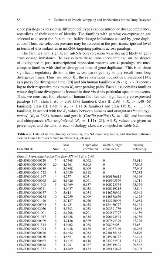
88 6 Evolution of Protein Wrapping and Implications for the Drug Designer
since paralogs expressed in different cell types cannot introduce dosage imbalance,regardless of their extent of identity. The families with paralog co-expression areselected to discern the factors that buffer dosage imbalance caused by gene dupli-cation. Thus, the selection pressure may be assessed at the post-transcriptional levelin terms of dissimilarities in miRNA-targeting patterns across paralogs.
The families with significant mRNA co-expression were deemed likely to gen-erate dosage imbalance. To assess how these imbalances impinge on the degreeof divergence in post-transcriptional repression patterns across paralogs, we mustcompare families with similar divergence time of gene duplicates. This is so sincesignificant regulatory dissimilarities across paralogs may simply result from longdivergence times. Thus, we adopt Ks, the synonymous nucleotide divergence [14],as a proxy for divergence time [20] and bin human families with < η >> 0 accord-ing to their respective maximum Ks over paralog pairs. Each class contains familieswhose duplicate divergence is located in time vis-á-vis particular speciation events.Thus, we construct four classes of human families with significantly co-expressedparalogs [17]: class I: Ks > 2.98 (378 families); class II: 2.98 > Ks > 1.48 (68families); class III: 1.48 > Ks > 1.11 (6 families) and class IV: Ks < 1.11 (5families), in accord with the Ks values between humans and orangutan (Pongo pyg-maeus) (Ks = 2.98), humans and gorilla (Gorilla gorilla) (Ks = 1.48), and humansand chimpanzee (Pan troglodytes) (Ks = 1.11) [21]. All Ks values are given aspercentages and the data for each orthology class are compiled in Table 6.2.
Table 6.2 Data set of evolutionary, expression, miRNA-based regulation, and structural informa-tion on human families binned in different Ks classes
Ensembl ID SizeMax.Ks
Expressioncorrelation
miRNA targetcoincidence
Packingdeficiency
Class I: Representative families from 378 with Ks> 2.98>ENSF00000000529 7 4.7368 0.092 0 58.611>ENSF00000000100 20 4.3361 0.105 0 57.905>ENSF00000000139 4 3.5898 0.009 0 57.905>ENSF00000001722 3 4.5429 0.112 0 57.235>ENSF00000001167 4 4.257 0.031 0.100156612 49.166>ENSF00000000095 26 4.8626 0.091 0.100505579 41.711>ENSF00000001308 3 4.5669 0.137 0.100723554 53.574>ENSF00000000711 4 4.0037 0.058 0.100919215 43.801>ENSF00000000137 10 5.618 0.054 0.146230992 40.942>ENSF00000000284 10 4.2759 0.099 0.146538636 41.175>ENSF00000001426 4 3.7137 0.026 0.183940909 31.682>ENSF00000000564 5 4.4951 0.052 0.184436777 38.162>ENSF00000002030 3 4.5383 0.032 0.203391736 44.664>ENSF00000001001 2 3.1208 0.201 0.204847727 41.439>ENSF00000001947 3 4.5458 0.355 0.206692562 44.336>ENSF00000000469 6 4.2126 0.369 0.207894326 47.726>ENSF00000001542 3 4.9462 0.088 0.20806405 33.63>ENSF00000001394 3 4.4438 0.185 0.215967149 40.265>ENSF00000000676 4 5.1652 0.052 0.220155165 32.679>ENSF00000000750 4 4.555 0.032 0.249786777 31.144>ENSF00000000662 6 4.1415 0.148 0.252284504 33.377>ENSF00000000678 3 4.398 0.077 0.259919421 29.562>ENSF00000001387 4 4.6489 0.132 0.265183678 33.705
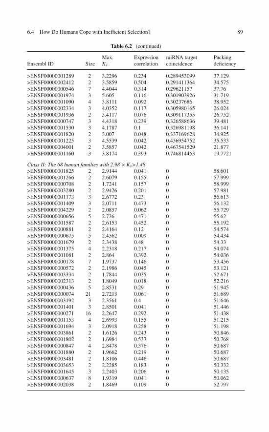
6.4 How Do Humans Cope with Inefficient Selection? 89
Table 6.2 (continued)
Ensembl ID SizeMax.Ks
Expressioncorrelation
miRNA targetcoincidence
Packingdeficiency
>ENSF00000001289 2 3.2296 0.234 0.289453099 37.129>ENSF00000002412 2 3.5859 0.504 0.291411364 34.575>ENSF00000000546 7 4.4044 0.314 0.29621157 37.76>ENSF00000001974 3 5.605 0.116 0.301903926 31.719>ENSF00000001090 4 3.8111 0.092 0.30237686 38.952>ENSF00000002334 3 4.0352 0.117 0.305980165 26.024>ENSF00000001936 2 5.4117 0.076 0.309117355 26.752>ENSF00000000747 3 4.4318 0.239 0.326588636 39.481>ENSF00000001530 3 4.1787 0.1 0.326981198 36.141>ENSF00000001820 2 3.007 0.048 0.337169628 34.925>ENSF00000001225 3 4.5539 0.042 0.436954752 35.533>ENSF00000004001 2 3.5857 0.042 0.467541529 21.877>ENSF00000001160 3 3.8174 0.393 0.746814463 19.7721
Class II: The 68 human families with 2.98 > Ks>1.48>ENSF00000001825 2 2.9144 0.041 0 58.601>ENSF00000001266 2 2.6079 0.155 0 57.999>ENSF00000000708 2 1.7241 0.157 0 58.999>ENSF00000003280 2 2.9426 0.201 0 57.981>ENSF00000001173 3 2.6772 0.23 0 56.613>ENSF00000001409 3 2.0711 0.473 0 56.132>ENSF00000004229 2 2.0857 0.062 0 55.729>ENSF00000000656 5 2.736 0.471 0 55.62>ENSF00000001587 2 2.6153 0.452 0 55.192>ENSF00000000881 2 2.4164 0.12 0 54.574>ENSF00000000675 5 2.4562 0.009 0 54.434>ENSF00000001679 2 2.3438 0.48 0 54.33>ENSF00000001375 4 2.2318 0.217 0 54.074>ENSF00000001081 2 2.864 0.392 0 54.036>ENSF00000000178 7 1.9737 0.146 0 53.456>ENSF00000000572 2 2.1986 0.045 0 53.121>ENSF00000003334 2 1.7844 0.035 0 52.671>ENSF00000002313 2 1.8049 0.018 0 52.216>ENSF00000000436 5 2.8531 0.29 0 51.945>ENSF00000000074 21 2.7213 0.061 0 51.689>ENSF00000003192 3 2.3561 0.4 0 51.646>ENSF00000001401 3 2.8501 0.041 0 51.446>ENSF00000000271 16 2.2647 0.292 0 51.438>ENSF00000001153 4 2.6993 0.155 0 51.215>ENSF00000001694 3 2.0918 0.258 0 51.198>ENSF00000003861 2 1.6126 0.243 0 50.846>ENSF00000001802 2 1.6984 0.537 0 50.768>ENSF00000000847 4 2.8478 0.376 0 50.687>ENSF00000001880 2 1.9662 0.219 0 50.687>ENSF00000003481 2 1.8106 0.446 0 50.687>ENSF00000003653 2 2.2285 0.183 0 50.332>ENSF00000001645 3 2.2403 0.206 0 50.135>ENSF00000000637 8 1.9319 0.041 0 50.062>ENSF00000002038 2 1.8469 0.109 0 52.797
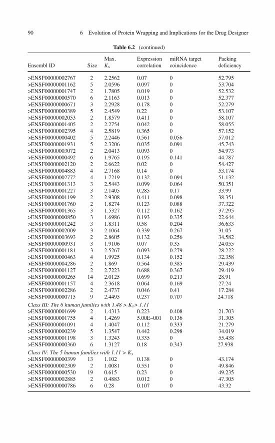
90 6 Evolution of Protein Wrapping and Implications for the Drug Designer
Table 6.2 (continued)
Ensembl ID SizeMax.Ks
Expressioncorrelation
miRNA targetcoincidence
Packingdeficiency
>ENSF00000002767 2 2.2562 0.07 0 52.795>ENSF00000001162 5 2.0596 0.097 0 53.704>ENSF00000001747 2 1.7805 0.019 0 52.532>ENSF00000000570 6 2.1163 0.013 0 52.377>ENSF00000000671 3 2.2928 0.178 0 52.279>ENSF00000000389 5 2.4549 0.22 0 53.107>ENSF00000002053 2 1.8579 0.411 0 58.107>ENSF00000001405 2 2.2754 0.042 0 58.055>ENSF00000002395 4 2.5819 0.365 0 57.152>ENSF00000000402 5 2.2446 0.561 0.056 57.012>ENSF00000001931 5 2.3206 0.035 0.091 45.743>ENSF00000003072 2 2.0413 0.093 0 54.973>ENSF00000000492 6 1.9765 0.195 0.141 44.787>ENSF00000002120 2 2.6622 0.02 0 54.427>ENSF00000004883 4 2.7168 0.14 0 53.174>ENSF00000002772 4 1.7219 0.132 0.094 51.132>ENSF00000001313 3 2.5443 0.099 0.064 50.351>ENSF00000001227 3 2.1405 0.285 0.17 33.99>ENSF00000001199 2 2.9308 0.411 0.098 38.351>ENSF00000001760 2 1.8274 0.123 0.088 37.322>ENSF00000001365 3 1.5327 0.112 0.162 37.295>ENSF00000000850 3 1.6986 0.193 0.335 22.644>ENSF00000001242 3 1.8311 0.58 0.204 36.633>ENSF00000002009 3 2.1064 0.339 0.267 31.05>ENSF00000003693 2 2.8605 0.132 0.256 34.582>ENSF00000000931 3 1.9106 0.07 0.35 24.055>ENSF00000001181 3 2.5267 0.093 0.279 28.222>ENSF00000000463 4 1.9925 0.134 0.152 32.358>ENSF00000004286 2 1.869 0.564 0.385 29.439>ENSF00000001127 2 2.7223 0.688 0.367 29.419>ENSF00000000265 14 2.0125 0.699 0.213 28.91>ENSF00000001157 4 2.3618 0.064 0.169 27.24>ENSF00000002286 2 2.4737 0.046 0.41 17.284>ENSF00000000715 9 2.4495 0.237 0.707 24.718
Class III: The 6 human families with 1.48 > Ks> 1.11>ENSF00000001699 2 1.4313 0.223 0.408 21.703>ENSF00000001755 4 1.4269 5.00E–001 0.136 31.305>ENSF00000001091 4 1.4047 0.112 0.333 21.279>ENSF00000000239 5 1.3547 0.442 0.298 34.019>ENSF00000001198 3 1.3243 0.335 0 55.438>ENSF00000000360 6 1.3127 0.18 0.343 27.938
Class IV: The 5 human families with 1.11 > Ks>ENSF00000000399 13 1.102 0.138 0 43.174>ENSF00000002309 2 1.0081 0.551 0 49.846>ENSF00000000530 19 0.615 0.23 0 49.235>ENSF00000002885 2 0.4883 0.012 0 47.305>ENSF00000000786 6 0.28 0.107 0 43.32
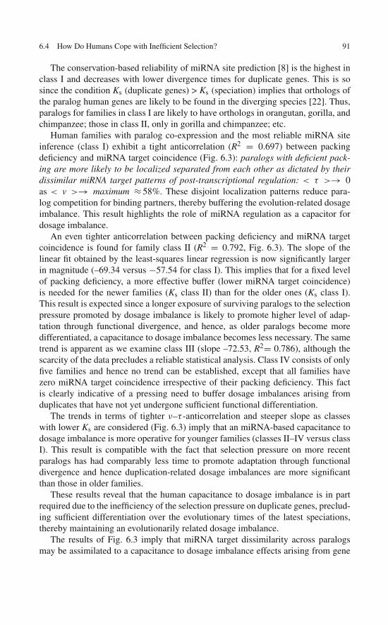
6.4 How Do Humans Cope with Inefficient Selection? 91
The conservation-based reliability of miRNA site prediction [8] is the highest inclass I and decreases with lower divergence times for duplicate genes. This is sosince the condition Ks (duplicate genes) > Ks (speciation) implies that orthologs ofthe paralog human genes are likely to be found in the diverging species [22]. Thus,paralogs for families in class I are likely to have orthologs in orangutan, gorilla, andchimpanzee; those in class II, only in gorilla and chimpanzee; etc.
Human families with paralog co-expression and the most reliable miRNA siteinference (class I) exhibit a tight anticorrelation (R2 = 0.697) between packingdeficiency and miRNA target coincidence (Fig. 6.3): paralogs with deficient pack-ing are more likely to be localized separated from each other as dictated by theirdissimilar miRNA target patterns of post-transcriptional regulation: < τ >→ 0as < v >→ maximum ≈ 58%. These disjoint localization patterns reduce para-log competition for binding partners, thereby buffering the evolution-related dosageimbalance. This result highlights the role of miRNA regulation as a capacitor fordosage imbalance.
An even tighter anticorrelation between packing deficiency and miRNA targetcoincidence is found for family class II (R2 = 0.792, Fig. 6.3). The slope of thelinear fit obtained by the least-squares linear regression is now significantly largerin magnitude (–69.34 versus −57.54 for class I). This implies that for a fixed levelof packing deficiency, a more effective buffer (lower miRNA target coincidence)is needed for the newer families (Ks class II) than for the older ones (Ks class I).This result is expected since a longer exposure of surviving paralogs to the selectionpressure promoted by dosage imbalance is likely to promote higher level of adap-tation through functional divergence, and hence, as older paralogs become moredifferentiated, a capacitance to dosage imbalance becomes less necessary. The sametrend is apparent as we examine class III (slope –72.53, R2= 0.786), although thescarcity of the data precludes a reliable statistical analysis. Class IV consists of onlyfive families and hence no trend can be established, except that all families havezero miRNA target coincidence irrespective of their packing deficiency. This factis clearly indicative of a pressing need to buffer dosage imbalances arising fromduplicates that have not yet undergone sufficient functional differentiation.
The trends in terms of tighter ν–τ -anticorrelation and steeper slope as classeswith lower Ks are considered (Fig. 6.3) imply that an miRNA-based capacitance todosage imbalance is more operative for younger families (classes II–IV versus classI). This result is compatible with the fact that selection pressure on more recentparalogs has had comparably less time to promote adaptation through functionaldivergence and hence duplication-related dosage imbalances are more significantthan those in older families.
These results reveal that the human capacitance to dosage imbalance is in partrequired due to the inefficiency of the selection pressure on duplicate genes, preclud-ing sufficient differentiation over the evolutionary times of the latest speciations,thereby maintaining an evolutionarily related dosage imbalance.
The results of Fig. 6.3 imply that miRNA target dissimilarity across paralogsmay be assimilated to a capacitance to dosage imbalance effects arising from gene
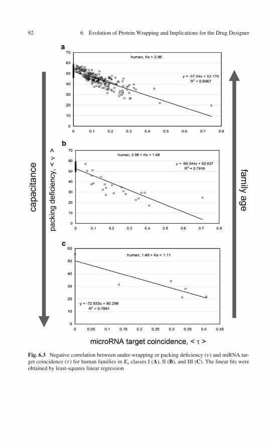
92 6 Evolution of Protein Wrapping and Implications for the Drug Designer
Fig. 6.3 Negative correlation between under-wrapping or packing deficiency (ν) and miRNA tar-get coincidence (τ ) for human families in Ks classes I (A), II (B), and III (C). The linear fits wereobtained by least-squares linear regression

6.5 Human Capacitance to Dosage Imbalances 93
duplication. The severity of such effects is in turn quantified by packing defi-ciency: Dosage imbalances are less tolerated for deficiently packed proteins forcingparalogs to be localized separately from each other.
6.5 Human Capacitance to Dosage Imbalancesin the Concentrations of Under-Wrapped Proteins
In this Section we examined dosage imbalances that have an evolutionary origin.Thus, gene duplication events generate dosage imbalances that impose selectionpressure on paralogs and the magnitude of the effects of this pressure depend onthe extent of under-wrapping or packing deficiency of the gene product. However,this dependence varies widely from unicellular to higher eukaryotes, with humansbeing particularly insensitive to dosage imbalances. In humans, there is a signifi-cant amount of genes with packing deficiency which are nevertheless extensivelyduplicated. This suggests that humans are resilient to evolution-related dosageimbalances, a capacitance that may be rationalized in terms of escape routes avail-able to humans but not to unicellular organisms, where dosage imbalances haveclear deleterious effects.
In the absence of expression dissimilarity, the initially identical paralogs of defi-ciently packed proteins are subject to high selection pressure because they competefor binding partners needed to maintain structural integrity. Conversely, tight proteinpacking reduces dosage sensitivity, thereby curbing selection pressure. Cross exam-ination of genetic and structural data reveals that humans have a built-in resilienceor capacitance to dosage imbalances. The determinant of this human capacitance istraced in this chapter to the paralog-discriminatory power of miRNA regulatory pat-terns. In this way, dissimilar paralog localization governed by post-transcriptionalregulation of protein levels mitigates the competition of paralogs for common bind-ing partners that become obligatory for proteins of low packing quality. In otherwords, dissimilarity in paralog localization operative through miRNA control offersan escape route to dosage imbalances created by gene duplication, and this escaperoute becomes more necessary as protein packing deficiency makes these dosageimbalances less tolerable.
If selection is indeed inefficient in humans as Lynch and co-workers proposed[3], one may wonder how miRNA-based capacitance could be achieved throughrandom genetic drift. The removal of an miRNA-binding site is readily achiev-able through a single deleterious mutation in one paralog and is unlikely to occurat the same binding site in another paralog. For instance, if M non-overlappingmiRNA-binding sites are present in the 3′-UTR of two paralogs (1 << M <156),the probability that a pair of mutations (one in each paralog) will discriminate par-alogs is (M–1)/M, while the probability that they both occur at the same site inthe two paralogs and hence do not contribute to increase the capacitance is 1/M.Thus, dissimilarity and even orthogonality in post-transcriptional repressive patternsis readily achievable through widespread random mutation and hence may be theresult of the nonadaptive forces prevalent in humans.
94 6 Evolution of Protein Wrapping and Implications for the Drug Designer
6.6 Why Should the Drug Designer Be Mindfulof Molecular Evolution?
As surveyed in this chapter, in spite of an uncanny structural similarity arising fromcommon ancestry, both ortholog and paralog proteins exhibit vast dissimilaritiesin their wrapping patterns. However, the root of these variations is very differentdepending on whether orthologs or paralogs are considered. As indicated in theprevious sections, differences in selection efficiency across species appear to beresponsible for wrapping differences across orthologs. On the other hand, in the caseof paralogs, significant dosage imbalances would arise if paralogs were identical,simply because paralogs coexist in the same individual. To avert such imbalancesand their fitness consequences, sufficient divergence of the original duplicate genesmust take place through dissimilar expression regulation. Thus, in the previoussections we showed how paralog segregation may be achieved.
An alternative evolutionary route to differentiate paralogs as needed to bufferthe dosage imbalances they create is through dissimilar wrapping of their otherwisehighly similar 3D structures. As demonstrated in the subsequent chapters, this phe-nomenon has profound consequences for drug design because it enables designersto discriminate paralogs guided by their different dehydron patterns, exploiting theidea that a drug may function as an external wrapper of the packing defects in thetarget protein.
Differences in wrapping patterns may be significant even for close paralogs withhigh levels (∼88%) of sequence identity, such as the kinase domains of the insulinreceptor (IR) and that of the insulin-like growth factor receptor (IGF1R), as shownin Fig. 6.4. From the perspective of molecular targeted cancer therapy, there is avested interest in engineering drugs able to discriminate between these two par-alogs: IGF1R is probably a major target in the treatment of breast cancer [23],while inhibition of the IR kinase is likely to have devastating consequences, eventhe risk of inducing a diabetic coma in the patient. Yet, harnessing on the wrap-ping design concept, it should be possible to target differences in the wrappingpatterns of both kinases (Fig. 6.4) to enhance therapeutic efficacy against breastcancer.
On the other hand, the observation that there are wrapping differences acrossorthologous proteins should also have a significant impact for the drug designer.For example, the therapeutic efficacy of drugs is routinely tested in animal mod-els through xenografts and other assays, always under the unwarranted assumptionthat the affinity and specificity of the drug for its target in humans would be thesame or very close to its affinity and specificity for the animal ortholog of the tar-geted protein. In light of the wrapping differences across orthologs, there is simplyno assurance that this scenario is indeed valid. This observation could potentiallyundermine the very foundation on which animal testing is predicated.
Yet, there is a positive implication of the observed wrapping differences acrossorthologs, and it relates to the drug-based treatment of infectious diseases suchas AIDS. It is indeed highly advantageous for the drug designer to detect wrap-ping differences between the target protein in the infecting species and its human

References 95
Fig. 6.4 Comparison of structures (ribbon representation) and wrapping patterns of the humanparalog kinases for the IR (insulin receptor) and the IGF1R (insulin-like growth factor 1 receptor)
ortholog, if any: A drug purposely designed to wrap specific dehydrons in the proteinof the infecting species that are not present in the human ortholog is likely to befar less toxic than a drug that cannot tell apart the two orthologs. Species selec-tivity is hardly a concern in an AIDS treatment geared at targeting the reversetranscriptase of the HIV-1 retrovirus, simply because this protein is not encodedin the human genome. On the other hand, a molecular design rooted in wrappingdifferences would be very helpful if the goal is to target a retroviral protein thatis also represented in the human proteome. Species selectivity matters in this lastcontext.
References
1. Fernández A, Scott R, Berry RS (2004) The nonconserved wrapping of conserved foldsreveals a trend towards increasing connectivity in proteomic networks. Proc Natl Acad SciUSA 101:2823–2827
2. Fernández A, Berry RS (2004) Molecular dimension explored in evolution to promoteproteomic complexity. Proc Natl Acad Sci USA 101:13460–13465
96 6 Evolution of Protein Wrapping and Implications for the Drug Designer
3. Lynch M, Conery JS (2003) The origins of genome complexity. Science 302:1401–14044. Kondrashov FA, Koonin EV (2004) A common framework for understanding the origin of
genetic dominance and evolutionary fates of gene duplications. Trends Genet 20:287–2905. Liang H, Rogale-Plazonic K, Chen J, Li WH, Fernández A (2008) Protein under-wrapping
causes dosage sensitivity and decreases gene duplicability. PLoS Genet 4:e116. Papp B, Pal C, Hurst LD (2003) Dosage sensitivity and the evolution of gene families in yeast.
Nature 424:194–1977. Fernández A, Scheraga H (2003) Insufficiently dehydrated hydrogen bonds as determinants
for protein interactions. Proc Natl Acad Sci USA 100:113–1188. Bartel D (2009) MicroRNAs: Target recognition and regulatory functions. Cell 136:215–2339. Fernández A (2004) Keeping dry and crossing membranes. Nat Biotechnol 22:1081–1084
10. Veitia RA (2002) Exploring the etiology of haploinsufficiency. Bioessays 24:175–18411. Veitia RA (2004) Gene dosage balance: Deletions, duplications and dominance. Trends Genet
21: 33–3512. Su AI, Wiltshire T, Batalov S et al (2004) A gene atlas of the mouse and human protein-
encoding transcriptomes. Proc Natl Acad Sci USA 101:6062–606713. Birney E, Andrews D, Caccamo M et al (2006) Ensembl 2006. Nucleic Acids Res 34:
D556–D56114. Yang Z, Nielsen R (2000) Estimating synonymous and nonsynonymous substitution rates
under realistic evolutionary models. Mol Biol Evol 17:32–4315. Friedman RC, Farth KK, Burge CB, Bartel DP (2009) Most mammalian mRNAs are
conserved targets of microRNAs. Genome Res 19:92–10516. Lewis B, Burge C, Bartel D (2005) Conserved seed pairing, often flanked by adenosines,
indicates that thousands of human genes are microRNA targets. Cell 120:15–2017. Fernández A, Chen J (2009) Human capacitance to dosage imbalance: coping with inefficient
selection. Genome Res 19:2185–219218. Grimson A, Farth KK, Johnston WK et al (2007) MicroRNA target specificity in mammals:
Determinants beyond seed pairing. Mol Cell 27:91–10519. Aloy P, Ceulemans H, Stark A, Russell RB (2003) The relationship between sequence and
interaction divergence in proteins. J Mol Biol 332:989–99820. Gu Z, Nicolae D, Lu HH, Li W-H (2002) Rapid divergence in expression between duplicate
genes inferred from microarray data. Trends Genet 18:609–61321. Chen F, Li W-H (2001) Genomic divergences between humans and other hominoids and the
effective population size of the common ancestor of humans and chimpanzees. Am J HumGenet 68:444–456
22. Gao L, Innan H (2004) Very low gene duplication rate in the yeast genome. Science 306:1367–1370
23. Surmacz E, Bartucci M (2005) Role of estrogen receptor alpha in modulating IGF-I receptorsignaling and function in breast cancer. J Exp Clin Cancer Res 23:385–394

Chapter 7Wrapping as a Selectivity Filter for MolecularTargeted Therapy: Preliminary Evidence
The conservation of structure across homolog proteins often diffuses the impact ofdrug-based inhibition by promoting alternative protein–ligand associations that mayultimately lead to toxic side effects. This problem becomes particularly acute whenattempting to interfere with signaling pathways involved in cell fate and cell prolif-eration, the type of molecular intervention often exploited in molecular anticancertherapy. This is because the clinically relevant targets, the kinases, evolved fromeach other and hence share an uncanny structural resemblance. However, as shownin the previous chapter, the sticky wrapping defects or dehydrons are typically notconserved across proteins of common ancestry, making them valuable a priori tar-gets to enhance specificity. Thus, nonconserved packing defects may be utilizedas selectivity switches across homolog targets. This chapter explores this paradig-matic concept and its ramifications for the rational design of drugs with controlledspecificity.
7.1 The Specificity Problem in Drug Design
Creating inhibitors with controlled specificity and without undesirable side effectsis a major goal of drug design. Drug-induced enzymatic inhibition may have at leasttwo kinds of side effects: the drug may interfere with pathways other than the one oftherapeutic relevance because of the manifold roles of the target protein or paralogproteins, being structurally similar, may alternatively associate with the inhibitor.Building on the evolutionary knowledge acquired from the previous chapter, thischapter introduces new and paradigmatic concepts to minimize drug interactionswith toxicity-related targets. In principle, the control of specificity is enabled as weexploit characteristics of protein–ligand interactions that are not preserved acrossparalogs, despite the similarity of their 3D structures [1–3]. Thus, the insights fromthe previous chapter prompt us to focus on a singular feature, the dehydron, a stickypacking defect shown to be of paramount importance in promoting protein asso-ciations [4–6]. Quantifiable features of these interactions allow us to introduce atarget phylogeny that we can exploit to infer drug cross-reactivities and modulatethe inhibitory impact of a drug, funneling it onto the realm of clinical relevance.
97A. Fernández, Transformative Concepts for Drug Design: Target Wrapping,DOI 10.1007/978-3-642-11792-3_7, C© Springer-Verlag Berlin Heidelberg 2010
98 7 Wrapping as a Selectivity Filter for Molecular Targeted Therapy
Considerable research has helped clarify many aspects of protein–ligand inter-action [7]. Various methods enable one to predict such interactions, such as theassessment of packing defects [8], analysis of protein interface geometry [9], dock-ing dynamics [10], modeling free energy [11], computational molecular probing[12], sequence threading [13], and learning theory approaches [14]. The first four ofthese are based on properties of protein interfaces; the latter two are based only onsequences and attempt to “learn” features encoded in those sequences. Combinedmethodologies are also being developed [14]. Some of these techniques seek fea-tures of protein surfaces that promote protein–protein interactions. In this chapterwe show how features with a quantifiable metric can be used to compare paralogsin a way that provides a guidance to develop more selective drugs.
As described in the previous chapters, to function, soluble proteins must maintainrelatively stable structures, a condition often requiring associations [15–17]. Isolatedstructures with packing defects arising as poorly protected hydrogen bonds typicallydo not prevail in water [3, 18]. On the other hand, such defects are inherentlysticky, promoting removal of surrounding water through protein associations [8],as indicated in Chap. 1. Accordingly, in this chapter we show that dehydrons maybe targeted in a new generation of highly selective drug-based inhibitors. Knownstructures of protein–inhibitor complexes [19–24] provide the foundation to designinhibitors that in effect become wrappers or protectors of dehydrons.
As shown in Chap. 6, while folds are typically conserved across homolog pro-teins, wrapping defects are typically not conserved [3], and this property providesthe main motivation for advocating a paradigmatic shift in drug design. Whileevery targetable protein family may in principle be subject to the same analysis,we shall narrow down the scope of the discussion for the sake of clarity. Thus,we shall restrict ourselves to the goal of designing molecular therapeutic agentsthat selectively interfere with cell signaling. In this context, structure-based designbecomes particularly daunting because the basic signal transducers in the cell,the kinases, share a common molecular ancestry and hence possess an uncannystructural similarity. This fact turns drug specificity into a major issue.
Kinase inhibitors are designed to impair the ATP-dependent signal transduction(Fig. 7.1a, b). Hence they should be capable of binding to the target competitively ornoncompetitively vis-à-vis the natural kinase ligand ATP. The main structural fea-tures of the binding of ATP to a kinase are illustrated in Fig. 7.1a, b, where an active(phosphorylated) tyrosine kinase, that of the insulin receptor (IR), is shown boundto ATP and to a substrate peptide. The kinase substrate becomes phosphorylated at atyrosine residue through a trans-esterification reaction involving transference of theγ-phosphate of ATP (Fig. 7.1b). Thus, a major challenge to design ATP competitiveinhibitors is the fact that the ATP-binding regions of most kinases are structurallyvery similar (with an RMSD <1.2 Å in backbone coordinates) and have a very highlevel of amino acid conservation.
Contrasting with this troublesome aspect, the lack of conservation of the wrap-ping (dehydron) pattern across kinases has paramount potential to promote drugspecificity, as illustrated in Fig. 7.1c, d. Thus, the structural alignment of the focaladhesion kinase (FAK, PDB.2J0L) and the insulin receptor kinase (PDB.1GAG)
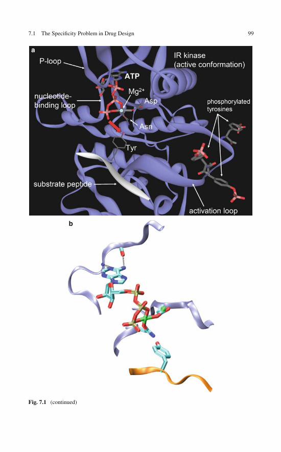
7.1 The Specificity Problem in Drug Design 99
a
b
Fig. 7.1 (continued)
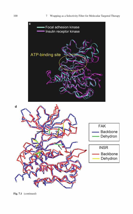
100 7 Wrapping as a Selectivity Filter for Molecular Targeted Therapy
c
d
Fig. 7.1 (continued)

7.1 The Specificity Problem in Drug Design 101
e
Fig. 7.1 (a) Generic structural features of a ternary complex involving a phosphorylated (active)kinase, its natural ligand ATP, and its phosphorylation substrate peptide (white). The phospho-rylation event (red arrow) involves trans-phosphoesterification of the substrate tyrosine withtransference of the γ-phosphate from ATP to the substrate, concurrent dismantling of the anchoringMg(II)-coordination complex, and finally, release of ADP. (b) Detailed rendering of the ATP posi-tion with respect to the phosphorylation substrate. Atoms are conventionally colored with Mg(II)in green, substrate peptide in gold, and IR kinase backbone in light blue. Structural (c) and wrap-ping (d) comparison between the focal adhesion kinase (FAK, PDB.2J0L) and the insulin receptorkinase (INRK, PDB.1GAG). The dehydrons in the ATP-binding region that are unique to FAKare marked by arrows. (e) Drug paradigmatically represented as a wrapper of pre-formed dehy-drons in the target protein. Conventional nonbonded interactions between drug and target such ashydrophobic contacts and intermolecular hydrogen bonds typically foster drug promiscuity as theyinvolve conserved features in the target protein
reveal a significant similarity, with RMSD (root mean square displacement) <1.3 Å(Fig. 7.1c). Yet, there are crucial differences in the dehydron patterns of bothproteins (Fig. 7.1d), particularly in their ATP-binding pockets, where the ATP com-petitive inhibitors bind [18]. These differences can be exploited with considerabletherapeutic advantage: FAK is a target of major clinical relevance to treat cancermetastasis and yet the probable activity of a purported FAK inhibitor against thestructurally related insulin receptor kinase is likely to have potentially devastat-ing effects, arising mainly from the induction of a diabetic coma in the patient[18]. However, there are dehydrons unique to FAK and not found in its paralog(arrows in Fig. 7.1d) which may be targeted to satisfy the therapeutic imperative ofspecificity. This peculiar way of targeting a molecular feature materializes as inter-molecular wrapping, as illustrated in Fig. 7.1e. The example shown in Fig. 7.1c, ddramatically reveals the difficulty in telling apart targets of clinical relevance from

102 7 Wrapping as a Selectivity Filter for Molecular Targeted Therapy
toxicity-related targets in molecular targeted therapy and unravels a possible avenueto solve this problem.
Thus, in principle, side effects resulting from off-target ligand binding may beminimized by selectively targeting nonconserved dehydrons unique to the desiredtarget protein with the guidance of a measure of wrapping proximity between struc-tures. In this regard, this chapter reveals that packing defects may act not only asselectivity filters operative across homolog proteins but also as selectivity switches,to re-engineer a protein–inhibitor to redirect its impact toward another target. Thislatter role requires taking advantage of the spatial displacement of packing defectsacross aligned homologous structures.
Functional differences across homolog proteins sharing a common fold are veryfrequent in biology. Previous studies revealed that the wrapping constitutes a molec-ular dimension explored when evolution is constrained to preserve the fold forfunctional reasons. Thus, it is likely that wrapping differences across paralogs areindicative of the functional fine-tuning needed to avoid dosage imbalances (seeChap. 6). It is precisely this fine-tuning that needs to be discriminated in drug-basedmolecular therapy. As this work reveals, such wrapping differences offer a new con-trol in constructing the selectivity filters and selectivity switches that may endow thewrapping technology with a high therapeutic value.
Structural descriptors of protein-binding sites, such as hydrophobicity [25], cur-vature [26], and accessibility [27], are routinely used to guide inhibitor design.However, examination of the 814 nonredundant protein–inhibitor PDB complexesreveals that 488 of them have binding cavities with only average hydrophobicity[25], not significantly higher than the rest of the surface. In such cases, ligand affin-ity is attributed to the intermolecular hydrogen-bonding propensities of the inhibitor,inferred from protein–substrate transition state mimetics [21–24]. However chargescreening in water renders putative intermolecular hydrogen bonds unlikely promot-ers of protein–ligand association, unless water removal is favored at the interface bysome pre-existing feature on the surface of the protein [8].
One obvious such feature is the solvent-exposed nonpolar moiety, except that, asstated above, such features occur infrequently on binding sites and if they do, theyare highly conserved across homologs. A more common and less troublesome water-excluding feature has been recently identified. We have reported [3, 8, 18, 28, 29]that dehydrons constitute sticky sites with a propensity to become dehydrated as aresult of protein associations.
To explore the potential of dehydrons as promoters of drug selectivity, we startby noting that in most PDB protein–inhibitor complexes, the ligand is in effect awrapper of protein dehydrons, although it was not purposely designed to fulfillthis role. In this way, the available bioinformatics data support the novel designconcept. Ultimately, we shall develop a wrapping technology and provide evi-dence supporting the idea that targeting dehydrons that are not conserved acrossparalogs constitute a useful strategy to enhance the selectivity of the inhibitor.Thus, the notion of wrapping (packing) similarity will be rigorously introducedand used selectively to modify a multiple-target inhibitor to achieve a higher
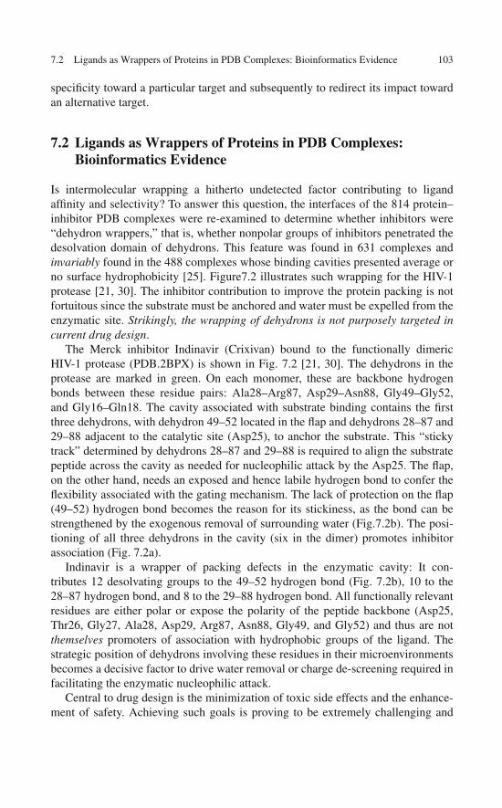
7.2 Ligands as Wrappers of Proteins in PDB Complexes: Bioinformatics Evidence 103
specificity toward a particular target and subsequently to redirect its impact towardan alternative target.
7.2 Ligands as Wrappers of Proteins in PDB Complexes:Bioinformatics Evidence
Is intermolecular wrapping a hitherto undetected factor contributing to ligandaffinity and selectivity? To answer this question, the interfaces of the 814 protein–inhibitor PDB complexes were re-examined to determine whether inhibitors were“dehydron wrappers,” that is, whether nonpolar groups of inhibitors penetrated thedesolvation domain of dehydrons. This feature was found in 631 complexes andinvariably found in the 488 complexes whose binding cavities presented average orno surface hydrophobicity [25]. Figure7.2 illustrates such wrapping for the HIV-1protease [21, 30]. The inhibitor contribution to improve the protein packing is notfortuitous since the substrate must be anchored and water must be expelled from theenzymatic site. Strikingly, the wrapping of dehydrons is not purposely targeted incurrent drug design.
The Merck inhibitor Indinavir (Crixivan) bound to the functionally dimericHIV-1 protease (PDB.2BPX) is shown in Fig. 7.2 [21, 30]. The dehydrons in theprotease are marked in green. On each monomer, these are backbone hydrogenbonds between these residue pairs: Ala28–Arg87, Asp29–Asn88, Gly49–Gly52,and Gly16–Gln18. The cavity associated with substrate binding contains the firstthree dehydrons, with dehydron 49–52 located in the flap and dehydrons 28–87 and29–88 adjacent to the catalytic site (Asp25), to anchor the substrate. This “stickytrack” determined by dehydrons 28–87 and 29–88 is required to align the substratepeptide across the cavity as needed for nucleophilic attack by the Asp25. The flap,on the other hand, needs an exposed and hence labile hydrogen bond to confer theflexibility associated with the gating mechanism. The lack of protection on the flap(49–52) hydrogen bond becomes the reason for its stickiness, as the bond can bestrengthened by the exogenous removal of surrounding water (Fig.7.2b). The posi-tioning of all three dehydrons in the cavity (six in the dimer) promotes inhibitorassociation (Fig. 7.2a).
Indinavir is a wrapper of packing defects in the enzymatic cavity: It con-tributes 12 desolvating groups to the 49–52 hydrogen bond (Fig. 7.2b), 10 to the28–87 hydrogen bond, and 8 to the 29–88 hydrogen bond. All functionally relevantresidues are either polar or expose the polarity of the peptide backbone (Asp25,Thr26, Gly27, Ala28, Asp29, Arg87, Asn88, Gly49, and Gly52) and thus are notthemselves promoters of association with hydrophobic groups of the ligand. Thestrategic position of dehydrons involving these residues in their microenvironmentsbecomes a decisive factor to drive water removal or charge de-screening required infacilitating the enzymatic nucleophilic attack.
Central to drug design is the minimization of toxic side effects and the enhance-ment of safety. Achieving such goals is proving to be extremely challenging and
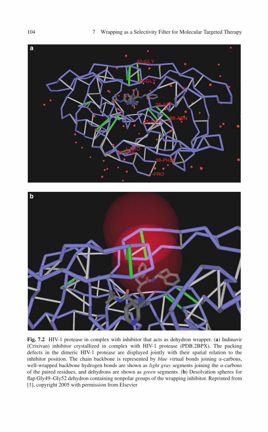
104 7 Wrapping as a Selectivity Filter for Molecular Targeted Therapy
a
b
Fig. 7.2 HIV-1 protease in complex with inhibitor that acts as dehydron wrapper. (a) Indinavir(Crixivan) inhibitor crystallized in complex with HIV-1 protease (PDB.2BPX). The packingdefects in the dimeric HIV-1 protease are displayed jointly with their spatial relation to theinhibitor position. The chain backbone is represented by blue virtual bonds joining α-carbons,well-wrapped backbone hydrogen bonds are shown as light gray segments joining the α-carbonsof the paired residues, and dehydrons are shown as green segments. (b) Desolvation spheres forflap Gly49–Gly52 dehydron containing nonpolar groups of the wrapping inhibitor. Reprinted from[1], copyright 2005 with permission from Elsevier
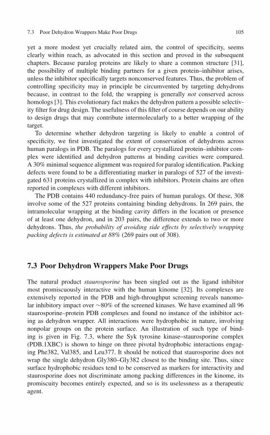
7.3 Poor Dehydron Wrappers Make Poor Drugs 105
yet a more modest yet crucially related aim, the control of specificity, seemsclearly within reach, as advocated in this section and proved in the subsequentchapters. Because paralog proteins are likely to share a common structure [31],the possibility of multiple binding partners for a given protein–inhibitor arises,unless the inhibitor specifically targets nonconserved features. Thus, the problem ofcontrolling specificity may in principle be circumvented by targeting dehydronsbecause, in contrast to the fold, the wrapping is generally not conserved acrosshomologs [3]. This evolutionary fact makes the dehydron pattern a possible selectiv-ity filter for drug design. The usefulness of this filter of course depends on our abilityto design drugs that may contribute intermolecularly to a better wrapping of thetarget.
To determine whether dehydron targeting is likely to enable a control ofspecificity, we first investigated the extent of conservation of dehydrons acrosshuman paralogs in PDB. The paralogs for every crystallized protein–inhibitor com-plex were identified and dehydron patterns at binding cavities were compared.A 30% minimal sequence alignment was required for paralog identification. Packingdefects were found to be a differentiating marker in paralogs of 527 of the investi-gated 631 proteins crystallized in complex with inhibitors. Protein chains are oftenreported in complexes with different inhibitors.
The PDB contains 440 redundancy-free pairs of human paralogs. Of these, 308involve some of the 527 proteins containing binding dehydrons. In 269 pairs, theintramolecular wrapping at the binding cavity differs in the location or presenceof at least one dehydron, and in 203 pairs, the difference extends to two or moredehydrons. Thus, the probability of avoiding side effects by selectively wrappingpacking defects is estimated at 88% (269 pairs out of 308).
7.3 Poor Dehydron Wrappers Make Poor Drugs
The natural product staurosporine has been singled out as the ligand inhibitormost promiscuously interactive with the human kinome [32]. Its complexes areextensively reported in the PDB and high-throughput screening reveals nanomo-lar inhibitory impact over ∼80% of the screened kinases. We have examined all 96staurosporine–protein PDB complexes and found no instance of the inhibitor act-ing as dehydron wrapper. All interactions were hydrophobic in nature, involvingnonpolar groups on the protein surface. An illustration of such type of bind-ing is given in Fig. 7.3, where the Syk tyrosine kinase–staurosporine complex(PDB.1XBC) is shown to hinge on three pivotal hydrophobic interactions engag-ing Phe382, Val385, and Leu377. It should be noticed that staurosporine does notwrap the single dehydron Gly380–Gly382 closest to the binding site. Thus, sincesurface hydrophobic residues tend to be conserved as markers for interactivity andstaurosporine does not discriminate among packing differences in the kinome, itspromiscuity becomes entirely expected, and so is its uselessness as a therapeuticagent.
106 7 Wrapping as a Selectivity Filter for Molecular Targeted Therapy
Fig. 7.3 Staurosporine does not wrap dehydrons and thus, it does not discriminate proteins withdifferent packing, which makes it highly promiscuous within the kinome. The staurosporine–Sykcomplex (PDB.1XBC) reveals a binding mode determined exclusively by hydrophobic interactionsengaging residues Leu377, Phe382, Val385. The closest Gly380–Gly383 dehydron is not wrappedby staurosporine
7.4 Wrapping as a Selectivity Filter
The subtle structural differences among paralogs of a kinase target may be quanti-fied by introducing a “wrapping homology.” Assessing wrapping similarity acrossparalogs requires comparing the microenvironments of backbone hydrogen bonds[8] that can be effectively carried out following the structural alignment [33].This “wrapping homology” becomes a useful tool to control specificity by tar-geting a packing defect. For a protein chain of length N, a matrix of dehydronsDij, i, j = 1, 2, . . . , N may be constructed by choosing Dij = 1 if residues i andj are paired by a dehydron and Dij = 0, otherwise. This representation is illus-trated in Fig. 7.4a. When the dehydron matrices for two proteins m and n arealigned, they are trimmed by restricting them to those residues that have beenstructurally aligned in a one-to-one correspondence. Then a Hamming distanceMH(m, n) = �i<j|Dij(m) − Dij(n)| may be computed to serve as an indicatorof the wrapping similarity between proteins m, n. Given a collection of paralogsm, n = 1, . . . , F, and its corresponding packing-similarity matrix MH = [MH(m, n)],a wrapping similarity tree (WST) may be easily constructed by adopting the premisethat the minimal pathway between protein nodes m and n crosses a number of nodesequal to MH(m, n).
A comparison of dehydron matrices for proteins with aligned structure enablesan automated digitalization of the drug selectivity problem. The alignment of dehy-dron matrices reveals the pattern of a priori cross-reactivities: Thus, targeting a

7.5 Wrapping as a Selectivity Filter: An Exercise in Drug Design 107
Fig. 7.4 Dehydron matrices and a priori cross-reactivities derived from their alignment. (a) Threedifferent representations of a folded peptide chain of length N = 17: fold, wrapping, and dehydronmatrix. The gray entries in the matrix indicate well-wrapped hydrogen bonds and the green entriesrepresent dehydrons. (b) A priori cross-reactivities across three proteins (1, 2, 3) of drugs/ligandsdesigned to wrap specific dehydrons (a, b) in protein 1. The cross-reactivities are inferred from thealignment of the dehydron matrices
specific dehydron in one particular protein is likely to promote cross-reactivitieswith other targets where this dehydron is found at the aligned position and precludeinhibitory activity whenever the dehydron aligns with a well-wrapped hydrogenbond (Fig. 7.4b).
7.5 Wrapping as a Selectivity Filter: An Exercise in Drug Design
We now present an exercise on controlling selectivity based on the wrapping designconcept. Because of the evolutionary proximity of kinases, side effects arising from

108 7 Wrapping as a Selectivity Filter for Molecular Targeted Therapy
off-target ligand binding often arise with kinase inhibitors, especially in cancertherapy [32]. Thus, a need arises to sharpen the binding affinity within the phar-macokinome associated with a specific drug. For example, the selective inhibitionof the constitutively active or deregulated Brc-Abl (Abl: Abelson tyrosine kinase),the fusion product of an aberrant chromosomal translocation, has proven decisiveto treat chronic myeloid leukemia (CML) [34]. Brc-Abl has been proven to be aprimary target for the potent inhibitor imatinib (Gleevec) [34] but is not its onlytarget [32]. Of the alternative targets with reported structure, the C-Kit tyrosinekinase has been recognized as a binding partner with clinical relevance, makingGleevec a therapeutic agent to treat certain solid tumors like gastrointestinal stro-mal tumor [35, 36]. In addition Gleevec binds tightly to the lymphocyte-associatedkinase (Lck) [37], an immunosuppression-related target probably undesirable, andto the platelet-derived growth factor receptor (PDGFR) kinase, a close homolog ofKit. Thus, we sought to modify Gleevec to improve its selectivity for Brc-Abl bytargeting dehydrons not conserved across paralogs.
The protein–inhibitor complex PDB.1FPU (Fig. 7.5a) reveals three electrostaticinteractions in Brc-Abl, the dehydrons Gly249–Gln252, Gln300–Glu316, and thesalt bridge Lys271–Glu286 that can be better wrapped by methylating Gleevec atthe positions indicated. Thus, methylation at positions I, II, and III would pre-sumably improve the wrapping of dehydrons 249–252, 300–316, and salt bridge271–286, respectively. A structural alignment of the paralogs of Brc-Abl wasperformed using the program Cn3D [33] to investigate the microenvironment con-servation for these intramolecular interactions. The alignment of six paralog proteinsis shown in Fig. 7.5b. The dehydron 249–252 (crankshaft-like kink marked in yel-low on Fig. 7.5b) is not conserved in any of the six paralogs of Brc-Abl, while thedehydron 300–316 becomes well wrapped in the paralogs. On the other hand, themicroenvironment of the salt bridge 271–286 is conserved.
The WST for Abl and its paralogs is shown in Fig. 7.5c. We have also restrictedthe WST computation to the hydrogen bonds/dehydrons in Brc-Abl which becomewrapped by Gleevec upon association. Adopting this restricted measure, the clos-est proteins to Brc-Abl become Lck, C-Kit, and PDGFR, precisely the alternativeGleevec targets with reported structure.
In order to enhance affinity and selectivity for Brc-Abl, we modified the inhibitormethylating at positions I and II (Fig. 7.5d). The synthesis of the wrapping prototyperecapitulates imatinib synthesis [38], as described in [39]. To test whether the speci-ficity and affinity for Brc-Abl improved, we conducted a spectrophotometric kineticassay to measure the phosphorylation rate of peptide substrates in the presence ofthe kinase inhibitor at different concentrations. This assay couples production ofadenosine diphosphate (ADP), the byproduct of downstream phosphorylation, withthe concurrent detectable oxidation of reduced nicotinamide adenosine dinucleotide(NADH). The oxidation results upon transfer of phosphate from PEP (phospho-enolpyruvate) to ADP followed by the NADH-mediated reduction of PEP to lactate.Thus, phosphorylation activity is monitored by the decrease in 340 nm absorbancedue to the oxidative conversion NADH→NAD+ [34, 39].

7.5 Wrapping as a Selectivity Filter: An Exercise in Drug Design 109
Fig. 7.5 (continued)
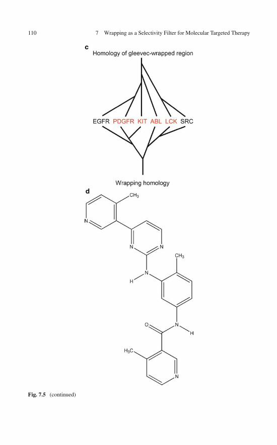
110 7 Wrapping as a Selectivity Filter for Molecular Targeted Therapy
Fig. 7.5 (continued)

7.5 Wrapping as a Selectivity Filter: An Exercise in Drug Design 111
Fig. 7.5 Wrapping as selectivity filter in inhibitor drug design: Modifications of Gleevec gearedat improving selectivity and affinity for Brc-Abl. (a) Three possible sites for Gleevec methylation(I–III) aimed at selectively improving the wrapping of packing defects of Brc-Abl (PDB.1FPU). (b)Structural alignment of Brc-Abl and its six paralogs using the program Cn3D. The yellow regioncorresponds to residues 247–257 in Abl. (c) Wrapping similarity tree (WST) for the six structurallyaligned paralogs including Brc-Abl. The WST restricted to the alignments of the Gleevec-wrappedregion in Brc-Abl is shown above. The paralogs in red have the most similar wrapping in the regionthat aligns with the Gleevec-wrapped region in Brc-Abl. (d) The modified Gleevec-based moleculemethylated at sites I and II and assayed in vitro. (e) Phosphorylation rates of Brc-Abl (blue), C-Kit(green), Lck (red), PDGFR (purple) and Src (brown) in the presence of Gleevec (triangles) andin the presence of the I- and II-methylated modified Gleevec (squares). The latter compound wasdesigned to better wrap the nonconserved dehydrons in Brc-Abl. To measure the rate of phosphory-lation due to kinase activity in the presence of inhibitors, a standard spectrophotometric assay hasbeen adopted in which the adenosine diphosphate (ADP) production, a marker of phosphorylationactivity, is coupled to NADH (reduced nicotinamide adenosine dinucleotide) oxidation and deter-mined by absorbance reduction at 340 nm, as described in [34, 39]. Basically, ADP production iscoupled to the final steps of glycolysis, so that the enzymatic reaction of ADP with reactant phos-phoenol pyruvate generates pyruvate whose production can be measured by reducing it to lactateusing redoxagent NADH. Reprinted from [1], copyright 2005 with permission from Elsevier
As indicated in Fig. 7.5e, the spectrophotometric assay revealed the inhibitionof the unphosphorylated Brc-Abl by the wrapper of the 249–252 and 300–316dehydrons (the I- and II-methylation product) improved over Gleevec levels.Furthermore, the inhibitory impact of the dehydron wrapper became selective forBrc-Abl vis-à-vis C-Kit and Lck. Dehydrons 249–252 and 300–316 are absent inthe latter kinases and, consistently, the drug designed to better wrap them has verylow inhibitory impact against C-Kit and Lck.

112 7 Wrapping as a Selectivity Filter for Molecular Targeted Therapy
Fig. 7.6 Selectivity switch determined by packing differences across paralog proteins. The struc-tural alignment (Fig. 7.5b), reveals that dehydron Gly249–Gln252 in Brc-Abl maps onto thespatially displaced dehydron Cys673–Gly676 in C-Kit. (a) Relative position of 673–676 dehy-dron of C-Kit with respect to Gleevec in protein–inhibitor complex (PDB.1T46). The desolvationdomain of the dehydron is displayed by two intersecting spheres. By methylating Gleevec at thepara-position (IV) rather than the ortho-position (I, Fig. 7.5a), the inhibitor becomes a selectivewrapper of the packing defect in C-Kit. (b) Phosphorylation rates from spectrophotometric assayon the kinases indicated in Fig. 7.5e inhibited by Gleevec methylated at positions II and IV. Thesquares correspond to the Gleevec variant methylated at positions II and IV. Notice the selec-tive and enhanced inhibition of C-Kit. Reprinted from [1], copyright 2005 with permission fromElsevier

References 113
7.6 Wrapping-Based Selectivity Switch
Can we really control imatinib specificity through a reworking of the parent com-pound as suggested in Fig. 7.4b? If this is indeed the case, we should be capableof re-funneling the inhibitory impact toward any of the alternative primary targetsof imatinib. A structural alignment of the Abl and Kit kinases reveals that dehy-dron Gly249–Gln252 in Abl (Fig. 7.5a) maps onto dehydron Cys673–Gly676 in Kit(Fig. 7.6a), spatially displaced with respect to the former. Given the relative posi-tion of the 673–676 dehydron of Kit with respect to Gleevec in the protein–inhibitorcomplex (PDB.1T46), we may determine which modification of Gleevec wouldselectively direct its impact toward C-Kit. By methylating Gleevec at the para-position (IV) in the terminal ring, rather than at the ortho-position (I, Fig. 7.5a),the inhibitor becomes a selective wrapper of the packing defect in C-Kit, while it nolonger wraps the 249–252 dehydron in Brc-Abl. The inhibitory impact of Gleevecmethylated at positions II and IV becomes apparent in the phosphorylation ratesobtained from the spectrophotometric assay on the same kinases studied previously(Figs. 7.5e and 7.6b). The inhibitory impact of the II and IV Gleevec variant is nowconcentrated on C-Kit, rather than Brc-Abl. Thus, by redesigning Gleevec accordingto the wrapping differences between Brc-Abl and C-Kit, we redirected the inhibitoryimpact from one kinase to the other. Taken together, both design exercises revealthat wrapping technology may enable us to introduce a selectivity switch in inhibitordesign.
References
1. Fernández A (2005) Incomplete protein packing as a selectivity filter in drug design. Structure13:1829–1836
2. Fernández A, Scott R, Berry RS (2004) The nonconserved wrapping of conserved foldsreveals a trend towards increasing connectivity in proteomic networks. Proc Natl Acad SciUSA 101:2823–2827
3. Fernández A, Berry RS (2004) Molecular dimension explored in evolution to promoteproteomic complexity. Proc Natl Acad Sci USA 101:13460–13465
4. Fernández A, Berry RS (2002) Extent of hydrogen-bond protection in folded proteins: Aconstraint on packing architectures. Biophys J 83:2475–2481
5. Fernández A, Berry RS (2003) Proteins with hydrogen-bond packing defects are highlyinteractive with lipid bilayers: Implications for amyloidogenesis. Proc Natl Acad Sci USA100:2391–2396
6. Fernández A, Rogale K, Scott LR, Scheraga HA (2004) Inhibitor design by wrapping packingdefects in HIV-1 proteins. Proc Natl Acad Sci USA 101:11640–11645
7. Deremble C, Lavery R (2005) Macromolecular recognition. Curr Opin Str Biol 15:171–1758. Fernández A, Scheraga HA (2003) Insufficiently dehydrated hydrogen bonds as determinants
of protein interactions. Proc Natl Acad Sci USA 100:113–1189. Ban Y-E, Edelsbrunner H, Rudolph J (2006) Interface surfaces for protein-protein complexes.
J Assoc Comp Mach 53:361–37810. Adesokan AA, Roberts VA, Lee KW, Linz RD, Briggs JM (2004) Prediction of HIV-1 inte-
grase/viral DNA interactions in the catalytic domain by fast molecular docking. J Med Chem47:821–828
114 7 Wrapping as a Selectivity Filter for Molecular Targeted Therapy
11. Kortemme T, Baker D (2004) Computational design of protein-protein interactions. Curr OpinChem Bio 8:91097
12. Kortvelyesi T, Silberstein M, Dennis S, Vajda S (2003) Improved mapping of protein bindingsites. J Comput Aided Mol Des 17:173–186
13. Lu L, Lu H, Skolnick J (2002) Multiprospector: An algorithm for the prediction of protein-protein interactions by multimeric threading. Proteins Struct Funct Genet 49:350–364
14. Sen TZ, Kloczkowski A, Jernigan RL et al (2004) Predicting binding sites of hydrolase-inhibitor complexes by combining several methods. BMC Bioinform 5:205–215.
15. Huber R (1979) Conformational flexibility in protein molecules. Nature 280:538–53916. Dunker KA, Brown C, Obradovic Z (2002) Identification and function of usefully disordered
proteins. Adv Protein Chem 62:25–4917. Verkhivker G, Bouzida D, Gehehaar D, Rejto P, Freer ST, Rose P (2003) Simulating disorder-
order transition in molecular recognition of unstructured proteins: Where folding meetsbinding. Proc Natl Acad Sci USA 100:5148–5153
18. Fernández A, Crespo A (2008) Protein wrapping: A molecular marker for association,aggregation and drug design. Chem Soc Rev 37:2372–2382
19. Stevens RC (2004) Long live structural biology. Nat Struct Mol Biol 11:293–29520. Fauman EB, Hopkins A, Groom C (2003) Structural bioinformatics in drug discovery. In:
Structural Bioinformatics, Bourne P, Weissig H, eds, Wiley-Liss, New York21. Wlodawer A, Vondrasek J (1998) Inhibitors of HIV-1 protease: A major success of structure-
assisted drug design. Annu Rev Biophys Biomol Struct 27:249–28422. Arkin MR, Wells JA (2004) Small-molecule inhibitors of protein-protein interactions: pro-
gressing towards the dream. Nat Rev Drug Discov 3:301–31723. Katz BA (2000) Structural basis for selectivity of a small molecule, S1-Binding, submicro-
molar inhibitor of urokinase-type plasminogen activator. Chem Biol 7:299–30724. Steinmetzer T, Hauptmann J, Sturzebecher J (2001) Expert Opin Invest Drugs 10:845–86425. Nicholls A, Sharp KA, Honig B (1991) Protein folding and association: Insights from the
interfacial and thermodynamic properties of hydrocarbons. Proteins Struct Funct Genet 11:281–196
26. Liang J, Edelsbrunner H, Woodward C (1998) Anatomy of protein pockets and cavities:Measurement of binding site geometry and implications for ligand design. Protein Sci7:1884–1897
27. Lee B, Richards FM (1971) The interpretation of protein structures: Estimation of staticaccessibility. J Mol Biol 55:379–400
28. Fernández A, Scott LR (2003) Adherence of packing defects in soluble proteins. Phys RevLett 91:18102
29. Fernández A, Scott LR (2003) Dehydron: A structure encoded signal for protein interactions.Biophys J 85:1914–1928
30. Munshi S, Chen Z, Li Y et al (1998) Rapid X-ray diffraction analysis of HIV-1 protease-inhibitor complexes: Inhibitor exchange in single crystals of the bound enzyme. ActaCrystallogr D 54:1053–1060
31. Mount DW (2001) Bioinformatics, Cold Spring Harbor Laboratory Press, New York32. Karaman MW, Herrgard S, Treiber DK et al (2008) A quantitative analysis of kinase inhibitor
selectivity. Nat Biotechnol 26:127–13233. NIH/NCBI website for the structural alignment program Cn3D. http://www.ncbi.nlm.nih.gov/
structure/CN3D/cn3d.shtml34. Schindler T, Bornmann W, Pellicena P et al (2000) Structural mechanism for STI-571
inhibition of Abelson tyrosine kinase. Science 289:1938–194235. Attoub S, Rivat C, Rodrigues S et al (2002) The C-kit tyrosine kinase inhibitor STI-571 for
colorectal cancer therapy. Cancer Res 62:4879–488336. Skene RJ, Kraus ML, Scheibe DN (2004) Structural basis for autoinhibition and STI-571
inhibition of C-kit tyrosine kinase. J Biol Chem 279:31655–31663

References 115
37. Perlmutter RM, Marth JD, Lewis DB et al (1988) Structure and expression of lck transcriptsin human lymphoid cells. J Cell Biochem 38:117–126
38. Li J, Johnson D, Sliskovic D, Roth B (2004) Contemporary Drug Synthesis. WileyInterscience, Hoboken
39. Fernández A, Sanguino A, Peng Z et al (2007) An anticancer C-kit kinase inhibitor is re-engineered to make it more active and less cardiotoxic. J Clin Invest 117:4044–4054

“This page left intentionally blank.”

Chapter 8Re-engineering an Anticancer Drug to MakeIt Safer: Modifying Imatinib to CurbIts Side Effects
As indicated in the previous chapter, molecular therapeutic approaches to cancer areoften geared at interfering with signaling pathways that govern cell fate or prolifera-tion. These strategies target kinases, the quintessential signal transducers in the cell.This approach remains challenging because kinases are evolutionarily and there-fore structurally related and thus kinase inhibitors often lack specificity or possessuncontrolled cross-reactivities, which may lead to toxic side effects. This chapterillustrates the power of the wrapping concept in developing safer kinase inhibitors,thus unraveling a rational approach to fulfill this therapeutic imperative. The focusof this chapter is the redesign of the anticancer drug imatinib (Gleevec) used totreat chronic myeloid leukemia, where its primary target is the chimeric Bcr-Ablkinase, as well as certain solid tumors based on its impact on the C-Kit kinase.Imatinib also has potentially cardiotoxic effects traceable to its impact on the Ablkinase in off-target cells. This chapter describes a wrapping modification of ima-tinib purposely designed to prevent inhibitory impact against Abl (and Bcr-Abl), tore-focus the impact toward C-Kit and to promote inhibition of an additional target,JNK, required to reinforce the prevention of cardiotoxicity. The molecular blueprintfor target discrimination, already suggested in Chaps. 4 and 6, is now establishedthrough assays that span a vast testing ground, from in silico assessment and test-tube probes to animal models. The findings surveyed in this chapter identify thewrapping-based re-engineered imatinib as an agent to treat gastrointestinal stromaltumors with reduced side effects and hence illustrate the potentiality of the wrappingapproach.
8.1 Rational Control of Specificity: Toward a Safer Imatinib
The possibility of molecular interference with signaling pathways that controlcell fate and cell proliferation opened up many therapeutic opportunities for thetreatment of malignancy. Thus, protein kinases, the signal transducers of the cell,have become paradigmatic targets for drug-based cancer therapy [1–13]. However,their evolutionary – and hence structural – relatedness often leads to unforeseenand uncontrollable cross-reactivities [13–16]. Although the relationship betweenspecificity, anticancer activity, and therapeutic efficacy remains opaque to our under-standing, a lack of controlled specificity toward targets of therapeutic relevance
117A. Fernández, Transformative Concepts for Drug Design: Target Wrapping,DOI 10.1007/978-3-642-11792-3_8, C© Springer-Verlag Berlin Heidelberg 2010

118 8 Re-engineering an Anticancer Drug to Make It Safer
typically underlies toxic side effects [5, 14]. Undesired and even health-threateningside effects can even be traced to inhibitory impact on a primary molecular targetin off-target (i.e., normal) cells. This scenario is superbly illustrated by the reportedpotential cardiotoxicity of imatinib (STI571, Gleevec) attributed to its impact onBcr-Abl [15], the chimeric Abelson (Abl) kinase target in the treatment of CML(chronic myeloid leukemia) already introduced in the preceding chapter.
In this chapter we survey a rational drug redesign strategy to control and re-focus the inhibitory impact but adopt a more sophisticated and physically rigorousblueprint than the dehydron pattern to guide molecular design [14]. Furthermore,for validation of the design strategy, we present a broader testing ground, from insilico assay to animal model, than that presented in the preceding chapter.
While wrapping remains a structure-based target discriminator, we now exploitits link to the physically more tangible and unique hydration structure of the targetprotein (Chap. 4, [16]) to guide our molecular design. Thus, exploiting the targetdehydration or de-wetting hot spots as a blueprint for drug design, we modifiedimatinib to significantly reduce its impact on Bcr-Abl, prima facie curbing its car-diotoxicity, while retaining the anticancer activity associated with its other primarytargets.
In concrete terms, by sculpting into the ligand the de-wetting differences acrossimatinib targets, we are able to focus the inhibitory impact specifically on the C-Kitkinase, a target for GIST (gastrointestinal stromal tumor) [9–11] while suppressingBcr-Abl inhibition. Finally, we promote JNK inhibition, as needed to reinforce theprevention of cardiotoxicity [15]. The discriminating molecular design is reflectedin selective anticancer activity on GIST cell lines and a GIST animal model, anda prima facie reduction of cardiotoxicity. The program for imatinib redesign issketched in Fig. 8.1.
Fig. 8.1 Imatinib targets and specific goals of its wrapping modification
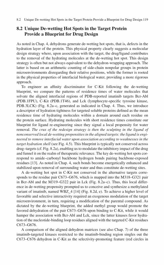
8.2 Unique De-wetting Hot Spots in the Target Protein Provide a Blueprint for Drug Design 119
8.2 Unique De-wetting Hot Spots in the Target ProteinProvide a Blueprint for Drug Design
As noted in Chap. 4, dehydrons generate de-wetting hot spots, that is, defects in thehydration layer of the protein. This physical property clearly suggests a moleculardesign strategy where, upon association with the target, the drug/ligand contributesto the removal of the hydrating molecules at the de-wetting hot spot. This designstrategy is often but not always equivalent to the dehydron-wrapping approach. Thelatter is based on an arithmetic counting of side-chain nonpolar groups in proteinmicroenvironments disregarding their relative positions, while the former is rootedin the physical properties of interfacial biological water, providing a more rigorousapproach.
To engineer an affinity discriminator for C-Kit following the de-wettingblueprint, we compare the patterns of residence times of water molecules thatsolvate the aligned interfacial regions of PDB-reported imatinib targets Bcr-Abl(PDB.1FPU), C-Kit (PDB.1T46), and Lck (lymphocyte-specific tyrosine kinase,PDB.3LCK) (Fig. 8.2a–c, generated as indicated in Chap. 4. Thus, we introducea descriptor of hydration tightness for targeted soluble proteins defined as the meanresidence time of hydrating molecules within a domain around each residue onthe protein surface. Hydrating molecules with short residence times constitute ourblueprint for ligand re-engineering since they signal a local propensity for waterremoval. The crux of the redesign strategy is then the sculpting in the ligand ofnonconserved local de-wetting propensities in the aligned targets: the ligand is engi-neered to remove interfacial water upon association according to weaknesses in thetarget hydration shell (see Fig. 4.5). This blueprint is typically not conserved acrossdrug–targets (cf. Fig. 8.2a), enabling us to modulate the inhibitory impact of the drugand funnel it on the realm of therapeutic relevance. The key de-wetting hot spots cor-respond to amide–carbonyl backbone hydrogen bonds pairing backbone-exposedresidues [13]. As noted in Chap. 4, such bonds become energetically enhanced andstabilized upon removal of surrounding water and thus constitute de-wetting sites.
A de-wetting hot spot in C-Kit not conserved in the alternative targets corre-sponds to the residue pair C673–G676, which is mapped into the M318–G321 pairin Bcr-Abl and the M319–G322 pair in Lck (Fig. 8.2a–c). Thus, this local differ-ence in de-wetting propensity prompted us to conceive and synthesize a methylatedvariant of imatinib, named WBZ_4 [14] (Fig. 8.2d, e). To achieve a higher level offavorable and selective interactivity required an exogenous modulation of the targetmicroenvironment, in turn, requiring a modification of the parental compound. Asdictated by the de-wetting blueprint, the added methyl group would promote thefavored dehydration of the pair C673–G676 upon binding to C-Kit, while it wouldhamper the association with Bcr-Abl and Lck, since the latter kinases favor hydra-tion of the nucleotide-binding loop residues aligned with the targeted C-Kit residuesC673–G676.
A comparison of the aligned dehydron matrices (see also Chap. 7) of the threeimatinib-targeted kinases restricted to the imatinib-binding region singles out theC673–C676 dehydron in C-Kit as the selectivity-promoting feature (red circles in

120 8 Re-engineering an Anticancer Drug to Make It Safer
Fig. 8.2 (a) De-wetting propensities of C-Kit residues in contact with imatinib (PDB.1T46, greenbars) and of aligned residues in Bcr-Abl kinase (PDB.1FPU, red bars) and Lck (PDB.3LCK,black bars). Residue i is in contact with the ligand if an atom of the latter lies within its domainD(i). The de-wetting propensity is quantified by the mean lifetime of solvating water molecules.Error bars denote Gaussian dispersion over 5 MD (molecular dynamics) runs. (b) Pattern ofde-wetting hot spots that arise as backbone hydrogen-bonded residues in C-Kit kinase (ribbonbackbone representation in light blue and de-wetting hot spots in green), aligned with Bcr-Abl kinase (magenta backbone and de-wetting hot spots in red). The imatinib methylation siteleading to the expulsion of water from C-Kit C673–G676 de-wetting hot spot is highlighted (yel-low rectangle). Imatinib is thus modified to favorably expel interfacial water molecules fromthe C-Kit microenvironment, a feature not conserved in Bcr-Abl. Hydrogen bonds are repre-sented as segments joining α-carbons of the paired residues and those lacking the propensityfor dehydration are shown as light gray segments. Residues from C-Kit chain are labeled ingreen and those from Bcr-Abl are labeled in white. (c) Differences in de-wetting hot spotsupon alignment of C-Kit (light blue backbone and hot spots in green) and Lck kinase (goldbackbone and black hot spots). Labels for Lck residues are shown in white. (d) Prototypemolecule WBZ_4 (N-{5-[4-(4-methyl piperazine methyl)-benzoylamido]-2-methylphenyl}-4-[3-(4-methyl)-pyridyl]-2-pyrimidine amine). The added methyl group is indicated in red. (e) Totalsynthesis of WBZ_4 [14]. The wrapping modification (methylation) of imatinib is indicated bythe thick arrow. (f–h) Dehydron matrices of Kit, Abl, and Lck kinases, respectively, derived fromPDB entries 1T46, 1FPU, and 3LCK and restricted to the residues in contact with imatinib. Rowsand columns are labeled by residue numbers, well-wrapped bonds are displayed in gray, and dehy-drons are shown in green, red, and black for Kit, Abl, and Lck, respectively. The entry denotinga selectivity-promoting feature for the wrapping design is circled in red. (a–e) reprinted from the[14], copyright 2007 with permission from the American Society for Clinical Investigation
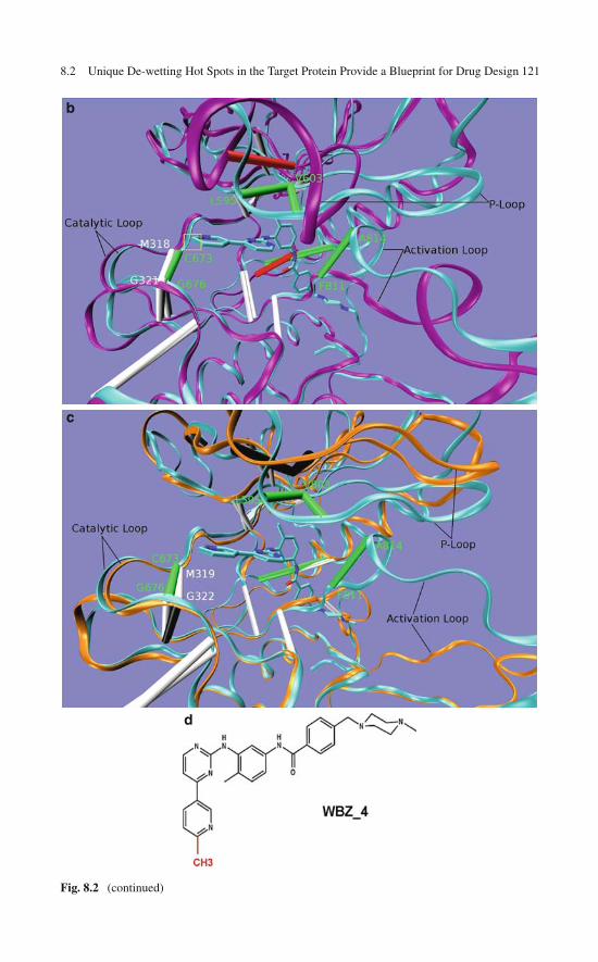
8.2 Unique De-wetting Hot Spots in the Target Protein Provide a Blueprint for Drug Design 121
b
c
d
Fig. 8.2 (continued)

122 8 Re-engineering an Anticancer Drug to Make It Safer
e
f
Fig. 8.2 (continued)
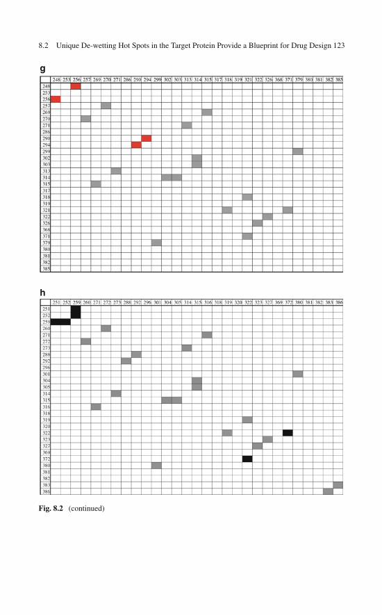
8.2 Unique De-wetting Hot Spots in the Target Protein Provide a Blueprint for Drug Design 123
g
h
Fig. 8.2 (continued)

124 8 Re-engineering an Anticancer Drug to Make It Safer
a
b
Fig. 8.3 (a) Aligned de-wetting patterns for C-Kit kinase (green) and JNK1 (blue) restricted to theC-Kit residues in contact with imatinib. (b) De-wetting hot spots arising as backbone hydrogen-bonded residues in C-Kit kinase (backbone in light blue and de-wetting hot spots in green), alignedwith JNK (blue backbone and de-wetting hot spots in yellow). The JNK residues M111 and N114,aligned with the de-wetting hot spot C673–G676 in C-Kit, are not paired by a hydrogen bond. Yet,M111 is a de-wetting hot spot for JNK (a), hence a harnessing spot for the designed imatininbmodification. Reprinted from the [14], copyright 2007 with permission from the American Societyfor Clinical Investigation

8.4 High-Throughput Screening: Test-Tube Validation of the Engineered Specificity 125
Fig. 8.2f–h), in good agreement with the de-wetting analysis. The dehydron matrixis precisely the representational tool that enables the simplest and fastest in silicoassessment of drug cross-reactivity across the entire human kinome, as described inChap. 9.
Reported assays on imatinib-promoted cardiomyocyte damage suggest that JNK(JNK1) inhibition may be beneficial to reduce the induced collapse of mitochon-drial membrane potential and preserve sarcolemmal integrity [15]. For this reason,we tested the binding of WBZ_4 as it anchors on the de-wetting pattern of JNK1(PDB.2G01). This analysis required alignment of JNK1 with the imatinib–C-Kitcomplex (PDB.1T46) (Fig. 8.3a, b). The high de-wetting propensity at JNK1 residueM111, the residue that aligns with C673 in C-Kit, instills confidence in the affinityof WBZ_4 for JNK.
8.3 In Silico Assays of the Water-Displacing Efficacyof a Wrapping Drug
As described in Chap. 4, molecular dynamics of the target-wetting patterns hasproven to be a useful tool to assess the efficacy of drugs or ligands in displacinginterfacial water from the target. In this regard, we have probed WBZ_4 in silicoby monitoring the spatio-temporal effect of the specific methylation on the kinase–inhibitor association through molecular dynamics (Fig. 8.4a, b). As C-Kit associateswith imatinib, the loop facing the para-position on the terminal ring (Figs. 8.2band 8.4a) was found to be unstable. In all five simulation trajectories, the back-bone hydrogen bond between Cys673 and Gly676 was irreversibly replaced bywater-mediated interactions within 1 ns (Fig. 8.4a). When WBZ_4 replaced ima-tinib within the C-Kit complex, the same loop was found to be stabilized due tothe improved dehydration of the hydrogen bond by the added methyl group onthe ligand (Fig. 8.4b). Thus, for C-Kit, WBZ_4 introduces favorable interactionsengaging the solvent-exposed pre-formed hydrogen bond and consequently stabi-lizes the inhibitor binding. The effect of WBZ_4 is the opposite on Bcr-Abl. Thesimulations demonstrate that in this case, the imatinib complex has a better stabilityin the same loop region, while WBZ_4 significantly destabilizes the loop. This isdue to the fact that the Met318–Gly321 backbone hydrogen bond on the Bcr-Ablloop is well dehydrated intramolecularly and positioned closer to imatinib; thus thisbond becomes well protected [17] by imatinib and the addition of the methyl groupclashes sterically with the loop. Thus, molecular dynamics provides a rationale forthe discriminatory power of WBZ_4 relative to the parent drug.
8.4 High-Throughput Screening: Test-Tube Validationof the Engineered Specificity
Screening methodologies for kinase inhibitors have been revolutionized in theirbreadth with the advent of bacteriophage libraries of kinase display [6], enabling

126 8 Re-engineering an Anticancer Drug to Make It Safer
Fig. 8.4 (a) Snapshot of the molecular dynamics simulation for C-Kit kinase bound to imatinib at1 ns. The main chain hydrogen bond between Cys673 and Gly676 is competitively and irreversiblyreplaced by hydrogen bonding to a water molecule, revealing the instability of the intramolecu-lar interaction. (b) Snapshot of the simulation of C-Kit kinase in complex with WBZ_4 at 1 ns.The main chain hydrogen bond between Cys673 and Gly676 is stabilized by the water expulsionpromoted by the added methyl on the inhibitor. Reprinted from the [14], copyright 2007 withpermission from the American Society for Clinical Investigation
a huge amplification of the affinity signal of the test compound. With panels nowcovering 442 of the 519 kinases that comprise the human kinome, this screeningmethodology has become the benchmark of high-throughput assays in the test tube.Thus, kinases are expressed on the capside of bacteriophages and this display con-struct is subject to a competitive binding assay. In the assay, the test compound andthe kinase display construct are in solution and the high-affinity and promiscuous
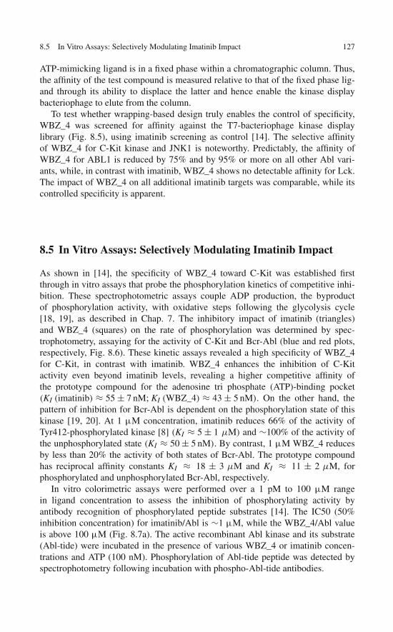
8.5 In Vitro Assays: Selectively Modulating Imatinib Impact 127
ATP-mimicking ligand is in a fixed phase within a chromatographic column. Thus,the affinity of the test compound is measured relative to that of the fixed phase lig-and through its ability to displace the latter and hence enable the kinase displaybacteriophage to elute from the column.
To test whether wrapping-based design truly enables the control of specificity,WBZ_4 was screened for affinity against the T7-bacteriophage kinase displaylibrary (Fig. 8.5), using imatinib screening as control [14]. The selective affinityof WBZ_4 for C-Kit kinase and JNK1 is noteworthy. Predictably, the affinity ofWBZ_4 for ABL1 is reduced by 75% and by 95% or more on all other Abl vari-ants, while, in contrast with imatinib, WBZ_4 shows no detectable affinity for Lck.The impact of WBZ_4 on all additional imatinib targets was comparable, while itscontrolled specificity is apparent.
8.5 In Vitro Assays: Selectively Modulating Imatinib Impact
As shown in [14], the specificity of WBZ_4 toward C-Kit was established firstthrough in vitro assays that probe the phosphorylation kinetics of competitive inhi-bition. These spectrophotometric assays couple ADP production, the byproductof phosphorylation activity, with oxidative steps following the glycolysis cycle[18, 19], as described in Chap. 7. The inhibitory impact of imatinib (triangles)and WBZ_4 (squares) on the rate of phosphorylation was determined by spec-trophotometry, assaying for the activity of C-Kit and Bcr-Abl (blue and red plots,respectively, Fig. 8.6). These kinetic assays revealed a high specificity of WBZ_4for C-Kit, in contrast with imatinib. WBZ_4 enhances the inhibition of C-Kitactivity even beyond imatinib levels, revealing a higher competitive affinity ofthe prototype compound for the adenosine tri phosphate (ATP)-binding pocket(KI (imatinib) ≈ 55 ± 7 nM; KI (WBZ_4) ≈ 43 ± 5 nM). On the other hand, thepattern of inhibition for Bcr-Abl is dependent on the phosphorylation state of thiskinase [19, 20]. At 1 μM concentration, imatinib reduces 66% of the activity ofTyr412-phosphorylated kinase [8] (KI ≈ 5 ± 1 μM) and ∼100% of the activity ofthe unphosphorylated state (KI ≈ 50 ± 5 nM). By contrast, 1 μM WBZ_4 reducesby less than 20% the activity of both states of Bcr-Abl. The prototype compoundhas reciprocal affinity constants KI ≈ 18 ± 3 μM and KI ≈ 11 ± 2 μM, forphosphorylated and unphosphorylated Bcr-Abl, respectively.
In vitro colorimetric assays were performed over a 1 pM to 100 μM rangein ligand concentration to assess the inhibition of phosphorylating activity byantibody recognition of phosphorylated peptide substrates [14]. The IC50 (50%inhibition concentration) for imatinib/Abl is ∼1 μM, while the WBZ_4/Abl valueis above 100 μM (Fig. 8.7a). The active recombinant Abl kinase and its substrate(Abl-tide) were incubated in the presence of various WBZ_4 or imatinib concen-trations and ATP (100 nM). Phosphorylation of Abl-tide peptide was detected byspectrophotometry following incubation with phospho-Abl-tide antibodies.
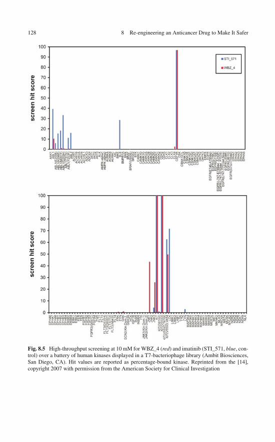
128 8 Re-engineering an Anticancer Drug to Make It Safer
Fig. 8.5 High-throughput screening at 10 mM for WBZ_4 (red) and imatinib (STI_571, blue, con-trol) over a battery of human kinases displayed in a T7-bacteriophage library (Ambit Biosciences,San Diego, CA). Hit values are reported as percentage-bound kinase. Reprinted from the [14],copyright 2007 with permission from the American Society for Clinical Investigation

8.5 In Vitro Assays: Selectively Modulating Imatinib Impact 129
Fig. 8.5 (continued)
Fig. 8.6 Kinetic inhibitory impact of compounds WBZ_4 and imatinib determined by measuringphosphorylation rates through spectrophotometric assays of C-Kit and Bcr-Abl kinase activ-ity. The kinases are inhibited by WBZ_4 (squares) and by the parental compound (triangles).Phosphorylation rate plots are given for Bcr-Abl (red) and C-Kit (blue). The open red symbols cor-respond to inhibition of unphosphorylated Bcr-Abl, while the full red symbols correspond to theTyr412-phosphorylated form. Error bars represent dispersion over five runs for each kinetic assay.Reprinted from the [14], copyright 2007 with permission from the American Society for ClinicalInvestigation
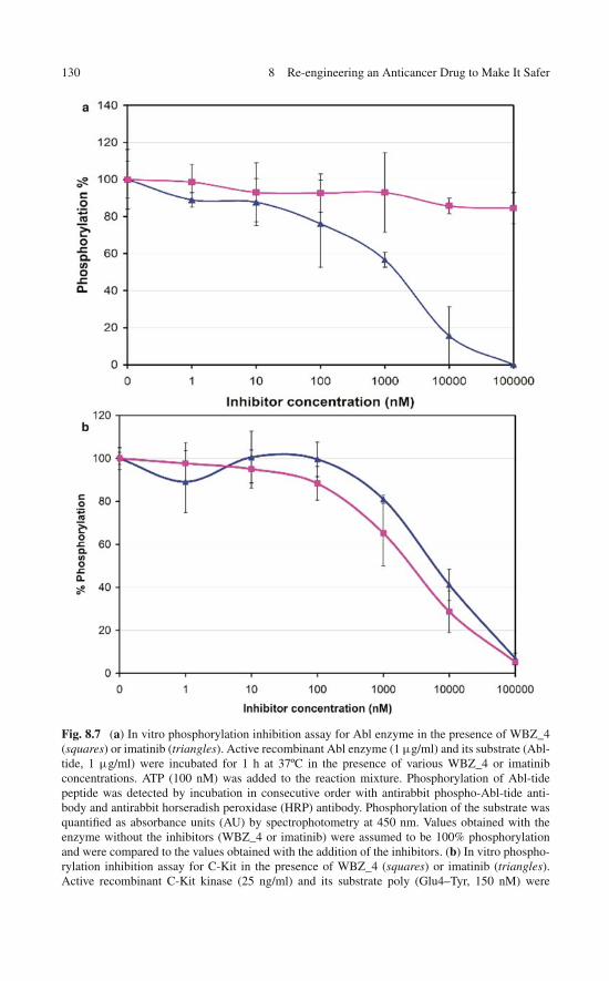
130 8 Re-engineering an Anticancer Drug to Make It Safer
Fig. 8.7 (a) In vitro phosphorylation inhibition assay for Abl enzyme in the presence of WBZ_4(squares) or imatinib (triangles). Active recombinant Abl enzyme (1 μg/ml) and its substrate (Abl-tide, 1 μg/ml) were incubated for 1 h at 37ºC in the presence of various WBZ_4 or imatinibconcentrations. ATP (100 nM) was added to the reaction mixture. Phosphorylation of Abl-tidepeptide was detected by incubation in consecutive order with antirabbit phospho-Abl-tide anti-body and antirabbit horseradish peroxidase (HRP) antibody. Phosphorylation of the substrate wasquantified as absorbance units (AU) by spectrophotometry at 450 nm. Values obtained with theenzyme without the inhibitors (WBZ_4 or imatinib) were assumed to be 100% phosphorylationand were compared to the values obtained with the addition of the inhibitors. (b) In vitro phospho-rylation inhibition assay for C-Kit in the presence of WBZ_4 (squares) or imatinib (triangles).Active recombinant C-Kit kinase (25 ng/ml) and its substrate poly (Glu4–Tyr, 150 nM) were

8.6 In Vitro Assay of the Selective Anticancer Activity of the Wrapping Design 131
The specificity of the inhibitory impact on C-Kit is significantly enhanced asWBZ_4 substitutes imatinib (Fig. 8.7a, b), while C-Kit inhibition is 23 ± 12%greater for WBZ_4 (Fig. 8.7b). The impact of the wrapping prototype ligand onalternative imatinib targets, such as the platelet-dependent growth factor receptor(PDGFR) kinase, could not be modulated since such proteins are not reported inPDB and thus, no de-wetting pattern can be reliably identified (cf. high-throughputscreening of WBZ_4 versus imatinib in Fig. 8.5). On the other hand, as shown inChap. 7, the PDGFR kinase is a close paralog of the Kit kinase and hence it is largelyexpected that any Kit kinase inhibitor would be cross-reactive with the PDGFRkinase.
8.6 In Vitro Assay of the Selective Anticancer Activityof the Wrapping Design
The controlled inhibitory impact of wrapper prototype WBZ_4 was further testedthrough in vitro assays on cancer cell lines representative of CML and GIST [14].The initial tests entailed treating the GIST882 [12] cell line and the CML K562 [7]cell line with WBZ_4 and contrasting its inhibitory impact on cell proliferation andin-cell specificity with those associated with imatinib. Because of its relative insol-ubility, WBZ_4 was incorporated into liposomes to promote cellular delivery. Theproliferation of C-Kit-expressing GIST882 cells treated with WBZ_4 was signifi-cantly inhibited in a quantitative dose-dependent manner similar to imatinib, withmaximum impact at 1 μM bulk concentration (Fig. 8.8a). By contrast, the inhibitoryimpact of WBZ_4 on CML K562 cells at the same bulk physiological dose of 1 μMis almost negligible (cell proliferation less than 10% lower than the proliferation ofuntreated cells), while 1 μM imatinib promotes a decrease in cell proliferation ofapproximately 66% (Fig. 8.8b). These results demonstrate the higher specificity inanticancer activity of WBZ_4.
A western blot assay on treated cancer-derived cell lines (Fig. 8.9a, b) was per-formed to determine the in-cell specificity of WBZ_4. The immunoblots revealedspecificity toward C-Kit consistent with the selective anticancer activity on theGIST882 cell line that expresses C-Kit. Thus, the activating phosphorylation ofC-Kit at sites Tyr703 and Tyr721 in ST882 cells is inhibited by WBZ_4 in adose-sensitive manner similar to imatinib (Fig. 8.9a). By contrast, phosphorylationof Bcr-Abl at Tyr 412 (22) in K562 cells was not significantly inhibited (<15%)by WBZ_4, while densitometry revealed an imatinib-induced inhibition of ∼85%(Fig. 8.9b).
�
Fig. 8.7 (continued) incubated for 1 h at 37ºC in the presence of various WBZ_4 or imatinibconcentrations. ATP (100 nM) was added to the reaction mixture. Phosphorylation of poly (Glu4–Tyr) peptide was detected by incubation in consecutive order with antiphosphotyrosine antibodyand antirabbit horseradish peroxidase (HRP) antibody. Reprinted from the [14], copyright 2007with permission from the American Society for Clinical Investigation

132 8 Re-engineering an Anticancer Drug to Make It Safer
Fig. 8.8 (a) Cell proliferation assay for GIST 882 cells. WBZ_4 inhibits cell proliferation of C-Kit-positive ST-882 cells. GIST cancer cells ST882 (GIST 882) were seeded in 96 wells plates ata density of 8 × 10³ cells per well. The cells were treated with various concentrations of WBZ_4(dark gray) and imatinib (light gray) for an additional 48 h. Cell proliferation is expressed as thepercentage of proliferating cells relative to untreated cells. (b) Cell proliferation assay for K562cells. WBZ_4 does not significantly inhibit cell proliferation of Bcr-Abl-expressing K562 cells.K562 cells were seeded in 96-well plates at a density of 1 × 10⁴ cells per well in 50 μl of medium.Two hours later, 50 μl medium containing different concentrations 0.01, 0.1, 1 μM of liposome-encapsulated WBZ_4 or soluble imatinib were added to the wells to reach a final volume of 100 μlper well. Following 48 h of exposure, the Alamar blue assay was performed. Plates were read atdual wavelength (570–595 nm) in an Elisa plate reader. Reprinted from the [14], copyright 2007with permission from the American Society for Clinical Investigation
Since imatinib is a micromolar inhibitor of phosphorylated Bcr-Abl (Fig. 8.6),the attack on CML cells at 1 μM bulk concentration (growth decrease 63%,Fig. 8.8b) must be attributed to the inhibition of phosphorylated Bcr-Abl (Fig. 8.9b)combined with the effective nanomolar inhibition of the unphosphorylated form(Fig. 8.6). By contrast, WBZ_4 hinders the phosphorylation of Bcr-Abl only par-tially (Fig. 8.9b) and in any case, it is an ineffective (micromolar) inhibitor of bothforms of Bcr-Abl (Fig. 8.6). Hence, its antitumor activity is predictably minimalon CML cells (Fig. 8.8b). In the case of GIST cells, comparable antitumor activityof both compounds (Fig. 8.8a) is likely to arise from comparable inhibitory impact(∼85%, Fig. 8.9a) on C-Kit phosphorylation, and the fact that both compounds arenanomolar-affinity inhibitors of C-Kit (Fig. 8.6).
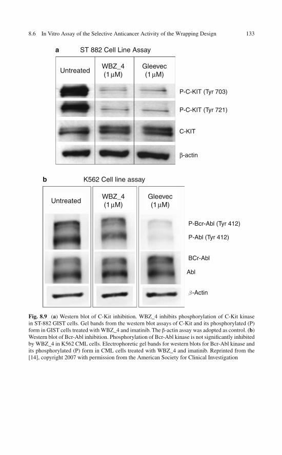
8.6 In Vitro Assay of the Selective Anticancer Activity of the Wrapping Design 133
a
P-C-KIT (Tyr 703)
ST 882 Cell Line Assay
UntreatedWBZ_4(1 μM)
Gleevec(1 μM)
P-C-KIT (Tyr 721)
C-KIT
β-actin
UntreatedWBZ_4(1 μM)
Gleevec(1 μM)
K562 Cell line assay
P-Bcr-Abl (Tyr 412)
P-Abl (Tyr 412)
BCr-Abl
Abl
β -Actin
b
Fig. 8.9 (a) Western blot of C-Kit inhibition. WBZ_4 inhibits phosphorylation of C-Kit kinasein ST-882 GIST cells. Gel bands from the western blot assays of C-Kit and its phosphorylated (P)form in GIST cells treated with WBZ_4 and imatinib. The β-actin assay was adopted as control. (b)Western blot of Bcr-Abl inhibition. Phosphorylation of Bcr-Abl kinase is not significantly inhibitedby WBZ_4 in K562 CML cells. Electrophoretic gel bands for western blots for Bcr-Abl kinase andits phosphorylated (P) form in CML cells treated with WBZ_4 and imatinib. Reprinted from the[14], copyright 2007 with permission from the American Society for Clinical Investigation

134 8 Re-engineering an Anticancer Drug to Make It Safer
8.7 Enhanced Safety of the Wrapping Redesign in AnimalModels of Gastrointestinal Stromal Tumor
To assay WBZ_4 for anticancer activity in vivo, an animal model for GIST growthbased on female C.B-17/IcrHsd-PrkdcSCID mice was developed de novo [14]. Themodel involved the subcutaneous injection of GIST882 cells [21]. The efficacy ofWBZ_4 was found to be comparable to that of imatinib, as determined by thedecrease in tumor volume and weight (Fig. 8.10a, b). No obvious toxicities wereobserved in the animals during treatment. Selectivity of WBZ_4 in the animalmodel was corroborated by assaying on the xenograft induced by CML cells K562(Fig. 8.10c). While imatinib is shown to significantly impair tumor growth (p <0.01), the prototype WBZ_4 has virtually no effect, in accord with its engineeredspecificity and hence lack of inhibitory impact on Bcr-Abl kinase.
Previous studies of imatinib-induced cardiotoxicity identified a protective JNK(JNK1) inhibition as a means to reduce the collapse of mitochondrial membrane
Fig. 8.10 Xenograft models of anticancer activity. (a) Effect of WBZ_4 or imatinib therapy onin vivo GIST growth determined by longitudinal tumor volume measurements. Mice were ran-domized to treatment with either control (normal PBS and empty liposomes give indistinguishableresults within experimental uncertainty), imatinib, or liposome-formulated WBZ_4. (b) Effect ofWBZ_4 or imatinib therapy on in vivo GIST growth determined by weight measurements. Animalsfrom all groups were sacrificed after 6 weeks of therapy, tumors were excised, and the weightwas recorded. (c) Effect of WBZ_4 or imatinib therapy on in vivo CML growth induced througha xenograft of K562 tumor cells, determined by longitudinal tumor volume measurement. TheWBZ_4 selectivity is hereby corroborated in vivo. Reprinted from the [14], copyright 2007 withpermission from the American Society for Clinical Investigation

8.7 Enhanced Safety of the Wrapping Redesign in Animal Models 135
Fig. 8.10 (continued)

136 8 Re-engineering an Anticancer Drug to Make It Safer
potential [15]. Accordingly, WBZ_4 was engineered to also inhibit JNK, a fea-ture preliminarily corroborated in vitro (Fig. 8.5). This protective role of WBZ_4is also validated in vivo. Western blotting of cell extracts derived from the sameimatinib- and WBZ_4-treated cardiomyocytes (Fig. 8.11a) revealed that both drugsincreased the phosphorylation state of ERK1/2 and p38MAPK as compared tountreated cultures. In contrast, cells treated with WBZ_4 showed reduced levelsof JNK activation at each concentration tested, as compared to untreated cells, orcells treated with imatinib. These results confirm other observations [15] demon-strating that imatinib induces the endoplasmic reticulum (ER) stress response andactivates JNKs in neonatal rat ventricular myocytes (NRVM). JNK activation hasbeen clearly associated with cardiomyocyte apoptosis via collapse of the mitochon-drial membrane potential and release of cytochrome c [22]. It was further confirmedthat imatinib (and WBZ_4) had little effect on p38MAPK activation at concentra-tions below 50 μM and actually increased ERK activation in these cultured cells.In contrast, WBZ_4 appeared to reduce levels of pJNK at each dose tested, sug-gesting that upstream activation of JNKs was inhibited by WBZ_4. Thus, a directinhibitory effect on JNK activity would predict that WBZ_4 should have reducedcardiotoxicity in vivo.
This expectation is supported by other observations using the surrogatemarker brain natriuretic peptide (BNP), a most sensitive indicator of myocardialhypertrophy and cardiac impairment [23]. The expected curbing of cardiotoxic-ity in WBZ_4 anticancer therapy was confirmed by examining the mRNA levelsof BNP in the left ventricle of mice from the same groups assayed for anticanceractivity. The BNP mRNA levels were about 58% higher in the ventricles fromimatinib-treated animals (Fig. 8.11b), while no significant difference was detected
�Fig. 8.11 Cardiotoxicity assays. (a) Western blot of JNK inhibition in cardiomyocytes. Westernblots were probed with primary antibodies specific for the phosphorylated forms of ERKs, JNKs,and p38MAPK. The position of molecular weight standards is indicated to the left of each blot. (b)Effect of WBZ_4 or imatinib therapy on mouse heart–brain natriuretic peptide (BNP). The mRNAlevels of BNP (a sensitive marker of myocardial hypertrophy and cardiac impairment) were exam-ined in the left ventricle of mice from the groups in Fig. 8.10a. The BNP mRNA levels were about58% higher in the ventricles from imatinib-treated animals (p = 0.02), but no significant differencewas noted in the WBZ_4-treated animals. (c) Comparison of left ventricular ejection fraction after6 weeks of control (groups treated with either PBS or empty liposomes), imatinib, or WBZ_4 ther-apy in mice. (d) Magnetic resonance imaging of cardiac function. Assessment of left ventricularfunction (red circle, lower cavity) was performed as follows: representative in vivo axial imagesof the left ventricle in diastole and systole of control and test mice were acquired in a 4.7 T MRscanner cardiac gating using a magnetization-prepared spoiled gradient echo sequence. To assesscardiac function, short axis cardiac cine images were acquired using a magnetization-prepared,cardiac-gated spoiled gradient echo sequence (TE/TR 2.1 ms/∼23.5 ms; 16 phases covering 1.5R–R cycles; in-plane resolution 312 μm × 312 μm; 1.25 mm slice thickness). For assessing EF,regions of interest encompassing the cavity of the left ventricle were drawn. For each animal, theaverage region of interest of four central slices in systole was divided by the average region ofinterest of four central slices in diastole taken at the same location in the left ventricle to derive theEF. (a–c) reprinted from the [14], copyright 2007 with permission from the American Society forClinical Investigation
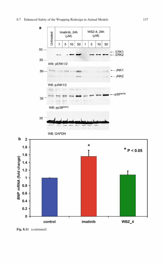
8.7 Enhanced Safety of the Wrapping Redesign in Animal Models 137
Fig. 8.11 (continued)

138 8 Re-engineering an Anticancer Drug to Make It Safer
Fig. 8.11 (continued)
References 139
in the WBZ_4-treated animals when compared with untreated mice. Finally, thereduced cardiotoxicity of WBZ_4 was directly tested in mice by determination ofthe percentage ejection fraction (EF) in the left ventricle by magnetic resonance(MR) imaging of treated mice subject to imatinib or WBZ_4 therapy (Fig. 8.11c, d).Following 6 weeks of therapy, the cardiac EF was significantly lower in the imatinibgroup compared to controls (p = 0.02), which is consistent with previous findings[15]. Remarkably, WBZ_4 treatment had no effect on cardiac EF despite prolongedtherapy (Fig. 8.11c, d).
8.8 Controlled Specificity Engineered Through RationalDesign: Concluding Remarks
The top-down approach presented in this chapter is centered in a rational ligandre-design sculpting target-specific molecular features. The approach harnesses fun-damental concepts pertaining to the discrimination of proteins with a commonancestry, sketched in Chaps. 4 and 6. These discriminatory features are associatedwith unique dehydration or de-wetting hot spots found on the protein/water inter-face of the purported target. The de-wetting propensities are in turn promoted bydehydrons in the protein structure, and hence the hydration blueprint of the tar-get becomes a design tool complementing the wrapping concept. In this particularillustration, the re-design had three goals: (a) to re-focus the primary impact onC-Kit kinase; (b) to reduce the inhibitory impact on Bcr-Abl kinase; and (c) to pro-mote JNK inhibition in order to reduce cardiotoxicity. These three goals have beenachieved [14], as attested by the in vitro and in vivo results described in this chap-ter, clearly revealing that drug specificity can be rationally controlled. The imatinibre-design turned the wrapping ligand into a selective proto-therapeutic agent to treatGIST with a prima facie reduction of cardiotoxicity.
References
1. Dancey J, Sausville EA (2003) Issues and progress with protein kinase inhibitors for cancertreatment. Nat Rev Drug Discov 2:296–313
2. Levitski A, Gazit A (1995) Tyrosine kinase inhibition: An approach to drug development.Science 267:1782–1788
3. Tibes R, Trent J, Kurzrock R (2005) Tyrosine kinase inhibitors and the dawn of molecularcancer therapeutics. Annu Rev Pharmacol Toxicol 45:357–84
4. Gibbs J, Oliff A (1994) Pharmaceutical research in molecular oncology. Cell 79:193–198
5. Donato NJ, Talpaz M (2000) Clinical use of tyrosine kinase inhibitors: Therapy for chronicmyelogenous leukemia and other cancers. Clin Cancer Res 6:2965–2966
6. Fabian MA, Biggs WH, Treiber DK et al (2005) A small molecule kinase interaction map forclinical kinase inhibitors. Nat Biotechnol 23:329–336
7. Gambacorti-Passerini C, le Coutre P, Mologni L et al (1997) Inhibition of the ABL kinaseactivity blocks the proliferation of BCR/ABL+ leukemic cells and induces apoptosis. BloodCells Mol Dis 23:380–394
140 8 Re-engineering an Anticancer Drug to Make It Safer
8. Schindler T, Bornmann W, Pellicena P et al (2000) Structural mechanism for STI-571inhibition of Abelson tyrosine kinase. Science 289:1938–1942
9. Attoub S, Rivat C, Rodrigues S et al (2002) The C-kit tyrosine kinase inhibitor STI-571 forcolorectal cancer therapy. Cancer Res 62:4879–4883
10. DeMatteo RP (2002) The GIST of targeted cancer therapy: A tumor (gastrointestinal stro-mal tumor), a mutated gene (c-kit), and a molecular inhibitor (STI571). Ann Surg Oncol 9:831–839
11. Skene RJ, Kraus ML, Scheibe DN et al (2004) Structural basis for autoinhibition and STI-571inhibition of C-kit tyrosine kinase. J Biol Chem 279:31655–31663
12. Tuveson DA, Willis NA, Jacks T et al (2001) STI571 inactivation of the gastrointestinal stro-mal tumor C-kit oncoprotein: Biological and clinical implications. Oncogene 20:5054–5058
13. Chen JP, Zhang X, Fernández A (2007) Molecular basis for specificity in the druggablekinome: Sequence-based analysis. Bioinformatics 23:563–572
14. Fernández A, Sanguino A, Peng Z et al (2007) An anticancer C-kit kinase inhibitor is re-engineered to make it more active and less cardiotoxic. J Clin Invest 117:4044–4054
15. Kerkela R, Grazette L, Yacobi R et al (2006) Cardiotoxicity of the cancer therapeutic agentimatinib mesylate. Nat Med 12:908–916
16. Druker BJ (2004) Molecularly targeted therapy: Have the floodgates opened? Oncologist9:357–360
17. Fernández A, Scheraga HA (2003) Insufficiently dehydrated hydrogen bonds as determinantsof protein interactions. Proc Natl Acad Sci USA 100:113–118
18. Barker S, Kassel D, Weigl D et al (1995) Characterization of pp60c-src tyrosine kinaseactivities using a continuous assay: Autoactivation of the enzyme is an intermolecularautophosphorylation process. Biochemistry 34:14843–14851
19. Songyang Z, Carraway KL, Eck M et al (1995) Catalytic specificity of protein-tyrosine kinasesis critical for selective signalling. Nature 373:536–539
20. Clarkson B, Strife A, Wisniewski D, Lambek CL, Liu C (2003) Chronic myelogenousleukemia as a paradigm of early cancer and possible curative strategy. Leukemia 17:1211–1262
21. Prenen H, Deroose C, Vermaelen P, Sciot R, Debiec-Rychter M (2006) Establishment of amouse gastrointestinal stromal tumour model and evaluation of response to Imatinib by smallanimal positron emission tomography. Anticancer Res 26:1247–1252
22. Baines CP, Molkentin JD (2005) Stress signaling pathways that modulate cardiac myocyteapoptosis. J Mol Cell Cardiol 38:47–62
23. Scheuermann-Freestone M, Simon Freestone N, Langenickel T et al (2001) A new modelof congestive heart failure in the mouse due to chronic volume overload. Eur J Heart Fail3:535–543
Chapter 9Wrapping Patterns as Universal Markersfor Specificity in the Therapeutic Interferencewith Signaling Pathways
Chapter 8 illustrated the power of the wrapping concept as a guidance to engineerspecificity and enhance safety in a kinase inhibitor. Yet, only a universal selectivityfilter, applicable to the entire human kinome – even to kinases with unreported struc-ture – would be truly useful for the drug designer. This chapter thematically belongsto the bioinformatics realm and addresses this issue at the broadest possible level.The surveyed findings reveal that targeting the polar dehydration patterns definedby protein dehydrons heralds a new generation of drugs that enable a tighter con-trol of specificity. The universality of this selectivity filter in the field of therapeuticinterference with cell signaling is thus established.
9.1 The Need for a Universal Selectivity Filter for RationallyDesigned Kinase Inhibitors
As indicated in Chaps. 7 and 8, kinase targeting is a central theme of molecularcancer therapy [1–5]. Be as it may, a structural basis for the rational design of kinaseinhibitors appears to be sorely absent [1, 3]. In practice, most leads and scaffoldsfor drug development are actually discovered through screening techniques and aregenerally transformed into therapeutic tools through a painstaking trial-and-errorprocess that often renders a final product plagued with clinical uncertainties.
While the paradigm of target specificity may be shifting to controlled multi-target drugs [3], the structural factors determining cross-reactivities are not yet fullyunderstood, in spite of some progress. For instance, the solvent-accessible nonpo-lar surface on the surface of a protein, frequently invoked as a major determinantof protein associations [6, 7], actually fosters promiscuity in the context of kinaseinhibition [3, 8]. This is so because nonpolar groups on the surface of proteins tendto be highly conserved across paralog proteins and even within the entire kinasesuperfamily [9, 10]. Progress in controlling specificity is also hampered by the lackof structural information. For instance, only about 22% of the human kinome isrepresented in the PDB.
In this chapter we focus on assessing the universality of wrapping as the molec-ular feature controlling nonpromiscuous drug targeting. To fulfill this objective
141A. Fernández, Transformative Concepts for Drug Design: Target Wrapping,DOI 10.1007/978-3-642-11792-3_9, C© Springer-Verlag Berlin Heidelberg 2010
142 9 Wrapping Patterns as Universal Markers
an extensive bioinformatics exploration of drug profiles is required. Recent high-throughput screenings of commercially available kinase inhibitors lead us to anoperational definition of promiscuity as a significant cross-reactivity (dissociationconstant Kd < 100 nM) extended over >30% of the sampled kinome. The problemis complicated due to the scarcity of affinity-profiled kinases with reported structure:To date, only about 24–25% of the human kinome (123 out of the ∼519 discov-ered protein kinases) is reported in the PDB. Furthermore, kinase homology modelsare quite unreliable to make inferences on such detailed features as wrapping pat-terns since the level of pairwise sequence identity across the kinase superfamily istypically low (<30%) [11].
To mitigate these problems, we take advantage of the high degree of topologicalconservation of the kinase folds, a known consequence of their common ancestry.Thus, reliable sequence-based attributes such as disorder propensity [12] are heremapped onto structurally threaded models to make inferences about dehydron pat-terns and ultimately about drug specificity/promiscuity patterns. As described inChap. 5, these cross-validations of dehydron inferences in proteins with unreportedstructure take advantage of the crucial link between disorder propensity and thedegree of exposure of the protein backbone in the native fold [13]. The latter isitself a measure of the propensity of backbone hydration, the main factor competingwith the hydrophobic collapse of the polypeptide chain.
Thus we seek a sequence-based attribute that enables a classification of kinasespace in a way that accurately reproduces similarities/differences in affinity profil-ing against a background of drugs [14, 15]. The classification introduces a partitioninto disjoint sets of kinases within which pharmacological differences are accuratelypredicted from molecular differences. This methodology could be implementedbecause the relevant classification of kinases was performed by comparing theregions deprived of adequate packing or intramolecular dehydration, i.e., the flexi-ble regions, which are precisely the markers differentiating kinases at the structurallevel [4].
The chapter is organized as follows: First, we determine the type of kinase molec-ular similarity that promotes promiscuity whenever targeted. Thus, we demonstratethat solvent-exposed nonpolar regions promoting ligand associations foster promis-cuity. To draw statistics at a kinomic scale, we formally define a region, the nonpolarhull, enabling comparison of targeted exposed nonpolar regions across differentkinases. Then, we examine other targeted features in molecular design aimed atfostering pairwise interactions between ligand and kinase and show that the highdegree of conservation of the partner groups on the protein surface does not enablea suitable control of specificity. Subsequently, we introduce a means of calculatingdehydration propensities on polar-paired regions on the protein surface and formallydefine a region, the so-called environmental (wrapping) hull, enabling a comparisonof polar dehydration propensities across targets. We demonstrate that this molecu-lar attribute is responsible for controlling specificity. To carry out the analysis at asequence level, a technique named environmental alignment is implemented. Thistechnique enables the identification of residues whose microenvironments are likelyto be perturbed by ligand association. Such residues are identified by aligning the
9.2 Computational Tool Box for Comparative Analysis of Molecular Attributes 143
kinase sequence against a background of sequences of homologous kinase–ligandcomplexes reported in the PDB.
9.2 Computational Tool Box for Comparative Analysisof Molecular Attributes Across the Human Kinome
9.2.1 Wrapping Inferences on Proteins with Unreported Structure
To establish the universal validity of wrapping as a selectivity filter, the comparativeanalysis of molecular attributes in target paralogs cannot be constrained to PDB-reported kinases (32 out of the 119 assayed in [14]). Homology-based predictions ofnative structures [16, 17] become plausible given the extent of PDB representation(∼24%) of paralogs within the kinase superfamily and also because dehydrons maybe directly inferred from sequence (Chap. 5, [13]) and ultimately contrasted with thehomology-based predictions for mutual validation. Thus, the disorder-based predic-tion of dehydrons hinges on the fact that native disorder arises essentially from theimpossibility to dehydrate intramolecular hydrogen bonds, as noted in Chap. 5. Thechoice of disorder score to benchmark homology predictions is an obvious one giventhe particularly low accuracy of the latter on loopy or flexible regions [10].
The disorder propensity is given by a score determined by the programPONDR R© (predictor of native disorder), a neural-network predictor of native disor-der. Only 0.4% of more than 900 nonhomologous PDB proteins give false-positivepredictions in regions with 40 or more consecutive sites of predicted disorder. Eventhis 0.4% of false positives is an overestimation, as many disordered regions inmonomeric chains become ordered upon ligand binding or in crystal contacts [12].The false-negative error rate (∼11% for regions of 40 or more consecutive predictedordered residues) is also compelling in regard to the predictor quality.
The correlation between solvent exposure of hydrogen bonds and disorderpropensity implies that it is possible to predict dehydrons directly from sequence[13]: It suffices to determine the PONDR-generated pattern associated with thedesired feature. The correlation implies that the propensity to adopt a nativelydisordered state becomes pronounced for proteins which, due to a chain compo-sition reflecting high hydrophilicity, cannot protect the backbone hydrogen bondseven minimally. Thus, we can infer the existence of dehydrons from the PONDRscore (fd) with 92% accuracy in regions with fd > 0.35 provided such regions areflanked by well-protected regions (fd < 0.35), to ensure the existence of structure.The accuracy of this sequence-based dehydron predictor was established by infer-ring the location of dehydrons in proteins with reported structure, for which themicroenvironment of each hydrogen bond can be determined unambiguously [13].The false negatives constitute 368 of the 8,215 dehydrons in a PDB database of1,466 proteins free from structural redundancy and less than 25% sequence iden-tity in pairwise alignment. The false positives correspond to 2,721 of the 133,623backbone hydrogen bonds examined.
144 9 Wrapping Patterns as Universal Markers
9.2.2 Alignment of Targetable Molecular FeaturesAcross the Human Kinome
In order to assess differences in the exposed nonpolar regions of kinase targets thatinteract with different ligands we define a common region, named the nonpolar hull.A residue a is defined as making contact with a ligand L within a PDB-reportedcomplex if a side chain heavy atom (H excluded) is found to be within 3.6 Å (upperbound for any bond length) of a heavy atom in the ligand. The nonpolar hull forprotein chain i, Hnp(i), is dependent on a “structural background set” of chains,S(i), which includes all homolog chains that align with chain i [18] for which thereare protein–ligand complexes with reported structure. Thus, the nonpolar hull isdefined as Hnp (i) = ∪j∈S(i)�i
(Rnp (j)
), where �i (a), with a ∈ chain j, is the residue
in chain i that aligns with residue a in chain j, and Rnp(j) is the set of nonpolarresidues in chain j in contact with its respective ligand Lj. For any pair i, j, thefollowing relation holds: �i
(Hnp (j)
) = Hnp (i), enabling a comparison of kinasesby examining differences in nonpolar hulls.
To assess differences in the dehydration propensities of polar regions for pur-ported kinase targets, we introduce a common region for comparison namedenvironmental hull. First, the set Renv(j) is defined for protein chain j as the setof residues paired by dehydrons in chain j within a protein–ligand complex withreported structure subject to the following condition: ligand Lj contributes to thedehydration of the dehydron, that is, it has some carbonaceous nonpolar groupwithin the dehydration domain of the dehydron. Then, the set Eenv(j) is defined asthe set of residues from chain j that contribute to the dehydration microenvironmentof a dehydron contained in Renv(j), that is, they are either paired by the dehydron orthey contain a side chain nonpolar group within the microenvironment of the dehy-dron. Then, the environmental hull for chain j, Henv(j), is defined as the union of theresidues in chain j that align with residues framing the environments of dehydronsthat in turn are environmentally affected by ligands in PDB-reported complexes:Henv (j) = ∪i∈S(j)�j (Eenv (i)) (notation has been followed consistently, the struc-tural background S(j) is defined above). As with nonpolar hulls, for any pair i, j, thefollowing relation also holds: �i (Henv (j)) = Henv (i). This property is needed toactually compare environments of different proteins.
9.3 Is Wrapping Pharmacologically Relevant?A Bioinformatics Analysis
In this section we report on previous attempts to correlate different molecularattributes of the protein targets with available drug-screening data for a sizable setof kinase targets. Essentially, the scope of this section is to contrast differencesin the pharmacological profiles of kinases with structure-based dissimilarities thattake into account specific molecular attributes. The large drug/kinase panel experi-mentally assayed [14] and exploited in our analysis is highly underreported in the

9.3 Is Wrapping Pharmacologically Relevant? A Bioinformatics Analysis 145
PDB. Consequently, a reliable sequence-based predictor of the relevant molecularattributes of kinases had to be implemented as shown in the previous sections.
We first define a pharmacological distance, dphar, that quantifies differences inthe affinity profiling of kinases against a background of available drugs. This met-ric is the Euclidean distance between affinity vectors with entries given in negativelogarithmic or G/RT units (G = free energy change for protein–ligand associa-tion, R = universal gas constant, T = absolute temperature). A positive cutoff valueG/RT = ln10 ≈ 2.3 is adopted for affinities reported as “no hit” in the screening(Kd > 10 μM). Figure 9.1a displays the matrix Dphar = [
dphar (i, j)]
for all pairs(i, j) from the 119 assayed kinases. The affinity profiling adopted included 19 of the20 drugs originally screened [14]: only the promiscuous staurosporine was excludedsince it does not belong to the pharmacology realm.
Fig. 9.1 (a) Pharmacological distance matrix Dphar = [dphar (i, j)
]for all pairs (i, j) from the
119 kinases assayed through affinity profiling against a background of 19 drugs [14]:SB202190; SB203580; sp600125; imatinib (Gleevec); VX-745; BIRB 796; BAY-43-9006;GW-2016; gefitinib; erlotinib; CI-1033; EKB-569; ZD-6474; Vatalanib; SU11248; MLN-518;LY-333531; roscovitine/CYC202; and flavopiridol. The distance is given by dphar (i, j) =[∑
m∈inhibitors (K (i, m) − K (j, m))2]1/2, where K (i, m) , K (j, m) represent, respectively, the nega-
tive logarithm of equilibrium constants for complexation of kinase i and kinase j with drug inhibitorm. (b) Aligned backbones
(RMSD ≈ 0.33Å
)for paralog kinases PDK1 (blue) and CHK1 (lilac)
in their active folds. The structures were reported in complex with ligands BIM8 (PDB.1UVR)and 3A3 (PDB.2GCU), respectively. The nonpolar hulls are depicted in yellow. (c) Nonpolar dis-tance matrix Dnp = [
dnp (i, j)]
over the 119 assayed kinases. (d) Plot of nonpolar distance versuspharmacological distance. Each circle represents a kinase pair. No correlation is observed, whilethere is some bimodality in each dimension. (e) Correlation between pseudopharmacological dis-tance (including staurosporine in the drug-screening background) and nonpolar distance betweenkinases. The sole outliers are pairs involving the EGFR kinase, the kinase whose affinity vector isnot dominated by staurosporine [14]. Reprinted from [10] with permission from Oxford Universitypress

146 9 Wrapping Patterns as Universal Markers
C
Fig. 9.1 (continued)

9.3 Is Wrapping Pharmacologically Relevant? A Bioinformatics Analysis 147
Fig. 9.1 (continued)
To determine whether pharmacological differences are dictated by differencesin nonpolar accessible surfaces of the targets within ligand-binding sites, a nonpo-lar distance, dnp (i, j), between the affinity-assayed kinases i, j is introduced. Thednp (i, j) is determined by differences in accessible nonpolar surface areas of therespective nonpolar hulls, Hnp (i) , Hnp (j). As rigorously defined in the previous
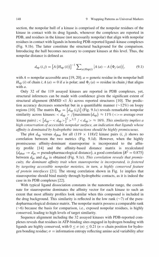
148 9 Wrapping Patterns as Universal Markers
section, the nonpolar hull of a kinase is comprised of the nonpolar residues of thekinase in contact with its drug ligands, whenever the complexes are reported inPDB, and residues in the kinase (not necessarily nonpolar) that align with nonpolarresidues in contact with ligands in homolog PDB-reported ligand–kinase complexes(Fig. 9.1b). The latter constitute the structural background for the comparison.Introducing the hull becomes necessary to compare kinases at this level. Thus, thenonpolar distance is defined as
dnp (i, j) = [A
(Hnp (i)
)]−1 ∑a∈Hnp(i)
∣∣A (a) − A(�j (a)
)∣∣, (9.1)
with A = nonpolar accessible area [19, 20]; a = generic residue in the nonpolar hullHnp (i) of chain i; A (a) = 0 if a is polar; and �j (a) = residue in chain j that alignswith a.
Only 32 of the 119 assayed kinases are reported in PDB complexes, yet,structural inferences can be made with confidence given the significant extent ofstructural alignment (RMSD <1 Å) across reported structures [10]. The predic-tion accuracy decreases somewhat but in a quantifiable manner (∼12%) on loopyregions [10]. The matrix Dnp = [
dnp (i,j)]
(Fig. 9.1c) reveals remarkable nonpolarsimilarity across kinases: < dnp > /
(maximum
(dnp
) ≈ 11% (<>= average over
kinase pairs) ;<[dnp−< dnp >
]2>1/2 /<dnp > ≈ 16%. This similarity implies a
high conservation of accessible nonpolar surface, an indication that ligands whoseaffinity is dominated by hydrophobic interactions should be highly promiscuous.
The plot dnp versus dphar for all (119 × 118)/2 kinase pairs (i, j) shows nocorrelation between the two metrics (Fig. 9.1d). However, when the highlypromiscuous affinity-dominant staurosporine is incorporated to the affin-ity profile [14] and the affinity-based distance matrix is recalculated(dphar → dps = pseudopharmacological distance
), a good correlation
(R2 = 0.875
)between dps and dnp is obtained (Fig. 9.1e). This correlation reveals that promis-cuity, the dominant affinity trait when staurosporine is incorporated, is fosteredby targeting accessible nonpolar moieties, in turn, a highly conserved featureof protein interfaces [21]. The strong correlation shown in Fig. 1e implies thatstaurosporine should bind mainly through hydrophobic contacts, as it is indeed thecase in its PDB complexes [22].
With typical ligand dissociation constants in the nanomolar range, the coordi-nate for staurosporine dominates the affinity vector for each kinase to such anextent that most affinity profiles look similar when this compound is included inthe drug background. This similarity is reflected in the low rank (∼7) of the pseu-dopharmacological distance matrix. The nonpolar matrix possess a comparable rank(∼6) because the basis for comparison, i.e., exposed nonpolar residues, is highlyconserved, leading to high levels of target similarity.
Sequence alignment including the 32 assayed kinases with PDB-reported com-plexes reveals that residues in ATP-binding sites engaged in hydrogen bonding withligands are highly conserved, with 0 ≤ σ (n) ≤ 0.21 (n = chain position for hydro-gen bonding residue; σ = information entropy reflecting amino acid variability after
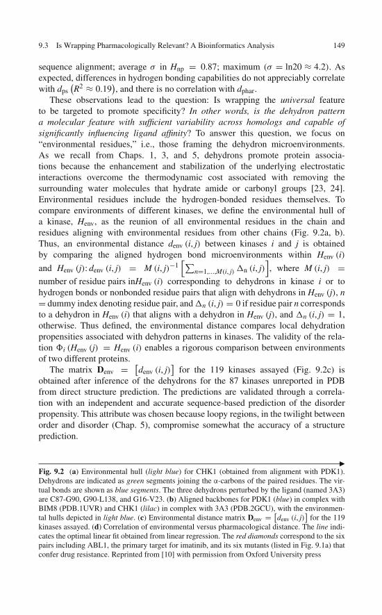
9.3 Is Wrapping Pharmacologically Relevant? A Bioinformatics Analysis 149
sequence alignment; average σ in Hnp = 0.87; maximum (σ = ln20 ≈ 4.2). Asexpected, differences in hydrogen bonding capabilities do not appreciably correlatewith dps
(R2 ≈ 0.19
), and there is no correlation with dphar.
These observations lead to the question: Is wrapping the universal featureto be targeted to promote specificity? In other words, is the dehydron patterna molecular feature with sufficient variability across homologs and capable ofsignificantly influencing ligand affinity? To answer this question, we focus on“environmental residues,” i.e., those framing the dehydron microenvironments.As we recall from Chaps. 1, 3, and 5, dehydrons promote protein associa-tions because the enhancement and stabilization of the underlying electrostaticinteractions overcome the thermodynamic cost associated with removing thesurrounding water molecules that hydrate amide or carbonyl groups [23, 24].Environmental residues include the hydrogen-bonded residues themselves. Tocompare environments of different kinases, we define the environmental hull ofa kinase, Henv, as the reunion of all environmental residues in the chain andresidues aligning with environmental residues from other chains (Fig. 9.2a, b).Thus, an environmental distance denv (i, j) between kinases i and j is obtainedby comparing the aligned hydrogen bond microenvironments within Henv (i)
and Henv (j): denv (i, j) = M (i, j)−1[∑
n=1,...,M(i, j) n (i, j)], where M (i, j) =
number of residue pairs inHenv (i) corresponding to dehydrons in kinase i or tohydrogen bonds or nonbonded residue pairs that align with dehydrons in Henv (j), n= dummy index denoting residue pair, and n (i, j) = 0 if residue pair n correspondsto a dehydron in Henv (i) that aligns with a dehydron in Henv (j), and n (i, j) = 1,otherwise. Thus defined, the environmental distance compares local dehydrationpropensities associated with dehydron patterns in kinases. The validity of the rela-tion �i (Henv (j) = Henv (i) enables a rigorous comparison between environmentsof two different proteins.
The matrix Denv = [denv (i, j)
]for the 119 kinases assayed (Fig. 9.2c) is
obtained after inference of the dehydrons for the 87 kinases unreported in PDBfrom direct structure prediction. The predictions are validated through a correla-tion with an independent and accurate sequence-based prediction of the disorderpropensity. This attribute was chosen because loopy regions, in the twilight betweenorder and disorder (Chap. 5), compromise somewhat the accuracy of a structureprediction.
�Fig. 9.2 (a) Environmental hull (light blue) for CHK1 (obtained from alignment with PDK1).Dehydrons are indicated as green segments joining the α-carbons of the paired residues. The vir-tual bonds are shown as blue segments. The three dehydrons perturbed by the ligand (named 3A3)are C87-G90, G90-L138, and G16-V23. (b) Aligned backbones for PDK1 (blue) in complex withBIM8 (PDB.1UVR) and CHK1 (lilac) in complex with 3A3 (PDB.2GCU), with the environmen-tal hulls depicted in light blue. (c) Environmental distance matrix Denv = [
denv (i, j)]
for the 119kinases assayed. (d) Correlation of environmental versus pharmacological distance. The line indi-cates the optimal linear fit obtained from linear regression. The red diamonds correspond to the sixpairs including ABL1, the primary target for imatinib, and its six mutants (listed in Fig. 9.1a) thatconfer drug resistance. Reprinted from [10] with permission from Oxford University press
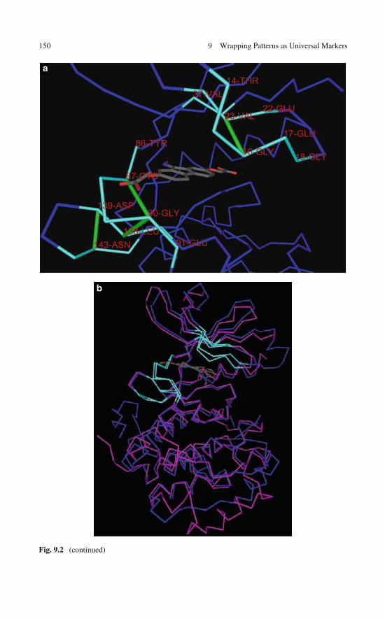
150 9 Wrapping Patterns as Universal Markers
a
b
Fig. 9.2 (continued)

c
d
Fig. 9.2 (continued)
152 9 Wrapping Patterns as Universal Markers
The strong correlation between denv and dphar(R2 ≈ 0.917
)(Fig. 9.2d) reveals
that the impact of drugs on the human kinome is dictated by differences in hydrationmicroenvironments or dehydron patterns across the ligand-binding regions of thekinases. The diversity in hydration microenvironments needed to yield specificityacross paralog kinases results from the variability (<σ>≈ 1.38) of environmentalresidues (<σ >≈ 0.21 when the average is restricted to residues paired by hydrogenbonds that are environmentally affected by the ligands).
The strong correlation shown in Fig. 9.2d provides compelling evidence in sup-port of the universality of protein wrapping as the selectivity filter for moleculartherapeutic approaches targeting the human kinome.
9.4 A Target Library for the Human Kinome:Broadening the Technological Basis of Drug Discovery
As shown in the previous section, the wrapping pattern of a target kinase providesa universal selectivity filter for the drug designer. This fact begets the obvious ques-tion: How do we take advantage of our selectivity filter to actually broaden thetechnological basis of drug discovery? To address this question we envision theconstruction of a dehydron library in drug-targeted proteins and then exploit thisbioinformatics library to generate a map of controlled cross-reactivity that resultswhen such specialized patterns are targeted. The controlled cross-reactivity mapwill reflect the superior level of specificity that may be achieved by targeting pack-ing patterns. This resource will exploit the matrix representation of the wrappingpattern of a protein that enables comparative alignment across an entire target super-family and will digitalize information on subtle packing differences across proteintargets. The long-term application of this resource is the control of side effects inmolecular-targeted therapy adopting the new drug design paradigm, the drug asdehydron wrapper.
As shown in Chaps. 7 and 8, specific drugs may be chemically modified toturn them into wrappers of dehydrons, protecting them from water attack. Thistype of ligand optimization has been shown in specific cases to retain anticanceractivity while reducing potential life-threatening side effects. Thus, the molecularbasis for target discrimination was delineated in vitro and through in vivo assays forantitumor activity, albeit over limited sets of purported targets.
Against this background, it becomes essential to introduce bioinformatics resultsof wide applicability that precisely map out the optimizing chemical modificationsfor all drug chemotypes and all kinase targets with the goal of controlling andenhancing inhibitor specificity across the entire human kinome. Thus, in principleat least, the technological basis of drug design can indeed be broadened contin-gent on our ability to construct a comparative and annotated dehydron library. Thislibrary should compile the cross-reactivity map that will contain “raw” informationon wrapping defects as selectivity-promoting features and contain the annotated

9.5 Useful Annotations of a Library of Specificity-Promoting Target Features 153
information to guide the drug designer to develop superior drugs with controlledspecificity. The raw data should include the following:
• Identification of all packing defects – dehydrons – targetable by ATP competinginhibitors for each human kinase.
• Comparative alignment of the dehydron pattern of one kinase against the respec-tive patterns in all the others to obtain a map of a priori cross-reactivities fordehydron-wrapping drugs.
• Identification of dehydrons with the lowest levels of conservation for each kinasetogether with the kinases where the dehydron is replaced by a well-wrappedhydrogen bond upon alignment. Thus, the optimal specificity-controlling dehy-drons will be determined through a “conservation score.”
• Determination of accessibility of optimal dehydrons vis-à-vis wrapping/targetingwithin a given drug chemotype and the set of kinases that is discriminated asthe optimal dehydrons are targeted. Obviously, a specificity-promoting dehydronwould be of no practical use if its exogenous wrapping is not accessible from theparticular lead chemotype considered.
Thus, the results expounded in this chapter pave the way for the construction ofthe matrix-based library of dehydrons for human kinases enabling direct compara-tive alignment across the structure-reported kinome. This library is destined to beused to decompose PDB-reported target space into clusters of cross-reactive kinases,enabling direct assessment of the full potential of the novel selectivity filter on tar-gets with reported structure. The results can be validated by examining compoundsindependently screened using a phage display kinase library and contrasting theiraffinity (KD) profile against predicted cross-reactivity clusters. The results shouldalso be cast in usable format to guide the engineering of safer inhibitors with con-trolled specificity. Dehydrons should be prioritized according to their decreasinglevels of conservation across homologs in the human kinome, hence serving asthe most reliable molecular features to be targeted in order to control and reducecross-reactivity.
9.5 Useful Annotations of a Library of Specificity-PromotingTarget Features
In the first stages of the resource building, we envision a comparative analysisconfined to kinases with PDB-reported structures, thus guaranteeing unambiguousdehydron identification. The structural alignment required for comparative analysiswill be largely facilitated by the kinase fold conservation resulting from commonancestry. Targetable dehydrons should be reported for each kinase jointly withseveral annotations instrumental to the drug optimization strategy: (a) extent ofdehydron conservation across the superfamily and expected cross-reactivity arisingif targeted by a wrapping ligand; (b) accessibility of the dehydron vis-à-vis a specificdrug chemotype; (c) activation state of the kinases under comparison; (d) varia-tions in dehydron patterns due to different induced fits of the same target; and (e)
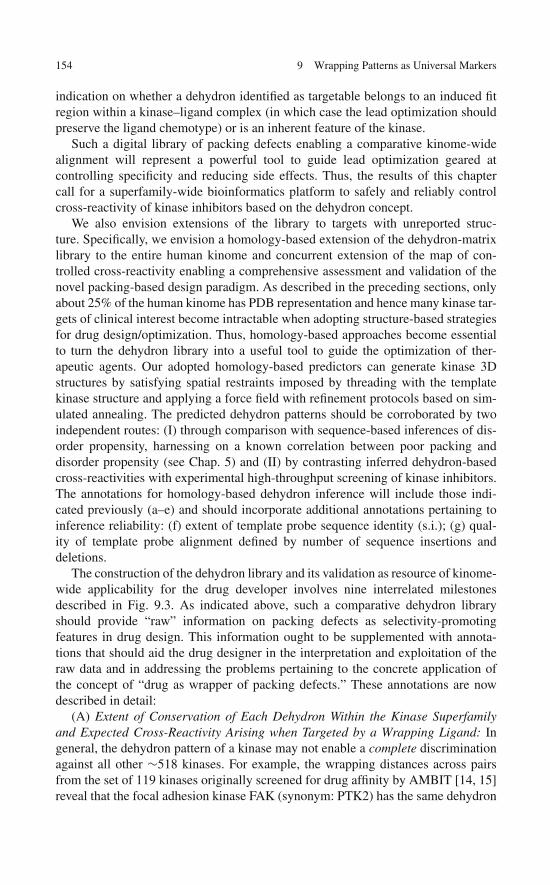
154 9 Wrapping Patterns as Universal Markers
indication on whether a dehydron identified as targetable belongs to an induced fitregion within a kinase–ligand complex (in which case the lead optimization shouldpreserve the ligand chemotype) or is an inherent feature of the kinase.
Such a digital library of packing defects enabling a comparative kinome-widealignment will represent a powerful tool to guide lead optimization geared atcontrolling specificity and reducing side effects. Thus, the results of this chaptercall for a superfamily-wide bioinformatics platform to safely and reliably controlcross-reactivity of kinase inhibitors based on the dehydron concept.
We also envision extensions of the library to targets with unreported struc-ture. Specifically, we envision a homology-based extension of the dehydron-matrixlibrary to the entire human kinome and concurrent extension of the map of con-trolled cross-reactivity enabling a comprehensive assessment and validation of thenovel packing-based design paradigm. As described in the preceding sections, onlyabout 25% of the human kinome has PDB representation and hence many kinase tar-gets of clinical interest become intractable when adopting structure-based strategiesfor drug design/optimization. Thus, homology-based approaches become essentialto turn the dehydron library into a useful tool to guide the optimization of ther-apeutic agents. Our adopted homology-based predictors can generate kinase 3Dstructures by satisfying spatial restraints imposed by threading with the templatekinase structure and applying a force field with refinement protocols based on sim-ulated annealing. The predicted dehydron patterns should be corroborated by twoindependent routes: (I) through comparison with sequence-based inferences of dis-order propensity, harnessing on a known correlation between poor packing anddisorder propensity (see Chap. 5) and (II) by contrasting inferred dehydron-basedcross-reactivities with experimental high-throughput screening of kinase inhibitors.The annotations for homology-based dehydron inference will include those indi-cated previously (a–e) and should incorporate additional annotations pertaining toinference reliability: (f) extent of template probe sequence identity (s.i.); (g) qual-ity of template probe alignment defined by number of sequence insertions anddeletions.
The construction of the dehydron library and its validation as resource of kinome-wide applicability for the drug developer involves nine interrelated milestonesdescribed in Fig. 9.3. As indicated above, such a comparative dehydron libraryshould provide “raw” information on packing defects as selectivity-promotingfeatures in drug design. This information ought to be supplemented with annota-tions that should aid the drug designer in the interpretation and exploitation of theraw data and in addressing the problems pertaining to the concrete application ofthe concept of “drug as wrapper of packing defects.” These annotations are nowdescribed in detail:
(A) Extent of Conservation of Each Dehydron Within the Kinase Superfamilyand Expected Cross-Reactivity Arising when Targeted by a Wrapping Ligand: Ingeneral, the dehydron pattern of a kinase may not enable a complete discriminationagainst all other ∼518 kinases. For example, the wrapping distances across pairsfrom the set of 119 kinases originally screened for drug affinity by AMBIT [14, 15]reveal that the focal adhesion kinase FAK (synonym: PTK2) has the same dehydron
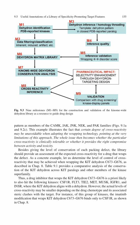
9.5 Useful Annotations of a Library of Specificity-Promoting Target Features 155
Fig. 9.3 Nine milestones (M1–M9) for the construction and validation of the kinome-widedehydron library as a resource to guide drug design
pattern as members of the CAMK, JAK, JNK, NEK, and PAK families (Figs. 9.1aand 9.2c). This example illustrates the fact that certain degree of cross-reactivitymay be unavoidable when adopting the wrapping technology, pointing at the verylimitations of this approach. The whole issue then becomes whether the particularcross-reactivity is clinically tolerable or whether it provides the right compromisebetween activity and toxicity.
Besides giving the level of conservation of each packing defect, the libraryshould provide an assessment of the expected cross-reactivity for a drug that wrapsthe defect. As a concrete example, let us determine the level of control of cross-reactivity that may be achieved when wrapping the KIT dehydron C673–G676, asdescribed in Chap. 8. Table 9.1 provides a comparative analysis of the conserva-tion of the KIT dehydron across KIT paralogs and other members of the kinasesuperfamily.
Thus, a drug inhibitor that wraps the KIT dehydron C673–G676 is a priori likelyto also hit the following kinases: CSF1R, FLT3, TIE2, MET, MUSK, IGFR1, andINSR, where the KIT dehydron aligns with a dehydron. However, the actual levels ofcross-reactivity may be smaller depending on the drug chemotype and its associatedsteric clashes with the target. For instance, of this group of kinases, the imatinibmodification that wraps KIT dehydron C673–G676 binds only to CSF1R, as shownin Chap. 8.
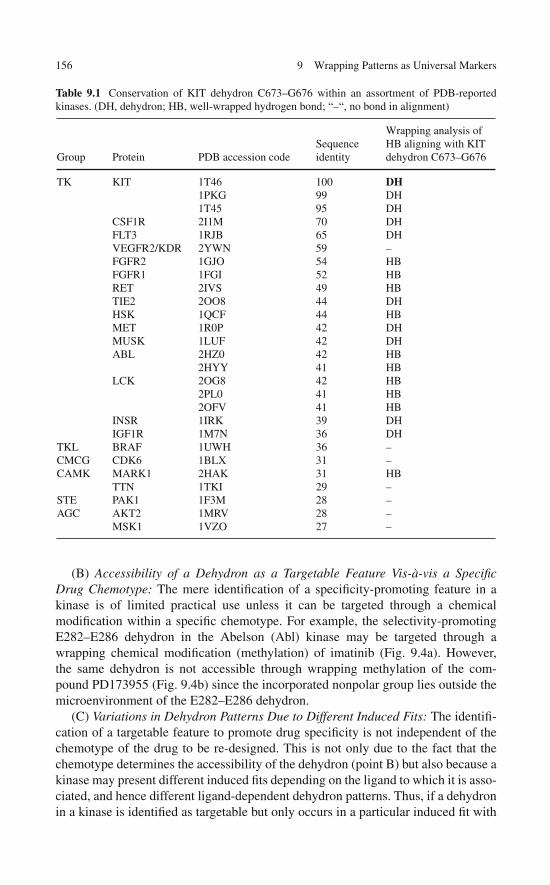
156 9 Wrapping Patterns as Universal Markers
Table 9.1 Conservation of KIT dehydron C673–G676 within an assortment of PDB-reportedkinases. (DH, dehydron; HB, well-wrapped hydrogen bond; “–“, no bond in alignment)
Group Protein PDB accession codeSequenceidentity
Wrapping analysis ofHB aligning with KITdehydron C673–G676
TK KIT 1T46 100 DH1PKG 99 DH1T45 95 DH
CSF1R 2I1M 70 DHFLT3 1RJB 65 DHVEGFR2/KDR 2YWN 59 –FGFR2 1GJO 54 HBFGFR1 1FGI 52 HBRET 2IVS 49 HBTIE2 2OO8 44 DHHSK 1QCF 44 HBMET 1R0P 42 DHMUSK 1LUF 42 DHABL 2HZ0 42 HB
2HYY 41 HBLCK 2OG8 42 HB
2PL0 41 HB2OFV 41 HB
INSR 1IRK 39 DHIGF1R 1M7N 36 DH
TKL BRAF 1UWH 36 –CMCG CDK6 1BLX 31 –CAMK MARK1 2HAK 31 HB
TTN 1TKI 29 –STE PAK1 1F3M 28 –AGC AKT2 1MRV 28 –
MSK1 1VZO 27 –
(B) Accessibility of a Dehydron as a Targetable Feature Vis-à-vis a SpecificDrug Chemotype: The mere identification of a specificity-promoting feature in akinase is of limited practical use unless it can be targeted through a chemicalmodification within a specific chemotype. For example, the selectivity-promotingE282–E286 dehydron in the Abelson (Abl) kinase may be targeted through awrapping chemical modification (methylation) of imatinib (Fig. 9.4a). However,the same dehydron is not accessible through wrapping methylation of the com-pound PD173955 (Fig. 9.4b) since the incorporated nonpolar group lies outside themicroenvironment of the E282–E286 dehydron.
(C) Variations in Dehydron Patterns Due to Different Induced Fits: The identifi-cation of a targetable feature to promote drug specificity is not independent of thechemotype of the drug to be re-designed. This is not only due to the fact that thechemotype determines the accessibility of the dehydron (point B) but also because akinase may present different induced fits depending on the ligand to which it is asso-ciated, and hence different ligand-dependent dehydron patterns. Thus, if a dehydronin a kinase is identified as targetable but only occurs in a particular induced fit with

9.5 Useful Annotations of a Library of Specificity-Promoting Target Features 157
Fig. 9.4 Differences in accessibility of ABL dehydron E282–E286 according to drug chemotype.(a) Dehydron-wrapping methylation of imatinib and (b) inaccessibility of dehydron to wrappingmethylation in compound PD173955
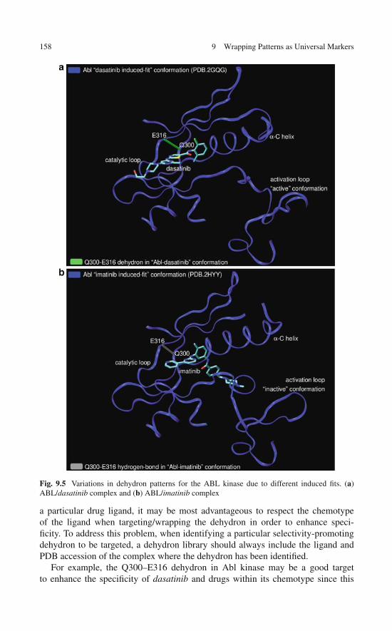
158 9 Wrapping Patterns as Universal Markers
Fig. 9.5 Variations in dehydron patterns for the ABL kinase due to different induced fits. (a)ABL/dasatinib complex and (b) ABL/imatinib complex
a particular drug ligand, it may be most advantageous to respect the chemotypeof the ligand when targeting/wrapping the dehydron in order to enhance speci-ficity. To address this problem, when identifying a particular selectivity-promotingdehydron to be targeted, a dehydron library should always include the ligand andPDB accession of the complex where the dehydron has been identified.
For example, the Q300–E316 dehydron in Abl kinase may be a good targetto enhance the specificity of dasatinib and drugs within its chemotype since this

9.6 The Dehydron Library as a Technological Resource 159
dehydron is present in the induced fit of the Abl upon complexation with dasatinib(Fig. 9.5a). However, that dehydron becomes a well-wrapped hydrogen bond in theinduced fit resulting upon Abl complexation with imatinib (Fig. 9.5b). Thus, thelibrary should contain annotation guarding against adopting the imatinib chemo-type when targeting the Q300–E316 dehydron in Abl, since this dehydron is notpresent upon imatinib association.
9.6 The Dehydron Library as a Technological Resource
The dehydron library will generate chemotype-dependent decompositions of tar-get space (human kinome) into cross-reactivity clusters. These results are crucialto assess the full potential of the packing-based drugs as therapeutic agents ofsuperior controllable selectivity. Cross-reactivity clusters include only kinases thatare inhibited by the same wrapping modification within a fixed lead chemotype. Thewrapping modification, in turn, targets a dehydron in a particular kinase, and allother kinases presenting a dehydron at the aligned position will belong to the samechemotype-dependent cross-reactivity cluster. The cartoon in Fig. 9.6 represents the
Fig. 9.6 Schematic representation of the generation of a chemotype-dependent cross-reactivitymap. A dehydron (green) in a particular protein (1) that may be wrapped (dashed-line arrow) by achemical substitution of a lead within a specific chemotype (red) will be singled out for comparisonacross the kinome and to define the cross-reactivity cluster to which this protein belongs. The extentof conservation of the dehydron will then be established by comparative alignment of dehydronmatrices within the bioinformatics library. Conservation of the dehydron (across proteins 1 and2 in the scheme) promotes cross-reactivity of the wrapping compound, defining the cluster forprotein 1 within the chemotype-dependent decomposition of target space (red rectangle). Proteinsthat lack drug-accessible dehydrons or those that contain unique (not conserved) drug-accessibledehydrons constitute singleton clusters. Validation is readily obtained by examining compoundsindependently screened using AMBIT’s high-throughput kinase panels [14, 15] and contrastingthe predicted cross-reactive clusters with the reported affinity (KD) values

160 9 Wrapping Patterns as Universal Markers
construction/validation of the cross-reactivity map combining the dehydron libraryand structural information on a single kinase/inhibitor complex for the particulardrug chemotype.
The cross-reactivity map is thus obtained by a straightforward procedure thatcombines structural input from a kinase/inhibitor complex with a kinome-wide com-parative alignment of packing defects provided by the bioinformatics library. Theresults will be validated by examining compounds independently screened usingthe T7-phage display kinase library (∼360 kinases in latest panels) [15] and con-trasting their affinity (KD) profile with their predicted cross-reactivity cluster: if acompound is identified to wrap a particular dehydron in a kinase, then this com-pound should be cross-reactive with all other kinases for which the dehydron ispresent at the aligned position except for kinases where structural alignment revealssteric clashes. This target space decomposition will assess the full potential of thewrapping-based design motif.
References
1. Bain J, McLauchlan H, Eliott M, Cohen P (2003) The specificities of protein kinase inhibitors:An update. Biochem J 371:199–204
2. Druker BJ (2004) Molecularly targeted therapy: Have the floodgates opened? Oncologist9:357–360
3. Hopkins AL, Mason JS, Overington JP (2006) Can we rationally design promiscuous drugs?Curr Opin Str Biol 16:127–136
4. Huse M, Kuriyan J (2002) The conformational plasticity of protein kinases. Cell 109:275–2825. Knight ZA, Shokat KM (2005) Features of selective kinase inhibitors. Chem Biol 12:
621–6376. Chothia C (1974) Hydrophobic bonding and accessible surface area in proteins. Nature
248:338–3397. Whittle PJ, Blundell TL (1994) Protein structure-based drug design. Annu Rev Biophys
Biomol Struct 23:349–3758. Feng BY, Shelat A, Doman TN, Guy RK, Shoichet BK (2005) High throughput assays for
promiscuous inhibitors. Nat Chem Biol 1:146–1489. Zhang X, Crespo A, Fernández A (2008) Trung promiscuous kinase inhibitors into safer
drugs. Trends Biotechnol 26:295–30010. Chen JP, Zhang X, Fernández A (2007) Molecular basis for specificity in the druggable
kinome: sequence-based analysis. Bioinformatics 23:563–57211. Manning G, Whyte DB, Martinez R, Hunter T, Sudarsanam S (2002) The protein kinase
complement of the human genome. Science 298:1912–193412. Braken C, Iakoucheva LM, Romero PR, Dunker AK (2004) Combining prediction, com-
putation and experiment for the characterization of protein disorder. Curr Opin Struct Biol14:570–576
13. Fernández A, Berry RS (2004) Molecular dimension explored in evolution to promoteproteomic complexity. Proc Natl Acad Sci USA 101:13460–13465
14. Fabian MA, Biggs WH, Treiber DK et al (2005) A small molecule kinase interaction map forclinical kinase inhibitors. Nat Biotechnol 23:329–336
15. Karaman MW, Herrgard S, Treiber DK et al (2008) A quantitative analysis of kinase inhibitorselectivity. Nat Biotechnol 26:127–132
16. Bonneau R, Straus CE, Rohl CA et al (2002) De novo prediction of three-dimensionalstructures for major protein families. J Mol Biol 322:65–78

References 161
17. Chivian D, Kim DE, Malmstrom L et al (2005) Prediction of CASP6 structures usingautomated Robetta protocols. Proteins 61 (Suppl 7):157–166
18. Higgins DG, Thompson JD, Gibson TJ (1996) Using CLUSTAL for multiple sequencealignments. Method Enzymol 266:383–402
19. Fraczkiewicz R, Braun W (1998) Exact and efficient analytical calculation of the accessiblesurface areas and their gradient for macromolecules. J Comput Chem 19: 319–333
20. Ooi T, Oobatake M, Nemethy G, Scheraga HA (1987) Accessible surface area as a mea-sure of the thermodynamic parameters of hydration of peptides. Proc Natl Acad Sci USA 84:3086–3090
21. Ma B, Elkayam T, Wolfson T, Nussinov R (2003) Protein-protein interactions structurallyconserved residues distinguish between binding sites and exposed protein surfaces. Proc NatlAcad Sci USA 100:5772–5777
22. Fernández A, Maddipati S (2006) A priori inference of cross reactivity for drug-targetedkinases. J Med Chem 49:3092–3100
23. Fernández A, Sosnick TR, Colubri A (2002) Dynamics of hydrogen bond desolvation inprotein folding. J Mol Biol 321:659–675
24. Fernández A (2004) Keeping dry and crossing membranes. Nat Biotechnol 22:1081–1084

“This page left intentionally blank.”
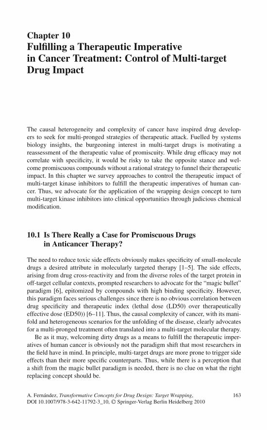
Chapter 10Fulfilling a Therapeutic Imperativein Cancer Treatment: Control of Multi-targetDrug Impact
The causal heterogeneity and complexity of cancer have inspired drug develop-ers to seek for multi-pronged strategies of therapeutic attack. Fuelled by systemsbiology insights, the burgeoning interest in multi-target drugs is motivating areassessment of the therapeutic value of promiscuity. While drug efficacy may notcorrelate with specificity, it would be risky to take the opposite stance and wel-come promiscuous compounds without a rational strategy to funnel their therapeuticimpact. In this chapter we survey approaches to control the therapeutic impact ofmulti-target kinase inhibitors to fulfill the therapeutic imperatives of human can-cer. Thus, we advocate for the application of the wrapping design concept to turnmulti-target kinase inhibitors into clinical opportunities through judicious chemicalmodification.
10.1 Is There Really a Case for Promiscuous Drugsin Anticancer Therapy?
The need to reduce toxic side effects obviously makes specificity of small-moleculedrugs a desired attribute in molecularly targeted therapy [1–5]. The side effects,arising from drug cross-reactivity and from the diverse roles of the target protein inoff-target cellular contexts, prompted researchers to advocate for the “magic bullet”paradigm [6], epitomized by compounds with high binding specificity. However,this paradigm faces serious challenges since there is no obvious correlation betweendrug specificity and therapeutic index (lethal dose (LD50) over therapeuticallyeffective dose (ED50)) [6–11]. Thus, the causal complexity of cancer, with its mani-fold and heterogeneous scenarios for the unfolding of the disease, clearly advocatesfor a multi-pronged treatment often translated into a multi-target molecular therapy.
Be as it may, welcoming dirty drugs as a means to fulfill the therapeutic imper-atives of human cancer is obviously not the paradigm shift that most researchers inthe field have in mind. In principle, multi-target drugs are more prone to trigger sideeffects than their more specific counterparts. Thus, while there is a perception thata shift from the magic bullet paradigm is needed, there is no clue on what the rightreplacing concept should be.
163A. Fernández, Transformative Concepts for Drug Design: Target Wrapping,DOI 10.1007/978-3-642-11792-3_10, C© Springer-Verlag Berlin Heidelberg 2010
164 10 Fulfilling a Therapeutic Imperative in Cancer Treatment
Uncertain clinical outcome resulting from health-threatening side effects is a cen-tral issue for therapies based on multi-target kinase inhibitors since kinases playdifferent signal transduction roles in different cellular contexts and a favorable inhi-bition in one scenario may prove fatal in another [12]. For instance, a specificinhibitor of the Abelson (Abl) kinase may be assumed to be most efficacious in treat-ing chronic myeloid leukemia (CML), since an aberrantly deregulated Abl kinase isa recognized primary target for treating this malignancy [5]. However, as extensivelydescribed in Chaps. 7 and 8, a systems biology assessment [12] has recently revealedthat Abl inhibition is also a likely culprit of cardiotoxicity: Abl inhibition recruitsa signaling cascade that leads to mitochondrial depolarization, ATP depletion, andultimately apoptosis in cardiomyocytes [12]. Thus, a more cross-reactive drug, i.e.,one that also inhibits another kinase along the pro-apoptotic pathway triggered byAbl inhibition (hence suppressing it) is expected to have a higher therapeutic effi-cacy in treating CML [13]. In this regard, an inhibitor that also impacts JNK wouldbe highly desirable, as described in Chap. 8. The rationale for this assertion is thathigher therapeutic doses may be tolerable due to the removal of cardiotoxicity, albeitat the expense of sacrificing specificity [13–15]. One possible downturn of such anapproach is that JNK inhibition may introduce uncertain clinical outcomes in off-target cells (i.e., cells other than cardiomyocytes). Given the downstream positionof JNK in most signal transduction cascades, this is not likely to be the case, as JNKmay be replaced by alternative and redundant signal transducers in other cellularcontexts.
The causal complexity of cancer and other diseases has motivated much effortdirected at reassessing the therapeutic value of promiscuity. This conceptual shiftis in part motivated by telling cases. For instance, multi-target kinase inhibitorssuch as Sutent (sunitinib) or Nexavar (sorafenib) have recently received FDAapproval, albeit with important caveats highlighting their side effects, especiallytheir cardiotoxicity [7, 12]. Thus, the possibility of exploiting drug promiscuity asa therapeutic opportunity is being considered in novel approaches to treat complexdisorders such as cancer, depression, and cardiovascular disease [7]. The simultane-ous modulation of multiple targets is often required to alter a clinical phenotype asbiological redundancies and alternative pathways can often bypass the inhibition ofa single target or of multiple targets along a single pathway [16]. Thus, in contrastwith the magic bullet, a “magic shotgun” targeting multiple proteins may in someinstances possess a higher therapeutic index than a specific drug [6].
Another argument favoring promiscuity arises with the observation that highlycross-reactive drugs may be more resilient against drug-resistant mutations [17].Cross-reactivity arises because, upon association with the target protein, such drugstypically make interactions with evolutionarily conserved residues and with back-bone groups, and less interactions with the residues that mutate to confer drugresistance [17, 18]. Targeting conserved residues or unspecific parts of the chain(backbone) begets promiscuity.
The safety of less selective drugs, and especially that of promiscuous kinaseinhibitors, is often addressed by assessing the full extent of their cross-reactivity[19]. This assessment has been facilitated by novel high-throughput screening
10.2 Cleaning Dirty Drugs with Selectivity Filters: Basic Insights 165
assays such as (a) the kinase assay using the T7-phage expression panels fromAmbit Biosciences [20, 21]; (b) a thermal stability shift assay using a 60-Ser/Thrkinase panel [22]; and (c) the living-cell assays of pathway inhibition that assess theimpact of the drug on the protein-recruiting capability of the target [23].
In vitro assays of cross-reactivity are affected by the complexities of tissue dis-tribution and, generally, by pharmacodynamic issues [24]. Yet, target affinity, beinggoverned by thermodynamics of ligand binding, represents a telling parameter inde-pendent of in vivo heterogeneities, except for allosteric antagonism. On the otherhand, crowding, membrane adsorption, and other effects can modulate in vivo drugconcentrations, increasing local levels in different spatial locations. Thus, certaincross-reactivities undetectable in vitro may surface in vivo, causing unexpected sideeffects [24, 25]. Conversely, tissue and subcellular distribution may prevent a lig-and from binding an in vitro-established target, introducing another caveat in theinterpretation of high-throughput screening results.
In spite of timely efforts to establish a paradigm shift, promiscuous drugsobviously carry a higher risk of inducing side effects than their more spe-cific counterparts. Even the successful anticancer drug Gleevec (imatinib), witha reduced spectrum of primary targets, i.e., the kinases Bcr-Abl, C-Kit, Lck,PDGFR, and CSF1R [20, 21], has been recently shown to be potentially cardiotoxic[12, 26] and labeled as such by the FDA (see: http://www.fda.gov/medwatch/safety/2006/Gleevec_DHCP_10-19-2006.htm). Not surprisingly, the more promis-cuous anticancer kinase inhibitors sunitinib and sorafenib are also cardiotoxic, to alarger extent than imatinib [12, 26]. In Ambit screenings [20] sunitinib was shownto bind 79 kinases out of 119 assayed, while sorafenib binds to 41.
Hence, it is forbiddingly risky to welcome promiscuous compounds into the ther-apeutic arena without a rational strategy to control their specificity and therapeuticindex. Such a control may be achieved if we can identify features in the target struc-ture that are unique to the target, i.e., “selectivity filters,” and drug modifications thatpromote interactions with such nonconserved features. Thus, selectivity filters mayserve as guidance to rational drug design [13–15, 27–29]. We advocate for this typeof control of therapeutic impact to clean less selective drugs following an integralassessment of the diverse functional roles of targeted proteins.
10.2 Cleaning Dirty Drugs with Selectivity Filters:Basic Insights
As described in Chaps. 6 and 9, most protein drug targets have paralogs, that is,proteins that share a common ancestor with the target and have diverged away fromit after speciation [30]. Thus, kinases belong to common ancestry groups (families)which typically share the same fold and basic structural features that often yieldunexpected cross-reactivities [20, 21, 31].
In principle, much of this cross-reactivity could be removed by drug redesignguided by the identification of the common structural features that promotepromiscuity and the nonconserved features that enable paralog discrimination. As

166 10 Fulfilling a Therapeutic Imperative in Cancer Treatment
shown in Chap. 8, this approach has been successfully attempted on the cross-reactive drug imatinib with promising results [13–15]. Its further utilization to cleantruly promiscuous drugs requires further analysis, as described in the next section,and will enable us to propose its extended use to clean kinase inhibitors.
A number of paralog-discriminating nonconserved features different from wrap-ping have been identified and, although not amenable of systematic application,some can be exploited as selectivity filters [27, 28]. One approach uses high-resolution crystal structures of kinases in complex with non-ATP ligands to identifyunique structural motifs in the purported targets. For example, a unique helical inserthas been found in the activation loop of the NEK2 and MPSK1 kinases adjacent tothe highly conserved DFG catalytic triad [27].
Specific inhibitors that bind to the inactive kinase conformations have also beendeveloped targeting the “DFG-out” conformation [32]. In this unique conformation,the position of the phenylalanine (F) residue which is located at the start of the acti-vation loop is flipped with respect to the active conformation, so that it points inwardwithin the ATP pocket. This mode of association has been confirmed by the bind-ing of imatinib to the inactive Abl kinase [33]. In addition to the DFG-out motif,nonconserved structural features within the inactive ensemble should perhaps beexploited to achieve paralog specificity. The inactive conformations of kinases makethem more discernible, while the active conformation reveals fewer discriminatoryfeatures since it is constrained to be catalytically functional and hence more con-served. The substrate-discriminatory amino acid variations that define the differentkinase functions are mostly located in loopy regions framing the ATP pocket, ratherthan in the pocket itself, making them less accessible targets. While targeting theinactive conformation may be a logical choice, there are also advantages in target-ing the active conformation. The latter requires structure conservation for functionalpurposes, and hence it is less tolerant to drug-resistant mutations. Thus, a kinasethat binds to a drug in the active conformation will seldom develop drug-resistantmutations since the latter can compromise the activity.
The selectivity filter advocated throughout this book is in principle of universalvalidity, as shown in Chap. 9, and may be used systematically. It arises from com-parison of the wrapping patterns of protein targets [30]. The wrapping pattern maybe turned into an operational selectivity filter for two reasons: (a) dehydrons maybe targeted by drugs that further wrap them by bringing nonpolar groups to theirproximity upon association and (b) dehydrons are not conserved across paralogs, asshown in Chap. 9. Thus, differences in dehydron distribution constitute promisingselectivity filters to clean promiscuous inhibitors.
10.3 Cleaning Dirty Drugs by Exploiting the Wrapping Filter:Proof of Concept
In principle, most kinase inhibitors are susceptible of being turned into selectivewrappers of packing defects through minor chemical modification and withoutaltering their chemotype. Thus, clinically relevant compounds with considerable

10.3 Cleaning Dirty Drugs by Exploiting the Wrapping Filter: Proof of Concept 167
cross-reactivities such as sunitinib (inhibiting 55% of the 290 kinases screened[21]), dasatinib (30%), EKB-569 (20%), sorafenib (20%), erlotinib (15%), gefitinib(6%), and imatinib (4%) may be redesigned into drugs with enhanced specificityusing the wrapping design concept. The generic strategy consists in modifying theparental compound to turn it into a wrapper of unique dehydrons while remov-ing potential sources of cross-reactivity. The latter arise since chemical groups inthe ligand often interact with conserved structural features in the protein such asbackbone moieties or side chains that remain invariant within the target family. Thewrapping strategy may in principle be applied to enhance the specificity of promis-cuous drugs like sunitinib, dasatinib, EKB-569, sorafenib, and erlotinib towardtheir respective primary targets: KIT/VEGFR2, ABL/SRC, EGFR, VEGFR2,EGFR [21].
To illustrate the operational value of this approach, we focus on redesigning atruly promiscuous drug, EKB-569 (Wyeth-Ayerst, [34]), to significantly enhance itsselectivity. This drug was launched as a major inhibitor of the epidermal growth fac-tor receptor (EGFR) kinase (IC50 = 38.5 nM). It gained major therapeutic interestfor the treatment of non-small cell lung cancer (NSCLC), colorectal neoplasia, andother EGFR-dependent solid tumors. Phase I and II trials for such therapeutic appli-cations are currently in progress and closed to new patients [34, 35]. Preliminaryhigh-throughput screening using a battery of 119 T7-phage-expressed kinases [20]revealed 25 sub-micromolar targets for EKB-569, making it a promiscuous drugwith likely side effects. Other compounds such as Iressa (gefitinib) and Tarceva(erlotinib) share the same “4-anilinoquinoline” chemotype [36], yet they are morespecific EGFR inhibitors [20]. The latter two gained FDA approval as anti-NSCLCagents.
EKB-569 promiscuity can be traced to its intermolecular interactions with highlyconserved residues within the EGFR kinase family. As shown in Fig. 10.1a, the ter-minal acryl group in the ligand is a source of promiscuity because it reacts withthe conserved residues Cys/Ser in the EGFR paralogs, acting as electrophile in anirreversible Michael adduct. Another source of EKB-569 promiscuity is the inter-molecular electrostatic interaction between its cyanide group and the gatekeeperresidue (Thr or Met), typically conserved within the family (Fig. 10.1a).
Thus, we may remove these sources of promiscuity by introducing the followingmodifications:
(a) Replace the double bond (the Michael acceptor) in the acryl group with a singlebond
(b) Replace the cyanide group with a methyl to retain the chemotype whileremoving the electrostatic interaction with the conserved gatekeeper
To promote selectivity, we may introduce a wrapping modification to targeta nonconserved dehydron in the primary target EGFR. A comparative structuralanalysis of the EGFR family reveals that the least-conserved EGFR dehydron isAsp831–Gly833. To wrap this dehydron, we may append a methyl group at position3 of the terminal benzene ring (Fig. 10.1b). This wrapping modification is likely

168 10 Fulfilling a Therapeutic Imperative in Cancer Treatment
to possess inhibitory activity exclusively against the EGFR paralogs that retain thedehydron in its original position: EGFR, Aurora2 (STK6), CLK3, ERBB2, PIM1,PIM2, PTK2 (FAK), SLK, and STK10. The modified drug is expected to binda subset of this target group since steric hindrances may arise as a result of thechemical medication. We thus provide a rational approach to clean the promiscu-ous inhibitor EKB-569 based on a selectivity filter, reducing its inhibitory spectrumfrom 25 confirmed targets [20] to only 9.
To validate this design strategy, we established a correlation between the affinitiesof EKB-569 for the 38 paralogs of EGFR reported in PDB and the extent of residueconservation at the Michael reaction site and at the gatekeeper position. To do so, wealigned each paralog structure with the EGFR structure and examined residues thatalign with Cys773 (Michael reactant) and Thr766 (gatekeeper). A statistical modelwas built to assess such correlation and revealed that the EKB-569 affinity is indeeddictated by these two sources of promiscuity (p-value = 0.0003). Thus, the terminalacryl group and the cyanide group in EKB-569 (Fig. 10.1a) are indeed the “dirty”moieties responsible for promiscuity.
The four dehydrons within the binding pocket of EGFR are Asp831–Gly833,Gly697–Phe699, Gly697–Gly700, and Gly772–Val821. By examining the conser-vation of these four dehydrons across the 38 EGFR paralogs, we found the leastconserved is dehydron Asp831–Gly833, retained only in the nine paralogs men-tioned above. Thus, we choose this dehydron as the nonconserved selectivity featureto be targeted. We then appended a methyl group at position 3 of the terminalbenzene ring that acts as a wrapper or protector of such feature (Fig. 10.1b).This redesigned EKB-569 molecule was synthesized following a pathway thatrecapitulates the EKB-569 synthesis [37], as shown in Fig. 10.1c.
Based on the selectivity filter provided by the wrapping patterns, we predict theaffinity profile of our prototype. The prediction is based on the conservation ofthe EGFR dehydron Asp831–Gly833 wrapped by the prototype (but not by EKB-569) and the existence of steric hindrances with nonpolar groups in the catalyticloop of the target structures. We predicted as “hits” only those targets with a con-served dehydron in such position and no steric hindrance. Only six hits are predicted(Table10.1): CLK3, EGFR, ERBB2, PTK2 (FAK), SLK, and STK10. In the caseswhere the residues aligning with EGFR’s Asp831-Gly833 are not engaged in a dehy-dron or a well-wrapped hydrogen bond, we examined whether such dehydron can beinduced upon ligand binding with minimal structural adaptation (this issue is furtherdiscussed in Chap. 11). Only in BTK and LCK kinases this dehydron can be induced
�
Fig. 10.1 Structural features promoting promiscuity and selectivity in EGFR kinase guidingEKB-569 cleaning redesign. (a) Structural alignment of EGFR kinase (blue ribbon representa-tion and atoms in licorice) and the paralog TNK2 kinase (red ribbon representation and atomsin balls and sticks), complexed with EKB-569 (licorice). Atoms are depicted following standardcolor convention (chlorine in green and fluorine in light green). The wrapping pattern of EGFRincludes the poorly conserved Asp831–Gly833 dehydron that may be targeted to achieve selectiv-ity since TNK2 lacks this dehydron. TNK2 contains the same two promiscuity-fostering features,

10.3 Cleaning Dirty Drugs by Exploiting the Wrapping Filter: Proof of Concept 169
Fig. 10.1 (continued) while lacking the dehydron at the locus where EGFR contains thespecificity-promoting feature. Thus, targeting the latter will ensure a discriminatory binding ofEGFR without hitting TNK2. (b) EGFR kinase structure (same representation as above) complexedwith the prototype EKB-569 redesigned inhibitor (licorice representation). To reduce EKB-569promiscuity, the acrylic double bond (Michael electrophile) is replaced by a single bond andthe gatekeeper-interacting cyanide is replaced by a methyl. To selectively target EGFR, a methylgroup is added to the terminal benzene ring as a wrapper of the barely conserved Asp831–Gly833dehydron. (c) Retro-synthetic pathway of the redesigned EKB-569 inhibitor. The synthesis reca-pitulates the EKB-569 pathway, albeit with different reactants [36]. Reprinted from (Trends inBiotechnology 26, [37]), copyright 2008 with permission from Elsevier
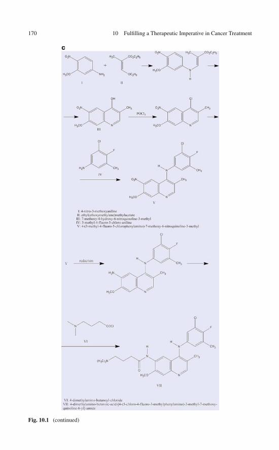
170 10 Fulfilling a Therapeutic Imperative in Cancer Treatment
Fig. 10.1 (continued)

10.3 Cleaning Dirty Drugs by Exploiting the Wrapping Filter: Proof of Concept 171
Table 10.1 Predicted and experimental affinity profiles for the “clean version” of kinase inhibitorEKB-569
KinaseDehydronconservation Steric hindrance Predicted affinity
Experimentalaffinity
ABL1 − − −ACK1(TNK2) − − −Aurora2(AURKA) + − − −Aurora3(AURKC) − − −BTK Possibly induced + Possible hit +CDK2 − − −CDK5 − − −CLK1 − − −CLK3 + + + +CSK − − −DAPK2 Possibly induced − − −DAPK3 − − −EGFR + + + +EPHA2 − − −ERBB2 + + + +FGFR1 Possibly induced − − −FGFR2 − − −HCK − − −INSR − − −JAK2 − − −JNK1 − − −JNK3 − − −KIT − − −LCK Possibly induced + Possible hit +MKNK2 − − −NEK2 − − −PAK1 − − −PAK4 − − −PAK6 − − −PAK7(PAK5) Possibly induced − − −PIM1 + − − −PIM2 + − − −PTK2(FAK) + + + +RPS6KA5 − − −SLK + + + +SRC − − −STK10 + + + +
upon drug binding with no steric hindrance with the catalytic loop, so they repre-sent other “possible” hits (Table 10.1). The experimentally obtained affinity profile(Fig. 10.2) for the prototype agrees almost perfectly with our predicted profile,since the drug actually binds only to the six hits inferred with certainty and the twolikely hits.
In this exercise, we have successfully cleaned the dirty inhibitor EKB-569using an approach based on a selectivity filter. This was accomplished by first
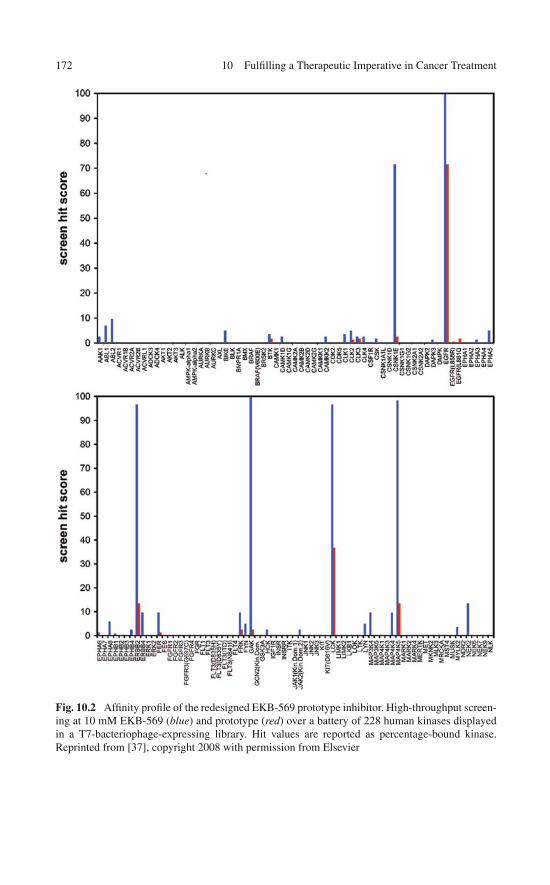
172 10 Fulfilling a Therapeutic Imperative in Cancer Treatment
Fig. 10.2 Affinity profile of the redesigned EKB-569 prototype inhibitor. High-throughput screen-ing at 10 mM EKB-569 (blue) and prototype (red) over a battery of 228 human kinases displayedin a T7-bacteriophage-expressing library. Hit values are reported as percentage-bound kinase.Reprinted from [37], copyright 2008 with permission from Elsevier

10.4 Cleaning Staurosporine Through a Wrapping Modification: A Stringent Test 173
Fig. 10.2 (continued)
removing the chemical features that promote promiscuity. Subsequently, we intro-duced a wrapping modification to target a non conserved dehydron in the intendedtarget and made the prototype more selective than the parental compound: Out ofthe 211 kinases experimentally screened [17, 25], our compound binds at nanomolarlevels to 6 kinases only, whereas EKB-569 binds to 19.
10.4 Cleaning Staurosporine Through a WrappingModification: A Stringent Test
In a more stringent test, staurosporine, the most promiscuous kinase inhibitor knownto man [21] was reengineered to elicit an inhibitory impact with controlled andenhanced specificity [38]. Staurosporine binds (KD < 3 μM) to ∼90% of the 290human kinases recently screened [21]. To pursue a structure-based “cleaning” ofstaurosporine through a wrapping modification of the ligand, we compared all28 PDB structures of kinases in complex with the ligand. A comparative wrap-ping analysis of their respective aligned ATP pockets prompted us to focus on theSrc dehydron Q250–E267 which is the least conserved dehydron in this structuralassortment. This dehydron is conserved in 7 out of 28 kinase structures, SRC, ABL,LCK, EGFR, ASK, TAO2, and GSK3, as indicated in Table10.2 and Fig. 10.3a, b.The dehydron has been targeted by wrapping it through a specific methylation ofstaurosporine at the imide N6-position of the indole ring (Fig. 10.3a) [38]. Thus,

174 10 Fulfilling a Therapeutic Imperative in Cancer Treatment
Table 10.2 Wrapping comparison of PDB-reported kinases in complex with staurosporine at posi-tion aligned with Src dehydron Q250–E267 (DH, dehydron; HB, well-wrapped hydrogen bond).The wrapping variant is predicted to bind only to the kinases that have a dehydron at the alignedposition and the prediction is contrasted with the experimental screening of the wrapping variantof staurosporine
Groupa Protein PDBWrappingclassification
Hit inscreening
Match predic-tion/experiment
TK SRC 1BYG DH Yes YesABL 2HZ4 DH Yes YesLCK 1QPD DH Yes YesEGFR 2ITW (wt) DH Yes Yes
2ITQ (G719S) HB Not screened2ITU (L858R) HB Not screened
FYN 2DQF HB No YesJAK3 1YVJ HB Not screenedSYK 1XBC HB No YesZAP70 1U59 HB No YesITK 1SNU HB No Yes
1SM2 HBTKL IRAQ4 2OIC HB Not screened
2NRY HBAGC PDK1 1OKY HB No Yes
PKAα 1STC HB No YesPKCθ 1XJD HB Not screened
STE ASK(MAP3K5)
2CLQ DH No No
TAO2 2GCD DH Not screenedCAMK CHK1 1NRV HB No Yes
MAPKAPK2 1NXK HB Not screened2PZY HB
MNK2 2HW7 HB No YesDAPK1 1WVY HB Not screenedPIM1 1YHS HB No Yes
CMCG CDK2 1AQ1 HB No YesGSK3β 1Q3D DH Not screened
Unclassified MPSK1(STK16)
2BUJ No HB Not screened
aGroup names: AGC: containing PKA, PKG, PKC families; CAMK: calcium/calmodulin-dependent protein kinase; CK1: casein kinase 1; CMGC: containing CDK, MAPK, GSK3, CLKfamilies; STE: homologs of yeast Sterile 7, Sterile 11, Sterile 20 kinases; TK: tyrosine kinase;TKL: tyrosine kinase-like
by redesigning staurosporine to turn it into a wrapper of the Src Q250–E267 dehy-dron, we can significantly restrict its inhibitory impact to the seven kinases thatshare the dehydron at the aligned position. The screening of the wrapping deriva-tive of staurosporine is shown in Fig. 10.4a–c: only ∼11%, or 26 out of the 223kinases assayed represent a hit, indicating a massive enhancement in specificity.Most significantly, our structure-based prediction of affinity based on the presenceor absence of the dehydron at the aligned position has only one false positive and not
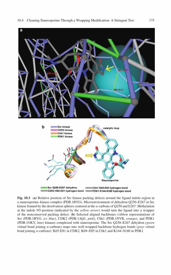
10.4 Cleaning Staurosporine Through a Wrapping Modification: A Stringent Test 175
Fig. 10.3 (a) Relative position of Src kinase packing defects around the ligand indole region ina staurosporine–kinase complex (PDB.1BYG). Microenvironment of dehydron Q250–E267 in Srckinase framed by the desolvation spheres centered at the α-carbons of Q250 and E267. Methylationat the indole N5-position (indicated by the yellow arrow) would turn the ligand into a wrapperof the nonconserved packing defect. (b) Selected aligned backbones (ribbon representation) ofSrc (PDB.1BYG, ice blue), CDK2 (PDB.1AQ1, pink), Chk1 (PDB.1NVR, orange), and PDK1(PDB.1OKY, lime) kinases complexed with staurosporine. The Src Q250–E267 dehydron (greenvirtual bond joining α-carbons) maps into well-wrapped backbone hydrogen bonds (gray virtualbond joining α-carbons): K65-E81 in CDK2, K69–E85 in Chk1 and K144–S160 in PDK1

176 10 Fulfilling a Therapeutic Imperative in Cancer Treatment
Fig. 10.4 (a–c) High-throughput screening at 10 mM for the wrapping variant of staurosporineover a battery of 223 human kinases displayed in a T7-bacteriophage library. Hit values are reportedas percentage-bound kinase

10.5 Systems Biology Insights into Wrapping-Directed 177
Fig. 10.4 (continued)
a single false negative over 15 instances (Table 10.2) where packing prediction canbe contrasted with experiment! These results demonstrate that the packing differ-ences across kinase homologs may guide molecular design to significantly enhancespecificity in a highly controllable manner and even for the most promiscuous drugs.
10.5 Systems Biology Insights into Wrapping-DirectedDesign of Multi-target Kinase Inhibitors
Systems biology, that is, the multi-scale integration of the functional componentsof organismic complexity, is likely to broaden the bioinformatics platform of drugdiscovery [39, 40]. The modulation of clinical targets in living systems remains acentral challenge and, for obvious reasons, systems biology is entrusted with provid-ing meaningful approaches to face this challenge [41]. This emerging discipline isultimately expected to bring meaningful insights to the issue of controlled promiscu-ity in anticancer therapy, yet its practical impact in this arena remains unclear and farfrom obvious. For all the interest in dirty or promiscuous therapeutic agents, drugdiscovery is still ingrained on the premise that successful therapeutic agents willresult from targeting single disease-causing molecules, as the paradigmatic exampleof imatinib illustrated.
Tumors are perceived to be complex systems whose survival and propagation isa consequence of their resilience to external perturbations [41]. In the context of this
178 10 Fulfilling a Therapeutic Imperative in Cancer Treatment
discussion, these perturbations materialize as drug-based therapeutic interferenceswith the cell signaling apparatus. Thus, when facing a drug-induced node failurearising from the specific inhibition of a signal transducer, the tumor is likely toresort to basic robustness-enhancing possibilities that will ensure survival. Theseoptions arise especially from the resilient architecture of the signaling network andthe genetic instability of the tumor [42], a feature that promotes sufficient targetheterogeneity to render most selective drugs useless.
From a systems biology perspective, the features conferring robustness includethe following [42–44]:
• Network modularity to hinder the propagation of effects arising from drug-induced node failure
• Network redundancy to maintain a biochemical flux in the face of a drug-inducednode failure
• Feedback adaptive controls that ensure the habilitation of alternative pathways asone pathway is blocked by drug action
• Heterogeneity in the tumor cell population, promoted by genetic instabilityand ensuring the adaptive survival of a cell subpopulation which defines adrug-resistant trait
Thus, since the tumor is a masterful survivor, capable of displaying such a gamutof drug-resistant adaptations and of introducing manifold obstacles to moleculartherapeutic intervention, it is hard to fathom what would be the advantage of asingle-target therapeutic agent. It surely appears that a single drug-induced nodefailure can be readily overcome by merely resorting to the adaptive resilience of thenetwork itself, unless the node happens to coincide with a point of network fragility.On the other hand, if the latter happens to be the case, the tumor can ultimatelyresort to its biological heterogeneity as an adaptive property. Thus, the genetic insta-bility of the tumor is likely to promote a heterogeneous cellular population wherebythe specific subpopulation that expresses drug-resistant kinase mutants or geneticvariants is likely to survive drug treatment [42].
These considerations lead to the perception that an effective drug should targetbiochemical fluxes embedded in networks rather than specific molecules [45]. Thisgoal may well be operationally unfeasible from the rational perspective of structure-based drug design. Furthermore, abandoning the single-target paradigm plunges usinto the daunting challenges of controlling side effects described in the previoussections of this chapter.
The multiplicity of context-dependent roles for a target protein poses a dan-ger of side effects even in highly selective drugs, such as the monoclonalantibodies trastuzumab (Herceptin, Genentech) [46] and bevacizumab (Avastin,Genentech/Roche) [47]. These therapeutic agents targeting, respectively, ERBB2and VEGFA kinases are cardiotoxic and exhibit additional side effects like neu-tropenia, hemorrhage, hypertension, and thromboembolism.
As suggested by the systems biology assessment of robustness, a simultaneousmodulation of multiple targets is probably necessary to alter a clinical phenotype as
10.6 Controlling the Cross-Reactivity of Sunitinib 179
biological redundancies and alternative pathways can often bypass the inhibition ofa single target. Thus, in spite of the issues pertaining to the control of side effects, a“magic shotgun” targeting multiple proteins may in some instances possess a highertherapeutic index than a more specific agent.
As noted previously in this chapter, multi-target drugs may be more resilientagainst drug-resistant mutations [48], providing another motivation to exploitpromiscuity. This assertion readily follows from the fact that cross-reactive drugstypically make interactions with evolutionarily conserved residues and fewer inter-actions with residues that need to mutate to confer drug resistance.
Even if systems biology advocates the exploitation of multi-target kinaseinhibitors, the latter probably entail a higher risk of promoting severe side effectsthan more specific compounds simply because they target more kinases. Yet, tocounterbalance the argument, the highly specific anti-VEGFA bevacizumab intro-duces multiple and severe toxicities (hemorrhage, hypertension, cardiotoxicity)comparable to sunitinib [12]. In fact, nearly all drug-targeted therapies directedagainst a constitutively deregulated kinase or a target downstream of such a kinasepresent side effects. This is likely due to inhibition of “bystander” targets not essen-tial for cancer cell killing or primary targets that may be essential to kill the cancercell but play crucial roles associated with cell survival in other contexts.
10.6 Controlling the Cross-Reactivity of Sunitinib to EnhanceTherapeutic Efficacy and Reduce Side Effects
From a systems biology perspective, the highly promiscuous but powerful anti-cancer agent sunitinib [49, 50] comes close to the ideal drug. Sunitinib actionillustrates the power of multi-targeted kinase inhibitors with a dual pharmacolog-ical impact covering tumor angiogenesis targets VEGFR1-3 and PDGFR and tumorproliferation/survival targets KIT, CSF1R, FLT3 and RET. This level of promiscuityobviously entails a greater risk of cardiotoxicity due to the likelihood of induc-ing cardiomyocyte apoptosis through activation of pathways that compromise ATPreplenishment or promote mitochondrial dysfunction [12, 51]. Intriguingly, treatedpatients with advanced GIST and metastatic renal carcinome often reveal signifi-cant reduction in left ventricular ejection fraction, a condition enhanced by othersunitinib-induced comorbidities, especially hypertension [51].
Thus, a wrapping-based redesign of sunitinib poses a far more serious challengethan that presented by imatinib (Chap. 8) because it is a far more promiscuous drug,because it targets major cancers (highly resilient solid tumors) and because of itsconfirmed severe side effects. We may regard the sunitinib redesign to substantiallydecrease its toxicity as the true proof of principle of the wrapping design concept.
A systems biology assessment of bystander sunitinib targets points to AMPKand RSK inhibition as culprit for cardiotoxicity [12]. Thus, RSK inhibitionreleases the pro-apoptotic factor BAD with activation of BAX and cytochrome crelease, in turn promoting apoptosis and ATP depletion. The inhibition of AMPK

180 10 Fulfilling a Therapeutic Imperative in Cancer Treatment
promotes ATP depletion by preventing downstream inhibition of EEF2, mTOR, andACC which are normally inhibited by AMPK in energy-compromising settings. Inaddition, AMPK inhibition affects cardiomyocyte survival if hypoxia occurs, sinceAMPK inhibits the energy-consuming processes of protein translation and lipidbiosynthesis. Finally, the exacerbated activity of mTOR and EEF2 leads to cardiachypertrophy [12, 51].
The differences in dehydron patterns across targets guide the redesign sunitinibinto a discriminating therapeutic agent capable of retaining the inhibitory impactagainst the clinically relevant sunitinib targets (VEGFR, PDGFR, KIT, CSF1R,FLT3, and RET), while significantly reducing the activity toward the cardiotoxicity-related targets AMPK and RSK. The challenge from a structural biology standpointis to identify a molecular feature that enables us to tell apart these two sets of targetsand to harness on this difference to construct a therapeutically selective KI.
At first glance, a comparison of the wrapping patterns of primary sunitinib tar-gets through structure alignment leads us to focus on a particular dehydron: theunburied backbone hydrogen bond N900–E917 in the VEGFR kinase, sunitinib’sprimary antiangiogenic target (Fig. 10.5a, b). This dehydron is conserved in allhigh-affinity targets relevant to angiogenesis, cancer-cell proliferation, and otherclinically relevant processes, including PDGFR, CAMK1G, CAMK1D, CSF1R,FLT3, KIT, and RET. By contrast, the dehydron aligns with the well-packed hydro-gen bond K78–E94 in AMPK2 (Fig. 10.6a, b) and with the well packed hydrogenbond T116–D132 in RSK. That is, differences in the packing of the region that aligns
a
Fig. 10.5 (continued)

10.6 Controlling the Cross-Reactivity of Sunitinib 181
b
Fig. 10.5 (a) Wrapping of VEGFR kinase (PDB.2P2H), in the usual representation with backbonein blue, well-wrapped hydrogen bonds in gray and dehydrons in green. All bonds, including thebackbone virtual bonds, are represented as lines joining the α-carbons of the paired residues. TheN900–E917 dehydron is a unique feature conserved across all sunitinib’s clinically relevant targetsbut aligned with well-wrapped hydrogen bonds in the cardiotoxicity-related targets AMPK andRSK. (b) Ribbon representation of VEGFR
a
Fig. 10.6 (continued)
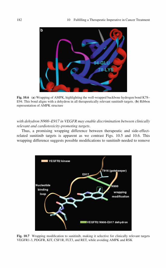
182 10 Fulfilling a Therapeutic Imperative in Cancer Treatment
b
Fig. 10.6 (a) Wrapping of AMPK, highlighting the well-wrapped backbone hydrogen bond K78–E94. This bond aligns with a dehydron in all therapeutically relevant sunitinib targets. (b) Ribbonrepresentation of AMPK structure
with dehydron N900–E917 in VEGFR may enable discrimination between clinicallyrelevant and cardiotoxicity-promoting targets.
Thus, a promising wrapping difference between therapeutic and side-effect-related sunitinib targets is apparent as we contrast Figs. 10.5 and 10.6. Thiswrapping difference suggests possible modifications to sunitinib needed to remove
Fig. 10.7 Wrapping modification to sunitinib, making it selective for clinically relevant targetsVEGFR1-3, PDGFR, KIT, CSF1R, FLT3, and RET, while avoiding AMPK and RSK

10.7 Is a Paradigm Shift in Drug Discovery Imminent? 183
its cardiotoxicity and at the same time probe the potential of AMPK and RSK ascardiotoxicity biomarkers.
According to the premises of the wrapping technology, the methylation at theposition indicated in Fig. 10.7 would turn the drug into a selective wrapper of dehy-dron N900–E917 in VEGFR. This drug is likely to bind selectively to all clinicallyrelevant targets since the latter contain a dehydron at the position that aligns withN900–E917 in VEGFR. By contrast, this wrapping modification will produce asteric clash upon association with kinases RSK and AMPK2. Following this reengi-neering, a series of experimental studies will be necessary to demonstrate the targetspecificity, efficacy (in vitro and in vivo), and safety (with primary emphasis oncardiac safety using in vitro and in vivo models).
10.7 Is a Paradigm Shift in Drug Discovery Imminent?
Mounting evidence from the treatment of complex disorders and malignanciesreveals that therapeutic efficacy does not correlate with drug specificity. Thisobservation has motivated a reassessment of the therapeutic value of promiscuityand may well trigger a paradigm shift, from magic bullets to multi-target ther-apies. These conceptual leaps are supported by novel high-throughput screeningtechnologies to assess cross-reactivity, hidden phenotypes, and side effects.
While a paradigm shift may be perceived as a necessity, it is far from becom-ing a reality. The major impediment arises because we do not have a clear pictureof the therapeutic advantages of the novel concepts that would replace drug speci-ficity. Knowing the realm of activity of “magic shotguns” is insufficient to warrantbroad therapeutic application. The clinical uncertainties and risks they introduceare likely broader than the more limited side effects associated with more specificdrugs.
Novel systems biology insights are leading to the belief that multi-target or evenpromiscuous drugs would be more effective to deal with complex diseases sincedynamical objects like biochemical fluxes and not the molecules themselves are nowperceived as the appropriate targets. However, the greater danger of side effects aris-ing in multi-target therapies clearly counterbalances the recent faith bestowed uponthem. Thus, we propose the use of selectivity filters to guide the rational control ofspecificity toward targets of clinical relevance within the systems biology frame ofinference that dictated the use of multi-target drugs in the first place. It is clear thattumors may be more efficiently tackled by opening several therapeutic fronts butonly a selectively promiscuous drug would be operative in such scenario.
At this juncture, multi-target molecular therapies could only be welcomed if theirtarget selectivity can be controlled to curb side effects and clinical uncertainties.Thus, there is a niche in emerging biotechnologies for novel approaches to cleanpromiscuous drugs in order to achieve tighter specificity control. We have shownthat such approaches are in principle feasible by redesigning highly cross-reactivekinase inhibitors, guided by novel selectivity filters. The drug redesign exercises and

184 10 Fulfilling a Therapeutic Imperative in Cancer Treatment
the proof of principle surveyed in this chapter suggest that cleaning a promiscuousdrug guided by basic new concepts is in principle possible and will hopefully inspirefurther efforts in this regard. Promiscuity may become a welcomed feature in drugdesign and may well propel the needed paradigm shift, but only if cross-reactivitycan be held under tight control through the effective implementation of selectivityfilters.
References
1. Dancey J, Sausville EA (2003) Issues and progress with protein kinase inhibitors for cancertreatment. Nat Rev Drug Discov 2:296–313
2. Levitski A, Gazit A (1995) Tyrosine kinase inhibition: An approach to drug development.Science 267:1782–1788
3. Tibes R, Trent J, Kurzrock R (2005) Tyrosine kinase inhibitors and the dawn of molecularcancer therapeutics. Annu Rev Pharmacol Toxicol 45:357–384
4. Gibbs J, Oliff A (1994) Pharmaceutical research in molecular oncology. Cell 79:193–1985. Donato NJ, Talpaz M (2000) Clinical use of tyrosine kinase inhibitors: Therapy for chronic
myelogenous leukemia and other cancers. Clin Cancer Res 6:2965–29666. Roth BL, Sheffler DJ, Kroeze WK (2004) Magic shotguns versus magic bullets:
Selectively non-selective drugs for mood disorders and schizophrenia. Nat Rev Drug Discov3:353–359
7. Frantz S (2005) Drug discovery: Playing dirty. Nature 437:942–9438. Keith CT, Borisy AA, Stockwell BR (2005) Multicomponent therapeutics for networked
systems. Nat Rev Drug Discov 4:71–789. Mencher SK, Wang LG (2005) Promiscuous drugs compared to selective drugs (promiscuity
can be a virtue). BMC Clin Pharmacol 5:3–910. McGovern SL, Helfand BT, Feng B, Shoichet BK (2003) A specific mechanism of nonspecific
inhibition. J Med Chem 46:4265–427211. Feng BY, Shelat A, Doman TN et al (2005) High-throughput assays for promiscuous
inhibitors. Nat Chem Biol 1:146–14812. Force T, Krause D, van Etten RA (2007) Molecular mechanisms of cardiotoxicity of tyrosine
kinase inhibition. Nat Rev Cancer 7:332–34413. Fernández A, Sanguino A, Peng Z et al (2007) An anticancer C-kit kinase inhibitor is re-
engineered to make it more active and less cardiotoxic. J Clin Invest 117:4044–405414. Demetri GD (2007) Structural reengineering of imatinib to decrease cardiac risk in cancer
therapy. J Clin Invest 117:3650–365315. Crunkhorn S (2008) Anticancer drugs: Redesigning kinase inhibitors. Nat Rev Drug Discov
7:120–12116. Hopkins AL, Mason J, Overington J (2006) Can we rationally design promiscuous drugs?
Curr Opin Struct Biol 16:127–13617. Hampton T (2004) ‘Promiscuous’ anticancer drugs that hit multiple targets may thwart
resistance. J Am Med Assoc 292:419–42218. Hopkins AL, Ren J, Milton J et al (2004) Design of non-nucleoside inhibitors of HIV-1 reverse
transcriptase with improved drug resistance properties. J Med Chem 47:5912–592219. Owens J (2006) Screening: Dirty drugs’ secrets uncovered. Nat Rev Drug Discov 5:54220. Fabian MA, Biggs WH, Treiber DK et al (2005) A small molecule kinase interaction map for
clinical kinase inhibitors. Nat Biotechnol 23:329–33621. Karaman MW, Herrgard S, Treiber DK et al (2008) A quantitative analysis of kinase inhibitor
selectivity. Nat Biotechnol 26:127–13222. Fedorov O, Marsden B, Pogacic V et al (2007) A systematic interaction map of validated
kinase inhibitors with Ser/Thr kinases. Proc Natl Acad Sci USA 104:20523–20528
References 185
23. MacDonald ML, Lamerdin J, Owens S et al (2006) Identifying off-targets effects and hiddenphenotypes of drugs in human cells. Nat Chem Biol 2:329–337
24. Brunton LL, Lazo JS, Parker KL (2006) Goodman & Gilman’s The Pharmacological Basis ofTherapeutics. 11th ed. McGraw-Hill, New York
25. Rishton GM (2005) Failure and success in modern drug discovery: guiding principles inthe establishment of high probability of success drug discovery organizations. Med Chem 1:519–527
26. Kerkela R, Grazette L, Yacobi R et al (2006) Cardiotoxicity of the cancer therapeutic agentimatinib mesylate. Nat Med 12:908–916
27. Fedorov O, Sundstrom M, Marsden B, Knapp S (2007) Insights for the development ofspecific kinase inhibitors by targeted structural genomics. Drug Discov Today 12:365–372
28. Bogoyevitch MA, Fairlie DP (2007) A new paradigm for protein kinase inhibition: blockingphosphorylation without directly targeting ATP binding. Drug Discov Today 12:622–633
29. Crespo A, Fernández A (2007) Kinase packing defects as drug targets. Drug Discov Today12:917–923
30. Chen J, Zhang X, Fernández A (2007) Molecular basis for specificity in the druggable kinome:Sequence-based analysis. Bioinformatics 23:563–572
31. Griffin JD (2005) Interaction maps for kinase inhibitors. Nat Biotechnol 23:308–30932. Liu Y, Gray NS (2006) Rational design of inhibitors that bind to inactive kinase conforma-
tions. Nat Chem Biol 2:358–36433. Noble ME, Endicott JA, Johnson LN (2004) Protein kinase inhibitors: Insights into drug
design from structure. Science 303:1800–180534. Torrance CJ, Jackson PE, Montgomery E et al (2000) Combinatorial chemoprevention of
intestinal neoplasia. Nat Med 6:1024–102835. Erlichman C, Hidalgo M, Boni JB et al (2006) Phase I study of EKB-569, an irreversible
inhibitor of the epidermal growth factor receptor, in patients with advanced solid tumors.J Clin Oncol 24:2252–2260
36. Wissner A, Overbeek E, Reich MF et al (2003) Synthesis and structure-activity relation-ships of 6,7-disubstituted 4-anilinoquinoline-3-carbonitriles. The design of an orally active,irreversible inhibitor of the tyrosine kinase activity of the epidermal growth factor receptor(EGFR) and the human epidermal growth factor receptor-2 (HER-2). J Med Chem 46:49–63
37. Zhang X, Crespo A, Fernández A (2008) Turning promiscuous kinase inhibitors into saferdrugs. Trends Biotechnol 26:295–301
38. Fernández A, Maddipati S (2006) The a-priori inference of cross reactivity for drug targetedkinases. J Med Chem 49:3092–3100
39. Butcher EC (2004) Systems biology in drug discovery. Nat Biotechnol 22:1253–125940. Butcher EC (2005) Can cell systems biology rescue drug discovery? Nat Rev Drug Discov
4:461–46741. Kitano H (2007) A robustness-based approach to systems-oriented drug design. Nat Rev Drug
Discov 6:202–20942. Volgestein B, Kinzler KW (2004) Cancer genes and the pathway they control. Nat Med
10:789–79943. Kitano H (2004) Biological robustness. Nat Rev Genet 5:826–83744. Carlson JM, Doyle J (2002) Complexity and robustness. Proc Natl Acad Sci USA 99:
2538–254545. Hellerstein MK (2008) Exploiting complexity and the robustness of network architecture for
drug discovery. J Pharmacol Exp Ther 325:1–946. Seidman A (2002) Cardiac dysfunction in the trastuzumab clinical trials experience. J Clin
Oncol 20:1215–122147. Martel CL (2006) Bevacizumab-related toxicities: Association of hypertension and protein-
uria. Commun Oncol 3:90–9348. Hampton T (2004) ‘Promiscuous’ anticancer drugs that hit multiple targets may thwart
resistance. J Am Med Assoc 292:419–422

186 10 Fulfilling a Therapeutic Imperative in Cancer Treatment
49. Motzer RJ, Michaelson MD, Redman BG et al (2006) Activity of SU11248, a multitargetedinhibitor of vascular endothelial growth factor receptor and platelet-derived growth factorreceptor, in patients with metastatic renal cell carcinoma. J Clin Oncol 24:16–24
50. Joensuu, H. (2007) Cardiac toxicity of sunitinib. Lancet 370:1978–198051. Chu TF, Rupnick MA, Kerkela R et al (2007) Cardiotoxicity associated with tyrosine kinase
inhibitor sunitinib. Lancet 370:2011–2019
Chapter 11Inducing Folding By Crating the Target
Kinases may be exploited as anticancer drug targets but their conformational plastic-ity often hinders the success of structure-based design. This is because of structuraladaptation: target structures may change or proteins may adopt new conformationsupon association with the ligand in unexpected and unpredictable ways. This maybe the main reason for the modest interest in rational drug design when targetingregions with high disorder propensity such as the activation loop of a protein kinase.Yet this region presents the largest amino acid variability within the family and thusconstitutes an attractive target to control specificity. In this chapter we advocate for astrategy to target flexible regions, offering a way to control the induced folding andturn it into a selectivity-promoting feature. Thus, drugs designed to wrap disorderedregions in the target may be used to steer induced folding in very specific ways.The results surveyed herald the paradigmatic design concept of protective drugs forstructurally adaptable targets.
11.1 Induced Folding: The Bête Noire of Drug Design
As described in Chaps. 7, 8, 9, and 10, kinases offer desirable therapeutic oppor-tunities because they transduce cell signals implicated in cancer progression anddevelopment and because they possess a natural binding pocket susceptible of beingtargeted by a man-made ligand [1–5]. These binding regions are framed by floppyregions (nucleotide-binding loop, activation loop, and catalytic loop) whose compo-sition determines the activation state of the kinase and its substrate specificity, henceconferring them their functional identity [5–10].
Precisely because of the conformational plasticity of the purported targets,structure-based design of kinase inhibitors has met with modest success. As aninhibitor binds to a kinase, it often promotes a largely unpredictable structuraladaptation of the floppy regions. This adaptation is referred to as the induced fold-ing. This phenomenon poses design problems since the stability of the drug–targetcomplex, and hence the drug affinity, cannot be meaningfully determined from thestructures of the isolated binding partners [6–11]. The ab initio prediction of theinduced fold remains forbiddingly difficult, often precluding a rational control of
187A. Fernández, Transformative Concepts for Drug Design: Target Wrapping,DOI 10.1007/978-3-642-11792-3_11, C© Springer-Verlag Berlin Heidelberg 2010
188 11 Inducing Folding By Crating the Target
cross-reactivity for drugs that interact with loopy regions [6, 7]. In fact, the inducedfolding problem itself is comparable to the protein folding problem in terms ofcomplexity and intractability [7–9].
However intractable, we are compelled to deal with this problem for a very sim-ple reason: the highest variability in amino acid composition across kinases occursprecisely in the floppy regions, more specifically in the activation loop [12]. Thus,the most significant specificity-promoting feature becomes in practice the mostdifficult to target!
As illustrated in this chapter, the induced folding problem may be tractable inspecial contexts that are relevant to drug design: there are restrictive instances wherestructural adaptation may be steered in a controllable and predictable way, and selec-tivity control may be achieved through the identification of targets that undergo asimilar induced fit upon association.
11.2 Wrapping the Target: A Tractable Case of InducedFolding
To deal with a tractable problem, we shall focus on small regions (four to eightresidues) of the protein chain which are unstructured in the uncomplexed state andrepresent extreme cases of wrapping deficiency, that is, they are unable to hinderbackbone hydration to any significant extent [11]. As discussed in Chap. 5, suchregions are unequivocally characterized as being unable to contribute with at leastseven nonpolar wrapping groups to the microenvironment of putative backbonehydrogen bonds. Since ρ = 7 is the threshold for existence of a backbone hydrogenbond, such regions are disordered with 100% certainty in the uncomplexed state ofthe protein. To distinguish the solvent shielding of induced hydrogen bonds fromthat of pre-formed hydrogen bonds, we shall introduce the term “crating” whenreferring to the former while retaining the usual term wrapping for the latter.
We shall focus on the problem of targeting regions that visit conformations wherethe backbone hydrogen bonds are too exposed to the solvent to be retained in sol-uble structure. The backbone amides and carbonyls in these regions are actuallyprone to become completely hydrated with concurrent loss of structure (Fig. 11.1).Thus, drug ligands may be specifically designed to “correct” the severe yet local-ized packing deficiency by removing surrounding water upon association, therebyinducing a hydrogen bond which is missing in the uncomplexed state of the protein.The design principle is illustrated in Fig. 11.1 and is based on ligand designs whichare protective of specific conformations visited but not retained by the target proteinin the unbound state.
As emphasized in previous chapters, in regions where kinase structure is retainedupon binding, drug selectivity may be achieved or at least controlled by targetingdehydrons [12]. This special type of targeting is realized by engineering a drugthat wraps the pre-formed intramolecular hydrogen bond [13–15], effectively con-tributing with nonpolar groups to the exclusion of water from the hydrogen bondmicroenvironment.

11.2 Wrapping the Target: A Tractable Case of Induced Folding 189
Fig. 11.1 Basic scheme of the folding-inducing drug. Due to insufficient crating in the unboundstate (ρ < 7), the target protein visits but does not retain a hydrogen-bonded conformation that isin dynamic equilibrium with the fully hydrated amide and carbonyl. Since the drug (gray sphere)is able to provide sufficient crating (ρ > 7), the target protein in the drug-bound state is ableto retain the hydrogen-bonded conformation. Since the proximity of a nonpolar moiety hindershydration, the drug has no affinity for the alternative conformation of the protein target, with itssolvent-exposed amide and carbonyl
Thus, a backbone hydrogen bond inferred from homolog structure alignmentmay be absent in a reported protein structure due to severe under-protection, i.e.,when ρ < 7 [11]. Yet, it may be selectively induced if a drug is appropriately con-structed to crate it, much like the drugs designed to “correct” wrapping defects intarget proteins [12]. The crating design concept on a structurally adaptable target isillustrated in Fig. 11.2, which displays a complex between a protective drug and akinase with a drug-induced folded region arising from the intermolecular crating ofa hydrogen bond. The F811–A814 dehydron within the activation loop (in red) ofthe C-Kit kinase is being crated and hence induced by a purposely designed modifi-cation of the anticancer drug imatinib. The latter is known to target the C-Kit kinase,while the modified version, known as WBZ_7 [16], provides additional crating tothe activation loop, enhancing affinity and selectivity beyond the levels achieved bythe parental compound [16]. In other words, in the unbound state of the protein,the activation loop is likely to visit the conformation that contains the F811–A814backbone hydrogen bond, but it is unlikely to retain it due to the local absence oftertiary structure. Yet, when the ligand is able to provide appropriate crating, thebond becomes inducible as the loop is able to adopt a specific conformation. Hencethere is a crucial difference between wrapping and crating: while wrapping refers toan extra level of shielding or protection of a pre-formed hydrogen bond retained inthe uncomplexed protein, crating provides exogenous shielding to a hydrogen bondthat would otherwise not survive in the uncomplexed state.
The design concept of drug as crater of a specific local conformation adoptedby a floppy region in the target has in fact been exploited in re-workings of ima-tinib [14]. As described in Chap. 8, this wrapping-based redesign not only targetsa unique dehydron in the intended target C-Kit but also crates an otherwise floppy
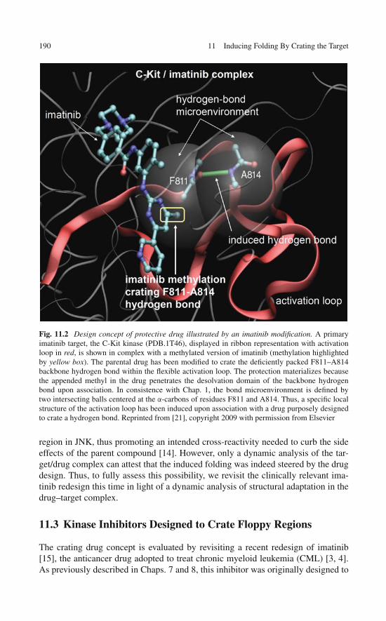
190 11 Inducing Folding By Crating the Target
Fig. 11.2 Design concept of protective drug illustrated by an imatinib modification. A primaryimatinib target, the C-Kit kinase (PDB.1T46), displayed in ribbon representation with activationloop in red, is shown in complex with a methylated version of imatinib (methylation highlightedby yellow box). The parental drug has been modified to crate the deficiently packed F811–A814backbone hydrogen bond within the flexible activation loop. The protection materializes becausethe appended methyl in the drug penetrates the desolvation domain of the backbone hydrogenbond upon association. In consistence with Chap. 1, the bond microenvironment is defined bytwo intersecting balls centered at the α-carbons of residues F811 and A814. Thus, a specific localstructure of the activation loop has been induced upon association with a drug purposely designedto crate a hydrogen bond. Reprinted from [21], copyright 2009 with permission from Elsevier
region in JNK, thus promoting an intended cross-reactivity needed to curb the sideeffects of the parent compound [14]. However, only a dynamic analysis of the tar-get/drug complex can attest that the induced folding was indeed steered by the drugdesign. Thus, to fully assess this possibility, we revisit the clinically relevant ima-tinib redesign this time in light of a dynamic analysis of structural adaptation in thedrug–target complex.
11.3 Kinase Inhibitors Designed to Crate Floppy Regions
The crating drug concept is evaluated by revisiting a recent redesign of imatinib[15], the anticancer drug adopted to treat chronic myeloid leukemia (CML) [3, 4].As previously described in Chaps. 7 and 8, this inhibitor was originally designed to
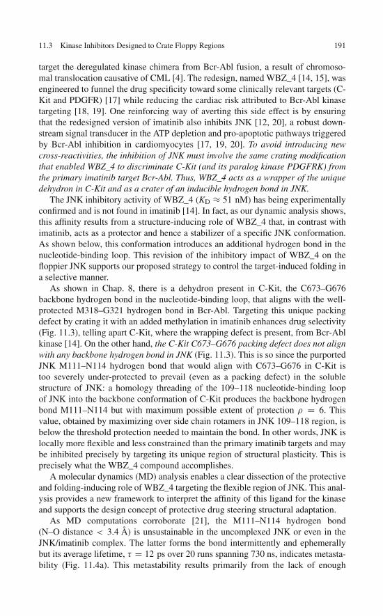
11.3 Kinase Inhibitors Designed to Crate Floppy Regions 191
target the deregulated kinase chimera from Bcr-Abl fusion, a result of chromoso-mal translocation causative of CML [4]. The redesign, named WBZ_4 [14, 15], wasengineered to funnel the drug specificity toward some clinically relevant targets (C-Kit and PDGFR) [17] while reducing the cardiac risk attributed to Bcr-Abl kinasetargeting [18, 19]. One reinforcing way of averting this side effect is by ensuringthat the redesigned version of imatinib also inhibits JNK [12, 20], a robust down-stream signal transducer in the ATP depletion and pro-apoptotic pathways triggeredby Bcr-Abl inhibition in cardiomyocytes [17, 19, 20]. To avoid introducing newcross-reactivities, the inhibition of JNK must involve the same crating modificationthat enabled WBZ_4 to discriminate C-Kit (and its paralog kinase PDGFRK) fromthe primary imatinib target Bcr-Abl. Thus, WBZ_4 acts as a wrapper of the uniquedehydron in C-Kit and as a crater of an inducible hydrogen bond in JNK.
The JNK inhibitory activity of WBZ_4 (KD ≈ 51 nM) has being experimentallyconfirmed and is not found in imatinib [14]. In fact, as our dynamic analysis shows,this affinity results from a structure-inducing role of WBZ_4 that, in contrast withimatinib, acts as a protector and hence a stabilizer of a specific JNK conformation.As shown below, this conformation introduces an additional hydrogen bond in thenucleotide-binding loop. This revision of the inhibitory impact of WBZ_4 on thefloppier JNK supports our proposed strategy to control the target-induced folding ina selective manner.
As shown in Chap. 8, there is a dehydron present in C-Kit, the C673–G676backbone hydrogen bond in the nucleotide-binding loop, that aligns with the well-protected M318–G321 hydrogen bond in Bcr-Abl. Targeting this unique packingdefect by crating it with an added methylation in imatinib enhances drug selectivity(Fig. 11.3), telling apart C-Kit, where the wrapping defect is present, from Bcr-Ablkinase [14]. On the other hand, the C-Kit C673–G676 packing defect does not alignwith any backbone hydrogen bond in JNK (Fig. 11.3). This is so since the purportedJNK M111–N114 hydrogen bond that would align with C673–G676 in C-Kit istoo severely under-protected to prevail (even as a packing defect) in the solublestructure of JNK: a homology threading of the 109–118 nucleotide-binding loopof JNK into the backbone conformation of C-Kit produces the backbone hydrogenbond M111–N114 but with maximum possible extent of protection ρ = 6. Thisvalue, obtained by maximizing over side chain rotamers in JNK 109–118 region, isbelow the threshold protection needed to maintain the bond. In other words, JNK islocally more flexible and less constrained than the primary imatinib targets and maybe inhibited precisely by targeting its unique region of structural plasticity. This isprecisely what the WBZ_4 compound accomplishes.
A molecular dynamics (MD) analysis enables a clear dissection of the protectiveand folding-inducing role of WBZ_4 targeting the flexible region of JNK. This anal-ysis provides a new framework to interpret the affinity of this ligand for the kinaseand supports the design concept of protective drug steering structural adaptation.
As MD computations corroborate [21], the M111–N114 hydrogen bond(N–O distance < 3.4 Å) is unsustainable in the uncomplexed JNK or even in theJNK/imatinib complex. The latter forms the bond intermittently and ephemerallybut its average lifetime, τ = 12 ps over 20 runs spanning 730 ns, indicates metasta-bility (Fig. 11.4a). This metastability results primarily from the lack of enough

192 11 Inducing Folding By Crating the Target
Fig. 11.3 Protective drug as folding inducer. Structural alignment of targets JNK (isoform 1)and C-Kit for the imatinib redesigned prototype drug WBZ_4 [14]. The JNK backbone fromPDB.1UKH is displayed as a thicker line than that of C-Kit (PDB.1T46). The C-Kit residues Y672and Y675 contribute to crate the “crankshaft” C673–G676 hydrogen bond (dashed line) in C-Kit.This bond gets exogenous protection from the drug/ligand which has been purposely engineered tocontribute to its crating. This crating capability represents the major difference between WBZ_4and the parental drug imatinib. Protective interactions contributing to the dehydration of the C673–G676 hydrogen bond are indicated as thin green lines. The JNK residues aligning with C673 andG676 in C-Kit are M111 and N114. Lacking enough intramolecular protection (the protectiveY675 in C-Kit is substituted for the poor protector A113 in JNK), they can only become paired byan induced backbone hydrogen bond (arrow) upon association with WBZ_4. This induced localfolding does not prevail upon binding to the parental drug imatinib since the latter lacks the cratinggroup at the terminal ring. Reprinted from [21], copyright 2009 with permission from Elsevier
intramolecular protection: the protective Y675 in C-Kit is substituted in JNK forthe poor protector A113 (Fig. 11.2 and Table11.1). By contrast, the hydrogen bondis induced upon association with WBZ_4 (Fig. 11.4a): the added methyl in WBZ_4brings extra protection upon binding.
�Fig. 11.4 Folding upon binding dynamics of JNK in complex with a protective drug. (a) Dynamicsof the M111–N114 backbone hydrogen bond formation in JNK (isoform 1) in the uncomplexedform, within the complex with imatinib, and within the complex with the crating ligand WBZ_4.The induced folding is monitored by the M111–N114 carbonyl–amide O–N distance and mate-rializes when this distance is below a hydrogen bond threshold (~3.4 Å, blue line). Twenty runswere generated for each case and the dynamics averaged over the trajectory ensemble is indi-cated by the red line plot. The black line plot corresponds to a single representative run. Forthe uncomplexed kinase, the runs covered an equilibration span of 500 ns (time extensions weredeemed unnecessary due to equilibration) and represented the control results. The folding dynam-ics for the complexes covered 730 ns. (b) Induced hydrogen bond dynamics for the N114D JNKmutant in complex with the crating drug WBZ_4. The stochastic boundary molecular dynam-ics (SBMD) method [21] was employed to reduce simulation times while capturing the localized

11.3 Kinase Inhibitors Designed to Crate Floppy Regions 193
a
b
Fig. 11.4 (continued) binding-folding dynamics at the ATP-binding site of the JNK kinase. Thesystem has a reaction zone of 42 residues and a reservoir region. The MD simulations wereperformed using the structural coordinates of free JNK (control), chimeric JNK/imatinib, andJNK/WBZ_4 complexes that resulted from alignment of the JNK1 crystal structure PDB.1UKHwith that of the C-Kit/imatinib complex PDB.1T46, followed by in silico removal of the Kitdomain. Hence, the simulations described the equilibration process of the JNK/imatinib andJNK/WBZ_4 complexes originally obtained from alignment and combination of the PDB files1UKH and 1T46. Reprinted from [21], copyright 2009 with permission from Elsevier

194 11 Inducing Folding By Crating the Target
The computational analysis supports and rationalizes the experimentally estab-lished affinity of WBZ_4 for JNK as being mainly due to its ability to crate andthus stabilize a particular JNK conformation. This conformation contains the M111–N114 backbone hydrogen bond that aligns with the packing defect C673–G676 inC-Kit and becomes an induced fit in JNK upon binding to WBZ_4.
To corroborate that the induction of the M111–N114 hydrogen bond is the dom-inant factor dictating the drug affinity for JNK, we examined the residues aligningwith the C-Kit C673 and G676 across paralogs of JNK(1/3) with PDB-reportedstructure: MAPK1, MAPK6, p38α, and p38γ (Table 11.1). As shown elsewhere[14], WBZ_4 does not have any detectable affinity for such paralogs. Indeed, weexpect that none of the paralogs is capable of maintaining the crankshaft hydro-gen bond because one of the putatively paired residues is charged (Asp) ratherthan the polar N114 in JNK (Table 11.1). Furthermore, the extent of crating of theJNK paralogs is comparable to that of JNK, and less than that of C-Kit, mainlybecause of the replacement of the C-Kit Y675 for poorer protectors (A, T). Thecharged Asp aligning with position N114 in JNK has higher hydrophilicity andis therefore disruptive of the backbone hydrogen bond. This is corroborated notonly by the absence of a crankshaft hydrogen bond in all reported structures ofJNK(1/3) paralogs (Table 11.1) but also dynamically (Fig. 11.4b). In contrast withwild-type JNK, the crating drug WBZ_4 fails to induce the crankshaft hydrogenbond in the N114D in silico mutated JNK (Fig. 11.4b). Thus, the substitution ofpolar N114 for charged D114 introduces disruptive negative design, preventing sidechain nonpolar groups from protectively clustering around the putative M111–D114bond.
Table 11.1 Across-paralog alignment of residues that either form or crate the inducible backbonehydrogen bond M111–N114 in JNK
Kinase Kit Jnk1 Jnk3 MAPK6 MAPK1 p38α p38γ
PDB code 1T46 1UKH 1PMN 2I6L 2OJI 1A9U 1CM8Gatekeeper THR670 MET108 MET146 GLN108 GLN103 THR106 MET109HB crating TYR672 LEU110 LEU148 TYR110 LEU105 LEU108 PHE111Crankshaft HB CYS673 MET111 MET149 MET111 MET106 MET109 MET112HB crating TYR675 ALA113 ALA151 THR113 THR108 ALA111 THR114Crankshaft HB GLY676 ASN114 ASN152 ASP114 ASP109 ASP112 ASP115Crankshaft HB
inducible?Yes Yes Yes No No No No
Crating drugbinding?
Yes Yes Yes No No No No
The alignment extends over all paralogs of JNK (isoforms 1 and 3) with PDB-reported structureand enables an inference of the feasibility of inducing the hydrogen bond. This feasibility dependson the purported extent of crating of the putative hydrogen bond and on the hydration propensityof the residues paired by the bond and becomes a necessary and sufficient condition for detectableaffinity of the protective drug WBZ_4

References 195
11.4 Steering Induced Folding with High Specificity:The Emergence of the Crating Design Concept
Their therapeutic appeal notwithstanding, from a structural biology perspective,kinase targets pose daunting problems to the drug designer: their floppy regionscan undergo considerable structural adaptation upon binding to the drug. Thus, dueto the often unpredictable nature of induced folding, structure-based drug designhas met limited success and is often relegated in favor of high-throughput screeningmethods and serendipitous engineering as the means to find or improve molecularleads. This leaves us with a conceptual conundrum since floppy regions, in partic-ular the activation loop, are the most unique in the kinase targets, i.e., the ones thatconfer the kinase its functional and contextual identity.
A rational control of induced folding is nevertheless possible and of course highlydesirable, as shown in this chapter. This control may be achieved by focusing onstructural adaptations of deficiently wrapped regions of the protein chain. Thus, wepropose a targeting approach suitable for floppy regions in kinases based on lig-and/drug designs which crate specific conformations of the protein that would notbe otherwise sustainable in the uncomplexed state. Such drugs contribute to improvethe wrapping of the target upon association with the point where the latter is nowcapable of retaining a specific conformation. Thus, the target affinity of folding-inducing drugs is due in part to the fact that they stabilize the drug/target complex.Support for this concept is found through re-examination of a recently re-engineeredversion of imatinib in light of a dynamic analysis. Our dynamic assessment showsthat the inhibitory activity of the compound results precisely from its ability to steerinduced folding by protecting a conformation in a floppy kinase. On the other hand,since packing deficiencies are fairly unique to kinase targets and typically not con-served across proteins of common ancestry, the specificity of such protective drugsis basically guaranteed.
On the horizon we envision a new generation of protective kinase inhibitors stem-ming from known chemical scaffolds that may induce specific local structuring ofthe activation loop, the most extensive flexible region. Such drugs may exquisitelycompete with ATP as they possess an extra anchoring site so far unexploited. Thispossibility may represent one of the few approaches to the problem of rationallyexploiting induced folding in floppy targets to steer drug cross-reactivity in waysthat provide clinical advantage.
References
1. Dancey J, Sausville EA (2003) Issues and progress with protein kinase inhibitors for cancertreatment. Nat Rev Drug Discov 2:296–313
2. Tibes R, Trent J, Kurzrock R (2005) Tyrosine kinase inhibitors and the dawn of molecularcancer therapeutics. Annu Rev Pharmacol Toxicol 45:357–384
3. Schindler T, Bornmann W, Pellicena P et al (2000) Structural mechanism for STI-571inhibition of Abelson tyrosine kinase. Science 289:1938–1942

196 11 Inducing Folding By Crating the Target
4. Schiffer CA (2007) BCR-ABL tyrosine kinase inhibitors for chronic myelogenous leukemia.N Engl J Med 357:258–265
5. Crespo A, Fernández A (2007) Kinase packing defects as drug targets. Drug Discov Today12:917–923
6. Teague S (2003) Implications of protein flexibility for drug discovery. Nat Rev Drug Discov2:527–541
7. Damm KL, Carlson HA (2007) Exploring experimental sources of multiple protein confor-mations in structure-based drug design. J Am Chem Soc 129:8225–8235
8. Hornak V, Simmerling C (2007) Targeting structural flexibility in HIV-1 protease inhibitorbinding. Drug Discov Today 12:132–138
9. Erickson J (2004) Lessons in molecular recognition: The effects of ligand and proteinflexibility on molecular docking accuracy. J Med Chem 47:45–55
10 Noble ME, Endicott JA, Johnson LN (2004) Protein kinase inhibitors: Insights into drugdesign from structure. Science 303:1800–1805
11. Pietrosemoli N, Crespo A, Fernández A (2007) Dehydration propensity of order-disorderintermediate regions in soluble proteins. J Proteome Res 6:3519–3526
12. Chen J, Zhang X, Fernández A (2007) Molecular basis for specificity in the druggable kinome:Sequence-based analysis. Bioinformatics 23:563–572
13. Fernández A (2004) Keeping dry and crossing membranes. Nat Biotechnol 22:1081–108414. Fernández A, Sanguino A, Peng Z et al (2007) An anticancer C-kit kinase inhibitor is re-
engineered to make it more active and less cardiotoxic. J Clin Invest 117:4044–405415. Crunkhorn S (2008) Anticancer drugs: Redesigning kinase inhibitors. Nat Rev Drug Discov
7:120–12116. Fernández A, Sanguino A, Peng Z et al (2007) Rational drug redesign to overcome drug
resistance in cancer therapy: Imatinib moving target. Cancer Res 67:4028–403317. Demetri GD, von Mehren M, Blanke CD et al (2002) Efficacy and safety of imatinib mesylate
in advanced gastrointestinal stromal tumors. N Engl J Med 347:472–48018. Kerkela R, Grazette L, Yacobi R et al (2006) Cardiotoxicity of the cancer therapeutic agent
imatinib mesylate. Nat Med 12:908–91619. Force T, Krause D, van Etten RA (2007) Molecular mechanisms of cardiotoxicity of tyrosine
kinase inhibition. Nat Rev Cancer 7:332–34420. Demetri GD (2007) Structural reengineering of imatinib to decrease cardiac risk in cancer
therapy. J Clin Invest 117:3650–365321. Fernández A, Bazan S, Chen J (2009) Taming the induced folding of drug-targeted kinases.
Trends Pharmacol Sci 30: 66–71

Chapter 12Wrapper Drugs as Therapeutic Editorsof Side Effects
Wrapping designs have some limitations. While they enable control of specificity,they may not be able to exclude a toxicity-related target if the latter shares the samewrapping pattern with the therapeutically relevant targets. In such circumstances,we may need to resort to a multicomponent therapy where one drug acts synergis-tically with the other while selectively antagonizing it in the specific context wherethe action of the first drug promotes toxicity. This chapter explores these a prioritherapeutic possibilities.
As previously described, due to their ability to interfere with signal transductionevents controlling cell proliferation and fate, kinase inhibitors hold promise as anti-cancer agents. Nevertheless, the functional role of a kinase depends on the cellularcontext and hence kinase inhibition in an off-target cell may lead to side effects.Motivated by these observations, we explore a mode of “therapeutic editing” whereone drug – the editor – suppresses the side effect promoted by the primary drugas it impacts off-target cells. Editor and primary drug have overlapping therapeuticimpact, while the editor suppresses the downstream propagation of toxicity-relatedsignaling.
12.1 The Editor Concept
Side effects in molecular targeted anticancer therapy are common [1–5], hard toavoid [6], often unexpected [7], and potentially health-threatening [8], thereby intro-ducing daunting difficulties to drug development. At least two major problems mustbe dealt with when addressing side effects [4–10]: (a) target proteins have paralogswith similar 3D structure [9, 10] and (b) the target protein plays a role in off-targetcells that is different from the role it plays in target (cancer) cells [5–7]. Accordingly,undesired cross-reactivities are expected due to (a), while (b) implies that target inhi-bition may be therapeutically efficacious in cancer cells, but toxic in off-target cells.This chapter will be limited to kinase inhibitors (KIs), the drugs that interfere withcell-signaling pathways, because of their promising prospects for anticancer therapy[9–12] and because such treatments require addressing the problems outlined above[5, 11–13].
197A. Fernández, Transformative Concepts for Drug Design: Target Wrapping,DOI 10.1007/978-3-642-11792-3_12, C© Springer-Verlag Berlin Heidelberg 2010

198 12 Wrapper Drugs as Therapeutic Editors of Side Effects
Problem (a) motivated the exploitation of selectivity filters for drug design [9,10], leading to an efficacious control of specificity through paralog discrimination[13]. This problem has been dealt with extensively in Chaps. 7, 8, and 9. wherewe showed that dehydrons in proteins can be used to distinguish family members,and that such packing defects can be targeted using ligands engineered to “wrap” orshield them from interactions with water [10, 13], resulting in high specificity.
Dealing with problem (b) requires advanced network annotation [5, 14]. Theproblem can be dissected by comparing protein interaction networks across tissuesand placing the target protein in different interacting contexts represented in variouscell types [14]. In this chapter we focus on problem (b) and advocate a strategy toavert the resulting side effects.
The context-dependence problem has been effectively dealt with through for-mulations for tissue-specific delivery [15–17]. The strategies to enhance tissuespecificity include incorporating the drug molecules into nanosystems such as lipo-somes, coupling drugs to antibodies or ligands that target cell receptors, exploitingpermeation enhancers or passive or active translocators. These approaches often failto be completely specific and introduce additional problems like limited delivery tothe site of action.
In light of this situation, we explore an alternative systemic approach to dealwith the context-dependence problem: a mode of “therapeutic editing” using two-component free-drug treatments. One drug – the editor – suppresses the sideeffect promoted by the primary drug, while both drugs overlap in their impacton therapeutically relevant targets. Thus, the editor selectively suppresses down-stream propagation of toxicity-related signaling while circumventing the deliv-ery issues that plague the conventional approaches to the context-dependenceproblem.
12.2 Editing Drugs to Curb Side Effects
Context-dependent functionalities introduce a risk of toxicity in molecular targettherapy [4–7]. For example, the constitutively active chimera Bcr-ABL kinase is aclinical target for treating chronic myeloid leukemia (CML), as evidenced by thesuccess of the KI imatinib (Gleevec) [18], but ABL inhibition in cardiomyocytes(off-target cells in CML treatment) may introduce a cardiotoxicity risk [5–7], asshown in Fig. 12.1. This risk is more pronounced for promiscuous KIs like sunitinib[5, 8].
In general, prevention of side effects arising from context dependence requirestissue-specific delivery [15–17] to selectively avoid inhibiting a target kinase in off-target cells. In practice, this level of selectivity is difficult to achieve. In view ofthis problem, we propose a two-component treatment in which one drug modu-lates a downstream component of the toxicity-signaling pathway recruited by theother.
Two-component treatments have been exploited to improve therapeutic impact[19–21]. In this chapter we advocate an alternative application with the aim of
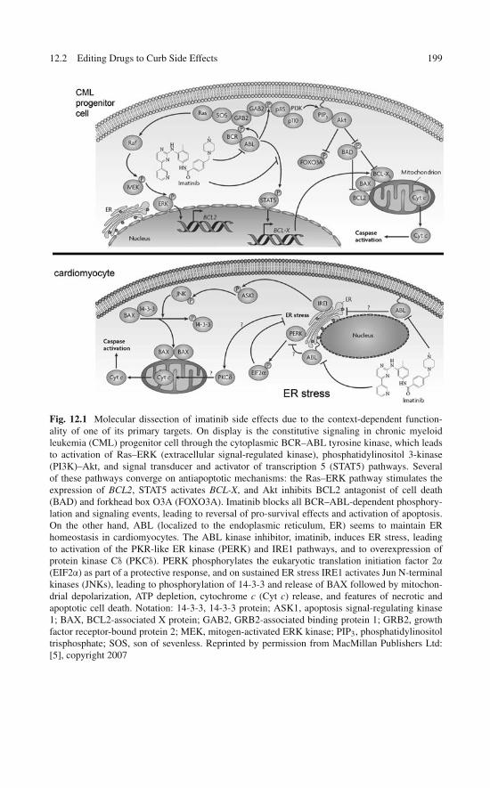
12.2 Editing Drugs to Curb Side Effects 199
Fig. 12.1 Molecular dissection of imatinib side effects due to the context-dependent function-ality of one of its primary targets. On display is the constitutive signaling in chronic myeloidleukemia (CML) progenitor cell through the cytoplasmic BCR–ABL tyrosine kinase, which leadsto activation of Ras–ERK (extracellular signal-regulated kinase), phosphatidylinositol 3-kinase(PI3K)–Akt, and signal transducer and activator of transcription 5 (STAT5) pathways. Severalof these pathways converge on antiapoptotic mechanisms: the Ras–ERK pathway stimulates theexpression of BCL2, STAT5 activates BCL-X, and Akt inhibits BCL2 antagonist of cell death(BAD) and forkhead box O3A (FOXO3A). Imatinib blocks all BCR–ABL-dependent phosphory-lation and signaling events, leading to reversal of pro-survival effects and activation of apoptosis.On the other hand, ABL (localized to the endoplasmic reticulum, ER) seems to maintain ERhomeostasis in cardiomyocytes. The ABL kinase inhibitor, imatinib, induces ER stress, leadingto activation of the PKR-like ER kinase (PERK) and IRE1 pathways, and to overexpression ofprotein kinase Cδ (PKCδ). PERK phosphorylates the eukaryotic translation initiation factor 2α
(EIF2α) as part of a protective response, and on sustained ER stress IRE1 activates Jun N-terminalkinases (JNKs), leading to phosphorylation of 14-3-3 and release of BAX followed by mitochon-drial depolarization, ATP depletion, cytochrome c (Cyt c) release, and features of necrotic andapoptotic cell death. Notation: 14-3-3, 14-3-3 protein; ASK1, apoptosis signal-regulating kinase1; BAX, BCL2-associated X protein; GAB2, GRB2-associated binding protein 1; GRB2, growthfactor receptor-bound protein 2; MEK, mitogen-activated ERK kinase; PIP3, phosphatidylinositoltrisphosphate; SOS, son of sevenless. Reprinted by permission from MacMillan Publishers Ltd:[5], copyright 2007
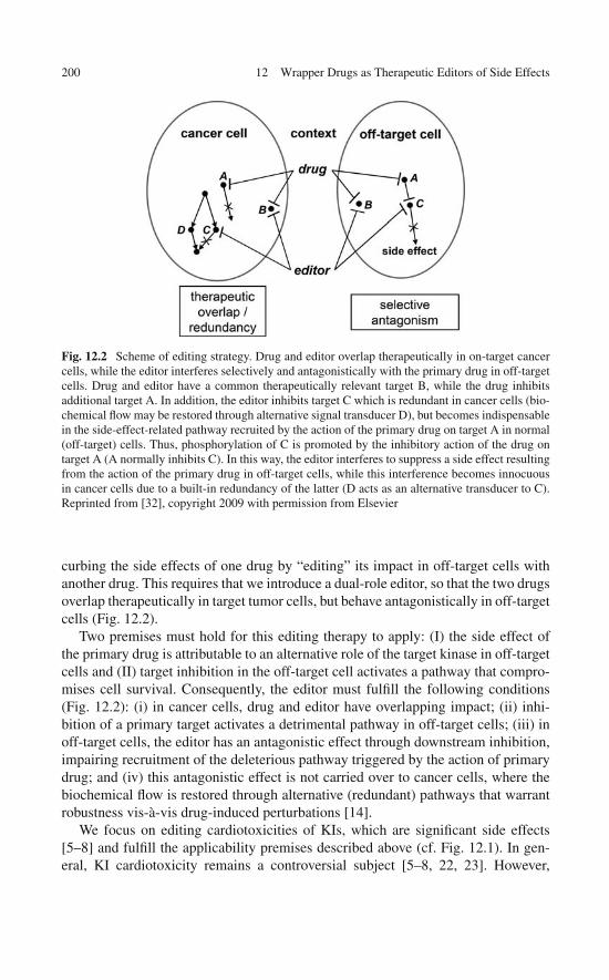
200 12 Wrapper Drugs as Therapeutic Editors of Side Effects
Fig. 12.2 Scheme of editing strategy. Drug and editor overlap therapeutically in on-target cancercells, while the editor interferes selectively and antagonistically with the primary drug in off-targetcells. Drug and editor have a common therapeutically relevant target B, while the drug inhibitsadditional target A. In addition, the editor inhibits target C which is redundant in cancer cells (bio-chemical flow may be restored through alternative signal transducer D), but becomes indispensablein the side-effect-related pathway recruited by the action of the primary drug on target A in normal(off-target) cells. Thus, phosphorylation of C is promoted by the inhibitory action of the drug ontarget A (A normally inhibits C). In this way, the editor interferes to suppress a side effect resultingfrom the action of the primary drug in off-target cells, while this interference becomes innocuousin cancer cells due to a built-in redundancy of the latter (D acts as an alternative transducer to C).Reprinted from [32], copyright 2009 with permission from Elsevier
curbing the side effects of one drug by “editing” its impact in off-target cells withanother drug. This requires that we introduce a dual-role editor, so that the two drugsoverlap therapeutically in target tumor cells, but behave antagonistically in off-targetcells (Fig. 12.2).
Two premises must hold for this editing therapy to apply: (I) the side effect ofthe primary drug is attributable to an alternative role of the target kinase in off-targetcells and (II) target inhibition in the off-target cell activates a pathway that compro-mises cell survival. Consequently, the editor must fulfill the following conditions(Fig. 12.2): (i) in cancer cells, drug and editor have overlapping impact; (ii) inhi-bition of a primary target activates a detrimental pathway in off-target cells; (iii) inoff-target cells, the editor has an antagonistic effect through downstream inhibition,impairing recruitment of the deleterious pathway triggered by the action of primarydrug; and (iv) this antagonistic effect is not carried over to cancer cells, where thebiochemical flow is restored through alternative (redundant) pathways that warrantrobustness vis-à-vis drug-induced perturbations [14].
We focus on editing cardiotoxicities of KIs, which are significant side effects[5–8] and fulfill the applicability premises described above (cf. Fig. 12.1). In gen-eral, KI cardiotoxicity remains a controversial subject [5–8, 22, 23]. However,

12.2 Editing Drugs to Curb Side Effects 201
in vivo and animal model studies [13] and clinical studies [8] corroborate theoriginal characterization of KI cardiotoxicities [5–7]. The discrepancy with alter-native reports that focus on imatinib [22, 23] is mainly due to the fact that the latterare not prospective studies, as patients did not have baseline functional studies andclinical endpoints were not delineated.
The therapeutic concept is illustrated by considering possible editors forsorafenib, a major KI successfully exploited to treat renal cell carcinoma (RCC)[24]. Sorafenib entails a cardiotoxicity risk traced to RAF1 inhibition in cardiomy-ocytes (Fig. 12.3 [5]). The drug binding to RAF1 hinders the MST2-inhibitoryRAF1–MST2 association [5, 25], as suggested by the results described below. Thus,RAF1 inhibition activates pro-apoptotic pathways mediated by MST2 and furtherdownstream by JNK, ultimately leading to cytochrome c release from mitochondria[5]. As shown in Fig. 12.3, two compounds are potentially capable of interferingwith this pathway, acting as sorafenib editors: the highly specific imatinib-derivedcompound WBZ_4, a JNK inhibitor with KD = 34nM [13], already introduced inChap. 8, and SUDE, a sunitinib-derived editor described below that inhibits MST2with KD = 11nM [9]. Both compounds overlap therapeutically with sorafenib whilethey can suppress its potential cardiotoxicity by blocking the sorafenib-recruitedpro-apoptotic pathway (Fig. 12.3a, b).
As shown in Chap. 8, WBZ_4 is a re-engineered version of imatinib withan enhanced specificity needed to curb imatinib’s potential cardiotoxicity. Theinhibitory impact of WBZ_4 is restricted to KIT, PDGFR, and JNK, all withnanomolar affinity, as corroborated by screening against a T7-bacteriophage libraryof kinase displays (Ambit Biosciences, CA) [13].
JNK is an indispensable downstream transducer of pro-apoptotic signals in car-diomyocytes [5, 25, 26] (Fig. 12.3), thus, its inhibition by WBZ_4 should antagonizesorafenib’s inhibition of RAF1. In cancer cells, both drugs overlap on therapeu-tic targets PDGFR and KIT [5, 13], while JNK inhibition by WBZ_4 interfereswith a redundant pro-apoptotic pathway that may be restored through p38 media-tion [26]. The surrogate role of p38 may not be universal, but holds in the MAPKcancer-related contexts of proliferation, differentiation, and apoptosis [26].
SUDE was designed using the wrapping-based selectivity filter [27] with twopurposes in mind: (A) decrease cross-reactivity and (B) target MST2. As a suni-tinib derivative [9], SUDE overlaps with sorafenib on therapeutic targets VEGFR,KIT, PDGFR, FLT1 and FLT3 [24, 28]. With MST2, these kinases constitute thecomplete target set for SUDE as corroborated by kinase phage-display screening,while SUDE becomes an MST2 inhibitor more potent than the parent compound(KD = 11nM versus 56nM). Thus, SUDE should edit sorafenib, counteracting itsdeleterious pathways in cardiomyocytes through its anti-MST2 activity (Fig. 12.3b).This editing role was experimentally validated for RCC, as shown below, but maynot hold for other cancers like ovarian carcinoma. In the latter, MST2 inhibitionmay actually promote cancer survival since the tumor suppressor LATS1 becomesactivated by MST2 [29]. This fact introduces a limitation in the editing applicabilityof SUDE.
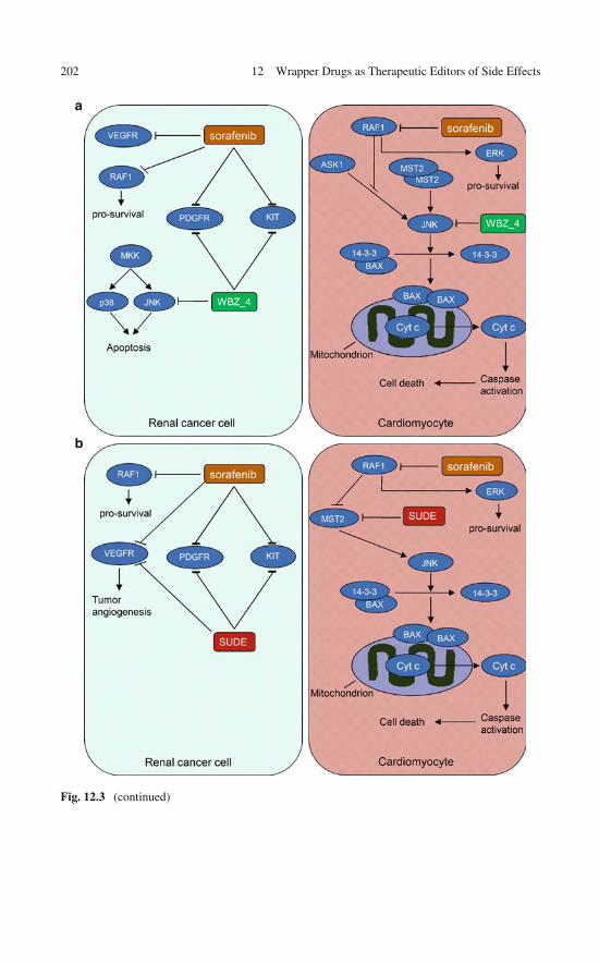
202 12 Wrapper Drugs as Therapeutic Editors of Side Effects
a
b
Fig. 12.3 (continued)
12.3 Designing a Therapeutic Editor Using the Wrapping Selectivity Filter 203
12.3 Designing a Therapeutic Editor Using the WrappingSelectivity Filter
SUDE was primarily engineered to edit sorafenib by inhibiting the kinase MST2,a downstream transducer in the pro-apoptotic pathway recruited by sorafenib incardiomyocytes (Fig. 12.3). Additionally, it was necessary for SUDE to overlaptherapeutically with sorafenib in cancer cells, to avoid disrupting anticancer activ-ity. Furthermore, SUDE needed to be specific enough to reduce the cardiotoxicityrisks of its parent drug sunitinib, adopted as the scaffold (see Chap. 10 for details).Sunitinib overlaps with sorafenib on the clinical targets VEGFR, KIT, PDGFR,FLT1, and FLT3 [5, 24, 28]. Sunitinib was redesigned guided by the wrappingselectivity filter defined in Chap. 9. The first goal is to make it selective and lesscardiotoxic, while the second is to turn it also into an MST2 inhibitor. To achievethe first goal, we exploit the wrapping differences across sunitinib’s reported targets[30]. As described in Chap. 10, alignment of wrapping patterns revealed a targetabledehydron in sunitinib’s clinical targets that is absent in the cardiotoxicity-related tar-gets AMPK and RSK (Fig. 12.4, [5, 8]). The distinct and targetable feature is thebackbone hydrogen bond pairing residues N900 and E917 in VEGFR, a primary tar-get of sunitinib (Fig. 12.4b), a bond significantly exposed to solvent in all primarytargets, but well wrapped in AMPK and RSK (Fig. 12.4a). This targeted dehydronis also present in MST2 and involves residues K69 and E85 at the aligned position(Fig. 12.4a). Thus, a methyl group was incorporated to sunitinib to provide a “wrap-ping” protection to the MST2 dehydron, also present in all clinically relevant targetsVEGFR, KIT, PDGFR, FLT1, and FLT3, but absent in AMPK and RSK. Thus, weredesigned sunitinib into SUDE (Fig. 12.4c), an MST2 inhibitor with controlledspecificity.
The predicted SUDE target set {MST2, VEGFR, KIT, PDGFR, FLT1, FLT3}was confirmed using the AMBIT high-throughput assay that uses a phage-displaykinase library [30]. With KD = 11nM, SUDE proved to be an MST2 inhibitor more
�
Fig. 12.3 Network level analysis of editing therapy, illustrated by sorafenib/WBZ_4 andsorafenib/SUDE combined activity. (a) Sorafenib edited by WBZ_4. In renal cancer cells sorafeniband WBZ_4 overlap therapeutically by inhibiting common clinically relevant targets (PDGFR andKIT). The inhibitory action of WBZ_4 on JNK confers editing power to this drug. This inhibitoryactivity is likely inconsequential for cancer cells, as the biochemical flow is restored through p38,while in cardiomyocytes, the inhibitory action of WBZ_4 on JNK antagonizes a pro-apoptoticpathway activated by upstream sorafenib-promoted inhibition of RAF1. This pathway leads to aBAX-mediated cytochrome c release to the cytosol triggering the caspase cascade activation. (b)Sorafenib edited by SUDE. SUDE overlaps therapeutically with sorafenib on VEGFR, PDGFR,and KIT in renal cancer cells, whereas in cardiomyocytes, the SUDE inhibition of MST2 impairsthe recruitment of pro-apoptotic pathways activated by the upstream sorafenib-promoted inhibitionof RAF1. Reprinted from [32], copyright 2009 with permission from Elsevier

204 12 Wrapper Drugs as Therapeutic Editors of Side Effects
Fig. 12.4 (continued)

12.4 Therapeutic Editing: Toward a Proof of Principle 205
Fig. 12.4 Molecular design of selective sunitinib-derived editor (SUDE) guided by struc-tural/wrapping comparison of sunitinib targets, by the need to remove cardiotoxicity-relatedanti-AMPK and anti-RSK activity and by the need to incorporate the new target MST2 for edit-ing purposes. Protein structures are also rendered in ribbon representation for visual aid. (a)Structures and dehydron patterns of sunitinib target AMPK (PDB.2H6D) and MST2 (homolo-gous to PDB.3CKX) kinase in complex with sunitinib. These targets need to be discriminated bySUDE, which should have affinity for MST2 but not for AMPK (or RSK). The K69–E85 dehydronin MST2 is conserved in the high-affinity targets (KIT, PDGFR, VEGFR, FLT1, and FLT3) relevantto angiogenesis and cancer cell proliferation. Crucially, it aligns with the well-wrapped hydrogenbond K78–E94 in AMPK2 (and with a well-wrapped hydrogen bond in RSK, not shown). Thisimportant packing difference suggests the modification to sunitinib needed to selectively targetMST2: methylation at the N1 indol position turns the drug into SUDE, a wrapper of the MST2dehydron K69–E85. Thus, by inhibiting MST2, a signal transducer in the pro-apoptotic pathwaystriggered by sorafenib, SUDE overlaps antagonistically with sorafenib. (b) SUDE retains activ-ity against clinically relevant sunitinib targets KIT, PDGFR, VEGFR, FLT1, and FLT3. Structureand packing pattern of VEGFR2 (PDB.2P2H) kinase in complex with sunitinib. The N900–E917dehydron in VEGFR2 is conserved in other clinically relevant targets of sunitinib and hence themodification to sunitinib serves as a wrapper to this common dehydron. (c) Chemical structure ofSUDE. The predicted SUDE cross-reactivities were confirmed using the AMBIT high-throughputassay that uses a phage-display kinase library. Reprinted from [32], copyright 2009 with permissionfrom Elsevier
potent than sunitinib (KD = 56nM, [30]). The higher affinity of SUDE is due to theadditional wrapping protection on MST2 dehydron K69–E85.
12.4 Therapeutic Editing: Toward a Proof of Principle
At a cellular level, the purported sorafenib editors should be assayed usingthe renal cancer cell line RCC-786-O and NRVMs (neonatal rat ventricularmonocytes). In RCCs, the sorafenib/WBZ_4 combination yields agonistic syn-ergy, marked by an increased inhibition of cell proliferation when comparedto sorafenib-alone levels at equivalent bulk concentrations (Fig. 12.5a): Thedose-dependent inhibition is greater than the Loewe-additive values [20, 21](Fig. 12.5b). By contrast, the sorafenib-induced recruitment of the pro-apoptoticpathway in cardiomyocytes should be impaired by the downstream interference ofWBZ_4 through JNK inhibition (Fig. 12.3a). Consequently, cytosolic release of

206 12 Wrapper Drugs as Therapeutic Editors of Side Effects
Fig. 12.5 (a) Cell proliferation assays for renal cancer cell line RCC 786-O for sorafenib/WBZ_4combination treatments. The cells were seeded in 96-well plates (4 × 10⁴ cells per well), culturedfor 24 h and treated for 48 h with sorafenib alone and in 50–50% combinations with WBZ_4 atoverall 0, 0.01, 0.1, 1, and 10 μM bulk concentrations. Cell proliferation was determined byAlamar Blue assay (Bio Source International, Camarillo, CA). Plates were read at dual wavelength(570–595 nm) in an Elisa plate reader (Molecular Devices Corporation, Sunnyvale, CA). Cellproliferation is expressed as the percentage of proliferating cells relative to untreated cells. Thesorafenib/WBZ_4 combination treatment led to an increased inhibition of cell proliferation whencompared to the sorafenib-alone levels at equivalent bulk concentrations. Also, the dose-dependentinhibition surpassed the Loewe-additive values indicating agonistic synergy. The sorafenib/SUDEcombination treatment (not shown) suppressed RCC proliferation to sorafenib-alone levels at iden-tical overall concentrations. In this case, the dose-dependent inhibition fits the Loewe-additivescheme indicating therapeutic reinforcement without synergy. (b) Agonistic synergy matrix forsorafenib/WBZ_4 combinations assayed in cell proliferation assays. Matrix elements are defined
12.4 Therapeutic Editing: Toward a Proof of Principle 207
cytochrome c should also be impaired. A dose-dependent study revealed antag-onistic synergy: If Y50 denotes the WBZ_4 concentration yielding 50% reduc-tion in sorafenib-induced cytochrome c release, we consistently obtained Y50 =Y50 (X) values (X = sorafenib concentration, 1μM ≤ X ≤ 10μM) ∼ 10% belowthe additive (linear) (X, Y50) plot. Furthermore, the cardioprotective effect of theeditor should become apparent as NRVM sarcolemmal integrity and ATP levels arerestored to normal values.
Alternatively, the sorafenib/SUDE combination suppressed RCC proliferation tosorafenib-alone levels at identical overall concentrations. This dose-dependent inhi-bition fits the Loewe-additive scheme [20, 21] indicating therapeutic reinforcementwithout synergy. In NRVMs, SUDE inhibits MST2, hence it should block the pro-apoptotic pathway triggered by sorafenib (Fig. 12.3b). The detailed mechanism ofthe sorafenib-induced activation of MST2 is not known precisely, but it is likely thatdrug binding to RAF1 impairs its association with MST2 known to inhibit the latter[25]. Furthermore, in the combined sorafenib/SUDE treatment we expect reductionof cytosolic release of cytochrome c, restored sarcolemmal integrity, and restoredATP levels all confirming SUDE’s role as sorafenib editor.
The clinical relevance of the editing therapy remains to be established. Thepotential sorafenib-induced cardiotoxicity [5] and the possibility of antagonist inter-ference exploiting WBZ_4 or SUDE hints at the therapeutic value of this mode ofside-effect removal. We already established the cardioprotective role of WBZ_4[13], while that of SUDE is expected to be established on the same premises:antagonistic interference on a pro-apoptotic pathway recruited by an upstreamkinase inhibition. A decisive test on the restoration of cardiac function throughediting therapy requires determining whether the left ventricular ejection frac-tion (LVEF) decreases significantly upon sorafenib treatment and is restored tonormal levels as sorafenib cardiotoxicity is antagonized downstream by WBZ_4or SUDE.
The editors impact a set of therapeutic targets that are more limited than that ofthe parent compounds [13], probably precluding the editors from serving as ther-apeutic agents by themselves. This is evident in RCC786-O-proliferation assays.Thus, in contrast with sorafenib effects (Fig. 12.5a), cell growth decreases by a
�
Fig. 12.5 (continued) as M(X,Y) = I(X,Y) − I′(X,Y), where X = sorafenib concentration inμM, Y = WBZ_4 concentration in μM, I(X, Y) = observed inhibition for the (X, Y) combi-nation, I′(X,Y) = predicted inhibition according to the Loewe-additive scheme [20, 21] whichrequires the (X, Y) concentration pair to satisfy the linear equation X/XI′ +Y/YI′ =1, where XI′sorafenib concentration yielding inhibition I′ and YI′= WBZ_4 concentration yielding inhibitionI ′. For clarification, a two-component treatment involving two drugs at respective concentra-tions X and Y is said to obey Loewe additivity if the concentration-dependent inhibition I (X,Y)
is equal to I′(X, Y), the predicted inhibition for the concentration pair (X, Y) that satisfies thelinear equation X/XI′ +Y/YI′ =1, where XI′ and YI′ are the single-agent effective concentra-tions that produce inhibition I′. Reprinted from [32], copyright 2009 with permission fromElsevier
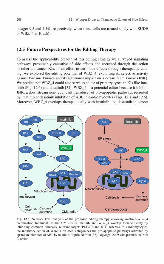
208 12 Wrapper Drugs as Therapeutic Editors of Side Effects
meager 9.5 and 4.5%, respectively, when these cells are treated solely with SUDEor WBZ_4 at 10 μM.
12.5 Future Perspectives for the Editing Therapy
To assess the applicability breadth of this editing strategy we surveyed signalingpathways presumably causative of side effects and recruited through the actionof other anticancer KIs. In an effort to curb side effects through therapeutic edit-ing, we explored the editing potential of WBZ_4, exploiting its selective activityagainst tyrosine kinases and its additional impact on a downstream kinase (JNK).We predict that WBZ_4 could also serve as editor of primary tyrosine KIs like ima-tinib (Fig. 12.6) and dasatinib [31]. WBZ_4 is a potential editor because it inhibitsJNK, a downstream non-redundant transducer of pro-apoptotic pathways recruitedby imatinib or dasatinib inhibition of ABL in cardiomyocytes (Figs. 12.1 and 12.6).Moreover, WBZ_4 overlaps therapeutically with imatinib and dasatinib in cancer
Fig. 12.6 Network level analysis of the proposed editing therapy involving imatinib/WBZ_4combination treatment. In the CML cells imatinib and WBZ_4 overlap therapeutically byinhibiting common clinically relevant targets PDGFR and KIT, whereas in cardiomyocytes,the inhibitory action of WBZ_4 on JNK antagonizes the pro-apoptotic pathways activated byupstream inhibition of ABL by imatinib. Reprinted from [32], copyright 2009 with permission fromElsevier

References 209
cells on the clinical targets PDGFR and KIT [13]. The synergies of a possibleimatinib/WBZ_4 treatment are described in Fig. 12.6.
To summarize, therapeutic editors may become a realistic engineering possibilityas we harness the molecular factors controlling drug specificity (cf. Chap. 9). Thus,the success of the editing examples described depends pivotally on the fact thatthe high-throughput screening of the engineered kinase inhibitors fit the modelingpredictions and the drug synergies found experimentally.
The therapeutic editor is a systemic concept whose clinical impact will ultimatelybe unraveled as the in vitro and in vivo assays described pave the way to clini-cal trials. This development may encounter difficulties arising, for instance, fromdifferences in bioavailability, half-life, and pharmacodynamics of drug and editor.Thus, the engineering of adequate delivery vehicles and possibly even drug redesignto tune these parameters will become an essential component of the editing strategyin the advanced stages of development.
References
1. Widakowich C, de Castro G, de Azambuja E, Dinh P, Awada A (2007) Review:Side effects of approved molecular targeted therapies in solid cancers. Oncologist 12:1443–1455
2. Schmidinger M, Zielinski CC, Vogl UM et al (2008) Cardiac toxicity of sunitinib andsorafenib in patients with metastatic renal cell carcinoma. J Clin Oncol 26:5204–5212
3. Lacouture ME (2006) Mechanisms of cutaneous toxicities to EGFR inhibitors. Nat RevCancer 6:803–812
4. Verheul HM, Pinedo HM (2007) Possible molecular mechanisms involved in the toxicity ofangiogenesis inhibition. Nat Rev Cancer 7:475–485
5. Force T, Krause D, van Etten RA (2007) Molecular mechanisms of cardiotoxicity of tyrosinekinase inhibition. Nat Rev Cancer 7:332–344
6. Force T, Kerkela R (2008) Cardiotoxicity of the new cancer therapeutics – mechanisms of,and approaches to, the problem. Drug Discov Today 13:778–784
7. Kerkela R, Grazette L, Yacobi R et al (2006) Cardiotoxicity of the cancer therapeutic agentimatinib mesylate. Nat Med 12:908–916
8. Chu TF, Rupnick MA, Kerkela R et al (2007) Cardiotoxicity associated with tyrosine kinaseinhibitor sunitnib. Lancet 370:2011–2019
9. Fernández A, Crespo A, Tiwari A (2009) Is there a case for selectively promiscuous anticancerdrugs? Drug Discov Today 14:1–5
10. Zhang X, Crespo A, Fernández A (2008) Turning promiscuous kinase inhibitors into saferdrugs. Trends Biotechnol 26:295–301
11. Dancey J, Sausville EA (2003) Issues and progress with protein kinase inhibitors for cancertreatment. Nat Rev Drug Discov 2:296–313
12. Levitzki A, Gazit A (1995) Tyrosine kinase inhibition: An approach to drug development.Science 267:1782–1788
13. Fernández A, Sanguino A, Peng Z et al (2007) An anticancer C-kit kinase inhibitoris re-engineered to make it more active and less cardiotoxic. J Clin Invest 117:4044–4054
14. Kitano H (2007) A robustness-based approach to systems-oriented drug design. Nat Rev DrugDiscov 6:202–209
15. Torchilin VP (2006) Multifunctional nanocarriers. Adv Drug Del Rev 58:1532–155516. Langer R (2001) Drug delivery: Drugs on target. Science 293:58–59

210 12 Wrapper Drugs as Therapeutic Editors of Side Effects
17. Allen TM, Cullis PR (2004) Drug delivery systems: Entering the mainstream. Science303:1818–1822
18. Deninger M, Buchdunger E, Druker BJ (2005) The development of imatinib as a therapeuticagent for chronic myeloid leukemia. Blood 105:2640–2653
19. Dancey JE, Chen HX (2006) Strategies for optimizing combinations of molecularly targetedanticancer drugs. Nat Rev Drug Discov 5:649–659
20. Keith CT, Borisky AA, Stockwell BR (2005) Multicomponent therapeutics for networkedsystems. Nat Rev Drug Discov 4:71–78
21. Zimmermann GR, Lehar J, Keith CT (2007) Multi-target therapeutics: When the whole isgreater than the sum of the parts. Drug Discov Today 12:34–42
22. Verweij J, Casali P, Kotasek D et al (2007) Imatinib does not induce cardiac left ventricularfailure in gastrointestinal stromal tumours patients: Analysis of EORTC-ISG-AGITG study62005. Eur J Cancer 43:974–978
23. Atallah E, Durand JB, Kantarjian H, Cortes J (2007) Congestive heart failure is a rare eventin patients receiving imatinib. Blood 110:1233–1237
24. Escudier B, Eisen T, Stadler WM et al (2007) Sorafenib in advanced clear-cell renal-cellcarcinoma. N Engl J Med 356:125–134
25. O’Neill E, Rushworth L, Baccarini M, Kolch W (2004) Role of the kinase MST2 insuppression of apoptosis by the Proto-Oncogene product Raf-1. Science 306:2267–2270
26. Dhillon AS, Hagan S, Rath O, Kolch W (2007) MAP kinase signalling pathways in cancer.Oncogene 26:3279–3290
27. Chen J, Zhang X, Fernández A (2007) Molecular basis for specificity in the druggable kinome:Sequence-based analysis. Bioinformatics 23:563–572
28. Faivre S, Demetri G, Sargent W, Raymond E (2007) Molecular basis for sunitinib efficacy andfuture clinical development. Nat Rev Drug Discov 6:734–745
29. O’Neill EE, Matallanas D, Kolch W (2005) Mammalian sterile 20-like kinases in tumorsuppression: An emerging pathway. Cancer Res 65:5485–5487
30. Karaman MW, Herrgard S, Treiber DK et al (2008) A quantitative analysis of kinase inhibitorselectivity. Nat Biotechnol 26:127–132
31. Kantarjian H, Jabbour E, Grimley J, Kirkpatrick P (2006) Dasatinib. Nat Rev Drug Discov5:717–718
32. Fernández A, Sessel S (2009) Selective antagonism of anticancer drugs for side-effectremoval. Trends Pharmacol Sci 30: 403–410

Chapter 13Wrapper Drugs for Personalized Medicine
Personalized molecular therapy (“the right drug for the right person”) is regardedas a major imperative of post-genomic medicine. This perception is reinforcedalmost daily as promising therapeutic agents are recalled because of idiosyncraticside effects detected in small subpopulations of patients. However pressing theneed, rational approaches to personalized drug therapy will ultimately and pivotallydepend on our ability to translate genomic individualities and variations into molec-ular biomarkers that can guide a patient-tailored design. This chapter addresses thisissue and describes how the wrapping design concept can be brought to fruitionin this area. The chapter introduces plausible scenarios in which genomic idiosyn-crasies and oncogenic variations may promote targetable differences in the wrappingpatterns of the gene products. Ultimately, the chapter extends an invitation to adoptand exploit protein wrapping as a molecular biomarker for personalized medicineand the wrapping technology as the enabling platform to tailor drugs to patientidiosyncrasies.
13.1 Wrapping as a Biomarker in Personalized Drug Therapy
The therapeutic imperative to make more efficacious and safer drugs by exploit-ing the enormous output of genomic forays has proven to be a daunting challenge,far more difficult than originally expected. No reliable rational approach has beenidentified so far to meet such a challenge. In addressing this imperative, the dearthof ideas of general applicability with the potential to become transformative andbroaden the technological base of the pharmaceutical industry is quite evident [1, 2].From the perspective of a rational drug designer, one can only speculate that a per-sonalized approach would require an effective translation of genomic idiosyncrasiesand disease-related variations into structural differences that may guide the molec-ular engineering of the therapeutic agent. While this conclusion may be inevitable,it has not been pursued beyond a limited number of anecdotal instances [3].
It is seldom the case that idiosyncratic or disease-related genomic differenceswould translate as significant structural changes in the gene products. The func-tional demands of the wild-type locus cannot generally be compromised without a
211A. Fernández, Transformative Concepts for Drug Design: Target Wrapping,DOI 10.1007/978-3-642-11792-3_13, C© Springer-Verlag Berlin Heidelberg 2010
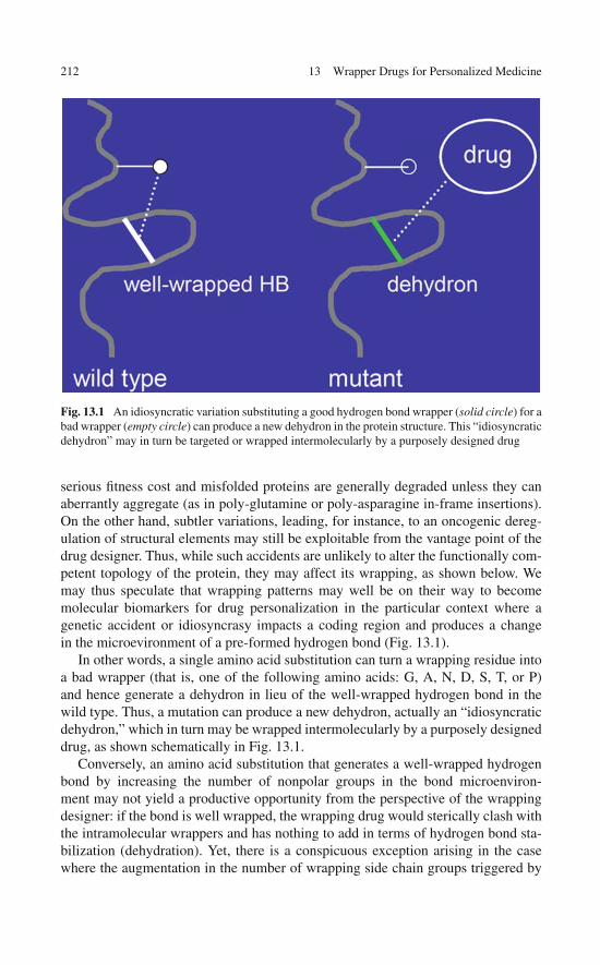
212 13 Wrapper Drugs for Personalized Medicine
Fig. 13.1 An idiosyncratic variation substituting a good hydrogen bond wrapper (solid circle) for abad wrapper (empty circle) can produce a new dehydron in the protein structure. This “idiosyncraticdehydron” may in turn be targeted or wrapped intermolecularly by a purposely designed drug
serious fitness cost and misfolded proteins are generally degraded unless they canaberrantly aggregate (as in poly-glutamine or poly-asparagine in-frame insertions).On the other hand, subtler variations, leading, for instance, to an oncogenic dereg-ulation of structural elements may still be exploitable from the vantage point of thedrug designer. Thus, while such accidents are unlikely to alter the functionally com-petent topology of the protein, they may affect its wrapping, as shown below. Wemay thus speculate that wrapping patterns may well be on their way to becomemolecular biomarkers for drug personalization in the particular context where agenetic accident or idiosyncrasy impacts a coding region and produces a changein the microevironment of a pre-formed hydrogen bond (Fig. 13.1).
In other words, a single amino acid substitution can turn a wrapping residue intoa bad wrapper (that is, one of the following amino acids: G, A, N, D, S, T, or P)and hence generate a dehydron in lieu of the well-wrapped hydrogen bond in thewild type. Thus, a mutation can produce a new dehydron, actually an “idiosyncraticdehydron,” which in turn may be wrapped intermolecularly by a purposely designeddrug, as shown schematically in Fig. 13.1.
Conversely, an amino acid substitution that generates a well-wrapped hydrogenbond by increasing the number of nonpolar groups in the bond microenviron-ment may not yield a productive opportunity from the perspective of the wrappingdesigner: if the bond is well wrapped, the wrapping drug would sterically clash withthe intramolecular wrappers and has nothing to add in terms of hydrogen bond sta-bilization (dehydration). Yet, there is a conspicuous exception arising in the casewhere the augmentation in the number of wrapping side chain groups triggered by
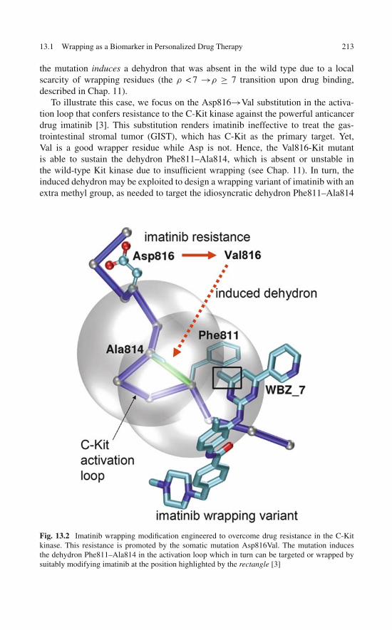
13.1 Wrapping as a Biomarker in Personalized Drug Therapy 213
the mutation induces a dehydron that was absent in the wild type due to a localscarcity of wrapping residues (the ρ < 7 →ρ ≥ 7 transition upon drug binding,described in Chap. 11).
To illustrate this case, we focus on the Asp816→Val substitution in the activa-tion loop that confers resistance to the C-Kit kinase against the powerful anticancerdrug imatinib [3]. This substitution renders imatinib ineffective to treat the gas-trointestinal stromal tumor (GIST), which has C-Kit as the primary target. Yet,Val is a good wrapper residue while Asp is not. Hence, the Val816-Kit mutantis able to sustain the dehydron Phe811–Ala814, which is absent or unstable inthe wild-type Kit kinase due to insufficient wrapping (see Chap. 11). In turn, theinduced dehydron may be exploited to design a wrapping variant of imatinib with anextra methyl group, as needed to target the idiosyncratic dehydron Phe811–Ala814
Fig. 13.2 Imatinib wrapping modification engineered to overcome drug resistance in the C-Kitkinase. This resistance is promoted by the somatic mutation Asp816Val. The mutation inducesthe dehydron Phe811–Ala814 in the activation loop which in turn can be targeted or wrapped bysuitably modifying imatinib at the position highlighted by the rectangle [3]
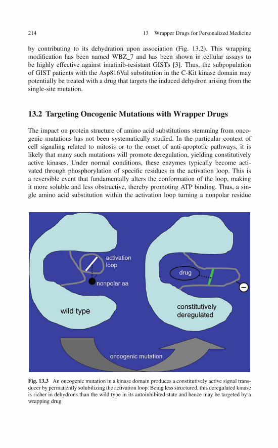
214 13 Wrapper Drugs for Personalized Medicine
by contributing to its dehydration upon association (Fig. 13.2). This wrappingmodification has been named WBZ_7 and has been shown in cellular assays tobe highly effective against imatinib-resistant GISTs [3]. Thus, the subpopulationof GIST patients with the Asp816Val substitution in the C-Kit kinase domain maypotentially be treated with a drug that targets the induced dehydron arising from thesingle-site mutation.
13.2 Targeting Oncogenic Mutations with Wrapper Drugs
The impact on protein structure of amino acid substitutions stemming from onco-genic mutations has not been systematically studied. In the particular context ofcell signaling related to mitosis or to the onset of anti-apoptotic pathways, it islikely that many such mutations will promote deregulation, yielding constitutivelyactive kinases. Under normal conditions, these enzymes typically become acti-vated through phosphorylation of specific residues in the activation loop. This isa reversible event that fundamentally alters the conformation of the loop, makingit more soluble and less obstructive, thereby promoting ATP binding. Thus, a sin-gle amino acid substitution within the activation loop turning a nonpolar residue
Fig. 13.3 An oncogenic mutation in a kinase domain produces a constitutively active signal trans-ducer by permanently solubilizing the activation loop. Being less structured, this deregulated kinaseis richer in dehydrons than the wild type in its autoinhibited state and hence may be targeted by awrapping drug
References 215
into a negatively charged one can promote the deregulation of the kinase, turn-ing it into a constitutively active signal transducer. This accident can of coursepromote tumor development and cancer progression [4], depending on whether thetransduced signal recruits pathways leading to mitosis, cell immortalization, focaladhesions, etc.
On the other hand, since the oncogenic substitution solubilizes the activationloop, it should promote a loss of structure. This process is likely to yield a loopconformation far worse wrapped than that of the autoinhibited state and hence, farricher in dehydrons (Fig. 13.3). Ergo, a constitutively deregulated kinase, should bemore susceptible to be targeted by wrapping/crating drugs than the wild type. Thisoperational hypothesis begs a validation in the light of the ideas described in thisbook.
13.3 Closing Remarks
By interfering with cell signaling pathways, single-molecule kinase inhibitors holdgreat promise as anticancer agents and probably hold potential for the moleculartargeted treatment of other diseases. Yet, their therapeutic efficacy and clinical ben-efit are typically limited to a fraction of the treated patients. Genomic variationsacross patients and idiosyncratic patterns of drug resistance are likely to contributesignificantly to such differences in clinical outcome. The acknowledgment of thesefacts has triggered the need for a personalized drug design. Perhaps this challengeis not so daunting in so far as we can translate idiosyncratic or disease-related vari-ations into molecular differences that can be systematically targeted by molecularengineers. The wrapping pattern may prove to be a transformative concept that maybe brought to fruition in this key area of molecular medicine.
References
1. Gazdar A (2009) Personalized medicine and inhibition of EGFR signaling in lung cancer. NewEngl J Med 361:1018–1020
2. Janne PA, Gray N, Settleman J (2009) Factors underlying sensitivity of cancers to smallmolecule kinase inhibitors. Nat Rev Drug Discov 8:709–723
3. Fernández A, Sanguino A, Peng Z et al (2007) Rational drug design to overcome drug resistancein cancer therapy: Imatinib moving target. Cancer Res 67:4028–4033
4. Hubbard SR (2004) Oncogenic mutations in B-Raf: Some losses yield gains. Cell 116, 764–766

“This page left intentionally blank.”
Chapter 14Last Frontier and Back to the Drawing Board:Protein–Water Interfacial Tension in DrugDesign
As we have advocated throughout this book, the protein–water interface is at thecrux of the drug design problem. Understanding this interface is essential whenaddressing the frontier problem of disrupting a protein–protein interaction with acompeting ligand (that is, as man tries to beat nature at its own recognition game).One lesson to be drawn from the preceding pages is that it is quite unproductiveto think of a drug and a target in a context where the solvent is merely regarded asbackground or as a statistical bath. Yet, little has been said about the thermodynamicnature of the protein–water interface and about the future exploitation of this knowl-edge to the advantage of molecular targeted design. This chapter squarely addressesthis problem, dealing with questions like What is the nature of the interfacial tensionbetween protein and water? What is the thermodynamic cost of creating a protein–water interface? How can we identify regions that beget large interfacial tension toguide targeting designs? In plain terms, which regions on the surface of the proteingenerate unstable interfaces that may be removed upon association with a drug?The results presented open up an avenue to implement a nanoscale thermodynamicsapproach to drug design, where the dehydron concept will play again a pivotal role.This chapter heralds the advent of drugs adapted to reduce the tension of protein–water interfaces, hence stabilizing the structure of the target. This strategy takes usback to the wrapping concept: Dehydron patches that generate significant interfacialtension between target and water may be favorably wrapped by the drug.
14.1 Interfacial Tension Between Protein and Water:A Missing Chapter in Drug Design
The physical nature of the protein–water interface remains elusive and yet, as advo-cated in the preceding chapters, this interface is truly at the crux of rational drugdesign. This shift in focus from protein structure to protein–water interface willsurely become instrumental to fulfill our expectations across the next tier of Frontierproblems in drug design, including interfering with a protein–protein association bydisrupting it with a small ligand. Chemical heterogeneities of the protein surface anda variety of local morphologies for the nanoscale confinement of interfacial water
217A. Fernández, Transformative Concepts for Drug Design: Target Wrapping,DOI 10.1007/978-3-642-11792-3_14, C© Springer-Verlag Berlin Heidelberg 2010
218 14 Last Frontier and Back to the Drawing Board
make it difficult to treat the protein–water interface using classical thermodynamicapproaches. Thus, we cannot readily introduce statistical thermodynamic conceptslike interfacial surface tension without extending the validity of the physical pic-ture to the nanoscale realm. Concepts like interfacial tension may be most usefulwhen dealing with the separation of homogeneous phases with smooth solution ofcontinuity but are far harder to define and apply when dealing with the complexmorphologies and heterogeneous surfaces of proteins. Thus, while the length-scaledependence of nonpolar hydration and hydrophobic interactions is relatively wellestablished [1], nontrivial complications arise mainly from the presence of nearbyhydrophilic groups that modulate the interfacial tension in a curvature-dependentmanner.
This chapter squarely addresses these problems and does so by introducing ananoscale thermodynamic treatment based on first principles. Thus, we deal withquestions like What is the thermodynamic cost of creating a protein–water interface?Which regions on the surface of the protein generate unstable interfacial patcheswith significant surface tension that may be removed upon drug association? Thetreatment lays the foundations of a nanoscale thermodynamics approach to drugdesign.
An intuitive picture emerges in the midst of the apparently murky problem: Giventhe heterogeneous chemical composition of the protein surface and its complicatedmorphology, the protein–water interfacial tension is likely to be patchy. Thus, weenvision hot spots defined by solvent cavities whose creation entails a positive incre-ment in interfacial free energy and regions of high solvent polarizability yieldingnegative free-energy increments (no tension spots). The positive free-energy patches(begetting tension) do not necessarily interface with hydrophobic moieties on theprotein surface. An analysis of the dielectric response under nanoscale confinementshows that polar groups with low-density hydration may also generate interfacialtension, as shown below.
Drawing inspiration from Chaps. 3 and 5, we note that a tractable problememerges when the extent of interfacial water confinement is described by a posi-tion (r)-dependent parameter = (r), the expected number of hydrogen bondpartnerships of a water molecule at position r ( = 4 for bulk water). Thus, thesolvent-accessible envelope of the protein surface may be covered by a minimal setW of water-confining osculating (first-order contact) spheres Djs, j ∈ W, each withits average value = j which depends on the radius θ = θj of Dj (the curvatureradius of the protein envelope) and the physicochemical nature of protein group(s)in the contact region (Fig. 14.1).
Complementing the information conveyed by j, a fugacity fj is introducedand defined as fj = Nj/Nj(b), where Nj is the expected number of watermolecules in Dj at equilibrium and Nj(b), the number associated with the samevolume in bulk solvent. Thus, the chemical potential μj of water in Dj becomesμj = kBT ln
[Nj/Nj(b)
](kB= Boltzmann constant). The θ -dependence of ,
f-values (Fig. 14.1) is obtained at equilibrium determined from classical trajecto-ries generated by molecular dynamics (MD) simulations within an NPT ensem-ble of the type described in Chap. 4. The computations start with the protein

14.1 Interfacial Tension Between Protein and Water: A Missing Chapter in Drug Design 219
Fig. 14.1 Scheme and plots illustrating the dependence of nanoscale-confinement parameters
and f on the radius θ of the osculating sphere at the protein–water interface. First-order contactswith a polar or nonpolar patch on the protein surface are treated individually. Values were deter-mined at equilibrium obtained by integrating Newton’s equations of motion in an NPT ensemblewith box size 103 nm3, starting with the PDB structure embedded in a pre-equilibrated cell of watermolecules. The box size was calibrated so that the solvation shell extended at least 10 Å from theprotein surface at all times. Simulations were performed as described in Chap. 4
structure embedded in a pre-equilibrated cell of explicitly represented watermolecules. A nonredundant set of 206 structures of monomeric proteins reportedin PDB (protein data bank) with chain lengths ranging from N= 34 (PDB.1QUZ) to223 (PDB.1AZ8) was used to generate the data shown in Fig. 14.1 (see Chap. 4 forcomputational details).
Nanoscale confinement significantly affects water polarizability P = P(r), whosedependence on may be obtained rigorously from first principles, starting fromthe Debye relation [2]: ∇. (ε0E + P) (r) = ρ(r) (ε0= vacuum permittivity, ρ(r)= charge density, E= electrostatic field). In Fourier-conjugate frequency space (ν-space) we get [2]
F(P)(ν) = Kp(ν)F(E)(ν) (14.1)
where F denotes 3D-Fourier transform F(g)(ν) = (2π )−3/2∫
eiν.rg(r)dr and thekernel Kp(ν) is the frequency Lorentzian Kp(ν) = (εb − ε0)/(1 + τ 2|ν|2), withτ= dielectric relaxation time (τ = τb ≈ 100 ps in bulk) and εb = bulk permittivity.Equation (14.1) yields the following equation in r-space:

220 14 Last Frontier and Back to the Drawing Board
∇.
[∫F−1(K)(r − r′)E(r′)dr′
]= ρ(r), (14.2)
with K(ν) = ε0 + Kp(ν), F−1(K)(r) = (2π )−3/2∫
e−iν.rK(ν)dν The readershould note that (14.2) is not the commonly used Poisson–Boltzmann equation,which requires an arbitrary proportionality between the fields E and P underthe incorrect assumption K(ν) ≡ constant. Rather, (14.2) includes the convolutionbetween F−1(K)(r) and E(r) that accurately translates in r-space the ν-dependentproportionality between E and P that rigorously holds in frequency space.
Upon water confinement, the dielectric relaxation undergoes a frequency redshift(a shift to lower frequencies or to larger relaxation times) arising from the reductionin hydrogen bond partnerships that translates in a reduction in orientational possibil-ities. Thus, τ = τb exp (B()/kBT), where the kinetic barrier B() = −kBT ln (/4)is obviously entropic, yielding τ = τb(/4)−1. In order to determine the-dependence of the polarization P, we introduce the electrostatic potential � =�(r), yielding E = ∇�. Thus, (14. 2) may be solved in frequency space:
F(�)(ν) = F(ρ)(ν)/[|ν|2 K(ν)
]. (14.3)
Taking the inverse Fourier transformation we obtain the electrostatic filed underwater confinement in explicit form:
E(r) = ∇F−1{F(ρ)(ν)/
[|ν|2 K(ν)]}
(r)
= (2π )−3/2∇∫
dνe−iν.rF(ρ)(ν)/[|ν|2 K(ν)]. (14.4)
This equation may be solved explicitly as we assume a net charge densitydistribution:
ρ(r) =∑
j∈W
∑n∈M(j)
4πqnδ(r − rn), (14.5)
where M(j) denotes the set of charges labeled by dummy index n, localized on theprotein surface and interfacing with Dj, and rn = position vector of charge n. Thus,from (14.3) and noting that
F(ρ)(ν) = (2π )−3/2∑
j∈W
∑n∈M(j)
4πqn exp (iν.rn), (14.6)
we obtain the explicit form of the electrostatic potential:
�(r) = ∑j∈W �j(r)
= (2π )−3∑
j∈W
∑n∈M(j)
∫dνe−iν.(r−rn)4πqn/
[|ν|2K(j)(ν)
],
(14.7)
14.1 Interfacial Tension Between Protein and Water: A Missing Chapter in Drug Design 221
with K(j)(ν) = ε0 + Kp,(j)(ν) = ε0 + (ε0 − ε0) /(
1 + τ 2j |ν|2
)and τj = τb(j/4)−1.
Combining (14.1), (14.4), and (14.6), we get explicitly the polarization of waterunder nanoscale confinement:
P(r) = ∫F−1(Kp)(r − r′)E(r′)dr′ = ∑
j∈W P(j)(r)
= (2π )−3∑
j∈W
∑n∈M(j)
∫dr′F−1 (
Kp,(j)) (
r − r′)
∇r′∫
dνe−iν.(r′−rn)4πqn/[|ν|2K(j)(ν)
],
(14.8)
with F−1(Kp,(j)
)(r) = ∫
dνe−iν.r (εb − ε0) /(
1 + τ 2j |ν|2
).
Thus, the overall free energy change G associated with creating the protein–water interface by transferring the water molecules from the bulk to the interface is
G = ∑j∈W
[(−1/2)
∫ ∣∣P(j)(r)∣∣2 dr − TSj
]
= −(1/2)∑
j∈W
∫dr
∣∣∣∣(2π)−3∑
n∈M(j)
∫dr′F−1 (
Kp,(j)) (
r − r′)
∇r′∫
dνe−iν.
(r′−rn
)4πqn/
[|ν|2K(j)(ν)
]∣∣∣∣2
−∑j∈W kBTcbNfj(4/3)πθ3
j ln (j/4)
. (14.9)
Since Nj = cbNfj(4/3)πθ3j (cb = 55.5 mol/(10−3 m3) = bulk water concentration
and N = 6.023 × 10²³/mol = Avogadro’s number), the last term in (14.9) rep-resents the sum of entropic contributions
(−�j∈WTSj), where the jth term in the
sum is associated with transferring Nj water molecules from bulk solvent (=4)to the interfacial osculating sphere Dj (with = j). Thus, we get the relation−TSj = −kBTcbNfj(4/3)πθ3
j ln (j/4) ≥ 0.Interfacial tension arises in an osculating sphere Dj when Gj > 0, that is, when
generating the interface region entails a thermodynamic cost translated in reversiblework performed on the system. As shown below, the most common patches gen-erating interfacial tension involve backbone hydrogen bonds of the protein thatare partially exposed to water, so that the amide or carbonyl is hydrated by low-density water. In other words, protein dehydrons generate interfacial tension. Thus,the most common osculating spheres contact polar patches arising from incom-plete shielding of the backbone. These spheres constitute 66% of the interfacialspheres that fulfill Gj > 0 in the database. They are described by the rangesθj = 2.3 ± 0.4Å, j = 1.2 ± 0.2, and fj = 0.75 ± 0.1 (Fig. 14.1), yielding a red-shifted relaxation τj = (3.42 ± 0.58)τb, with − TSj = (2.48 ± 0.45)kBT andGj = (1.54 ± 0.40) kBT . Thus, dehydrons yield interfacial tension throughinsufficient polar hydration.
222 14 Last Frontier and Back to the Drawing Board
Since the , f-parameters remain invariant for θ ≥ 7Å, interfacial polar regionswith curvature radius θ > 7Å are covered with disjoint spheres with θj = 7Å(Fig. 14.1). At θj = 7Å, we get j = 4 (Sj = 0), hence these osculatingspheres generate no interfacial tension. By contrast, nonpolar interfacial regionswith θ ≥ 7Å, may also be covered with disjoint spheres of radius θj = 7Å,(the corresponding values =3, f=1 remain invariant for θj ≥ 7Å) but in thiscase, these regions yield the highest interfacial tension at Gj ≈ −TSj =−kBTcbNf (4/3)π7310−30m3 ln (3/4) = 13.17kBT . The same contribution is asso-ciated with each disjoint θ = 7Å sphere covering convex regions
(θ < −2.7 Å
).
By contrast, the “clathrate” range 0 ≥ θ ≥ −2.7 Å, where nonpolar moieties canbe hydrated while preserving the tetrahedral hydrogen bond lattice (=4) do notgenerate tension: as expected this leads to S = 0.
To summarize, three conclusions transpire from the nanoscale thermodynam-ics results: (a) The interfacial tension between protein and water is patchy andthe result of both nanoscale confinement of interfacial water and local redshifts indielectric relaxation; (b) the poor hydration of polar groups (a curvature-dependentphenomenon) generates interfacial tension, a property previously attributed only tohydrophobic patches; and (c) because of its higher occurrence at protein–water inter-faces, the poorly hydrated dehydrons become collectively bigger contributors to theinterfacial tension than the rarer nonpolar patches on the protein surface.
These conclusions open up an avenue to actually implement a nanoscale thermo-dynamics approach to drug design, where no doubt the dehydron concept will playa pivotal role. This rigorous treatment heralds the advent of drugs adapted to reducethe tension of protein–water interfaces, hence stabilizing the structure of the proteintarget. This strategy takes us back to the wrapping concept: patches that generatesignificant interfacial tension between target and water may be favorably wrappedby the drug, and we already know how to do that.
14.2 Disrupting Protein–Protein Interfaces with Small Molecules
Protein–protein associations, be they ephemeral or obligatory, constitute core bio-logical events that enable basic cellular operations like signal transfer, catalyticactivity, oligomeric functionalization. Understandably, their disruption using man-made ligands that have the capacity to bind competitively has become an importantgoal in molecular therapeutics [3]. Rational approaches to this drug design problemare based on the identification of “hot spots” of protein–protein interfaces [3]. Thesehot spots consist of interfacial residues that make the most significant contribu-tion to the association free energy of the protein. In practice, they are operationallyidentified through “alanine scanning,” a thermodynamic assessment of the effect ofsite-specific substitution on the complexation free energy. Thus, specific interfacialresidues are mutated to alanine (essentially a truncation of the side chain) and theeffect is quantified by G, the differential free energy of association. This quantityis in turn obtained calorimetrically by comparing the association affinity of mutant

References 223
and wild-type protein. Alanine scanning has been used in conjunction with drugdesign with moderate success to selectively disrupt hormone-receptor associationsusing competitive man-made ligands [3].
By definition, patches that significantly contribute to protein–water interfacialtension must translate into hot spots for protein–protein associations. Hence thelaborious alanine scanning may be effectively replaced by a direct computationalidentification of patches of interfacial tension in the free subunits. This computa-tional advance will no doubt simplify the engineering of therapeutic interferencewith protein-protein associations.
References
1. Chandler D (2005) Interfaces and the driving force of hydrophobic assembly. Nature 437:640–647
2. Debye P (1929) Polar Molecules. Dover, New York3. Wells JA, McClendon CL (2007) Reaching for high-hanging fruit in drug discovery at protein–
protein interfaces. Nature 450:1001–1009

“This page left intentionally blank.”

Epilogue
This book advocates a rational translational approach to molecular-targeted therapybased on the wrapping concept. As such, it stands at the antipodes of drug discov-ery endeavors based on high-throughput screening and trial-and-error approaches.Before incarnating as a molecular design concept in Chaps. 7, 8, 9, 10, 11, 12, 13,and 14, wrapping is explored from architectural, biophysical, bioinformatics, andevolutionary perspectives in preparatory chapters 1, 2, 3, 4, 5, and 6. Wrapping is anovel category in structural biology, in which the 3D structure of the target proteinis examined in relation to the surrounding solvent. Thus, wrapping becomes particu-larly insightful as we attempt to examine the protein–water interface. The emergingdrug discovery platform is rooted in fundamental principles that shaped our currentunderstanding of biological water. This “wrapping technology” is articulated withthe aid of a bioinformatics toolbox that enables us to explore the biomolecular andevolutionary basis of drug specificity and encode the necessary information in adigitalized matrix format. In this regard, the book advocates that controlled speci-ficity is not only the safest and therapeutically most efficacious alternative but alsoa plausible one within the realm of rational design.
While the book portends to introduce transformative concepts, capable of broad-ening the technological base of the pharmaceutical industry, we are acutely awarethat a sobering note is in order. Wrapping designs are physically sound and seemto withstand the test of academic scrutiny and, yet, ultimately, only clinical trialscan fully assess their therapeutic value and safety. In this regard, and from a prac-tical perspective, current business models in the pharmaceutical R&D may not besufficiently resilient to accommodate this type of core innovation.
Current business models predicated around prevalent discovery paradigmsappear – at least to some of us – to be getting progressively obsolete. Thus, morethan ever, the lead in the pharmaceutical industry will depend on alternative mod-els with the suppleness to incorporate innovative research and harvest its fruits. Asfar as we can see, this type of innovation can come from only one source: funda-mental knowledge. On the other hand, the business context required for its effectiveexploitation remains elusive and perhaps requires a new breed of entrepreneurship.
Standing at the cross-roads of drug discovery and academic pursuit, thisbook squarely addresses basic translational and integrative problems in the phar-maceutical industry. It does so by introducing fundamental concepts in basic
225A. Fernández, Transformative Concepts for Drug Design: Target Wrapping,DOI 10.1007/978-0-387-88630-5, C© Springer-Verlag Berlin Heidelberg 2010

226 Epilogue
biomolecular research that hold potential to transform molecular engineering, withthe overarching goal of designing safer drugs with reduced or at least controllableside effects. The actual implementation of the purported innovations within currentor future business models lies outside the scope of this monograph, yet it would bedesirable – if not imperative – that both sides of the endeavor evolve harmoniouslytogether.
Index
AAberrant aggregation, 6, 49, 59–77ABL, 156–158, 167, 173–174, 198–199, 208Actin, 69–70, 133Activation loop, 166, 187–190, 195, 213–215Adiabatic, 33ADP, 101, 108, 111, 127Aggregation, 6, 49–50, 59–77, 83Agonist, 200, 205–207Alanine scanning, 222–223AMPK, 179–183, 203, 205Amyloid, 50, 59, 73–74, 76Angiogenesis, 179–180, 205Animal model, 79–80, 94, 117–118, 134–139,
201Antagonist, 199–200, 205, 207Apoptosis, 136, 164, 179, 199, 201ASK1, 199ATP, 98, 101, 127, 130–131, 148, 153, 164,
166, 173, 179–180, 191, 193, 195, 199,207, 214
Aurora, 168, 171
BBackbone hydration, 17, 20, 24–25, 27–29, 87,
142, 188Bacteriophage library, 128, 176, 201BAD, 179, 199Balance equation, 18, 20, 22–24, 26BAX, 179, 199, 203BCL2, 199Bevacizumab, 178–179Binding partner, 6, 9, 50, 59, 72, 74, 76, 82–83,
85, 91, 93, 105, 108, 187Binding site, 49, 85, 87, 93, 102, 105, 147–148,
193Bioavailability, 209Bioinformatics, 6, 24, 102–105, 141–142,
144–152, 154, 159–160, 177
Biomarker, 180, 211–214Brain natriuretic peptide, 136Brc-Abl, 108, 111–113Buttress, 18, 24, 72–73
CCalmodulin, 69–71, 174CAMK, 155–156, 174, 180Cardiac hypertrophy, 180Cardiac impairment, 136Cardiomyocyte, 125, 136, 164, 179–180, 191,
198–199, 201, 203, 205, 208Cardiotoxicity, 117–118, 134, 136, 139, 164,
179–181, 198, 200–201, 203, 205, 207Cell
fate, 97, 117immortalization, 215proliferation, 97, 117, 131–132, 180, 197,
205–206Chemotype, 152–160, 166–167Chimera, 191, 198Chronic myeloid leukemia, 108, 117–118, 164,
190, 198–199C-Kit, 108, 111–113, 117–120, 124–127,
129–133, 139, 165, 189–194, 213–214Clathrate, 52, 222Clinical trial, 1, 209, 225CLK, 174CML, 108, 118, 131–134, 164, 190, 198–199,
208Coarse grain, 30–31, 34, 38Constant of motion, 27, 31–32, 39, 41–45Cooperativity, 1–14, 27–46, 74Coulomb field, 10–11, 61Coulomb screening, 43, 54Crating, 187–195, 215Cross reactivity, 125, 142, 152–155, 159–160,
163–165, 167, 179–180, 182–183,188–190, 195, 201
227

228 Index
Crowding, 41, 45, 165CSF1R, 155–156, 165, 179–180, 183Cytochrome c, 136, 179, 199, 201, 203, 207
DDasatinib, 158–159, 167, 208Dehydron
cluster, 59–61, 68–72field, 10–12, 18, 28–29, 42, 45–46force, 11, 45, 50matrix, 107, 125, 154
Deleterious mutation, 80, 84, 93Desolvation, 4, 6–7, 9, 12–13, 29, 31–32, 34,
61–64, 71, 74–75, 103–106, 112, 175,190
De-wetting hot spot, 51, 55–56, 118–125, 139DFG catalytic triad, 166Dielectric
modulator, 61, 67, 70Dirty drug, 163, 165–173Disorder propensity, 60, 142–143, 149, 154,
187Disorder score, 60, 87, 143Disulfide bond, 17–25Dosage imbalance, 80, 83–85, 87–88, 91,
93–94, 102Dosage sensitivity, 83–85, 87, 93Drug
resistance, 149, 164, 179, 213, 215screening, 144–145
EEditor, 197–209EGFR, 145, 167–169, 171, 173–174EKB–569, 145, 167–169, 171–173Endoplasmic reticulum, 136, 199Environmental coordinates, 54Environmental distance, 149Environmental hull, 144, 149Epigenetic, 76–77ERBB, 168, 171, 178ERK, 136, 199Erlotinib, 145, 167Expression profile, 87Expression vector, 85, 87
FFAK, 98, 101, 154, 168, 171Fitness, 80, 83, 94, 212Flavopiridol, 145FLT, 155–156, 179–180, 183, 201, 203, 205Focal adhesion, 98, 101, 154, 215Folding machine, 32Folding trajectory, 31, 33, 41, 45–46
Fourier transform, 43–44, 219–220FOXO, 199Frequency space, 43, 219–220
GGastrointestinal stromal tumor, 108, 117–118,
134, 213Gatekeeper, 167–169, 194Gefitinib, 145, 167Gene duplication, 83–85, 88, 93Gene family, 83, 85GSK3, 173–174
HHamming distance, 106High-throughput screening, 1, 105, 125–128,
131, 154, 164–165, 167, 172, 176, 182,195, 209
HIV-1, 8–9, 103–104Homolog, 24–25, 79–80, 87, 97–98, 102,
105–106, 108, 142–144, 148–149,153–154, 174, 177, 189, 191, 205
Hot spot, 9, 50–51, 55–56, 61, 63, 68, 118–125,139, 218, 222–223
Hydration shell, 49–52, 54–55, 57, 70, 119Hydrophobic collapse, 3, 28–29, 31–32, 39,
45, 142Hypoxia, 180
IIdiosyncratic, 1, 14, 211–213, 215IGFR, 155Imatinib, 12–13, 57, 108, 113, 117–139, 145,
149, 155–159, 165–167, 177, 179,189–193, 195, 198–199, 201, 208,213–214
Imatinib resistance, 214Indinavir, 103–104Indole ring, 173Induced folding, 187–192, 195Insulin receptor, 94–95, 98, 101Interface, 2–3, 6, 9, 11, 42, 49–50, 52, 61,
63–64, 67–68, 82, 98, 102–103, 139,148, 217–219, 221–222, 225
Interfacial tension, 217–223Interfacial water, 49–50, 57, 59, 61, 64–65, 70,
119–120, 125, 217–218, 222Intramolecular interaction, 3, 27, 32, 108, 126In vitro assay, 127–133, 152, 165, 209Isotope studies, 41
JJNK, 117–118, 124–125, 127, 136, 139, 155,
164, 171, 190–194, 199, 201, 203, 205,208

Index 229
KKinase, 12–13, 71, 94–95, 97–98, 101,
105–108, 111–113, 117–120, 124–131,133–134, 139, 141–143, 144–145,147–149, 152–156, 158–160, 163–169,171–181, 183, 187–195, 197–201, 203,205, 207–208, 213–215
Kinase display, 125–126, 201Kinase inhibitor, 12–13, 98, 108, 117, 125,
131, 141–143, 154, 160, 163–166, 171,173, 177–179, 183, 187, 190–195, 197,199, 209, 215
Kinetic folding experiment, 41Kinome, 105–106, 108, 125–126, 141–144,
152–155, 159–160
LLactate, 108, 111Lactoglobulin, 32LCK, 108, 111, 119–120, 127, 156, 165, 171,
173–174Left ventricular ejection fraction, 136, 179, 207Ligand, 2, 6, 12, 49–50, 56–57, 59, 71, 97–98,
101–103, 105, 108, 118–120, 125, 127,131, 139, 142–145, 147–149, 152–154,156, 158–159, 165, 167–168, 173, 175,187–189, 191–192, 195, 217
Ligand affinity, 102–103, 149Loewe additive, 205–207Lorentz field, 64Lorentzian, 219
MMagic bullet, 163–164Magic shotgun, 164, 179Many-body interaction, 30, 34Many-body problem, 2, 27MAPK, 136, 174, 194, 201MEK, 199Michael adduct, 167Michael electrophile, 169MicroRNA, 84–85Misfolded protein, 212Misfolding, 32, 71–77, 83Mitochondrial depolarization, 164, 199Mitochondrial membrane, 125, 134, 136Mitosis, 214–215Modularity, 178Molecular cancer therapy, 141Molecular dynamics, 31, 41, 52, 63, 120,
125–126, 191–192, 218Molecular evolution, 94–95Molecular medicine, 215Molecular targeted therapy, 1–14, 97–113, 152
Molecular therapy, 2, 102, 163, 211MPSK, 166, 174MRI, 136, 139MST2, 201, 203, 205, 207MTOR, 180Multi target therapy, 163, 182–183
NNADH, 108, 111Nanoscale thermodynamics, 217–218, 222Neonatal rat ventricular myocyte, 136Neurotoxin, 18, 73Nonpolar distance, 145, 147–148Nonpolar hull, 142, 144–145, 147–148Nucleation, 37, 41
OOncogenic mutation, 214–215Ortholog, 24, 79–82, 88, 91, 94–95Osculating sphere, 219, 221–222
PP38, 86, 136, 194, 201, 203P53, 61–63, 65, 67–69Packing defect, 9, 53, 59, 61, 63, 66, 74, 94,
97–98, 102–106, 111–155, 160, 166,175, 191, 194, 198
Paralog, 80, 83, 85–88, 91, 93–95, 97, 101,105, 108, 112, 131, 141, 145, 152,165–166, 168, 191, 194, 198
PD173955, 156–157PDGFR, 108, 111, 131, 165, 179–180, 183,
191, 201, 203, 205, 208PDK1, 145, 149, 174–175Pearson correlation, 87Permittivity, 3–4, 11, 37, 43–44, 54, 59, 61, 64,
66–67, 219Personalized medicine, 211–215Personalized molecular therapy, 211Pharmacodynamics, 209Pharmacological distance, 145, 149Phosphorylation, 101, 108, 111–113, 127,
129–133, 136, 199–200, 214PIM, 168, 171, 174Poisson-Boltzmann equation, 220Poisson equation, 43Potassium channel, 17, 73Prion, 69–71, 73–74, 76Promiscuity, 101, 105, 141–142, 163–165,
167–169, 173, 177, 179–180, 183Promiscuous drug, 141, 163–167, 177, 179,
183Protein architecture, 24–26, 41

230 Index
Protein association, 2–3, 6–9, 11, 17, 49–50,55–56, 59, 67, 70, 97–98, 102, 141,149, 217, 222–223
Protein family, 17, 98Protein G, 34–35, 37–40, 46Protein interface, 61, 63, 67, 98Protein-ligand complex, 12, 144Protein-ligand interaction, 98Protein-protein interface, 49, 222–223Pyruvate, 111
RRAF1, 201, 203, 207Ramachandran basin, 33, 35, 38–39Ramachandran map, 33Ramachandran state, 30RAS, 199Recognition, 6, 49, 59, 61, 68–69, 127, 217Redshift, 220–222Renal cell carcinoma, 201Residence time, 50–53, 55–57, 119Roscovitine, 145RSK, 179–181, 183, 203, 205
SSarcolemmal integrity, 125, 207Screening, 1, 3, 28, 43, 54, 102–103, 105,
125–128, 131, 141, 144–145, 154,164–165, 167, 172, 174, 176, 182, 195,201, 209
Selection inefficiency, 84Selectivity filter, 97–113, 141–143, 152–153,
165–166, 168, 171, 183, 198, 201,203–205
Selectivity switch, 97, 102, 112–113Sequence identity, 25, 79, 94, 142–143, 156Severin, 70SH3 domain, 50–53, 55, 63–65Side effect, 1, 80, 97, 102–103, 105, 107,
117–139, 152, 154, 163–165, 167,178–180, 182–183, 190–191, 197–209,197–200, 202, 204, 206–208, 211, 226
Signaling pathway, 97, 117, 141–160,197–198, 208, 215
Signal transduction, 98, 164, 197SLK, 168, 171Small-molecule drug, 2, 163Small-molecule inhibitor, 2, 163Solid tumor, 108, 117, 167, 179Sorafenib, 164–165, 167, 201, 203, 205–207
SOS, 199Specificity, 1–2, 49, 57, 79, 94, 97–103,
105–106, 108, 113, 117–118, 125–127,131, 134, 139, 141–159, 163–167, 169,173–174, 177, 180, 182–183, 187–188,191, 195, 197–198, 201, 203, 209
Spectrophotometric assay, 111–113SRC, 111, 167, 171, 173–175STAT5, 199Staurosporine, 105–106, 145, 148, 173–177STE, 156, 174STK10, 168, 171Structural deficiency, 9, 18, 20, 24–25, 59,
72–73SUDE, 201, 203, 205–208Sunitinib, 164–165, 167, 179–180, 182–183,
198, 201, 203, 205Synergy, 205–207Synonymous nucleotide mutation, 88
TTAO2, 173–174
technology, 102, 113, 155, 180, 211, 225Tetrahedral lattice, 64Therapeutic editor, 203–205, 209Therapeutic index, 163–165, 179Three-body correlation, 34Three-body interaction, 34Transcription factor, 61, 67–68, 72Translational repression, 84, 87Trastuzumab, 178Tumor antigen, 63Tyrosine kinase, 98, 105, 108, 119, 174, 199
UUnfolded state, 17
VVatalanib, 145VEGFR, 180–181, 201, 203, 205
WWBZ_4, 119–120, 125–134, 136, 139,
191–194, 201, 203, 205–208WBZ_7, 189, 214Wrapping
electrostatics, 37homology, 106interaction, 4, 12–13, 29