transcriptomic characterisation and genomic glimps into ... · transcriptomic characterisation and...
TRANSCRIPT

Meyer et al. BMC Genomics (2015) 16:27 DOI 10.1186/s12864-014-1205-6
RESEARCH ARTICLE Open Access
Transcriptomic characterisation and genomicglimps into the toxigenic dinoflagellateAzadinium spinosum, with emphasis onpolykeitde synthase genesJan M Meyer1,2, Christian Rödelsperger2, Karsten Eichholz1,3, Urban Tillmann1, Allan Cembella1,Angela McGaughran2 and Uwe John1*
Abstract
Background: Unicellular dinoflagellates are an important group of primary producers within the marine planktoncommunity. Many of these species are capable of forming harmful algae blooms (HABs) and of producing potentphycotoxins, thereby causing deleterious impacts on their environment and posing a threat to human health. Therecently discovered toxigenic dinoflagellate Azadinium spinosum is known to produce azaspiracid toxins. These toxinsare most likely produced by polyketide synthases (PKS). Recently, PKS I-like transcripts have been identified in a numberof dinoflagellate species. Despite the global distribution of A. spinosum, little is known about molecular features. In thisstudy, we investigate the genomic and transcriptomic features of A. spinosum with a focus on polyketide synthesis andPKS evolution.
Results: We identify orphan and homologous genes by comparing the transcriptome data of A. spinosum with adiverse set of 18 other dinoflagellates, five further species out of the Rhizaria Alveolate Stramelopile (RAS)-group, andone representative from the Plantae. The number of orphan genes in the analysed dinoflagellate species averaged27%. In contrast, within the A. spinosum transcriptome, we discovered 12,661 orphan transcripts (18%). Thedinoflagellates toxins known as azaspiracids (AZAs) are structurally polyethers; we therefore analyse the transcriptomeof A. spinosum with respect to polyketide synthases (PKSs), the primary biosynthetic enzymes in polyketide synthesis.We find all the genes thought to be potentially essential for polyketide toxin synthesis to be expressed in A. spinosum,whose PKS transcripts fall into the dinoflagellate sub-clade in PKS evolution.
Conclusions: Overall, we demonstrate that the number of orphan genes in the A. spinosum genome is relatively smallcompared to other dinoflagellate species. In addition, all PKS domains needed to produce the azaspiracid carbonbackbone are present in A. spinosum. Our study underscores the extraordinary evolution of such gene clusters and, inparticular, supports the proposed structural and functional paradigm for PKS Type I genes in dinoflagellates.
BackgroundMarine dinoflagellates are distributed worldwide frompolar regions to tropical seas, where they typically con-stitute a considerable fraction of the plankton and thusaffect the global carbon balance. Although dinoflagellatesare often considered as phytoplankton because manyare photosynthetic primary producers, other species are
* Correspondence: [email protected] Chemistry, Alfred Wegener Institute for Polar and MarineResearch, Bremerhaven, GermanyFull list of author information is available at the end of the article
© 2015 Meyer et al.; licensee Biomed Central.Commons Attribution License (http://creativecreproduction in any medium, provided the orDedication waiver (http://creativecommons.orunless otherwise stated.
facultative or obligate heterotrophs. In addition, dino-flagellates are heavily represented among species knownto form “harmful algae blooms” (HABs), thereby havingstrong impacts on ecosystem services and functioningand potentially affecting human health as their toxinsare vectored through food webs into seafood [1].Dinoflagellates comprise a well-supported monophy-
letic group based on both molecular data and manyunique morphological characters [2]. The Dinophyceaebelong to the Alveolata, together with the Ciliata and Api-complexa. Compared to all other eukaryotes, the genome
This is an Open Access article distributed under the terms of the Creativeommons.org/licenses/by/4.0), which permits unrestricted use, distribution, andiginal work is properly credited. The Creative Commons Public Domaing/publicdomain/zero/1.0/) applies to the data made available in this article,

Meyer et al. BMC Genomics (2015) 16:27 Page 2 of 14
of dinoflagellates is highly unusual with respect to bothstructure and regulation [3-7]. The nucleus contains chro-mosomes that are permanently condensed throughout thecell cycle except during DNA replication [8,9], displayinga liquid crystalline state [10]. Dinoflagellate genomes areamong the largest known among eukaryotes, ranging insize from 1.5 to 245 Gbp [11]. The number of protein cod-ing genes is relatively high and is proposed to range from30,000 to 90,000 [12]. In addition to the structural peculi-arities, the large genome size, high gene copy numbers,and a high content of repetitive genomic elements havemade genome sequencing and assembly for dinoflagellatespecies a difficult task. Nevertheless, the draft assembly ofthe dinoflagellate, Symbiodinium minutum, which pos-sesses one of the smallest reported dinoflagellate genomes(1.5 Gbp) was recently published [7]. Additional Tran-scriptomic studies (e.g. [13]) are beginning to focus re-search towards in-depth analysis of gene content indinoflagellates.The dinoflagellate Azadinium spinosum [14] is respon-
sible for the production of potent toxins known as azaspir-acids (AZAs) [15], which can accumulate in a variety ofshellfish species, causing Azaspiracid Poisoning (AZP) – asevere gastrointestinal illness – in human consumers ofsuch contaminated shellfish. Since the first report of AZPin 1995, AZAs have been found in shellfish from manywestern European countries and also from African,Chilean, and Chinese coastlines [16-20]. The presenceof AZAs has been associated (in all confirmed cases)with members of the genus Azadinium, which now com-prises more than a dozen species [21]. These observationsindicate the widespread biogeographical distribution andpotentially important ecological and socio-economic roleof Azadinium.The AZPs in Azadinium are polyether compounds with
structural affinities to the polyketides, a complex anddiverse class of secondary metabolites. The polyketidescomprise not only potent toxins, but also many com-pounds with key biomedical functions, as antibiotics, in-secticides, and immunosuppressive and anti-tumor agents[22]. Polyketides are synthesized by specific enzymescalled polyketide synthases (PKS) through a series of con-densation and reduction steps of acyl monomers. PKSenzymes are multi-functional complexes consisting of aminimal set of catalytic domains, namely ketoacylsynthase(KS), acyl transferase (AT) and acyl carrier protein (ACP),which are required for function. Three further domains(ketoacylreductases (KR), dehydrases (DH) and enolreduc-tases (ER)) can be optionally present, and when present,these are responsible for the broad variety of polyketidestructures found in dinoflagellates [23].PKS enzymes can be classified into three groups accord-
ing to structural and functional elements. Type I PKSenzymes are large multifunctional proteins, comprising
either one big protein or organized into modules. In TypeII PKS the different domains are organized as individualproteins, which form complexes for polyketide synthesis.The monofunctional, homodimeric Type III PKS act dir-ectly on acetyl-Coenzyme A during synthesis without re-quirement for ACP [24].Type I PKS genes have been found in many prokaryotes
and eukaryotes, including numerous fungi and bacteria[22], apicomplexa [25], haptophytes [26-28], chlorophytes[26] and dinoflagellates [5,29,30]. Phylogenetic analysishas revealed the existence of a protistian PKS clade con-sisting of apicomplexa, haptophytes, chlorophytes and di-noflagellates [30]. Yet these relationships do not reflectthe proposed evolution of their source species [26,31] andtherefore little is known about the origin and emergenceof PKS genes. The widespread phylogenetic and biogeo-graphical distribution of PKS genes among species pro-vides an interesting tool to study evolution.The genome size of A. spinosum has been estimated to
be 9 Gbp [32] and therefore falls into the smaller end ofthe size spectrum among dinoflagellates. With its wide-spread occurrence, toxic potential and relatively smallgenome size, A. spinosum is an interesting candidate forstudy in an evolutionary context. Here, we aim to achievefirst insights into genome structure and organization of A.spinosum. We perform transcriptomic analysis, comparingdata of A. spinosum with that of 18 other dinoflagellates(The Marine Microbial Eukaryotic Transcriptome Sequen-cing Project [33]), and three diatom species (the hapto-phyte Emiliania huxleyi, the cryptomonad Guillardiatheta, and the chlorophyte Chlamydomonas reinhardtii)to identify orphan and homologous genes. Specifically, weanalyse the transcriptome with respect to the enzymes in-volved in polyketide synthesis (Type I PKS) and therebyconfirm that PKS from A. spinosum belong to the dinofla-gellate Type I KS sub-group. We further show that the A.spinosum genome is less complex, based on sequence re-peat structures within its noncoding regions, than that ofother dinoflagellates with larger genome sizes, but doesnot deviate in a major way from the structure andorganization typical of free-living dinoflagellates.
ResultsFeatures of the Azadinium spinosum genomeThe 454-shotgun sequencing on a GS Junior Systemyielded 103,860 high quality genomic reads (37 Mb)(GenBank accession number: SRR1576789). The datasetprovided substantial insights into the genomic structureand organization of A. spinosum, although the total cover-age was rather low for such a big genome. The distribu-tion of genomic features in A. spinosum based uponsequence analysis with the program RepeatMasker [34] isgiven in Table 1. This analysis revealed the overall gen-omic GC-content to be 49.7% which was lower than the

Table 1 Genomic characteristics of Azadinium spinosum
Genomic feature No. ofelements
Lengthoccupied (bp)
(%) sequenceanalysed
Small RNA* 163 30369 0.08
Simple repeats 27167 2357444 6.25
Low complexity 1949 164855 0.44
Retrovirus 69 64400 0.17
Protein-coding 860 374136 0.99
No Database hit - 34748219 92.07
Total 37739423 100
*Small RNA comprises snRNA and scRNA.
Table 2 Ten most common genes found in Azadiniumspinosum genomic sequences
Function Reads Blast2go annotation
Energy metabolism 41 uncharacterized mitochondrialprotein g00810 like
Energy metabolism 26 cytochrome c oxidase subunit 1*
Energy metabolism 11 cytochrome b complex III*
Energy metabolism 10 pentatricopeptiderepeat-containing protein*
Photosynthesis 56 photosystem q protein*
Photosynthesis 29 photosystem i p700 chlorophylla apoprotein*
Photosynthesis 16 photosystem i p700 chlorophylla apoprotein a1*
Pyrimidin metabolism 71 deoxyuridine 5 -triphosphatenucleotidohydrolase
Transcription factor 10 similar to copia protein
mRNA processing 10 splicing factor 3a subunit 2
*Indicates annotations from organelle DNA.
Meyer et al. BMC Genomics (2015) 16:27 Page 3 of 14
transcriptome GC content of 60.3%. About six percent ofthe assembled A. spinosum genome consisted of simplerepeats 2-10 bp in length. Of these, repeats (ATG)n,(TTG)n and (CAT)n were the most common, accountingfor 0.005% of the total number of bp. Low-complexity re-peats, primarily poly-purine/poly-pyrimidine stretches, orregions of extremely high AT or GC content, made up0.44% of the A. spinosum genome and, of these, A-rich re-peats were most abundant followed by GA-rich and thenG-rich repeats (Table 1).Annotation of the genomic sequences with blastx
against the Swissprot database using the blast2go tool re-sulted in a low annotation success of the genomic DNAdata. From a total of 103,860 genomic DNA reads, 860(0.8%) were identified as potential coding sequence. Thiscorresponds to 374,136 bp (0.99% from a total of 37 Mb;Table 1), with an average read length of 472 bp. Combinedwith a retrovirus content of 0.17%, the protein coding se-quence in A. spinosum therefore accounts for 1.16% of thetotal genomic DNA (Table 1).The ten most common genes found in the identified
protein coding sequence are given in Table 2. Almost allof them belong to energy and primary metabolic path-ways; exceptions are transcription factors and proteinsinvolved in mRNA processing (Table 2).
Features of the Azadinium spinosum transcriptomeA total of 75,455 contigs were assembled after IlluminaRNA sequencing, of which 844 were carrying the completespliced leader sequence (DCCGUAGCCAUUUUGG-CUCAAG, D =U, A or G). From these contigs, 69,956protein fragments were predicted, of which 28,357 (40%)could be assigned to proteins in the PFAM database withan e-value <0.001.The completeness of the transcriptomic dataset was de-
termined using the CEGMA tool, which represents a data-base of 458 highly conserved eukaryotic core genes presentin a wide range of eukaryotic taxa [35]. In the A. spinosumtranscriptome data, 430 (94%) of these conserved genefamilies were detected. Additionally, we searched forthe presence of key metabolic pathways enzymes (for
glycolysis, tricarboxylic acid [TCA], pentose phosphateand oxidative phosphorylation pathways) in our transcrip-tomic data. Among all enzymes checked, only hexokinase(for glycolysis) was absent; instead, we found glucokinase,an enzyme capable of performing a similar reaction to theconversion of glucose into glucose-6-phosphate. Furthertranscripts highly similar to all histone encoding genes(H2A, H2B, H3, H4), histone methyl-transferases and his-tone deacetylase were present (see Additional file 1). Noevidence supporting a weaker expression of these geneswas detected (median reads per base: histones 3.1, allother genes 2.9).Analysis of the translated protein sequences, searched
against the PFAM database revealed the most commonprotein domains in A. spinosum. Among these proteindomains, protein kinases and, ion transporters – bothcommon domains for protein-protein interaction andABC-transporters – were found (Figure 1, see Additionalfile 2).We compared the predicted protein data of A. spinosum
(expressed sequence tag, EST data) with EST or wholegenome data from 18 other dinoflagellate species (seeAdditional file 3). For comparison within the RhizariaAlveolate Stramelopile (RAS) group proteins associatedwith cellular signal transduction, we conducted a data-base comparison of three diatoms (Fragilariopsis cylindrus[36], Thalassiosira pseudonana [37] and Phaeodactylumtricornutum [38]), the haptophyte Emiliania huxleyi [39],and the cryptomonad Guillardia theta [40]. In addition,the chlorophyte Chlamydomonas reinhardtii [41] servedas a representative species of the Plantae. BLAST analysisrevealed that 18% of the proteins found in A. spinosum rep-resent orphan genes (proteins found in a given species thatlack homologues in other species), 37% have homologues

0
2000
4000
6000
8000 number of proteins with domaintotal number of proteins
Figure 1 The 20 most abundant protein domains in the Azadinium spinosum transcriptome. Black bars denote the number of proteins with acertain domain. Black and grey bars together indicate the total number of proteins for the first n domains. The subset and order of domains wasiteratively chosen to maximize the total number of proteins (black plus grey bars) for the first n domains. The 20 most abundant domains make up28% of the total proteome (28,357) that could be matched to the PFAM database (e-value 0.001).
Meyer et al. BMC Genomics (2015) 16:27 Page 4 of 14
in other dinoflagellate species, 19% are shared betweenA. spinosum and the other alveolate groups, and 26%are shared between A. spinosum and C. reinhardtii. Therelative complement of orphan genes varied widely amongthe tested dinoflagellate species (Figure 2). For example,A. spinosum, with 18% orphan genes, together withAmphidinium massartii, Symbiodinium minutum, Karlo-dinium micrum and Alexandrium ostenfeldii (19%, 17%,17% and 14%, respectively), grouped among the dinofla-gellates having the lowest percentage of orphan genes. Incontrast, the dinoflagellates Oxyrrhis marina and Scripp-siella trochoidea were found to have the highest percent-ages (44% and 42%, respectively) of orphan genes in thecomparisons of this study. The average percentage of or-phan genes within the dinoflagellates was 27%.A. spinosum produces polyketide toxins, and we there-
fore paid special attention to the enzymes involved in
polyketide biosynthesis. We identified a number of pro-teins putatively involved in this pathway, by both BLASTanalysis and by accessing the KEGG-database. Amongthese proteins, KS, KR, AT, ACP, ACPS, TE and MT wereidentified, whereas ER proteins were absent from ourdataset (Table 3).Next, we assembled six putative PKS mRNA sequences
out of the reads matching PKS sequences (GenBankAccession numbers: KM588916-KM588921; Table 4), ofwhich four were complete and two were partial (splicevariants). In five of the six cases, BLASTx analysis re-vealed high similarity to those of another dinoflagellate,A. ostenfeldii, for which three sequences exist in Gen-Bank (Ac0019, Ac0038 and 10-x_J14). The remainingsequence was found to be most similar to another dinofla-gellate sequence, Heterocapsa triquetra; strain HTE5908(Table 4).

Chlamydomonas reinhardtii
Chlorophytes
Dinoflagellates
Diatoms
orphanhomologues within phylumhomologues outside phylumhomologues in outgroup
Haptophytes
Emiliania huxleyi
Fragilariopsis cylindrus
Guillardia theta
Thalassiosira pseudonana
Cryptomonadales
Phaeodactylum tricornutum
Oxyrrhis marina
Outgroup
Ceratium fusus Karlodinium micrum Amphidinium massartii
Polarella glacialis Protoceratium reticulatum
Gymnodinium catenatum
Prorocentrum minimum Crypthecodinium cohnii
Scrippsiella trochoidea Glenodinium foliaceum Peridinium aciculiferum Dinophysis acuminata
Alexandrium andersoniiSymbiodinium minutum Alexandrium ostenfeldii
Karenia brevis
Heterocapsa triquetra
Azadinium spinosum
Figure 2 (See legend on next page.)
Meyer et al. BMC Genomics (2015) 16:27 Page 5 of 14

(See figure on previous page.)Figure 2 Transcriptomic data reveals proteins conserved between different dinoflagellates and outgroup species. Comparison between19 dinoflagellate species including A. spinosum, three diatoms, the haptophyte Emiliania huxleyi, and the cryptomonad Guillardia theta. The size ofthe pie charts is proportional to the number of sequences analysed. The colour code indicates the hierarchical classification into homology groupsdefined by descending evolutionary distance, e.g., if a sequence had BLAST hits in the outgroup species Chlamydomonas reinhardtii, it was classified as“homologues in outgroup” irrespective of potential homologues inside the phylum. The schematic phylogeny to the left of the figure represents therelationship between the species analysed. Details about origin and number of sequences analysed can be found in Additional file 3: Table S1.
Meyer et al. BMC Genomics (2015) 16:27 Page 6 of 14
Each of these six transcripts was also analysed in silico,yielding theoretical protein sizes of approximately 100KDa after translation for each transcript. Additionally, thepresence of a single ketide synthase (KS) domain in eachindividual transcript was supported by comparing thesetranscripts against the PFAM database. This analysis indi-cated the presence of a single ketide synthase (KS) domainin all transcripts, confirming that they are all PKS-relatedsequences. All transcripts except AS3D905 contained aDTACSS-motif which includes the conserved amino acids(aa) cysteine, histidine and lysine (Figure 3). The presenceof these conserved features supports catalytic activity inthese transcripts, as they are known to enable proteinfunction [42] in different phylogenetic lineages (e.g., chlor-ophytes, haptophytes, apicomplexa and dinoflagellates).In A. spinosum, the mean putative protein length among
the six identified PKS transcripts was estimated to be865 ± 84 aa (± s.e.m.). The average protein consisted ofan N-terminal region (330 ± 64 aa), a central KS domainwith an estimated size of 388 ± 29 aa, and a C-terminal re-gion (147 ± 29 aa). A conserved motif was found in the N-terminal region of all transcripts (Figure 4), which did notyield any database hits via BLAST and PFAM domainsearch algorithms. Nevertheless, the motif showed se-quence similarity to K. brevis, A. ostenfeldii and H. tri-queta sequences in the database. This sequence similarityvaried from 28 to 51% between K. brevis and A. spinosum,whereas the maximum sequence similarity between A. spi-nosum and H. triqueta (HTE 6310), and between A.
Table 3 Assembled Azadinium spinosum transcriptsputatively involved in polyketide synthesis, identifiedby BLAST analysis against the KEGG database
KEGG database hit (p-value < 0.001) Frequency in reads
Polyketide synthase (PKS) 22
Ketoacyl synthase (KS) 157
Ketoreductase (KR) 25
Acyltransferase (AT) 41
Acyl carrier protein (ACP) 27
Acyl carrier protein synthase (ACPS) 2
Thioesterase (TE) 19
Enoylreductase (ER) -
Methyltransferase (MT) 200
spinosum and A. ostenfeldii (Ac0038) sequences was 64%and 62%, respectively.
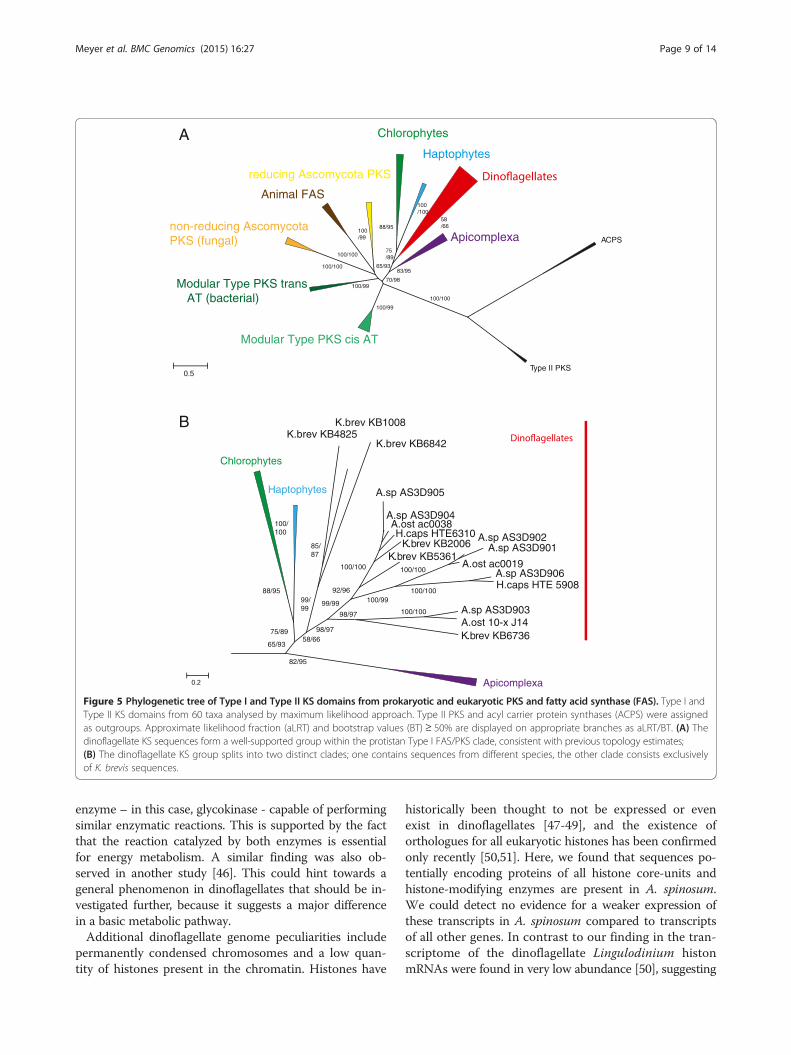
A. spinosum PKS transcripts are type I ‘Protistan’A phylogenetic maximum likelihood approach yielded in-sights into relationships of PKS sequences from A. spino-sum in the current study compared with additional PKSsequences from other dinoflagellates, with an acyl carrierprotein synthase (ACPS) sequence as out-group [30]. Theresulting overall tree topology was consistent with previ-ously known basic topology for dinoflagellates (Figure 5A).Both ACPS and Type II PKS sequences grouped consist-ently as out-group taxa with 100% approximate likelihoodratio (aLRT) and bootstrap support (BT). The transcriptsin the current study grouped with strong support along-side Type I PKS sequences, indicating that they are alsoType I (Figure 5A).Support for a ‘protistan’ clade of PKS Type I sequences,
consisting of apicomplexa, dinoflagellates, haptophytes,and chlorophyta was strong/moderate within the phyl-ogeny (98% aLRT/70% BT; Figure 5A). Each of thesemajor groups, however, formed a discrete sub-groupwithin the protistan clade. The dinoflagellate clade brokedown into two clearly separated sub-clades, one of whichincluded sequences from all dinoflagellate species evalu-ated in this study (including A. spinosum, A. ostenfeldii, H.triqueta and K. brevis). The other dinoflagellate sub-groupcontained only K. brevis KS sequences (Figure 5B).
N-terminal and C-terminal PKS evolution in A. spinosumThe phylogenetic relationship of the PKS N-terminal re-gion among the examined dinoflagellates is shown in amaximum likelihood phylogeny in Figure 4A. The treereflects the same dinoflagellate resolution found in theKS-based phylogeny (Figure 5B), with one sub-clade com-prising only K. brevis sequences, and the other consistingof sequences from A. spinosum, A. ostenfeldii, H. triquetaand K. brevis. The alignment of the N-terminal region re-vealed several conserved aa positions, including the highlyconserved motive ExExGYLG, which was altered in someof the A. spinosum sequences (Figure 4B). Specifically, inthe sequence AS3D901, the whole dinoflagellate-specificN-terminal region was missing. For AS3D903, the GYLpart of the motive was replaced by aliphatic aa.In a final phylogenetic analysis focusing on the PKS C-
terminal region, the BLAST algorithm identified 25%
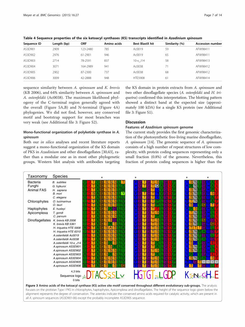
Table 4 Sequence properties of the six ketoacyl synthases (KS) transcripts identified in Azadinium spinosum
Sequence ID Length (bp) ORF Amino acids Best BlastX hit Similarity (%) Accession number
AS3D901 2909 123-2480 785 Ac0019 59 AFW98411
AS3D902 2976 61-2901 946 Ac0019 65 AFW98411
AS3D903 2714 78-2591 837 10-x_J14 58 AFW98413
AS3D904 3071 164-2989 941 Ac0038 71 AFW98412
AS3D905 2902 87-2300 737 Ac0038 68 AFW98412
AS3D906 3009 42-2888 948 HTE5908 61 AFW98414
Meyer et al. BMC Genomics (2015) 16:27 Page 7 of 14
sequence similarity between A. spinosum and K. brevis(KB 2006), and 64% similarity between A. spinosum andA. ostenfeldii (Ac0038). The maximum likelihood phyl-ogeny of the C-terminal region generally agreed withthe overall (Figure 5A,B) and N-terminal (Figure 4A)phylogenies. We did not find, however, any conservedmotif and bootstrap support for most branches wasvery weak (see Additional file 3: Figure S2).
Mono-functional organization of polyketide synthase in A.spinosumBoth our in silico analyses and recent literature reportssuggest a mono-functional organization of the KS domainof PKS in Azadinium and other dinoflagellates [30,43], ra-ther than a modular one as in most other phylogeneticgroups. Western blot analysis with antibodies targeting
V D T V C S S S L TV D T A C S S S L AL D T A C S S S L MV D S A C S S S L YV D T A C S S S L LI D T A C S S S L VL D T A C S S S I VI D T A C S A G L VI D T A C S S S L VI D T A C S S S L VI D T A C S S G L VI D T A C S A S L VA D T A C S A S L AI D T A C S S S L VC D T A C S S S L VI D T A C S S S L VV D T A C S A S L VC D T A C S S A L VC D T A C S S S L IA D T A C S A S L VI D T A C S S S L VI D T A C S S S L VA D T A C S A S L C
Taxonomy SpeciesBacteria B. subtilesFunghi G. fujikuroiAnimal FAS H. sapiens
B. moriC. elegans
Chlorophytes O. lucimarinusO. tauri
Haptophytes E. huxleyi
Apicomplexa T. gondiC. parvum
Dinoflagellates K. brevis KB 2006K. brevis KB 5361H. triquetra HTE 5908H. triquetra HTE 6310A.ostenfeldii Ac0019A.ostenfeldii Ac0038A.ostenfeldii 10-x_J14A.spinosum AS3D901A.spinosum AS3D902A.spinosum AS3D903A.spinosum AS3D904A.spinosum AS3D905A.spinosum AS3D906
Sequence logo4,3 bits
0 bits
Figure 3 Amino acids of the ketoacyl synthase (KS) active site motif confocuses on the protistan Type I PKS in chlorophytes, haptophytes, Apicomplealignment represents the degree of conservation. The asterisks indicate the coall A. spinosum sequences (AS3D901-06) except the probably incomplete AS3
the KS domain in protein extracts from A. spinosum andtwo other dinoflagellate species (A. ostenfeldii and H. tri-quetra) confirmed this interpretation. The blotting patternshowed a distinct band at the expected size (approxi-mately 100 kDA) for a single KS protein (see Additionalfile 3: Figure S1).
DiscussionFeatures of Azadinium spinosum genomeThe current study provides the first genomic characteriza-tion of the photosynthetic free-living marine dinoflagellate,A. spinosum [14]. The genomic sequence of A. spinosumconsists of a high number of repeat structures of low com-plexity, with protein coding sequences representing only asmall fraction (0.8%) of the genome. Nevertheless, thisfraction of protein coding sequences is higher than the
H G T G T S L G D P K S N I G H A EH G T G T Q A G D A K A N V G H A EH G P G T K V G D P K S N M G H P EH G T G T R V G D P K S N L G H S EH G T G T K V G D P K S N M G H A EH G T G T P L G D P K T N M G H A EH G T G T P L G D P K S H V C H T EH G T G T E L G D P K A S V G H A EH G T G T S L G D P K T N I G H L EH G T G T P L G D P K S N I G H L EH G T G T S L G D P K S N I C H G EH G T G T S L G D P K S N I G H C EH G T G T S L G D P K T T T G H L EH G T G T S L G G P K S N M A H G EH G T G T S L G D P K S N F G H L EH G T G T S L G D P K S N V A H G EH G T G T A L G D P K S H V G H E EH G T G T A L G D P K S N F G H L EH G T G T S L G D P K S N F G H L EH G T G T A L G D P K A H V G H E EH G T G T S L G D P K S N V A H S EH G T G T S L G D P R R G T - - - -H G T G T S L G D P K T H T G H L E
served throughout different evolutionary sub-groups. The analysisxa and dinoflagellates. The height of the sequence logo given below thenserved amino acids required for catalytic activity, which are present inD905 sequence.

KB4825
KB1008
HTE5908
AS3D906
AS3D902
AS3D901
Ac0019
AS3D905
AS3D904
HTE6310
Ac0038
KB2006
KB5361
10-x J14
AS3D903
L F R L P E E V E E G Y L G F G A K G K V A W L DL Q R F P E E V E E G Y L G Q G G R G K V A W M DL G R L A Q E V E E G Y L G K N G R A K V M W L D- - - - - - - - - - - - - - - - - - - - - M W L DF G R L C M E V E E G Y L G K N G R A K V M W V DF S R M P V E F E R G Y L G K D G S A K T L L L DF S R L A V E F E R G Y L G K G G S A K T L L L DF S R L A V E F E S G Y L G K E S T A K T L L L DF S R L A V E F E R G Y L G K E G S A K T L L L DF G R L A V E F E P G Y L G R G A G A K V L H L DF V R L P S E L E P G Y L G K D C T A K T M N L DF E K M K A E F E V G Y L G N E N I T R C A V I EY E G L R A E M E E A F L G K E N N S R V G L I E
Sequence logo
4.3 bit
0 bit
BA100
100
9999
98
90
85
92
90
71
Accession number
AFW98414
AFW98411
AFW98411
AFW98412
ABQ85800
AFW98413
ABQ85797
KM588916
KM588917
KM588918
KM588919
KM588920
KM588921
Figure 4 Phylogeny and multiple alignment of the truncated conserved N-terminal region of the dinoflagellate KS. (A) Maximum likelihooddendrogram of the N-terminus computed with 1,000 bootstrap replicates. Bootstrap support is indicated on the dendrogram branches; (B) Multiplealignment of the conserved N-terminal region. The height of the sequence logo shows the degree of conservation. Each line of the alignmentcorresponds to the dinoflagellate strain shown in the dendrogram. Outgroup sequences are not shown because they are lacking the N-terminal motif.
Meyer et al. BMC Genomics (2015) 16:27 Page 8 of 14
0.2% of protein coding regions of the genomes of both A.ostenfeldii (genome size: 105.3 Gb) and H. triquetra (gen-ome size: 18.6–23.6 Gb) [5,44].The ten most common genes in the genomic data were
identified based on read counts, suggesting that geneswhich are covered by more reads may have more copiesin the genome. Dinoflagellates are known for their largegene families with sometimes high gene copy numbers[12]. However, our analysis did not allow us to differenti-ate whether the reads we identified are derived from mul-tiple copies of the same gene within the genome or fromdifferent genes from the same gene family.A minor fraction (0.17%) of the A. spinosum sequenced
data was assigned to be of retroviral origin. This includesDNA transposons and retrotransposons, which have beenfound to a similar extent (0.15%) in the A. ostenfeldii gen-ome [5]. In the genome of S. minutum a slightly higheramount of DNA transposons and retrotransposons (0.5%and 1.1%, respectively) were found. Large tandem repeats,which account for at least 58% of the A. ostenfeldii gen-ome [5], were not found in A. spinosum and account for4.6% of the comparatively smaller S. minutum genome [7].Tandem repeats may play a role in chromatin packing andgenome organization, similar to what is known for telo-mere regions [45], thus they may play an important role inthe organization of the >50 Gbp genome of A. ostenfeldii.Meanwhile, their absence in A. spinosum might explain
the substantially smaller and less complex genome of thisspecies [32].
Features of the A. spinosum transcriptomeThe generated EST data of A. spinosum represents a highcoverage of the transcriptome. Among all identified tran-scripts, 844 carried the complete splice leader sequence.Almost all eukaryotic core genes (94% in CEGMAanalyses) and the essential enzymes from the key meta-bolic pathways were found. The difference in GC-contentwe observed between genome and transcriptome is com-parable to what was found previously in S. minutum [7].Unsurprisingly, protein kinase, DNA binding, cytoskel-
eton proteins, ion channels, and ABC transporter domainswere identified among the most abundant protein do-mains. These domains play important roles in maintainingessential functions in unicellular organisms, e.g., for sig-naling pathways, gene expression, cellular organization,and homeostasis of ions and small molecules. These find-ings are similar to published datasets from other dinofla-gellates, however in our analysis the number of domainsinvolved in metabolism seems to be less dominant withinthe top 20 domains [4,5].As an interesting exception, hexokinase, the key enzyme
of glycolysis, was absent from the A. spinosum transcrip-tome. The absence of the hexokinase is an indication thatenzymatic activity might have been replaced by another
Type II PKS
ACPSApicomplexa
Haptophytes
Chlorophytes
reducing Ascomycota PKS
Animal FAS
non-reducing Ascomycota PKS (fungal)
Modular Type PKS trans AT (bacterial)
Modular Type PKS cis AT
Chlorophytes
Haptophytes
Apicomplexa
K.brev KB4825K.brev KB1008
K.brev KB6842
A.sp AS3D905
A.sp AS3D904A.ost ac0038H.caps HTE6310
K.brev KB2006K.brev KB5361
A.sp AS3D902A.sp AS3D901
A.ost ac0019A.sp AS3D906H.caps HTE 5908
A.sp AS3D903A.ost 10-x J14K.brev KB673658/66
75/89
88/95
100/ 100
99/99
99/99
98/97
98/97
82/95
100/100
100/100100/99
100/100100/100
85/87
65/93
92/96
100/100
100/99
100/100
100/100
100/99
100/99
83/95
100/100
88/9558/66
65/93
75/89
A
B
70/98
Figure 5 Phylogenetic tree of Type I and Type II KS domains from prokaryotic and eukaryotic PKS and fatty acid synthase (FAS). Type I andType II KS domains from 60 taxa analysed by maximum likelihood approach. Type II PKS and acyl carrier protein synthases (ACPS) were assignedas outgroups. Approximate likelihood fraction (aLRT) and bootstrap values (BT) ≥ 50% are displayed on appropriate branches as aLRT/BT. (A) Thedinoflagellate KS sequences form a well-supported group within the protistan Type I FAS/PKS clade, consistent with previous topology estimates;(B) The dinoflagellate KS group splits into two distinct clades; one contains sequences from different species, the other clade consists exclusivelyof K. brevis sequences.
Meyer et al. BMC Genomics (2015) 16:27 Page 9 of 14
enzyme – in this case, glycokinase - capable of performingsimilar enzymatic reactions. This is supported by the factthat the reaction catalyzed by both enzymes is essentialfor energy metabolism. A similar finding was also ob-served in another study [46]. This could hint towards ageneral phenomenon in dinoflagellates that should be in-vestigated further, because it suggests a major differencein a basic metabolic pathway.Additional dinoflagellate genome peculiarities include
permanently condensed chromosomes and a low quan-tity of histones present in the chromatin. Histones have
historically been thought to not be expressed or evenexist in dinoflagellates [47-49], and the existence oforthologues for all eukaryotic histones has been confirmedonly recently [50,51]. Here, we found that sequences po-tentially encoding proteins of all histone core-units andhistone-modifying enzymes are present in A. spinosum.We could detect no evidence for a weaker expression ofthese transcripts in A. spinosum compared to transcriptsof all other genes. In contrast to our finding in the tran-scriptome of the dinoflagellate Lingulodinium histonmRNAs were found in very low abundance [50], suggesting

Meyer et al. BMC Genomics (2015) 16:27 Page 10 of 14
differences in abundance of histones between dinofla-gellate species.Approximately 40% of the predicted proteins of A. spi-
nosum could not be matched to the PFAM database.This is in line with previous findings in dinoflagellatesand might be an indication for novel pathways expressedin dinoflagellates [5,52-55]. On average a quarter of thegenes found in the dinoflagellate species studied herehad no recognizable homologues in other dinoflagellatesgenera. The estimated number of these ‘orphan’ genestotaled 27% of all genes for the dinoflagellates we stud-ied; this is within the range for other well-studied phyla(e.g. bacteria [56], and nematodes [57]), for which 2-50%of identified genes are orphans.Different evolutionary mechanisms can lead to the for-
mation of orphan genes. For example, duplication can freegene copies from evolutionary constraints - while the ori-ginal function is retained in the original gene, the dupli-cated gene copy can possess new protein functions [58].An additional source for orphan genes is horizontal genetransfer from other organisms, which has been shown tohave the potential to be a significant source of geneticinnovation in dinoflagellates [59].Taken together, a rather unique set of genes may be
characteristic for species within the dinoflagellates, po-tentially reflecting their complex life cycle and nutritionbecause most are mixo- or heterotrophs and only a feware obligate photoautotrophs [60-62].
Function and evolution of PKSAll polyketide biosynthetic enzyme functions needed tosynthesize the azaspiracid carbon backbone are present inA. spinosum (Table 3). This is to our knowledge the firstdinoflagellate dataset where all genes needed for polyke-tide synthesis were identified. Whether these PKS-relatedsequences are coding, are involved in fatty acid synthesisor toxin production, and/or have another function re-mains unclear. However, this set of genes opens new ave-nues to study toxin synthesis in A. spinosum and maybeother dinoflagellates also up to now it remains unknownin which cellular compartment toxin synthesis takes place.PKS enzymes have been shown to be expressed in bothchloroplast and cytoplasm, but we could not identify sig-nal peptide sequences which may hint towards a specificcellular compartment. However, co-immunoprecipitationstudies have shown that PKS functions in K. brevis are in-deed organized in a mono-modular way, meaning thateach domain comprises a single enzyme [29,43].The KS domain is essential to Type I polyketide synthe-
sis, which is carried out in a stepwise manner. Each KSdomain elongates the existing carbon chain by condensa-tion of the acetyl-CoA/malonyl-CoA building blocks, withtwo carbon atoms normally added in each elongation step.This means that Type I modular PKS must contain at
least 20 modules to create the common carbon backbonefor the diverse set of identified azaspiracid toxins [15]. Weanalysed six unique PKS transcripts each containing oneKS domain, in detail. Whether or not separate transcriptsare used or if the same transcripts are recycled during thepolyketide synthesis process remains unclear. In any case,the complete AZA structure could be synthesized withthe total number of KS, KR, AT and the other domainsidentified in our dataset.Analyses of A. spinosum full-length PKS transcripts
(Figure 3) and of the corresponding Western blots(Additional file 3: Figure S1) confirms the mono-modularfunctional Type I PKS structure, which might be acommon feature in dinoflagellates [29,30]. It has beenproposed that this structure likely diverged from multi-modular PKS around the time of origin of the dinoflagel-lates [30,43]. The other protistan groups studied here(haptophytes, chlorophytes and apicomplexa) exhibit themultimodular Type I form. The protist KS groups formwell-supported monophyletic clades in the current ana-lysis, consistent with earlier findings [26,30].The flanking regions of KS proteins identified in A. spi-
nosum contain less conserved motives than in other or-ganisms, but this sequence feature was also found in otherdinoflagellates [30]. In the C-terminal region, in particular,no conserved motif was identified in the alignment, sup-porting the assumption that this is a relict of the linker se-quences of the multi-modular PKS Type I. With thislinker structure, the single domains could have been sepa-rated to allow structural function of the complex enzyme.Adding A. spinosum sequences to the existing C-terminaldataset did not significantly alter the overall topologypresent in the N-terminal dataset, despite weaker phylo-genetic support. In contrast, within the N-terminal se-quences, a high degree of conservation was found in thealignment, arguing that the N-terminal has another func-tion rather than acts as a linker region. Instead, the N-terminal region might have an enzymatic or structuralfunction, or could act as a protein-protein binding do-main. The conserved N-terminal motif identified previ-ously [30] was found in four of our six A. spinosumsequences. However, we observed an alteration of theGYLG motif towards AFLG. Alterations of the GYLGmotif have been reported from the dinoflagellate, Gam-bierdiscus polynesiensis and also in some K. brevis strainswhere several variants of the GYLG motive were found[63]. The absence or alteration of the conserved N-terminal motif in the remaining two A. spinosum se-quences (AS3D901 and AS3D903) could be explainedin several ways. For example, these copies might havediverged to play a role in another pathway via geneduplication. Alternatively, these differences may lead toalterations in polyketide synthesis. Finally, these two se-quences may simply represent non-functional transcripts

Meyer et al. BMC Genomics (2015) 16:27 Page 11 of 14
or pseudogenes. Functional tests are required to elucidatethe role of the PKS genes as well as of the C- and N-terminal sequences.
ConclusionsWe characterized the transcriptome of the toxic dinofla-gellate A. spinosum, revealing that it possesses a lownumber of orphan genes compared to other tested dino-flagellate species. With respect to toxin synthesis, we de-tected transcripts of all genes essential for polyketidesynthesis in A. spinosum. As the number of transcriptomicdatasets for different dinoflagellate species increases, dee-per insights into the functional differences between toxicand non-toxic variants, even within a species, as well asinto mechanisms of HAB formation, will be gained.
MethodsMaintenance and harvesting of dinoflagellate culturesAzadinium spinosum cultures of strain 3D9 isolated fromthe Scottish east coast (57° 3.9′ N; 02°30.2′W) were grownas described in Tillmann et al. [14]. In brief, cultures weregrown at 20°C at a salinity of 32 PSU. All exponentiallygrowing cultures were then harvested by centrifugationfor 15 min at 3200 × g with fixed angle rotor (Eppendorf5810 R, Eppendorf, Hamburg, Germany).
Methods for DNA; extractionFollowing centrifugation, cell pellets were re-suspended inTissue and Cell Lysis Buffer (DNeasy Plant kit; Qiagen,Hamburg, Germany) and transferred into a 2 ml cryo-vial containing acid-washed glass beads (Sigma-Aldrich,Steinheim, Germany). Cell lysis was achieved with aBio101 FastPrep instrument (Thermo, Savant Illkirch,France) run at maximum speed (6.5 ms−1) for 45 s. DNAextraction proceeded according to the DNeasy Plant kitprotocol.
Shotgun sequencingPrior to sequencing, DNA was sheared and contaminantsand small fragments were removed with the Ampure BeadPCR purification system (Invitrogen, Karlsruhe, Germany)following the standard protocol for 454-library prepar-ation and shotgun sequencing (Roche, MannheimGermany). Sequencing was then carried out on a RocheGS Junior machine (Roche, Mannheim, Germany) fol-lowing standard protocols.
Sequencing post-run processingThe associated 454-sequencing software (Roche GS JuniorVersion 2.8) was used for quality control and contig as-sembly. All sequencing reads featuring inaccurate keysequences, chimeric sequences, biased nucleotides, or un-identified nucleotides were regarded as low quality reads
and were discarded. A total of 103,860 high quality reads,yielding 37,739,423 bp of genomic information, wereretained for analysis.
Sequence analysis of genomic DNA readsLow overall genomic coverage and a very large genomesize meant that assembly of the retained reads was unreal-istic. Reads were thus analysed with the programRepeatMasker ver. 4.0.5 [34] to establish the repeatstructure of the genome. To determine the potentialproportion of protein-coding sequence, annotation ofthe high quality reads was also performed. The Blast2goalgorithm (www.blast2go.org) was applied against theSwissprot database, and only reads with an e-value below10−7 were considered in the analysis.
Genome size estimationThe genome size of A. spinosum was estimated by DNAextraction and subsequent qPCR. The DNA contentwas calculated using a standard curve. For more detailssee [32].
RNA methods; extractionCell pellets obtained after centrifugation of exponen-tially growing cultures were immediately re-suspendedin 1 ml of 60°C hot TriReagent (Sigma-Aldrich, Steinheim,Germany) and transferred into a 2 ml cryovials contain-ing acid washed glass beads (Sigma-Aldrich, Steinheim,Germany). As for DNA extraction, cell lysis was achievedwith a Bio101 FastPrep instrument (Thermo SavantIllkirch, France) run at maximum speed (6.5 ms−1) for 45s. RNA isolation was then performed as described in [64].In brief, after thawing the cell lysate on ice, 200 μl of purechloroform was added to each sample. The sample wasvortexed thoroughly and incubated for 10 min at roomtemperature. The aqueous phase was separated by centri-fugation and transferred into a new vial together with100% isopropanol and incubated at -20°C to precipitatethe RNA. The pellet was collected by centrifugation at4°C, washed with 70% ethanol, air-dried and re-suspendedin RNase-free water (Qiagen, Hilden, Germany).Only RNA samples with high quality (OD 260/280 > 2
and OD260/230 > 1.8), determined with a NanoDrop ND-100 spectrometer (PeqLab, Erlangen, Germany), and highRNA integrity, checked with the Agilent RNA Nano ChipAssay (Agilent, Santa Clara, USA), were used for tran-scriptome sequencing.
RNA sequencingRNA sequencing and post-run processing was performedby The Marine Microbial Eukaryotic Transcriptome Se-quencing Project (MMETSP).

Meyer et al. BMC Genomics (2015) 16:27 Page 12 of 14
Analysis of transcriptome dataAn assembly pipeline from the MMETSP was to assemblehigh quality raw reads into contigs, which were translatedinto amino acid sequences and blasted against the PFAMdatabase (PFAM count (e-value < 0.001) file Supplement).
Homology analysisWe used the NCBI blast suite (version 2.2.28+) to com-pare the set of 69,956 predicted protein fragments withgene predictions and EST data from the following species:dinoflagellates: Alexandrium andersonii, Alexandriumostenfeldii, Amphidinium massartii, Ceratium fusus,Crypthecodinium cohnii, Dinophysis acuminate, Gleno-dinium foliaceum, Gymnodinium catenatum, Hetero-capsa triquetra, Karenia brevis, Karlodinium micrum,Oxyrrhis marina, Peridinium aciculiferum, Polarella gla-cialis, Prorocentrum minimum, Protoceratium reticulatem,Scrippsiella trochoidea, Symbiodinium minutum; dia-toms: Fragilariopsis cylindrus, Phaeodactylum tricornu-tum, Thalassiosira pseudonana; haptophyte: Emilianiahuxleyi; cryptomonad: Guillardia theta; and the chloro-phyte Chlamydomonas reinhardtii. We computed all pair-wise BLAST comparisons (BLASTp, tBLASTx, BLASTx,and tBLASTn, depending on the type of database) at athreshold of e-value < 0.001 and counted the number ofsequences with hits in the other species. For the compari-son to the CEGMA data set [35], we used the hmmerpackage (version 3.0) to detect hits (e-value < 0.001) toany of the CEGMA profile HMMs.
Phylogenetic analysesAdditional amino acid sequences were obtained from theNCBI GenBank and from [30]. A representative dataset of65 Type I and Type II PKS sequences covering the majorclades of prokaryota, fungi, animals, apicomplexa, hapto-phytes and chlorophytes was used for maximum likeli-hood phylogenetic analyses. A multi-sequence alignmentwas created in MEGA 5.2 [65] with the implementedMUSCLE algorithm [66]. A maximum likelihood tree wascomputed in PhyML 3.0 [67] under assumption of theLa-Gascuel amino acid replacement matrix [68]; likeli-hood ratio tests [69] and 1,000 bootstrap analyses wereperformed as a measure of the validity of each branch.
Analyses of the N- and C-terminal regionThe N- and C- terminal regions of the dinoflagellate KSsequences were truncated from the KS domain andblasted with tBLASTx against the NCBI and PFAM data-bases. No significant hits other than to known KS se-quences in the database were obtained by BLASTxanalysis. Multiple alignment and subsequent phylogen-etic trees were calculated as described for the KSdomain.
Western immunoblotting of dinoflagellate PKSWestern blot analysis of protein extracts from A. ostenfel-dii AOSH 2 and NCH 85, A. tamarense Atam5, H. tri-quetra SCCAP strain K-0481, E. huxleyi, Phaeodactylumsp., A. spinosum and S. trochoidea was performed accord-ing to [30]. In brief, 10 mg of total protein extract was sep-arated on a 10% SDS/polyacrylamide gel and transferredonto polyvinylidene fluoride membranes by Western Blot.Membranes were blocked with 5% skimmed milk in Tris-buffered saline with 0.5% Tween 20 (TBS-T) for 1 h. Pri-mary antibodies (Rabbit anti-K. brevis KS KB 2006 1:5000)were diluted in 5% skimmed milk in TBS-T and the mem-branes were incubated at 4°C overnight. Subsequently, themembranes were washed three times with TBS-T andincubated with the appropriate secondary antibody di-lution (Goat anti-rabbit 1:20000 Peroxidase-conjugated)(Sigma-Aldrich, Schnelldorf, Germany) in 5% skimmedmilk/TBS-T for 2 h at room temperature.
Additional files
Additional file 1: List of histon related contigs.
Additional file 2: Most common protein domains based on PFAMdatabase search: pfam_count.
Additional file 3: Table S1. – Origin and number of the sequencesused in the orphan analysis (Figure 2). Figure S1. – Presence of KS proteinin different microalgae. Western blot analysis of protein extracts from A.ostenfeldii AOSH 2 and NCH 85, A. tamarense Atam5, Heterocapsa triquetraSCCAP strain K-0481, Emiliania huxleyi, Phaeodactylum sp., Azadiniumspinosum and Scrippsiella trochoidea using a polyclonal rabbit anti- K. brevisKS antibody. Figure S2. – Phylogeny truncated C-terminal region of thedinoflagellate KS. Maximum likelihood dendrogram of the C-terminuscomputed with 1,000 bootstrap replicates, bootstrap values ≥ 50% aredisplayed on dendrogram branches.
Competing interestsThe authors declare that they have no competing interests.
Authors’ contributionsJMM and UJ designed the study. JMM and KE performed the experiments.JMM, CR and UJ performed the bioinformatics and analysed the data. Allauthors read, edited and approved the final manuscript.
AcknowledgementsThe authors are grateful to Dr. Frances Van Dolah (NOAA Center for Coastaland Environmental Health and Biomolecular Research, Charleston, SouthCarolina, USA) for the custom peptide polyclonal antibodies specific for Kareniabrevis KS domain (KB2006; GenBank accession no. EF410007). This research wasconducted within the PACES research program of the Alfred-Wegener-InstituteHelmholtz-Zentrum für Polar- und Meeresforschung as part of the Coastaltheme (WP2).
FundingFinancial support was provided by the PACES research program of theAlfred-Wegener-Institute Helmholtz-Zentrum für Polar- und Meeresforschung.The Gordon and Betty Moore Foundation funded sequencing, assembly, andpreliminary annotation through Grant GBMF2637 to the National Center forGenome Resources.
Author details1Ecological Chemistry, Alfred Wegener Institute for Polar and MarineResearch, Bremerhaven, Germany. 2Evolutionary biology, Max Planck Institutefor Developmental Biology, Tübingen, Germany. 3Adenoviridae: Receptors,
Meyer et al. BMC Genomics (2015) 16:27 Page 13 of 14
Trafficking and Vectorology, Institut de Génétique Moléculaire deMontpellier, Montpellier, France.
Received: 11 June 2014 Accepted: 24 December 2014
References1. Hallegraeff GM. A review of harmful algal blooms and their apparent global
increase. Phycologia. 1993;32:79–99.2. Harper JT, Waanders E, Keeling PJ. On the monophyly of chromalveolates using
a six-protein phylogeny of eukaryotes. Int J Syst Evol Micr. 2005;55:487–96.3. Moreno-Diaz-de-la-Espina S, Alverca E, Cuadrado A, Franca S. Organization of
the genome and gene expression in a nuclear environment lacking histonsand nucleosomes: the amazing dinoflagellates. Eur J Cell Biol. 2005;84:137–49.
4. Lowe CD, Mello LV, Samatar N, Martin LE, Montagnes DJS. The transcriptomeof the novel dinoflagellate Oxyrrhis marina (Alveolata: Dinophyceae): responseto salinity examined by 454 sequencing. BMC genomics. 2011;12:519.
5. Jaeckisch N, Yang I, Wohlrab S, Gloeckner G, Kroymann J, Vogel H, et al.Comparative genomic and transcriptomic characterization of the toxigenicmarine dinoflagellate Alexandrium ostenfeldii. PLoS ONE. 2011;6:e28012.doi:10.1371/journal.pone.0028012.
6. Jackson CJ, Gornik SG, Waller RF. The mitochondrial genome and transcriptomeof the basal dinoflagellate Hematodinium sp.: character evolution within thehighly derived mitochondrial genomes of dinoflagellates. Genome Biol Evol.2012;4:59–72.
7. Shoguchi E, Shinzato C, Kawashima T, Gyoja F, Mungpakdee S, Koyanagi R,et al. Draft assembly of the Symbiodinium minutum nuclear genome revealsdinoflagellate gene structure. Curr Biol. 2013;23:1399–408.
8. Dodge JD. The dinophyceae. The chromosome of the algae. Edited byGodward MBE. London: Arnold; 1966.
9. Rizzo PJ. Those amazing dinoflagellate chromosomes. Cell Res. 2003;13:215–7.10. Rill RL, Livolant F, Aldrich HC, Davidson MW. Electron microscopy of liquid
crystalline DNA: Direct evidence for cholesteric-like organization of DNA indinoflagellate chromosomes. Chromosoma. 1989;98:280–6.
11. Lin S. Genomic understanding of dinoflagellates. Res Microbiol. 2011;166:551–69.12. Hou Y, Lin S. Distinct gene number-genome size relationships for eukaryotes
and non-eukaryotes: gene content estimation for dinoflagellate genomes.PLoS ONE. 2009;4:e6978. doi:10.1371/journal.pone.0006978.
13. Beauchemin M, Roy S, Daoust P, Dagenais-Bellefeuille S, Bertomeu T, LetourneauL, et al. Dinoflagellate tandem array gene transcripts are highly conserved andnot polycistronic. PNAS. 2012;109:15793–8.
14. Tillmann U, Elbrächter M, Krock B, John U, Cembella A. Azadinium spinosumgen. et sp. nov. (Dinophyceae) identified as a primary producer of azaspiracidtoxins. Eur J Phycol. 2009;44:63–79.
15. Krock B, Tillmann U, John U, Cembella AD. Characterization of azaspiracids inplankton size-fractions and isolation of an azaspiracid-producing dinoflagellatefrom the North Sea. Harmful Algae. 2008; doi:10.1016/j.hal.200806.003.
16. James KJ, Furey A, Lehane M, Ramstad H, Aune T, Hovgaard P, et al. Firstevidence of an extensive Northern European distribution of AzaspiracidPoisoning (AZP) toxins in shellfish. Toxicon. 2002;40:909–15.
17. Magdalena AB, Lehane M, Krys S, Fernandez ML, Furey A, James KJ. The firstidentification of azaspiracids in shellfish from France and Spain. Toxicon.2003;42:105–8.
18. Taleb H, Vale P, Amanhir R, Benhadouch A, Sagou R, Chafik A. First detection ofazaspiracids in mussels. North West Afr J Shellfish Res. 2006;25:1067–70.
19. Twiner MJ, Rehmann N, Hess P, Doucette GJ. Azaspirazid shellfish poisoning:a review on the chemistry, ecology, and toxicology with emphasis onhuman health impacts. Marine Drugs. 2008;6:39–72.
20. Furey A, O’Doherty S, O’Callaghan K, Lehane M, James KJ. Azaspiracidpoisoning (AZP) toxins in shellfish: toxicological and health considerations.Toxicon. 2010;56:173–90.
21. Tillmann U, Salas R, Jauffrais T, Hess P, Silke J. Azaspiracids. The producingorganism(s): biology and trophic transfer. In: Botana L, editor. Seafood andfreswater toxins. Pharmacology, physiology, and detection, seafood andfreshwater toxins. Pharmacology, physiology, and detection. Boca Raton:CRC Press Boca Raton; 2014. p. 1197.
22. Staunton J, Weissman KJ. Polyketide biosynthesis: a millennium review.Nature Prod Rep. 2001;18:380–416.
23. Gokhale RS, Dipika T. Biochemistry of polyketide synthases. In: Rehm HJ,Reed G, editors. Biotechnology. 2nd ed. Weinheim, Germany: Wiley-VCHVerlag GmbH; 2001. p. 341–72.
24. Shen B. Polyketide biosynthesis beyond the type I, II and III polyketidesynthase paradigms. Curr Opin Chem Biol. 2003;7:285–95.
25. Zhu G, LaGier MJ, Stejskal F, Millership JJ, Cai X, Keithly JS. Cryptosporidiumparvum: the first protist known to encode a putative polyketide synthase.Gene. 2002;298:79–89.
26. John U, Beszteri B, Derelle E, Van de Peer Y, Read B, Moreau H, et al. Novelinsights into evolution of protistan polyketide synthases throughphylogenomic analysis. Protist. 2008;159:21–30.
27. John U, Beszteri S, Gloeckner G, Singh R, Medlin L, Cembella AD. Genomiccharacterisation of the ichthyotoxic prymnesiophyte Chrysochromulinapolylepis, and the expression of polyketide synthases genes in synchronisedcultures. Eur J Phycol. 2010;45:215–29.
28. Freitag M, Beszteri S, Vogel H, John U. Effects of physiological shocktreatments on toxicity and polyketide synthase gene expression inPrymnesium parvum (Prymnesiophyceae). Eur J Phycol. 2011;46:193–201.doi:10.1080/09670262.2011.591438.
29. Monroe E, Van Dolah FM. The toxic dinoflagellate Karenia brevis encodesnovel type I-like polyketide synthases containing discrete catalytic domains.Protist. 2008;159:471–82.
30. Eichholz K, Beszteri B, John U. Putative monofunctional type I polyketidesynthase units: a dinoflagellate-specific feature? PLoS ONE. 2012;7:e48624.doi:10.1371/journal.pone.0048624.
31. Keeling PJ, Burger G, Durnford DG, Lang BF, Lee RW, Pearlman RE, et al. Thetree of eukaryotes. Trends Ecol Evol. 2005;20:670–6.
32. Toebe K, Joshi A, Messtorff P, Tillmann U, Cembella A, John U. Moleculardiscrimination of taxa within the dinoflagellate genus Azadinium, the sourceof azaspiracid toxins. J Plankton Res. 2013;35:225–30.
33. Keeling PJ, Burki F, Wilcox HM, Allam B, Allen EE, Amaral-Zettler LA, et al.The Marine Microbial Eukaryote Transcriptome Sequencing Project(MMETSP): illuminating the functional diversity of eukaryotic life in theoceans through transcriptome sequencing. PloS Biol. 2014;12:e001889.
34. Smit AFA, Hubley R, Green P. RepeatMasker Open-3.0. http://www.repeat-masker.org.1996-2010.
35. Parra G, Bradnam K, Korf I. CEGMA: a pipline to accurately annotate coregenes in eukaryotic genomes. Bioinformatics. 2007;23:1061–7.
36. Lommer M, Specht M, Roy AS, Kraemer L, Andreson R, Gutowska MA, et al.Genome and low-iron response of an oceanic diatom adapted to chroniciron limitation. Genome Bio. 2012;13:R66. doi:10.1186/gb-2012-13-7-r66.
37. Armbrust EV, Berges JA, Bowler C, Green BR, Martinez D, Putnam NH, et al.The genome of the diatom Thalassiosira pseudonana: ecology, evolution,and metabolism. Science. 2004;306:79–86.
38. Bowler C, Allen AE, Badger JH, Grimwood J, Jabbari K, Kuo A, et al. ThePhaeodactylum tricornutum genome reveals the evolutionary history ofdiatom genomes. Nature. 2008;456:239–44.
39. Read BA, Kegel J, Klute MJ, Kuo A, Lefebvre SC, Maumus F, et al. Pangenome of the phytoplankton Emiliania underpins its global distribution.Nature. 2013;499:209–13.
40. Curtis BA, Tanifuji G, Burki F, Gruber A, Irimia M, Maruyama S, et al. Algalgenomes reveal evolutionary mosaicism and the fate of nucleomorphs.Nature. 2012;492:59–65.
41. Merchant SS, Prochnik SE, Vallon O, Harris EH, Karpowicz SJ, Witman GB,et al. The Chlamydomonas genome reveals the evolution of key animal andplant functions. Science. 2007;318:245–50.
42. von Wettstein-Knowles P, Olsen JG, McGuire K, Henriksen A. Fatty acidsynthesis -role of active site histidines and lysine in Cys-His-His-typebeta-ketoacyl-acyl carrier protein synthases. FEBS J. 2006;273:695–710.
43. Van Dolah FM, Zippay ML, Pezzolesi L, Rein KS, Johanson JG, Morey JS, et al.Subcellular localization of dinoflagellate polyketide synthases and fatty Acidsynthase activity. J Phycol. 2013;49:118–1127.
44. McEwan M, Raheel H, Claudio HS, Patrick JK. Nuclear genome sequencesurvey of the dinoflagellate Heterocapsa triquetra. J Eukaryot Microbiol.2008;55:530–5.
45. Nikitina T, Woodcock CL. Closed chromatin loops at the ends of chromosomes.J Cell Biol. 2004;166:161–5.
46. Butterfield ER, Howe CJ, Ellen R, Nisbet R. An analysis of dinoflagellatemetabolism using EST data. Protist. 2013;164:218–36.
47. Hackett JD, Anderson DM, Erdner DL, Bhattacharya D. Dinoflagellates:a remarkable evolutionary experiment. Am J Bot. 2004;91:1523–34.
48. Zhang H, Hou Y, Miranda L, Campbell DA, Sturm NR, Gaasterland T,et al. Spliced leader RNA trans-splicing in dinoflagellates. PNAS.2007;104:4618–23.

Meyer et al. BMC Genomics (2015) 16:27 Page 14 of 14
49. Lin S, Zhanga H, Zhuanga Y, Tranb B, Gillb J. Spliced leader–basedmetatranscriptomic analyses lead to recognition of hidden genomicfeatures in dinoflagellates. PNAS. 2010;107:20033–8.
50. Roy S, Morse D. A full suite of histones modifying genes are transcribed inthe dinoflagellate Lingulodinium. PloS One. 2012;7:e34340. doi:10.1371/journal.pone.0034340.
51. Gornik SG, Ford KL, Mulhern TD, Bacic A, McFadden GI, Waller RF. Lossofnucleosomal DNA condensation coincides with appearance of anovelnuclear protein in dinoflagellates. Curr Biol. 2012;22:2303–12.
52. John U, Groben R, Beszteri B, Medlin L. Utility of amplified fragment lengthpolymorphisms (AFLP) to analyse genetic structure within the Alexandriumtamarense species complex. Protist. 2004;155:169–79.
53. Van Dolah FM, Lidie KB, Monroe EA, Bhattacharya D, Campbell L, DoucetteGJ, et al. The Florida red tide dinoflagellate Karenia brevis: New insights intocellular and molecular processes underlying bloom dynamics. HarmfulAlgae. 2007;8:562–72.
54. Uribe P, Fuentes D, Valdés J, Shmaryahu A, Zuniga A, Holmes D, et al.Preparation and analysis of an expressed sequence tag library from thetoxic dinoflagellate Alexandrium catenella. Mar Biotechnol. 2008;10:692–700.
55. Yang I, John U, Beszerti S, Gloeckner G, Krock B, Goesmann A, et al.Comparative gene expression in toxic vs non-toxic strains of the marinedinoflagellate Alexandrium minutum. BMC genomics. 2010;11:248.
56. Fukuchi S, Nishikawa K. Estimation of the number of authentic orphangenes in bacterial genomes. DNA Res. 2004;11:219–31.
57. Roedelsperger C, Streit A, Sommer RJ. Structure, function and evolution ofthe nematode genome. In: eLS. Chichester: John Wiley & Sons, Ltd; 2013.doi:10.1002/9780470015902.a0024603.
58. Katju V, Lynch M. On the formation of novel genes by duplication in theCaenorhabditis elegans gemone. Mol Biol Evol. 2006;23:1056–67.
59. Wisecaver JH, Brosnahan ML, Hackett JD. Horizontal gene transfer is asignificant driver of gene innovation in dinoflagellates. Genome Biol Evol.2013;5:2368–81.
60. Jeong HJ, Yoo YD, Park JY, Song JY, Kim ST, Lee SH, et al. Feeding byphototrophic red-tide dinoflagellates: five species newly revealed and six speciespreviously known to be mixotrophic. Aquat Microb Ecol. 2005;40:133–50.
61. Jeong HJ, Yoo YD, Kim JS, Seong KA, Kang NS, Kim TH. Growth, feeding andecological roles of the mixotrophic and heterotrophic dinoflagellates in marineplanktonic food webs. Ocean Sci J. 2010;45:65–91.
62. Stoecker DK. Mixotrophy among dinoflagellates. J Eukaryot Microbiol.1999;46:397–401.
63. Pawlowiez R, Morey JS, Darius HT, Chinain M, Van Dolah FM. Transcriptomesequencing reveals single domain type I-like polyketide synthases in the toxicdinoflagellate Gambierdiscus polynesiensis. Harmful Algae. 2014;36:29–37.
64. Wohlrab S, Iversen M, John U. A molecular and co-evolutionary context forgrazer induced toxin production in Alexandrium tamarense. PLoS ONE.2010;5:e15039. doi:10.1371/journal.pone.0015039.
65. Tamura K, Peterson D, Peterson N, Stecher G, Nei M, Kumar S. MEGA5: molecularevolutionary genetics analysis using maximum likelihood, evolutionary distance,and maximum parsimony methods. Mol Biol Evol. 2011;28:2731–9.
66. Edgar RC. MUSCLE: multiple sequence alignment with high accuracy andhigh throughput. Nucleic acids res. 2004;32:1792–7.
67. Guindon S, Dufayard JF, Lefort V, Anisimova M, Hordijk W, Gascuel O. Newalgorithms and methods to estimate maximum-likelihood phylogenies:assessing the performance of PhyML 3.0. Syst Biol. 2010;59:307–21.
68. Le SQ, Gascuel O. An improved general amino acid replacement matrix. MolBiol Evol. 2008;25:1307–20.
69. Anisimova M, Gascuel O. Approximate likelihood-ratio test for branches:a fast, accurate, and powerful alternative. Syst Biol. 2006;55:539–52.
Submit your next manuscript to BioMed Centraland take full advantage of:
• Convenient online submission
• Thorough peer review
• No space constraints or color figure charges
• Immediate publication on acceptance
• Inclusion in PubMed, CAS, Scopus and Google Scholar
• Research which is freely available for redistribution
Submit your manuscript at www.biomedcentral.com/submit