transcription by a promoter site sensitive to s1 nuclease
TRANSCRIPT
Vol. 8, No. 10MOLECULAR AND CELLULAR BIOLOGY, Oct. 1988, p. 4174-41840270-7306/88/104174-11$02.00/0Copyright © 1988, American Society for Microbiology
Modulation of Epidermal Growth Factor Receptor Proto-OncogeneTranscription by a Promoter Site Sensitive to S1 Nuclease
ALFRED C. JOHNSON, YOSHIHIRO JINNO, AND GLENN T. MERLINO*Laboratory of Molecular Biology, National Cancer Institute, 9000 Rockville Pike, 3712D27, Bethesda, Maryland 20892
Received 23 February 1988/Accepted 24 June 1988
The epidermal growth factor (EGF) receptor is the functional target of the mitogen EGF and the cellularhomolog of the avian erythroblastosis virus erbB oncogene product. Regulation of expression of theproto-oncogene encoding the EGF receptor can be elucidated by studying the structure and function of the genepromoter outside the confines of the cell. Previously, we reported the isolation of the human EGF receptor genepromoter. The promoter is highly GC rich, contains no TATA or CAAT box, and has multiple transcriptionstart sites. An Si nuclease-sensitive site has now been found 80 to 110 base pairs (bp) upstream from the maijorin vivo transcription initiation site. Two sets of direct repeat sequences were found in this area; both conformto the motif TCCTCCTCC. When deletion mutations were made in this region of the promoter by using eitherBal 31 exonuclease or S1 nuclease, we found that in vivo activity dropped three- to fivefold, on the basis oftransient-transfection analysis. Examination of nuclear protein binding to normal and mutated promoter DNAsby gel retardation analysis and DNase I footprinting revealed that two specific factors bind to the direct repeatregion but cannot bind to the S1 nuclease-mutated promoter. One of the specific factors is the transcriptionfactor Spl. The results suggest that these nuclear trans-acting factors interact with the S1 nuclease-sensitiveregion of the EGF receptor gene promoter and either directly or indirectly stimulate transcription.
The epidermal growth factor (EGF) is a potent mitogencapable of stimulating protein and RNA synthesis, DNAreplication, and, ultimately, cellular proliferation (5, 19, 30).It elicits these cellular responses by binding to the N-terminus of a 170-kilodalton cell surface glycoprotein, theEGF receptor. The receptor possesses a very active tyrosinekinase at its C-terminus and is capable of phosphorylatingitself and other substrates upon EGF binding (4). By virtueof its extensive homology to the erbB oncogene product ofthe avian erythroblastosis virus, the EGF receptor is con-sidered the cellular erbB proto-oncogene (7, 33, 47, 53). Thisproto-oncogene has been found to be overexpressed andoccasionally amplified in a variety of malignant cell types (6,27, 31, 37, 38, 54). More recently, we demonstrated thatoverexpression of the normal EGF receptor in NIH 3T3 cellsby a retroviral vector induces malignancy and tumorigenicity(48).The production of a protein so deeply involved in cellular
proliferation and potentially responsible for malignant trans-formation must be carefully regulated in the normal cell.However, relatively little is known about control of EGFreceptor gene expression. Previously, we reported the iso-lation, sequence, and partial characterization of the humanEGF receptor gene promoter (23). The promoter has multi-ple transcription start sites and numerous protein-bindingsites, including at least five Spl-binding sites (24). Thepresence of multiple start sites may be due to the absence ofan obvious TATA box (23). A chimeric plasmid containingthe EGF receptor gene promoter ligated upstream of thechloramphenicol acetyltransferase (CAT) reporter gene (15)is active when transiently transfected into human KB epi-dermoid carcinoma or African green monkey kidney CV-1cell lines (17, 23, 24).
* Corresponding author.
Promoter regions of active genes are frequently hypersen-sitive to the endonucleases DNase I and S1 (10, 29, 50). Thissensitivity is thought to be the result of changes in thestructure of the active chromatin, possibly because of thedifferential dissociation of nucleosomes and local alterationin DNA superhelicity (29, 50). Isolated eucaryotic promoterregions cloned into plasmid vectors maintain sensitivity toS1 nuclease, provided the plasmid is supercoiled (12, 14, 34,35, 40, 44, 55).
Previously, we showed that the EGF receptor gene pro-moter chromatin is hypersensitive to DNase I in humanA431 epidermoid carcinoma cells, which are known tooverexpress the EGF receptor (13). Because the DNaseI-hypersensitive sites are often characterized by sensitivityto the single-strand-specific endonuclease Si (29, 50), wetested the cloned EGF receptor gene promoter for sensitivityto Si nuclease. We report in this paper the identification ofa site exquisitely sensitive to Si nuclease residing in thevicinity of two pairs of direct-repeat sequences conformingto the motif TCCTCCTCC. We demonstrate that this site isnecessary for optimal gene activity and is associated withnuclear proteins whose binding correlates with optimal tran-scription activity.
MATERIALS AND METHODS
Cell lines. The cultured cells used included a subclone ofhuman epidermoid carcinoma KB cells (KB3-1), humanepidermoid carcinoma A431 cells, and African green mon-key kidney CV-1 cells. All cell lines were maintained inDulbecco modified Eagle medium supplemented with 10%fetal bovine serum (GIBCO Laboratories).
Plasmid DNA manipulations. Si nuclease and DNA ligasewere purchased from Pharmacia. Most restriction endonu-cleases were obtained from New England Biolabs, Inc. The
4174
EGF RECEPTOR GENE PROMOTER Si NUCLEASE SENSITIVITY
HinLir
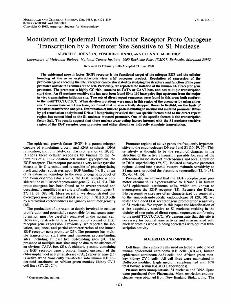
-~~~~~~~~~~~~~~~~~~~~~~~~~~~~~~~~~~~~~~~~~11 21 -350
GTCCCCGC TGCTGGTTCT TCCCT CCTTCA CTCCTCCTCCTCTGCTid III L _IA_1_W.nker A
6 Sau3A -300 23V - [email protected] V 0
CCTCC T CCTCCT GCCGCCTZ TCCCTCCT TCCCGCCCTGCCTCCCB
[~T~ -250
CGCGCCTCGGCCCGCGCGAGCTAGACGTCCGGGCAGCCCCCGGCGCAGCGCGGFIG. 1. EGF receptor gene promoter region. Sequences are numbered according to their positions relative to the translation start site (+ 1
at AUG). The major transcription start site (#1) is designated by the bent arrow at position -258. Heavy arrows underscore the location ofthe four sequences making up two sets of direct repeats (designated A and B). The cross-hatched box marks the general region sensitive toSi nuclease. The underlined sequences are those deleted by S1 nuclease in the mutated promoter plasmid pSld6. Boxed sequences designateputative Spl-binding sites. Numbered arrowheads mark the upstream limit of Bal 31 digestion in cloned promoter mutations (-387 for 9H11,-359 for 9H21, -331 for 9H6, and -297 for 9H23). The numbering has been adjusted relative to the previously published promoter sequence(23) because of the addition of nucleotides at positions -282, -221, and -186 in that sequence.
simian virus 40 (SV40) enhancer region was obtained frompSV2CAT (15, 16) by using PvuII and NcoI (converted toBamHI with linker DNA). A 180-base-pair (bp) SaII-SphIfragment containing six GC boxes was obtained from theplasmid pd141 (39).To generate Bal 31 deletion mutants, a EGF receptor
promoter-CAT construction containing a promoter fragmentfrom -388 to -16 (pERCAT9; 24) was digested with NdeI(in the vector near the promoter insertion HindIII site),digested for various times with Bal 31, filled in with theKlenow fragment ofDNA polymerase I, and ligated to XhoIlinkers. DNA fragments containing the mutated promoterregion, the CAT gene, and the RNA processing signals wereisolated after digestion with ApaI. These DNAs were thenligated to the missing XhoI-ApaI vector fragment obtainedfrom pERCAT2 (23), and the resulting complete plasmidswere cloned into HB101 bacteria. One copy of the SV4072-bp repeat enhancer sequence was then added into theBamHI site at the end of the RNA polyadenylation signal.To generate the Si nuclease-mutated plasmids pSld6 and
pSld7, a dimeric plasmid preparation of pGER9, consistingof the EGF receptor promoter fragment from -388 to -16,the CAT gene, and the RNA processing signals inserted intothe HindlIl and BamHI sites of the plasmid pGEM3 (Pro-mega), was digested with Si nuclease (see below for adescription of conditions). The linear monomeric form of theplasmid was then isolated from an agarose gel. The ends ofthese molecules were made blunt with Klenow fragment,religated, and cloned into N38 bacteria. One copy of theSV40 enhancer sequence was later inserted into the BamHIsite.
Deletion mutants were characterized by dideoxy DNAsequencing (43) by primer extension of RNA transcribedfrom the Sp6 promoter in the Gemini vector containing themutated EGF receptor promoter DNA.SI nuclease sensitivity assay. S1 digestion of supercoiled
plasmid DNA was performed as described elsewhere (29,55). Briefly, DNA at a concentration of 0.1 jig/ml was
digested in 30 mM sodium acetate (pH 4.5)-300 mM NaCl-0.2 mM EDTA-3 mM ZnCl2 with 5 U of Si nuclease per ,ugof DNA at 42°C for 25 min. At this time, more than half ofthe DNA was in a linear form. Digested DNA was extractedwith phenol-chloroform-isoamyl alcohol (25:24:1) and etha-nol precipitated. The Si nuclease-sensitive site was thenmapped by digesting Si nuclease-cleaved DNA with avariety of restriction enzymes according to manufacturerspecifications and electrophoresing on agarose or polyacryl-amide gels.DNA transfection and CAT assay. Supercoiled plasmid
DNAs purified by double banding in CsCl gradients weretransfected into either KB or CV-1 cells by calcium phos-phate coprecipitation (15, 16, 22). Basically, 10 ,ug of DNAprecipitated with calcium phosphate was added to culturedcells plated the day before at 4 x 10⁵ or 8 x 10⁵ cells per100-mm-diameter dish for KB or CV-1 cells, respectively,and fed 3 h earlier with fresh complete media. After 3.5 to 4h, the cells were glycerol shocked (30 s for KB cells and 3min for CV-1 cells) and fed with complete media. After 48 h,extracts were prepared from the transfected cells, and CATassays were performed by using acetyl-CoA and ['4C]chlor-amphenicol (15, 16). All assays were carried out with equalamounts of extract protein. Techniques were checked with aRous sarcoma virus-,-galactosidase plasmid as an internalcontrol.RNA isolation and primer extension. Total RNA was
isolated from monkey CV-1 cells (five 100-mm-diameterplates for each construction) at 48 h after plasmid DNAtransfection by solubilization in guanidine isothiocyanateand centrifugation through a CsCl cushion (3). RNA concen-trations were determined by UV absorbance and confirmedby formaldehyde-agarose gel electrophoresis. The lattertechnique was also used to check the quality of the RNA. A15-,ug sample of this RNA was hybridized to 0.1 pmol of a5'-end-labeled CAT-specific DNA 24-mer in 40% forma-mide-0.4 M NaCI-1 mM EGTA [ethylene glycol-bis(,-ami-noethyl ether)-N,N,N',N',-tetraacetic acid}-40 mM PIPES
VOL. 8, 1988 4175
4176 JOHNSON ET AL.
[piperazine N,N'-bis(ethanesulfonic acid); pH 6.4] at 42°Cfor 3 h. This primer, which hybridizes to the region betweenresidues 4920 and 4943 of pSV2CAT (15, 16), was thenextended by reverse transcriptase and analyzed by 8 Murea-polyacrylamide gel electrophoresis as described previ-ously (36). Extended primers marking the RNA start siteswere visualized by autoradiography.DNA-protein binding assays. The gel retardation assay was
performed by using a modification of the method of Singh etal. (45) as described elsewhere (24). Briefly, 0.5 to 1.0 ng ofa DNA fragment 5' end labeled with P-32 was incubated with10 to 30 ,ug of A431 nuclear protein extract at room temper-ature for 20 min in 20 ,ul of 50 mM Tris hydrochloride (pH7.5)-250 ,ug of bovine serum albumin per ml-5% glycerol-5mM EDTA-2.8 p.g of poly(dI-dC) - poly(dI-dC). DEAE-sepharose chromatography was used to create fractions BAand BC, as described elsewhere (26). The DNA-proteinmixture was analyzed on a native 4% polyacrylamide gel(45). For some competition experiments, excess cold DNAfragments were preincubated with the nuclear protein ex-tract at room temperature for 5 min before addition of thelabeled fragment. Polyacrylamide gels were dried beforeautoradiography.DNase I footprinting was performed as described else-
where (8, 9). A 469-bp EGF receptor promoter MstII-HindlIl fragment P-32 5' end labeled at the MstII site wasincubated with A431 nuclear extract previously fractionatedby heparin-agarose chromatography, as described elsewhere(24). Routinely, protein fractions eluted from the heparin-agarose at NaCl concentrations of 0.3 and 0.6 M wereincubated with end-labeled DNA, the mixture was digestedwith DNase I, and the resulting DNA was analyzed by 8 Murea-polyacrylamide gel electrophoresis and autoradiogra-phy. Affinity-purified Spl (2, 25) was generously provided byJames Kadonaga and Robert Tjian (University of California,Berkeley, Calif.).
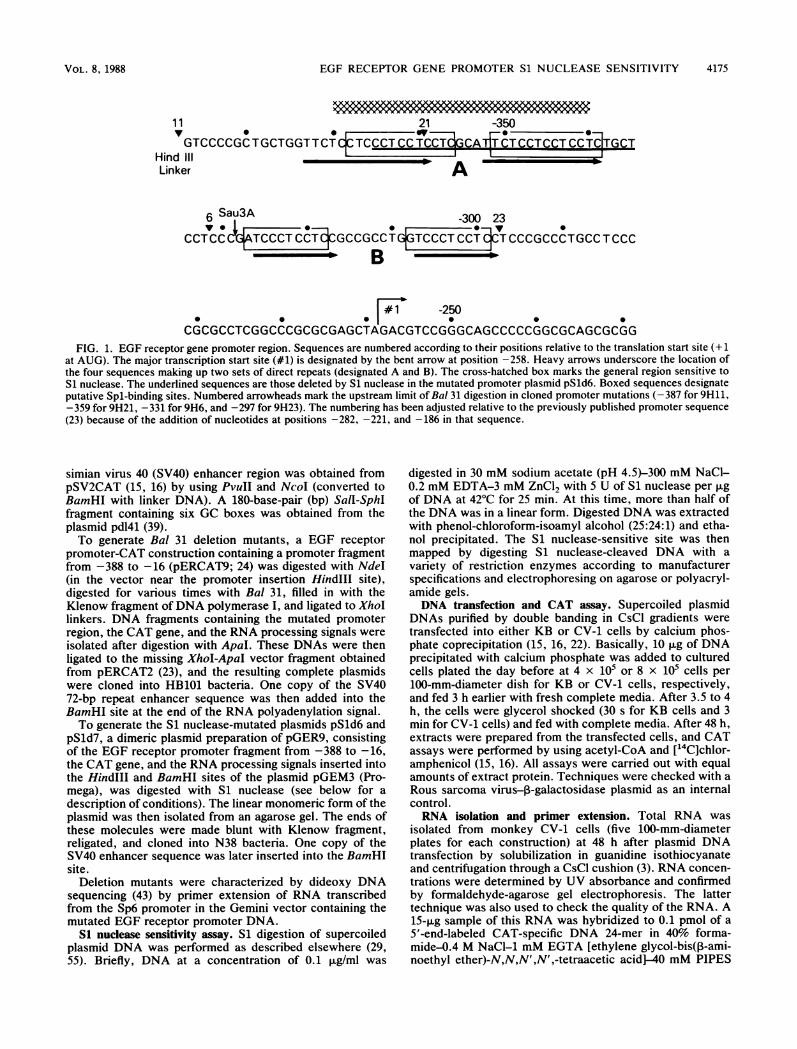
with the position of repeat A (Fig. 1). The same region wassensitive to the single-strand nuclease P1 as well (data notshown). It seems that sensitivity to Si nuclease requires thesupercoiled plasmid structure because EcoRI-restricted lin-ear DNA was not digested by Si nuclease (Fig. 3A, lane 4).DNA region containing Si nuclease-sensitive direct repeat is
necessary for optimal EGF receptor gene expression. To test
A2.9kbp
595bp
* 480bp
RESULTS
S1 nuclease-sensitive site in EGF receptor gene promoter.Upon examination of the EGF receptor gene promoter, twopairs of direct repeats were discovered (Fig. 1). One pair(designated B in Fig. 1), located between nucleotides -298and - 326 (relative to the translational start site), consisted ofa perfect 10-bp repeat sequence (39 to 67 bp upstream of themajor in vivo transcription initiation site). A second pair(designated A in Fig. 1), found between nucleotides -339and -373, shared 14 of 15 bp (80 to 114 bp upstream of themajor in vivo transcription start site). Both repeats had onlypyrimidine residues on one strand and purine residues on thecomplement strand, and both conformed to the motifTCCTCCTCC. DNA containing stretches of (dC-dT) - (dA-dG), singly or repeatedly, have been shown to be associatedwith sensitivity to the endonuclease Si both in vitro and invivo (see Discussion).To determine whether Si nuclease sensitivity could be
detected in this region, supercoiled plasmid DNA containingthe EGF receptor gene promoter (-388 to -16) was used.When the promoter-containing plasmid pGER9 was sequen-tially cut with Si nuclease and EcoRI, two new DNAfragments were generated that were not seen when EcoRIwas used alone (Fig. 2, lane 3). Two new bands were alsoobtained when Si nuclease digestion was followed by PvuIIcutting (Fig. 2, lane 4). This Si nuclease-sensitive site waslocalized to the region between -340 and -370, coincident
90bp
BP2
Si(A24I
1 2 3 4
#1 H
I 190
X3 P2 RI
__
AT'
480
595FIG. 2. Fine mapping of S1 nuclease-sensitive site in EGF re-
ceptor gene promoter. (A) Ethidium bromide-stained polyacryl-amide gel. Lane 1, HaeIII-digested (x marker DNA; lane 2,HindIII-digested A marker DNA; lane 3, plasmid pGER9 digestedsequentially with S1 nuclease and then with EcoRI, a procedureyielding two fragments of 2.9 kbp and 595 bp; lane 4, pGER9digested with S1 nuclease and then with PvuII, a procedure yieldingtwo additional fragments of 480 and 90 bp. (B) Schematic represen-tation of promoter region. The thick line is the vector DNA, and thethin line is promoter DNA. The box is the 250-bp HindIII-EcoRICAT fragment. The bent arrow marks the major transcription startsite (#1). The heavy vertical arrow indicates the Si nuclease-sensitive site derived from this data. P2, PvuII; H3, HindIlI; Ri,EcoRI; A2, Avall (lost during cloning).
MOL. CELL. BIOL.
EGF RECEPTOR GENE PROMOTER Si NUCLEASE SENSITIVITY
A
BSi
A BpGER6/9Hl1 I -O--O
C 9H11 9H21 9H6 9H23
Ac-Ch?
Chi*@ *9
2 3 4 5 6 7 8 9
#1
RelativeCAT -
Activity1.0 0.8 0.4 0.25
H3 RICATD
9H21 ---D-0 CATDE9H6 ---+-Q9H23---._ .iIThEI
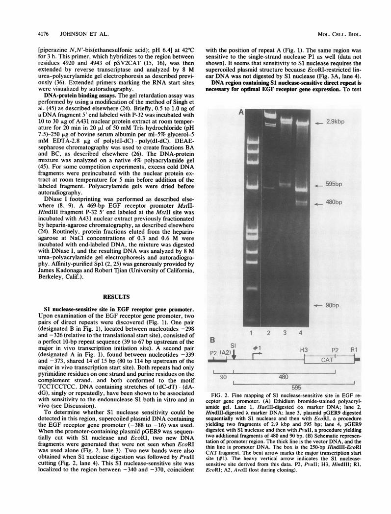
FIG. 3. Characterization of Bal 31 deletion mutations. (A) Ethidium bromide-stained agarose gel. Lanes 1 to 4, Supercoiled plasmidpGER6 was either untreated (lane 1), digested with EcoRI (RI) (lane 2), digested first with S1 nuclease and then with EcoRI (lane 3), ordigested first with EcoRI and then with S1 nuclease (lane 4). Lane 5, HindIII-digested X marker DNA. The left arrow shows one of the bandsgenerated by S1 nuclease. The smaller second band was electrophoresed off the gel. Lanes 6 to 9, Digestion of Bal 31-generated promotermutation plasmid 9H11 (lane 6), 9H21 (lane 7), 9H6 (lane 8), or 9H23 (lane 9) first with Si nuclease and then with EcoRI. (B) Structures ofBal 31 mutations. The CAT HindIII-EcoRI (H3-RI) 250-bp fragment is boxed, and the major (#1) transcription start site is marked by the bentarrow. Each circle represents one pair of the direct repeats A and B (Fig. 1), and the black box marks the location of the S1 nuclease-sensitiveregion. Dashed lines indicate the extent of digestion by Bal 31. The vector for these plasmids was pGEM4. (C) Activity of Bal 31-mutatedEGF receptor gene promoter DNA in a transient-transfection assay. Plasmid DNA (10 F±g) was transfected into human KB cells by calciumphosphate coprecipitation. After 48 h, extracts were prepared and CAT activity was determined. These plasmids contain one copy of the72-bp repeat SV40 enhancer region. CAT activity is presented relative to that of 9H11 (activity taken as 1.0). Chl, ['4C]chloramphenicol;Ac-Chl, acetylated ["'C] chloramphenicol.
whether the direct-repeat region is involved in EGF receptorgene expression, the promoter region, previously placedupstream of the bacterial CAT reporter gene, was digestedwith the exonuclease Bal 31. The resulting mutations elimi-nated different amounts of the two pairs of direct repeats(Fig. 1, arrowheads and Fig. 3B). As the sequences contain-ing the repeats were eliminated, the Si-sensitive site disap-peared (Fig. 3A, lanes 6 to 9). Each of the four mutatedplasmids was separately transfected into human KB cells,and after 48 h, extracts were prepared for determination ofCAT activity. As more of the repeat sequences were deleted,CAT activity continued to decrease (Fig. 3C). When bothpairs of repeats were deleted (9H23), activity dropped byabout fourfold. These results suggest that this DNA region isinvolved in EGF receptor gene regulation.To more directly test the role of the S1-sensitive site, S1
nuclease was used to mutate this region in pGER9. Afterblunt-end religation, DNAs were cloned into HB101. Twocloned plasmids, pSld6 and pSld7, were isolated and se-quenced. They were found to have 31- and 33-bp deletions inthe upstream direct repeat A, respectively, a finding thatconfirms the location of the S1-sensitive site. One of these,pSld6, was analyzed further. The parent plasmid pGER9and the mutant pSld6 are identical, except for the 31-bp
S1-sensitive sequences; both contain the CAT gene andRNA processing signals of pSVOCAT and the vectorpGEM3. In addition, another pair of plasmids was con-structed that contained one copy each of the SV40 72-bprepeat enhancer element. A comparison of the CAT activitygenerated by transfection of these two promoters into cul-tured cells should reveal any change in gene transcriptioncaused by the removal of repeat A.The normal and S1-mutated plasmids with or without
enhancers were transfected into monkey CV-1 cells. After 48h, extracts were prepared from the cells for determination ofCAT activity. The mutated promoter was 3.2-fold less activethan the normal promoter when both contained a single copyof the SV40 enhancer and 2.1-fold less active withoutenhancers. Because CV-1 cells are monkey cells and makevery low levels of EGF receptor, we used the normal andmutated plasmids (pGER9 and pSld6) to transfect humanKB epidermoid carcinoma cells, which contain about100,000 receptors per cell (54). The KB cells are less activethan CV-1 cells as transfection recipients, and we had to useenhancers in these plasmids to accurately compare CATactivities. The promoter with the S1 mutation was 4.7-foldless active than the normal promoter (Fig. 4), a resultcorroborating those obtained with CV-1 cells.
VOL. 8, 1988 4177
r-111,L
4178 JOHNSON ET AL.
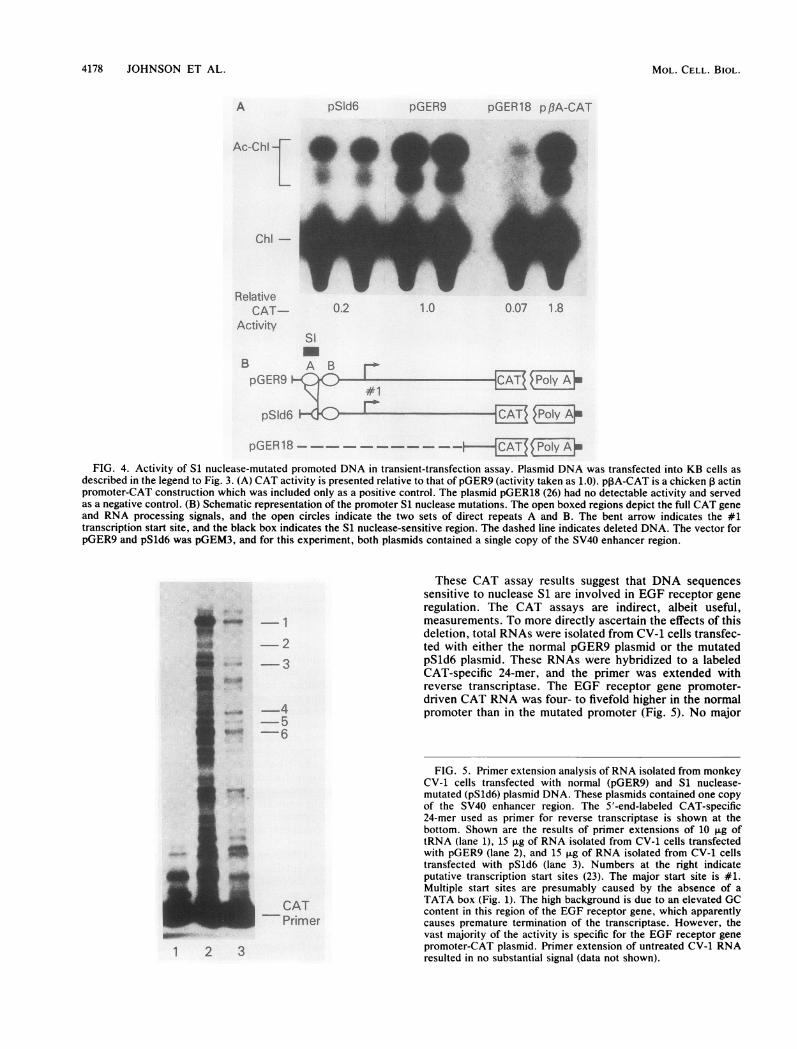
A pSld6 pGER9 pGER18 pf3A-CAT
Chl-
RelativeCAT 0.2 1.0 0.07 1.8
ActivitySi
B A B rApGER9 C PoIy AE
pSld6 r A
pGER18 -._--_-PolyA*
FIG. 4. Activity of S1 nuclease-mutated promoted DNA in transient-transfection assay. Plasmid DNA was transfected into KB cells asdescribed in the legend to Fig. 3. (A) CAT activity is presented relative to that of pGER9 (activity taken as 1.0). pPA-CAT is a chicken 3 actinpromoter-CAT construction which was included only as a positive control. The plasmid pGER18 (26) had no detectable activity and servedas a negative control. (B) Schematic representation of the promoter S1 nuclease mutations. The open boxed regions depict the full CAT geneand RNA processing signals, and the open circles indicate the two sets of direct repeats A and B. The bent arrow indicates the #1transcription start site, and the black box indicates the S1 nuclease-sensitive region. The dashed line indicates deleted DNA. The vector forpGER9 and pSld6 was pGEM3, and for this experiment, both plasmids contained a single copy of the SV40 enhancer region.
These CAT assay results suggest that DNA sequencessensitive to nuclease S1 are involved in EGF receptor generegulation. The CAT assays are indirect, albeit useful,measurements. To more directly ascertain the effects of thisdeletion, total RNAs were isolated from CV-1 cells transfec-
- 2 ted with either the normal pGER9 plasmid or the mutated- 3 pSld6 plasmid. These RNAs were hybridized to a labeled
CAT-specific 24-mer, and the primer was extended withreverse transcriptase. The EGF receptor gene promoter-
4driven CAT RNA was four- to fivefold higher in the normal
5 promoter than in the mutated promoter (Fig. 5). No major
* ~6
FIG. 5. Primer extension analysis ofRNA isolated from monkeyCV-1 cells transfected with normal (pGER9) and S1 nuclease-mutated (pSld6) plasmid DNA. These plasmids contained one copyof the SV40 enhancer region. The 5'-end-labeled CAT-specific24-mer used as primer for reverse transcriptase is shown at thebottom. Shown are the results of primer extensions of 10 p.g oftRNA (lane 1), 15 ,ug of RNA isolated from CV-1 cells transfected
3)>_"withpGER9 (lane 2), and 15 ,ug of RNA isolated from CV-1 cellstransfected with pSld6 (lane 3). Numbers at the right indicateputative transcription start sites (23). The major start site is #1.Multiple start sites are presumably caused by the absence of a
CAT TATA box (Fig. 1). The high background is due to an elevated GCPrimer content in this region of the EGF receptor gene, which apparently__Primer causes premature termination of the transcriptase. However, the
vast majority of the activity is specific for the EGF receptor gene
l 2 3 promoter-CAT plasmid. Primer extension of untreated CV-1 RNAresulted in no substantial signal (data not shown).
MOL . CELL . BIOL .

EGF RECEPTOR GENE PROMOTER Si NUCLEASE SENSITIVITY
changes in the transcription start sites were detected (Fig. 5).The RNA results were in complete agreement with the CATprotein data.
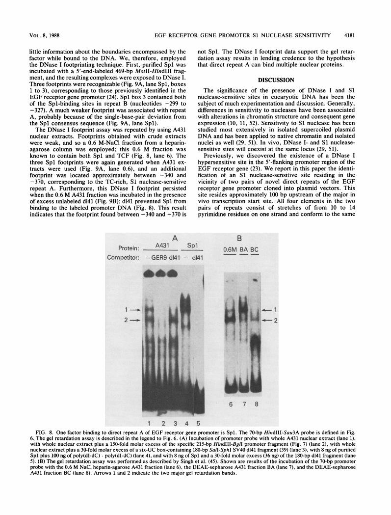
Binding of nuclear proteins to direct-repeat region of EGFreceptor gene promoter. The Si-sensitive region may beinvolved in gene regulation by providing binding sites fornuclear trans-acting factors. If so, DNA-binding assaysshould show that proteins which can bind to the normal EGFreceptor gene promoter cannot bind to the Si nuclease-mutated promoter. To test this idea, a 70-bp HindIII-Sau3Afragment containing direct repeat A was isolated from thenormal promoter, and a 39-bp HindIII-Sau3A fragment wasisolated from the mutated promoter (Fig. 6 and 7). Bothfragments were end labeled with P-32, separately incubatedwith nuclear extracts from A431 cells, and electrophoresedon a native polyacrylamide gel. Two prominent bands (Fig.6A, lane 2, arrows 1 and 2) associated with the normal 70-bppromoter appeared in the gel. Their migration would suggestthat they are DNA-protein complexes. Both bands representspecific binding because their intensity was substantiallydiminished when a 100-fold molar excess of unlabeled 90-bpfragment from the normal promoter (the 70-bp HindIll-Sau3A fragment cloned into pGEM3 and then cut withHindIII and EcoRI) was included in the DNA bindingreaction (Fig. 6A, lane 3). An unlabeled 59-bp fragment from
Molar Excess:
1-
2-
A
the pSld6-mutated promoter (the 39-bp HindIII-Sau3A frag-ment cloned into pGEM3) competed poorly for binding (Fig.6A, lanes 6 to 8); however, the 59-bp S1d6 fragment didcompete at high concentrations for band 2. The latter wasprobably caused by the recreation of half of direct repeat Ain the S1d6 mutation (TCCTCCCTCC; Fig. 1). Anotherunlabeled normal promoter fragment (the 215-bp HindIII-BglI fragment) was also used as a specific competitor (Fig.7). Interestingly, even a 10-fold molar excess of this largerfragment, which contains both the upstream and down-stream TCC repeat regions (repeats A and B, respectively)and two strong Spl-binding sites (24), successfully competedwith the labeled probe for binding to the unidentified pro-tein(s) (Fig. 7a, lanes 1 to 5).
In contrast to the DNA fragment containing repeat A fromthe normal promoter, the Si nuclease-mutated 39-bp DNAfragment was incapable of binding nuclear protein, as judgedby the absence of slowly migrating bands in the gel retarda-tion assay (Fig. 7b, lane 9).Spl is one of two separate factors that bind to Si nuclease-
sensitive promoter regions. A clue to the identity of one of thenuclear DNA-binding proteins was revealed upon closeexamination of the DNA sequence of the two pairs ofpromoter direct repeats. Repeat B contained two 10-bpsequences that completely match the Spl consensus se-
- 100 175 250 100 175 250 175 250
'1N& A
Free_70bp
1 2 3 4 5
LabeledProbe -*70bp
B B-(H3) S3A;90bp pGE(RI)
Competitor Ld622 ...J pGER9
6 7 8 9 10 11
#1r ''''
Al Al
l 62bp I
FIG. 6. Detection of specific EGF receptor gene promoter DNA-binding protein by gel retardation assay. (A) A431 nuclear extract wasincubated with a 5'-end-labeled 70-bp HindIII-Sau3A (H3-S3A) fragment, and the resulting DNA-protein complexes were resolved by nativepolyacrylamide gel electrophoresis and autoradiography. Two retarded bands are visualized (lane 2, bands 1 and 2). Competitor DNAsincluded a 90-bp subcloned normal promoter fragment (from pGER9; lanes 3 to 5), a 59-bp subcloned Si-deleted promoter fragment (frompSld6; lanes 6 to 8), and a nonspecific downstream 62-bp AvaI (Al) fragment (lanes 10 and 11). Lane 1, No extract was added; lane 9, 125ng of additional poly(dI-dC) * poly(dI-dC) was included. (B) Schematic representation of the probe and competitors relative to the promoter.Wavy lines are vector sequences. RI, EcoRI (cloning site); #1, major transcription start site. The two open circles indicate the two sets ofdirect repeats A and B. Asterisks indicate the positions where the probes were 5' end labeled.
VOL. 8, 1988 4179
I

4180 JOHNSON ET AL.
Probes:Competitors:
Molar Excess
apGER9
215bp 62bp
- 2.5 5.0 10 20 10 20*. *'90 .& % . , a
1- 6t " t Al t
2---o- 6
' I, "I e t 4
.&8A &A
Free70bp
Free39bp
1 2 3 4 5 6 7 8 9 10
pSld6
A. 170bp pGER9
HX H S3A215bp
#1
I I IBl Al Al
.j , 62bp j
FIG. 7. Comparison of nuclear protein binding to normal 70-bp (pGER9) (a) and deleted 39-bp (pSld6) (b) 5'-end-labeled promoter DNAby gel retardation assay. (a) The competitors included the specific 215-bp HindIII-BglI (H3-B1) fragment (lanes 2 to 5) and the nonspecific62-bp AvaI (Al) fragment (lanes 6 and 7). Lane 1, No competitor added; lane 8, no extract added. (b) The labeled pSld6 fragment was eitherincubated alone (lane 10) or with A431 extract (lane 9). (c) Schematic representation of probes and comnpetitors. S3A, Sau3A; #1, majortranscription start site. Two open circles mark the positions of the two sets of direct repeats A and B. Asterisks indicate the positions wherethe probes were 5' end labeled.
quence-binding site (2, 25). We previously reported thatpurified Spl can bind to these sites (24). Repeat A, which issensitive to Si nuclease, contained two additional potentialSpl-binding sites matching the consensus sequence in 9 of 10positions (Fig. 1).To test whether repeat A can bind Spl, gel retardation
assays were again used. The end-labeled 70-bp HindIII-Sau3A normal promoter fragment was incubated with theA431 nuclear extract in the absence or presence of a vastmolar excess of an unlabeled SV40 180-bp SalI-SphI com-
petitor fragment containing the six Spl-specific GC boxes(from plasmid pdl41 [39]). A 30-fold molar excess of theunlabeled d141 fragment effectively competed for the forma-tion of the complex designated band 1 (Fig. 8A, lane 3). Band2 was not affected by the presence of the Spl-bindingfragment (Fig. 8A, lane 3).
Purified Spl was then incubated with the end-labeled70-bp normal promoter fragment. Spl bound to the promoterDNA and formed a radioactive complex comigrating withband 1 (Fig. 8A, lane 4). Furthermore, a 30-fold molar excess
of unlabeled d141 completely blocked the formation of thiscomplex (Fig. 8A, lane 5). Band 2 did not appear whenpurified Spl was bound to this promoter fragment and ismost likely not the result of Spl binding. To prove thishypothesis, two chromatographically distinct A431 fractionswere used in the gel retardation assay. A431 nuclear extracthad previously been subjected to heparin-agarose andDEAE-sepharose column chromatography (26). BA, a 0.12M-KCl fraction known to contain Spl (26), formed a DNA-protein complex comigrating with band 1 but not with band2 (Fig. 8B, lane 7). BC, a 0.5 M-KCl fraction lacking Spl,formed a DNA-protein complex comigrating with band 2 butnot with band 1 (Fig. 8B, lane 8).These gel retardation assay results indicate that one of the
two factors capable of binding to the S1 nuclease-sensitivepromoter region was Spl. The other factor, which was notSpl, was associated with the TC-rich repeat A region and isreferred to as a TC factor or TCF.The gel retardation assay is a very quick and sensitive
technique for detecting DNA binding; however, it reveals
bpSld6
Extract+ _
cLabeleciProbes
Com petitorDNA
MOL. CELL. BIOL.
IL
EGF RECEPTOR GENE PROMOTER Si NUCLEASE SENSITIVITY
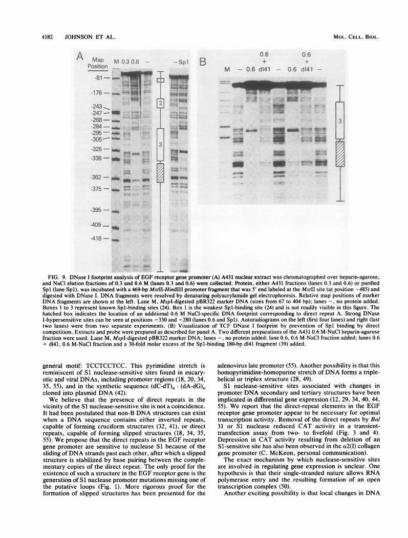
little information about the boundaries encompassed by thefactor while bound to the DNA. We, therefore, employedthe DNase I footprinting technique. First, purified Spl wasincubated with a 5'-end-labeled 469-bp MstII-HindIII frag-ment, and the resulting complexes were exposed to DNase I.Three footprints were recognizable (Fig. 9A, lane Spl, boxes1 to 3), corresponding to those previously identified in theEGF receptor gene promoter (24). Spl box 3 contained bothof the Spl-binding sites in repeat B (nucleotides -299 to-327). A much weaker footprint was associated with repeatA, probably because of the single-base-pair deviation fromthe Spl consensus sequence (Fig. 9A, lane Spl).The DNase I footprint assay was repeated by using A431
nuclear extracts. Footprints obtained with crude extractswere weak, and so a 0.6 M-NaCl fraction from a heparin-agarose column was employed; this 0.6 M fraction wasknown to contain both Spl and TCF (Fig. 8, lane 6). Thethree Spl footprints were again generated when A431 ex-tracts were used (Fig. 9A, lane 0.6), and an additionalfootprint was located approximately between -340 and-370, corresponding to the TC-rich, Si nuclease-sensitiverepeat A. Furthermore, this DNase I footprint persistedwhen the 0.6 M A431 fraction was incubated in the presenceof excess unlabeled d141 (Fig. 9B); d141 prevented Spl frombinding to the labeled promoter DNA (Fig. 8). This resultindicates that the footprint found between -340 and -370 is
AProtein: A431 Sp'
Competitor: -GER9 d141 - d
_4b
not Spl. The DNase I footprint data support the gel retar-dation assay results in lending credence to the hypothesisthat direct repeat A can bind multiple nuclear proteins.
DISCUSSION
The significance of the presence of DNase I and Sinuclease-sensitive sites in eucaryotic DNA has been thesubject of much experimentation and discussion. Generally,differences in sensitivity to nucleases have been associatedwith alterations in chromatin structure and consequent geneexpression (10, 11, 52). Sensitivity to Si nuclease has beenstudied most extensively in isolated supercoiled plasmidDNA and has been applied to native chromatin and isolatednuclei as well (29, 51). In vivo, DNase I- and S1 nuclease-sensitive sites will coexist at the same locus (29, 51).
Previously, we discovered the existence of a DNase Ihypersensitive site in the 5'-flanking promoter region of theEGF receptor gene (23). We report in this paper the identi-fication of an Si nuclease-sensitive site residing in thevicinity of two pairs of novel direct repeats of the EGFreceptor gene promoter cloned into plasmid vectors. Thissite resides approximately 100 bp upstream of the major invivo transcription start site. All four elements in the twopairs of repeats consist of stretches of from 10 to 14pyrimidine residues on one strand and conform to the same
BL 0.6M BA BC
I1 ---2-2 ---o-
II110.
I
3m
It Am<
6 7 8
1 2 3 4 5FIG. 8. One factor binding to direct repeat A of EGF receptor gene promoter is Spl. The 70-bp HindIII-Sau3A probe is defined in Fig.
6. The gel retardation assay is described in the legend to Fig. 6. (A) Incubation of promoter probe with whole A431 nuclear extract (lane 1),with whole nuclear extract plus a 150-fold molar excess of the specific 215-bp HindIII-BgIl promoter fragment (Fig. 7) (lane 2), with wholenuclear extract plus a 30-fold molar excess of a six-GC box-containing 180-bp SaIl-SphI SV40 d141 fragment (39) (lane 3), with 8 ng of purifiedSpl plus 100 ng of poly(dI-dC) poly(dI-dC) (lane 4), and with 8 ng of Spl and a 30-fold molar excess (36 ng) of the 180-bp d141 fragment (lane5). (B) The gel retardation assay was performed as described by Singh et al. (45). Shown are results of the incubation of the 70-bp promoterprobe with the 0.6 M NaCl heparin-agarose A431 fraction (lane 6), the DEAE-sepharose A431 fraction BA (lane 7), and the DEAE-sepharoseA431 fraction BC (lane 8). Arrows 1 and 2 indicate the two major gel retardation bands.
VOL. 8, 1988 4181
4182 JOHNSON ET AL.
A Map M 0.3 0.6Position
-81-
-176- ^
-243 __.,-247--268--284--295-
-305--A-325- _
-338- _ 8&. --
-362 - -af-375-_
m-_ *-'- - GM
_..
-395-_
-409--
-418 -
3
- Spl B
7.
t-4 -
6 4.
14: £*4
0.6 0.6
M - 0.6 d141 - 0.6 d141
a - s~~~~~~~~~~~~~~~~~~~~~~~~~~~~~~~~~~~~~~~~~~~~~~~~~~~~~~~~~.
__PI--_
-'' ._
__va-W_ %&.m r~~~. nI
I3
/6/6,
1 _
FIG. 9. DNase I footprint analysis of EGF receptor gene promoter (A) A431 nuclear extract was chromatographed over heparin-agarose,and NaCl elution fractions of 0.3 and 0.6 M (lanes 0.3 and 0.6) were collected. Protein, either A431 fractions (lanes 0.3 and 0.6) or purifiedSpl (lane Spl), was incubated with a 469-bp MstII-HindIII promoter fragment that was 5' end labeled at the MstII site (at position -485) anddigested with DNase I. DNA fragments were resolved by denaturing polyacrylamide gel electrophoresis. Relative map positions of markerDNA fragments are shown at the left. Lane M, MspI-digested pBR322 marker DNA (sizes from 67 to 404 bp); lanes -, no protein added.Boxes 1 to 3 represent known Spl-binding sites (24). Box 1 is the weakest Spl-binding site (24) and is not readily visible in this figure. Thehatched box indicates the location of an additional 0.6 M NaCI-specific DNA footprint corresponding to direct repeat A. Strong DNaseI-hypersensitive sites can be seen at positions -330 and -280 (lanes 0.6 and Spl). Autoradiograms on the left (first four lanes) and right (lasttwo lanes) were from two separate experiments. (B) Visualization of TCF DNase I footprint by prevention of Spl binding by directcompetition. Extracts and probe were prepared as described for panel A. Two different preparations of the A431 0.6 M-NaCl heparin-agarosefraction were used. Lane M, MspI-digested pBR322 marker DNA; lanes -, no protein added; lane 0.6, 0.6 M-NaCl fraction added; lanes 0.6+ d141, 0.6 M-NaCl fraction and a 30-fold molar excess of the Spl-binding 180-bp d141 fragment (39) added.
general motif: TCCTCCTCC. This pyrimidine stretch isreminiscent of Si nuclease-sensitive sites found in eucary-otic and viral DNAs, including promoter regions (18, 20, 34,35, 55), and in the synthetic sequence (dC-dT), (dA-dG)ncloned into plasmid DNA (42).We believe that the presence of direct repeats in the
vicinity of the Si nuclease-sensitive site is not a coincidence.It had been postulated that non-B DNA structures can existwhen a DNA sequence contains either inverted repeats,capable of forming cruciform structures (32, 41), or directrepeats, capable of forming slipped structures (18, 34, 35,55). We propose that the direct repeats in the EGF receptorgene promoter are sensitive to nuclease Si because of thesliding ofDNA strands past each other, after which a slippedstructure is stabilized by base pairing between the comple-mentary copies of the direct repeat. The only proof for theexistence of such a structure in the EGF receptor gene is thegeneration of Si nuclease promoter mutations missing one ofthe putative loops (Fig. 1). More rigorous proof for theformation of slipped structures has been presented for the
adenovirus late promoter (55). Another possibility is that thishomopyrimidine-homopurine stretch of DNA forms a triple-helical or triplex structure (28, 49).
Si nuclease-sensitive sites associated with changes inpromoter DNA secondary and tertiary structures have beenimplicated in differential gene expression (12, 29, 34, 40, 44,55). We report that the direct-repeat elements in the EGFreceptor gene promoter appear to be necessary for optimaltranscription activity. Removal of the direct repeats by Bal31 or Si nuclease reduced CAT activity in a transient-transfection assay from two- to fivefold (Fig. 3 and 4).Depression in CAT activity resulting from deletion of anS1-sensitive site has also been observed in the a2(I) collagengene promoter (C. McKeon, personal communication).The exact mechanism by which nuclease-sensitive sites
are involved in regulating gene expression is unclear. Onehypothesis is that their single-stranded nature allows RNApolymerase entry and the resulting formation of an opentranscription complex (50).Another exciting possibility is that local changes in DNA
MOL. CELL. BIOL.
EGF RECEPTOR GENE PROMOTER Si NUCLEASE SENSITIVITY
structure, as detected by DNase I and Si nuclease digestion,alter the interactions between promoter DNA and specificDNA-binding protein. It was, therefore, of great interest todetermine whether the Si nuclease-sensitive site in the EGFreceptor gene promoter is associated with a DNA-bindingprotein. Both gel retardation and DNase I footprintingassays were performed by using labeled EGF receptor genepromoter fragments and nuclear extracts from A431 cells.These experiments clearly showed that two proteins wereable to bind to repeat A, which was responsible for confer-ring Si nuclease sensitivity to the promoter (Fig. 6 to 9). Oneprotein is the positive transcription factor Spl (2, 8, 9, 25).The other protein is what we refer to as TCF. Uponobliteration of the structure of repeat A, neither Spl norTCF bound to the region (Fig. 7). This result establishes acorrelation between the binding of these proteins and opti-mal transcription activity as determined by transient-trans-fection assays. We believe that this paper is the first pub-lished report correlating Si nuclease sensitivity, trans-acting-factor binding, and altered gene expression.The EGF receptor gene promoter S1 nuclease-sensitive
site consisted of the two pairs of direct repeats A and B (Fig.1); all four conformed closely to the motif TCCTCCTCC,and all four contained potential Spl-binding sites. The rela-tionship between the two pairs of repeats may be significant.An unlabeled DNA fragment containing both sets of directrepeats competed for binding about ten times more effec-tively in the gel retardation assay than did a competitor DNAfragment possessing only the upstream set of direct repeats(repeat A) (Fig. 6 and 7). The results of these competitionexperiments raised the possibility that the same or similarfactors bind to both sets of repeats. This hypothesis is likelythe case for Spl, which bound strongly to the downstreamset of repeats (repeat B) (Fig. 9). Furthermore, it is conceiv-able that Spl and TCF interact, perhaps in a dynamic orcooperative fashion. This possibility is most intriguing be-cause the four repeat elements are separated from each otherby 19, 21, and 23 bp, or by about two full turns of the DNAhelix. The exact relationship between the binding of thesefactors and the structure of the DNA in this region ispresently unknown.Sequences resembling the human EGF receptor gene
promoter TCCTCCTCC region are found in several otherhuman receptor gene promoters, including the insulin andlow-density lipoprotein (LDL) receptor genes (1, 46). Al-though S1 nuclease sensitivity has not been examined inthese promoters, the TC-rich region in the LDL receptorgene is part of a sterol regulatory element and binds anuclear protein (46). It remains to be established whetherother S1 nuclease-sensitive promoter regions containingDNA sequences similar to that of the EGF receptor genepromoter TCCTCCTCC motif are associated with the bind-ing of Spl, TCF, or related trans-acting factors.
ACKNOWLEDGMENTS
We wish to thank Ira Pastan, Ryoichiro Kageyama, PamelaMarino, and Catherine McKeon for useful and stimulating discus-sions. We are indebted to Cliff Parkison for dideoxy sequencing,James Kadonaga and Robert Tjian for purified Spl, John Brady forpdl41, Bruce Paterson for p3A-CAT, and Ryoichiro Kageyama forA431 nuclear extract chromatographic fractions BA and BC. Weacknowledge Steven Neal for photographic assistance and JennieEvans and Althea Gaddis for editorial assistance.
LITERATURE CITED
1. Araki, E., F. Shimada, M. Uzawa, M. Mori, and Y. Ebina. 1987.Characterization of the promoter region of the human insulinreceptor gene. Evidence for promoter activity. J. Biol. Chem.262:16186-16191.
2. Briggs, M. R., J. T. Kadonaga, S. P. Bell, and R. Tjian. 1986.Purification and biochemical characterization of the promoter-specific transcription factor Spl. Science 234:47-52.
3. Chirgwin, J. M., A. E. Przybyla, R. J. MacDonald, and W. J.Rutter. 1979. Isolation of biologically active ribonucleic acidfrom sources enriched in ribonuclease. Biochemistry 18:5294-5299.
4. Cohen, S., H. Ushiro, C. Stoscheck, and M. Chinkers. 1982. Anative 170,000 epidermal growth factor receptor-kinase complexfrom shed plasma membrane vesicles. J. Biol. Chem. 257:1523-1531.
5. Covelli, I., R. Mozzi, R. Rossi, and L. Frati. 1972. The mecha-nism of action of the epidermal growth factor III. Stimulation ofthe uptake of labeled precursors into RNA, DNA and proteinsinduced by EGF in isolated tumor cells. Hormones 3:183-191.
6. Cowley, G., J. A. Smith, B. Gusterson, F. Hendler, and B.Ozanne. 1984. The amount of EGF receptor is elevated onsquamous cell carcinomas. Cancer Cells 1:5-10.
7. Downward, J., Y. Yarden, E. Mayes, G. Scarce, N. Totty, P.Stockwell, A. Ullrich, J. Schlessinger, and M. D. Waterfield.1984. Close similarity of epidermal growth factor receptor andv-erbB oncogene protein sequence. Nature (London) 307:521-527.
8. Dynan, W. S., and R. Tjian. 1983. Isolation of transcriptionfactors that discriminate between different promoters recog-nized by RNA polymerase II. Cell 32:669-680.
9. Dynan, W. S., and R. Tjian. 1983. The promoter-specific tran-scription factor Spl binds to upstream sequences in the SV40early promoter. Cell 35:79-87.
10. Elgin, S. C. R. 1981. DNase I-hypersensitive sites of chromatin.Cell 27:413-415.
11. Elgin, S. C. R. 1982. Chromatin structure, DNA structure.Nature (London) 300:402-403.
12. Evans, T., E. Schon, G. Gora-Maslak, J. Patterson, and A.Efstradiatis. 1984. S1-hypersensitive sites in eukaryotic pro-moter regions. Nucleic Acids Res. 12:8043-8058.
13. Fabricant, R. N., J. E. Delarco, and G. J. Todaro. 1977. Nervegrowth factor receptor on human melanoma cells in culture.Proc. Natl. Acad. Sci. USA 74:565-569.
14. Goding, C. R., and W. C. Russell. 1983. S1 sensitive sites inadenovirus DNA. Nucleic Acids Res. 11:21-36.
15. Gorman, C., L. Moffat, and B. Howard. 1982. Recombinantgenomes which express chloramphenicol acetyltransferase inmammalian cells. Mol. Cell. Biol. 2:1044 1051.
16. Gorman, C. M., G. T. Merlino, M. C. Willingham, I. Pastan,and B. H. Howard. 1982. The Rous sarcoma virus long terminalrepeat is a strong promoter when introduced into a variety ofeukaryotic cells by DNA-mediated transfection. Proc. Natl.Acad. Sci. USA 79:6777-6781.
17. Haley, J., N. Whittle, P. Bennett, D. Kinchington, A. Ullrich, andM. Waterfield. 1987. The human EGF receptor gene: structureof the 110 kb locus and identification of sequences regulating itstranscription. Oncogene Res. 1:375-3%.
18. Hentschel, C. C. 1982. Homocopolymer sequences in the spacerof a sea urchin histone gene repeat are sensitive to the S1nuclease. Nature (London) 295:714-716.
19. Hollenberg, M. D., and P. Cuatrecasas. 1973. Epidermal growthfactor: receptors in human fibroblasts and modulation of actionby cholera toxin. Proc. Natl. Acad. Sci. USA 70:2964-2968.
20. Htun, M., E. Lund, and J. E. Dahlberg. 1984. Human Ul RNAgenes contain an unusual nuclease S1 cleavage site within theconserved 3' flanking region. Proc. Natl. Acad. Sci. USA 81:7288-7292.
21. Ishii, S., J. T. Kadonaga, R. Tjian, J. N. Brady, G. T. Merlino,and I. Pastan. 1986. Binding of the Spl transcription factor bythe human Harvey ras 1 proto-oncogene promoter. Science 232:1410-1413.
VOL. 8, 1988 4183

4184 JOHNSON ET AL.
22. Ishii, S., G. T. Merlino, and I. Pastan. 1985. Promoter region ofthe human Harvey ras proto-oncogene: similarity to the EGFreceptor proto-oncogene promoter. Science 230:1378-1381.
23. Ishii, S., Y.-H. Xu, R. M. Stratton, B. A. Roe, G. T. Merlino,and I. Pastan. 1985. Characterization and sequence of thepromoter region of the human epidermal growth factor receptorgene. Proc. Natl. Acad. Sci. USA 82:4920-4924.
24. Johnson, A. C., S. Ishii, Y. Jinno, I. Pastan, and G. T. Merlino.1988. EGF receptor gene promoter: deletion analysis and iden-tification of nuclear protein binding sites. J. Biol. Chem. 263:5693-5699.
25. Kadonaga, J. T., and R. Tjian. 1986. Affinity purification ofsequence-specific DNA binding proteins. Proc. Natl. Acad. Sci.USA 83:5889-5893.
26. Kageyama, R., G. T. Merlino, and I. Pastan. 1988. Epidermalgrowth factor (EGF) receptor gene transcription: requirementfor Spl and an EGF receptor-specific factor. J. Biol. Chem. 263:6329-6336.
27. King, C. R., M. H. Kraus, L. T. Williams, G. T. Merlino, I. H.Pastan, and S. A. Aaronson. 1985. Human tumor cell lines withEGF receptor gene amplification in the absence of aberrantsized mRNAs. Nucleic Acid Res. 13:8477-8486.
28. Kohwi-Shigematsu, T., N. Scribner, and Y. Kohwi. 1988. Anultimate chemical carcinogen, N-acetoxy-2-acetylamino-fluorene detects non-B DNA structures that are reactive withchloroacetaldehyde in supercoiled plasmid DNA. Carcinogene-sis 9:457-461.
29. Larsen, A., and M. Weintraub. 1982. An altered DNA confor-mation detected by S1 nuclease occurs at specific regions inactive chick globin chromatin. Cell 29:609-622.
30. Lembach, K. J. 1976. Enhanced synthesis and extracellularaccumulation of hyaluronic acid during stimulation of quiescenthuman fibroblasts by mouse epidermal growth factor. J. Cell.Physiol. 89:277-288.
31. Libermann, T. A., H. R. Nasbaum, N. Razon, R. Kris, I. Lax, H.Soreg, N. Whittle, M. D. Waterfield, A. Ullrich, and J. Schles-singer. 1985. Amplification, enhanced expression and possiblerearrangment of EGF receptor gene in primary human braintumors of glial origin. Nature (London) 313:144-147.
32. Lilley, D. M. F. 1980. The inverted repeat as a recognizablestructural feature in supercoiled DNA molecules. Proc. Natl.Acad. Sci. USA 77:6468-6472.
33. Lin, C. R., W. S. Chen, W. Kruiger, L. S. Stolarsky, W. Weber,R. M. Evans, I. M. Verma, G. N. Gill, and M. G. Rosenfeld.1984. Expression cloning of human EGF receptor complemen-tary DNA: gene amplification and three related messenger RNAproducts in A431 cells. Science 224:843-848.
34. Mace, H. A. F., H. R. B. Pelham, and A. A. Travers. 1983.Association of an S1 nuclease sensitive structure with shortdirect repeats 5' of Drosophila heat shock genes. Nature(London) 304:555-557.
35. McKeon, C., A. Schmidt, and B. deCrombrugghe. 1984. Asequence conserved in both the chicken and the mouse a2(I)collagen promoter contains sites sensitive to S1 nuclease. J.Biol. Chem. 259:6636-6640.
36. Merlino, G. T., J. S. Tyagi, B. deCrombrugghe, and I. Pastan.1982. Transcription of the chicken a2 (type I) collagen gene byhomologous cell-free extracts. J. Biol. Chem. 257:7254-7261.
37. Merlino, G. T., Y.-H. Xu, S. Ishii, A. J. L. Clark, K. Semba, K.Toyoshima, T. Yamamoto, and I. Pastan. 1984. Amplificationand enhanced expression of the epidermal growth factor recep-tor gene in A431 human carcinoma cells. Science 224:417-419.
38. Merlino, G. T., Y.-H. Xu, N. Richert, A. J. L. Clark, S. Ishii, S.
Banks-Schlegal, and I. Pastan. 1985. Elevated epidermal growthfactor receptor gene copy number and expression in a squamouscarcinoma cell line. J. Clin. Invest. 75:1077-1079.
39. Mishoe, M., J. N. Brady, M. Radonovich, and N. P. Salzman.1984. Simian virus 40 guanine-cytosine-rich sequences functionas independent transcriptional control elements in vitro. Mol.Cell. Biol. 4:2911-2920.
40. Nickol, J. M., and G. Felsenfeld. 1983. DNA conformation at the5' end of the chicken adult ,B-globin gene. Cell 35:467-477.
41. Panayotatos, N., and R. D. Wells. 1981. Cruciform structures insupercoiled DNA. Nature (London) 289:466-470.
42. Pulleyblank, D. E., D. B. Haniford, and A. R. Morgan. 1985. Astructural basis for S1 nuclease sensitivity of double-strandedDNA. Cell 42:271-280.
43. Sanger, F., S. Nicklen, and A. R. Coulson. 1977. RNA sequenc-ing with chain-terminating inhibitors. Proc. Natl. Acad. Sci.USA 74:5463-5467.
44. Schon, E., T. Evans, J. Welsch, and A. Efstratiadis. 1983.Conformation of a promoter DNA: fine structure mapping ofS1-hypersensitive sites. Cell 35:837-848.
45. Singh, H., S. Ranijan, D. Baltimore, and P. A. Sharp. 1986. Anuclear factor that binds to a conserved sequence motif intranscriptional control elements of immunoglobulin genes. Na-ture (London) 319:154-158.
46. Sudhof, T. C., D. W. Russell, M. S. Brown, and J. L. Goldstein.1987. 42 bp element from LDL receptor gene confers end-product repression by sterols when inserted into viral TKpromoter. Cell 48:1061-1069.
47. Ullrich, A., L. Coussens, J. S. Hayflick, T. J. Dull, A. Gray,A. M. Tam, J. Lee, Y. Yarden, T. A. Libermann, J. Schles-singer, J. Downward, E. L. V. Mayes, N. Whittle, M. D.Waterfield, and P. H. Seeburg. 1984. Human epidermal growthfactor receptor cDNA sequence and aberrant expression of theamplified gene in A431 epidermoid carcinoma cells. Nature(London) 309:418-425.
48. Velu, T. J., L. Beguinot, W. C. Vass, M. C. Willingham, G. T.Merlino, I. Pastan, and D. R. Lowy. 1987. Epidermal growthfactor dependent transformation by a human EGF receptorproto-oncogene. Science 238:1408-1410.
49. Voloshin, 0. N., S. M. Mirkin, V. I. Lyamichev, B. P. Belotserk-ovskii, and M. D. Frank-Kamenetskii. 1988. Chemical probing ofhomopurine-homopyrimidine mirror repeats in supercoiledDNA. Nature (London) 333:475-476.
50. Weintraub, H. 1983. A dominant role for DNA secondarystructure in forming hypersensitive structures in chromatin. Cell32:1191-1203.
51. Weintraub, H. 1985. High-resolution mapping of S1- and DNaseI-hypersensitive sites in chromatin. Mol. Cell. Biol. 5:1538-1539.
52. Weisbrod, S. 1982. Active chromatin. Nature (London) 297:289-295.
53. Xu, Y.-H., S. Ishii, A. J. L. Clark, M. Sullivan, R. K. Wilson,D. P. Ma, B. A. Roe, G. T. Merlino, and I. Pastan. 1984. Humanepidermal growth factor receptor cDNA is homologous to avariety of RNAs overproduced in A431 carcinoma cells. Nature(London) 309:806-810.
54. Xu, Y.-H., N. Richert, S. Ito, G. T. Merlino, and I. Pastan. 1984.Characterization of epidermal growth factor receptor geneexpression in malignant and normal human cell lines. Proc.Natl. Acad. Sci. USA 81:7308-7312.
55. Yu, Y.-T., and J. L. Manley. 1986. Structure and function of theS1 nuclease-sensitive site in the adenovirus late promoter. Cell45:743-751.
MOL. CELL. BIOL.