toxicological profile for thorium · toxicological profile for thorium. agency for toxic substances...
TRANSCRIPT

TOXICOLOGICAL PROFILE FOR THORIUM
Agency for Toxic Substances and Disease Registry U.S. Public Health Service
In collaboration with:
U.S. Environmental Protection Agency
October 1990

ii
DISCLAIMER
The use of company or product name(s) is for identification only and does not imply endorsement by the Agency for Toxic Substances and Disease Registry.


1
1. PUBLIC HEALTH STATEMENT
The purpose of this statement is to provide you with information about thorium and to emphasize the human health effects that may result from exposure. At this time, thorium has been found at above background levels at 16 out of 1177 National Priorities List (NPL) hazardous waste sites. We do not know how many of the 1177 NPL sites have been evaluated for thorium. As EPA evaluates more sites, the number of sites at which thorium is found at above background levels may change. Because these sites are potential or actual sources of human exposure to thorium and because thorium may cause harmful health effects, this information is important for you to know.
When a radioactive chemical is released from a large area such as an industrial plant, or from a container such as a drum or bottle, it enters the environment as a radioactive chemical emission. This emission, which is also called a release, does not always lead to exposure. You are exposed only when you come into contact with the radioactive chemical. You can come into contact with it in the environment through breathing air, eating, drinking, or smoking substances containing the radioactive chemical. Exposure may also result from skin contact with the radioactive chemical alone, or with a substance containing it. Exposure can also occur by being near radioactive chemicals in concentrations that may be found at hazardous waste sites or at industrial accidents.
If you are exposed to a hazardous chemical, several factors determine whether harmful effects will occur and the type and severity of those health effects. These factors include the dose (how much), the duration (how long), the pathway by which you are exposed, the other chemicals to which you are exposed, and your individual characteristics such as age, sex, eating habits, family traits, and state of health.
1.1 WHAT IS THORIUM?
Thorium is a naturally-occurring, radioactive metal. Small amounts of thorium are present in all rocks, soil, above-ground and underground water, plants, and animals. These small amounts of thorium contribute to the weak background radiation for such substances. Soil commonly contains an average of about 6 parts of thorium per million parts (ppm) of soil. Rocks in some underground mines may also contain thorium in a more concentrated form. After these rocks are mined, thorium is usually concentrated and changed into thorium dioxide or other chemical forms. Thorium-bearing rock that has had most of the thorium removed from it is called "depleted" ore or tailings.
More than 99% of natural thorium exists in the form (isotope) thorium-232. Besides this natural thorium isotope, there are more than 10 other different isotopes that can be artificially produced. In the environment, thorium-232 exists in various combinations with other minerals, such as silica. Most thorium compounds commonly found in the environment do not dissolve easily in water and do not evaporate from soil or water into the air.

2
1. PUBLIC HEALTH STATEMENT
The thorium isotope-232 is not stable. It breaks down into two parts. This process of breaking down is called decay. The decay of thorium-232 produces a small part called "alpha" radiation and a large part called the decay product. The decay product of thorium-232 also is not stable. Like thorium-232, it in turn breaks down to an unstable isotope and the process continues until a stable product is formed. During these decay processes, the parent thorium-232, its decay products, and their next decay products produce a series of new substances (including radium and radon), alpha and beta particles, and gamma radiation. The alpha particles can travel only very short distances through most materials and cannot go through human skin. The gamma radiation can travel farther and can easily go through human skin. The decay of thorium-232 into its decay products happens very slowly. In fact, it takes about 14 billion years for half the thorium-232 to change into new forms. Fourteen billion years is called the radioactive half-life of thorium-232.
Due to the extremely slow rate of decay, the total amount of natural thorium in the earth remains almost the same, but it can be moved from place to place by nature and people. For example, when rocks are broken up by wind and water, thorium or its compounds becomes a part of the soil. When it rains, the thorium-containing soil can be washed into rivers and lakes. Also, activities such as burning coal that contains small amounts of thorium, mining or milling thorium, or making products that contain thorium also release thorium into the environment. Smaller amounts of other isotopes of thorium are produced usually as decay products of uranium-238, uranium-235, and thorium-232, and as unwanted products of nuclear reactions.
Thorium is used to make ceramics, lantern mantles, and metals used in the aerospace industry and in nuclear reactions. Thorium can also be used as a fuel for generating nuclear energy. More than 30 years ago thorium oxides were used in hospitals to make certain kinds of diagnostic x-ray photographs. Further information on the properties and uses of thorium can be found in Chapters 3 and 4 of this profile.
1.2 HOW MIGHT I BE EXPOSED TO THORIUM?
Since thorium is found almost everywhere, you will be exposed to small amounts of it in the air you breathe and in the food and water you eat and drink. Scientists know, roughly, the average amounts of thorium in food and drinking water. Most people in the United States eat some thorium with their food every day. Normally, very little of the thorium in lakes, rivers, and oceans gets into the fish or seafood we eat. The amounts in the air are usually so small that they can be ignored.
There may be more thorium than normal near an uncontrolled hazardous waste site in which thorium has not been disposed of properly, Consequently, you may be exposed to slightly more thorium if you live near

3
1. PUBLIC HEALTH STATEMENT
one of these sites because you could breathe windblown dust containing thorium or eat food grown in soil contaminated with thorium. Children playing near a waste site could get thorium into their bodies if they eat contaminated soil. You could also be exposed to more thorium than normal if you work in an industry that mines, mills, or manufactures products containing thorium, or work in a research laboratory performing experiments with thorium. Larger-than-normal amounts of thorium might also enter the environment through accidental releases from thorium processing plants. Further information on the potential for exposure to thorium can be found in Chapter 5 of this profile.
1.3 HOW CAN THORIUM ENTER AND LEAVE MY BODY?
Only a small amount of the thorium that you breathe or swallow in food, water, or soil enters your blood. One animal study has shown that thorium can enter the body if it is placed on the skin. After breathing thorium, you will usually sneeze, cough, or breathe out some of it within minutes. Some forms of thorium can stay in your lungs for long periods of time. However, in most cases, the small amount of thorium left in your lungs will leave your body in the feces and urine within days. After you eat or drink thorium, almost all of it leaves your body in the feces. The small amount of thorium left in your body may enter your bones from the blood and stay there for many years. The main way thorium will enter your body is by breathing dust contaminated with thorium. For further information on how thorium can enter and leave your body, see Chapter 2.
1.4 HOW CAN THORIUM AFFECT MY HEALTH?
Studies on thorium workers have shown that breathing thorium dust may cause an increased chance of developing lung disease and cancer of the lung or pancreas many years after being exposed. Changes in the genetic material of body cells have also been shown to occur in workers who breathed thorium dust. Liver diseases and effects on the blood have been found in people injected with thorium in order to take special x-rays. Many types of cancer have also been shown to occur in these people many years after thorium was injected into their bodies. Since thorium is radioactive and may be stored in bone for a long time, bone cancer is also a potential concern for people exposed to thorium. Animal studies have shown that breathing in thorium may result in lung damage. Other studies in animals suggest drinking massive amounts of thorium can cause death from metal poisoning. The presence of large amounts of thorium in your environment could result in exposure to more hazardous radioactive decay products of thorium, such as radium and thoron, which is an isotope of radon. Radium and radon are the subjects of separate toxicological profiles prepared by ATSDR. Thorium is not known to cause birth defects or to affect the ability to have children. For further information on the health effects of thorium, see Chapter 2.

4
1. PUBLIC HEALTH STATEMENT
1.5 WHAT LEVELS OF EXPOSURE HAVE RESULTED IN HARMFUL HEALTH EFFECTS?
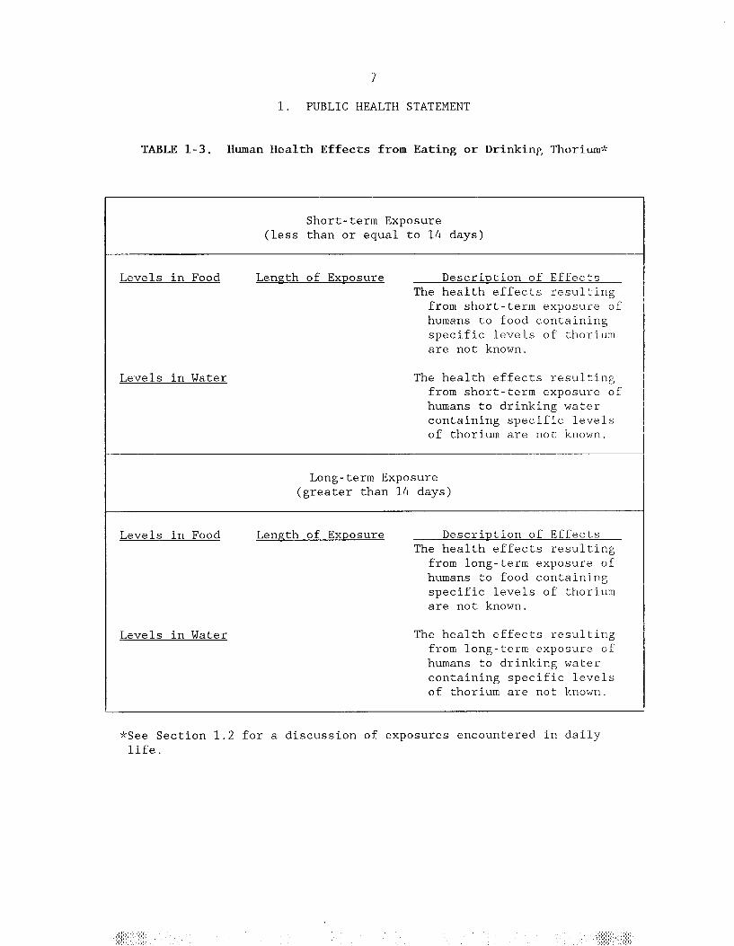
Thorium is odorless and tasteless, so you cannot tell if you are being exposed to thorium. As shown in Tables l-l through l-4, we know very little about specific exposure levels of thorium that result in harmful effects in people or animals. High levels of exposure have been shown to cause death in animals, but no direct cause of death could be determined and no other health effects have been reported. For more information, see Chapter 2.
1.6 IS THERE A MEDICAL TEST TO DETERMINE WHETHER I HAVE BEEN EXPOSED TO THORIUM?
Special tests that measure the level of radioactivity from thorium or thorium isotopes in your urine, feces, and air you breathe out can determine if you have been exposed to thorium. These tests are useful only if run within several days to a week after exposure. The tests cannot, however, tell you if your health will be affected by the exposure. The tests can be run only with special equipment and are probably not available at your local clinic or hospital. For more information, see Chapters 2 and 6.
1.7 WHAT RECOMMENDATIONS HAS THE FEDERAL GOVERNMENT MADE TO PROTECT HUMAN HEALTH?
The Environmental Protection Agency (EPA) requires that the Federal Government be notified if more than 1 millicurie (3.7x10⁷ Becquerels) of radioactivity from natural thorium is released into the environment. The Nuclear Regulatory Commission has issued Maximum Permissible Concentrations (MPC) in air and water for workplace exposure to thorium. For more information on government regulations and guidelines, see Chapter 7.
1.8 WHERE CAN I GET MORE INFORMATION?
If you have any more questions or concerns not covered here, please contact your State Health or Environmental Department or:
Agency for Toxic Substances and Disease Registry Division of Toxicology 1600 Clifton Road, E-29 Atlanta, Georgia 30333
This agency can also give you information on the location of the nearest occupational and environmental health clinics. Such clinics specialize in recognizing! evaluating, and treating illnesses that result from exposure to hazardous substances.


9
2. HEALTH EFFECTS
2.1 INTRODUCTION
This chapter contains descriptions and evaluations of studies and interpretation of data on the health effects associated with exposure to thorium. Its purpose is to present levels of significant exposure for thorium based on toxicological studies, epidemiological investigations, and environmental exposure data. This information is presented to provide public health officials, physicians, toxicologists, and other interested individuals and groups with (1) an overall perspective of the toxicology of thorium and (2) a depiction of significant exposure levels associated with various adverse health effects.
2.2 DISCUSSION OF HEALTH EFFECTS BY ROUTE OF EXPOSURE
To help public health professionals address the needs of persons living or working near hazardous waste sites, the data in this section are organized first by route of exposure -- inhalation, oral, and dermal -- and then by health effect -- death, systemic, immunological, neurological, developmental, reproductive, genotoxic, and carcinogenic effects. These data are discussed in terms of three exposure periods -- acute, intermediate, and chronic.
Levels of significant exposure for each exposure route and duration (for which data exist) are presented in tables and illustrated in figures. The points in the figures showing no-observed-adverse-effect levels (NOAELs) or lowest-observed-adverse-effect levels (LOAELS) reflect the actual levels of exposure used in the studies. LOAELs have been classified into "less serious" or "serious" effects. These distinctions are intended to help the users of the document identify the levels of exposure at which adverse health effects start to appear, determine whether or not the intensity of the effects varies with dose and/or duration, and place into perspective the possible significance of these effects to human health.
The significance of the exposure levels shown on the tables and figures may differ depending on the user's perspective. For example, physicians concerned with the interpretation of clinical findings in exposed persons or with the identification of persons with the potential to develop such disease may be interested in levels of exposure associated with "serious" effects. Public health officials and project managers concerned with response actions at Superfund sites may want information on levels of exposure associated with more subtle effects in humans or animals (LOAEL) or exposure levels below which no adverse effects (NOAEL) have been observed.
Thorium is a relatively reactive, metallic radioactive element. Because thorium is a radioactive element, evaluation of adverse health effects due to exposure to thorium requires a slightly different approach than with chemicals. Radiation is a health risk because radioactive elements can emit energetic particles or electromagnetic radiation that can damage cells. Radioactive elements are those that undergo spontaneous

10
2. HEALTH EFFECTS
disintegration (decay) in which energy is released (emitted) either in the form of particles, such as alpha or beta particles, or rays, such as gamma or x-rays. This disintegration or decay results in the formation of new elements, some of which may themselves be radioactive, in which case they will also decay. The process continues until a stable (non-radiative) state is reached (see Appendix B for more information). The rate of emission of alpha particles from thorium is low, and the rate of emission of gamma rays is very low (see Chapter 3). Alpha particles are unable to deeply penetrate skin, but can travel short distances in the body (about 4 to 6 cell diameters) if they are emitted from within the body. The intensity and energy of alpha particles emitted depends on the particular isotope of thorium in question. Several isotopes of thorium exist. By mass, the most predominant ones in the environment are thorium-230 (a decay product of uranium-238) and natural thorium (thorium-232) (see Chapter 3). The number of particles emitted is related to the radioactive half-life of the isotope, which is about 14 billion years for natural thorium (thorium-232). The other type of radiation hazard is from gamma rays, which can penetrate the body and pass through the air. However, natural thorium has a very low gamma activity, which means there is little danger from this type of radiation from natural thorium. Daughter products of thorium, however, may emit more gamma radiation than natural thorium (see Chapter 3).
When thorium emits alpha particles, it disintegrates into other daughter radionuclides (radioactive materials), such as radium-226 and radon-222 (from thorium-230 in the uranium-238 decay series) or radium-228 and thoron (radon-220 from thorium-232 in the thorium decay series). It eventually decays to stable lead-208 or -206, which is not radioactive. More information about the decay of thorium can be found in Chapter 3. The toxicological characteristics of radon, radium, and lead are the subject of separate ATSDK Toxicological profiles.
The decay rate or activity of radioactive elements has traditionally been specified in curies (Ci). The curie is approximately 37 billion disintegrations (decay events) per second (3.7x10¹⁰ dps). In discussing thorium, a smaller unit,1x10-l2 Ci. the picocurie (pCi) is used, where pCi is equal to In international usage, the S.I. unit (the International System of Units) for activity is the Becquerel (Bq), which is equal to 1 disintegration per second or about 27 pCi. (Information for conversion between units is given in Appendix B.) Measurements of radioactivity, expressed as nCi (nanocurie), in the environment are more sensitive than units of mass. For this reason, amounts of thorium are expressed in pCi units in Chapter 5. In animal studies, the exposure levels were usually reported in mg (milligrams), but have been converted to activity units (nCi and Bq) for presentation in Chapter 2. The absorbed dose from radiation can be expressed in units of rads or it can be stated in terms of dose equivalent, which includes a modification to reflect the quality of the radiations, for radiation protection purposes, and is expressed in terms of rems. For alpha radiations a quality factor, Q, of 20 is used to convert absorbed dose to dose equivalent.

11
2. HEALTH EFFECTS
Both large and small amounts of radiation are damaging to health. Current scientific consensus is radiation can also increase the probability of cancer, and a conservative assumption is no threshold level exists below which there is no additional risk of cancer. There is considerable debate about how great the cancer risks are when people are chronically exposed to very low levels of radiation. Since everyone is environmentally exposed to a small amount of radiation, the minimum amount of additional radiation that may constitute a health hazard is not well known.
The following sections summarize the health effects associated with thorium. Evidence exists that most, if not all, effects of thorium may be due to its radiological, and not chemical, effects. The mechanism of toxicity for all effects are not well understood. For more information about radiation, see Appendix B.
2.2.1 Inhalation Exposure
2.2.1.1 Death
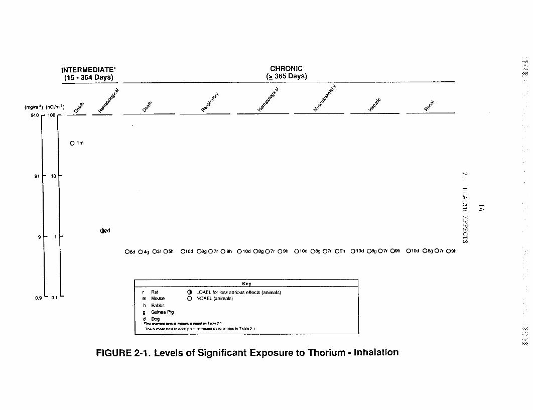
Two epidemiology studies have examined mortality among thorium workers neither found significant excess mortality. The standard mortality ratio (SMR) for all causes of death in a cohort of 3039 male workers in a thorium processing plant was 1.05 in comparison to United States white males (Polednak et al. 1983). The estimated radiation levels to the workers for inhalation intake ranged from 0.003-0.192 nCi/m
3 (0.001-0.007 Bq/m3) for a
period of l-33 years. No evidence of overt industrial disease was found in a cohort of 84 workers at a thorium refinery exposed to <0.045-450 nCi/m3
(<0.002-0.02 Bq/m3) for <l-20 years (Albert et al. 1955). In both studies, the workers were exposed to other toxic compounds (uranium dust) as well as other radioactive materials (thoron, uranium daughters, thorium daughters, cerium).
No compound-related mortality was found in mice exposed to 114-330 Mg/m3(12.54-36.3 nCi/m
3 = 464-1343 Bq/m3) thorium nitrate intermittently
for 18 weeks (Patrick and Cross 1948). No compound-related mortality was found in rats, guinea pigs, rabbits, or dogs exposed intermittently for 1
3 3 3year to 5 mg thorium/m (0.550 nCi/m = 20 Bq/m ) as thorium dioxide (Hodge et al. 1960). These NOAEL values are reported in Table 2-l and plotted in Figure 2-l.
2.2.1.2 Systemic Effects
Respiratory Effects. Although the SMR for respiratory diseases was 1.31 among workers at a thorium refinery (Polednak et al. 1983), the increase may have been attributable in part to smoking. Exposure level
3estimates for inhalation intakes ranged from 0.003-0.192 nCi/m (0.001-0.007
Bq/m ) for a period of 1-33 years. Because the workers were exposed to 3


15
2. HEALTH EFFECTS
other toxic compounds (uranium dust) as well as other radioactive metals, toxic effects cannot necessarily be attributed to thorium. Therefore, no quantitative information from the study is reported in Table 2-l or Figure 2-l.
Progressive cirrhosis of the lungs was found in a subchronic inhalation study in rats (Likhachev et al. 1973a). Rats were exposed intermittently for 6-9 months to an inert aerosol (control), to the inert aerosol enriched with 10% or 49% insoluble thorium dioxide, or to thorium dioxide (100%) alone. The severity of the lung cirrhosis was directly related to the radiation dose and the amount of thorium dioxide. Cirrhosis of the lungs became evident in 3-6 months in the 100% thorium dioxide group, in 9-12 months in the 49% thorium dioxide group, in 12-15 months in the 10% thorium dioxide group, and in 18-24 months in the inert aerosol control group. At lung exposures of up to 150 rad, reticulosarcoma was found, while at lung exposures of 100-2700 rad, glandular cancerous tumors were found (see Section 2.2.1.8). The tumors may have been caused by thorium dioxide; the exact amount of thorium administered was not clear from the report, so the results of the study do not appear in Table 2-l or Figure 2-l.
No histopathological effects on the lungs were found in rats, guinea pigs, rabbits,or dogs exposed intermittently for 1 year to 5 mg thorium/m3
(0.550 nCi/m3 = Bq/m3) as thorium dioxide (Hodge et al. 1960). This NOAEL value is presented in Table 2-l and plotted in Figure 2-l.
Hematological Effects. A complete blood count (CBC) was done on a cohort of 273 male monazite sand refinery workers to determine the effect of thorium on the hematological system. The measured body burden (calculated from in vivo detection of external gamma rays emitted by daughter products of thorium still in the subject's body and from thoron in expired air) of thorium was higher in those workers exposed for a longer time period, but the blood count did not correlate with the body burden of thorium (Conibear 1983). A correlation was found, however, between the blood count and cigarette smoking habits. Exposure level estimates for inhalation intakes of nicotine or thorium were not reported, and the external gamma-ray exposure rate was between 0.5 and 5.0 mR/hour. Because the workers were exposed to other toxic compounds (silica, yttrium, acid and alkali fumes) as well as other sources of radioactivity, toxic effects cannot necessarily be attributed to thorium. Therefore, the results of the study do not appear in Table 2-l or Figure 2-l.
Effects on hematological parameters (abnormal forms of monocytes, lymphocytes and granulocytes, hypoplastic bone marrow, red cell count depression, macrocytosis, increase in immature granulocytes) were found in dogs exposed 6 hours/day, 5 days/week to various chemical forms of thorium:
3 3thorium nitrate tetrahydrate for 60 days (4 nCi/m = 150 Bq/m ); thorium
3 3dioxide for 60 days (4.8 nCi/m = 180 Bq/m ); thorium tetrafluoride for 304
3 3 3days (0.9 nCi/m = 33 Bq/m ); thorium oxalate for 270 days (1.4 nCi/m = 52
Bq/m ) (Hall et al. 1951). Differences in the degree of toxicity of the 3
16
2. HEALTH EFFECTS
various chemical forms of thorium on hematological parameters could not be determined from this study, although gagging, retching, and occasional vomiting were found periodically in the dogs exposed to thorium nitrate
3tetrahydrate. The lowest LOAEL, thorium tetrafluoride (0.9 nCi/m = 33
3Bq/m ), is reported on Table 2-l and plotted on Figure 2-l.
No effects on hematological parameters, blood nonprotein nitrogen (NPN), or the histopathology of the spleen were found in rats, guinea pigs,
3 3rabbits, or dogs exposed for 1 year to 5 mg/thorium m (0.550 nCi/m = 20
3Bq/m ) as thorium dioxide (Hodge et al. 1960). This NOAEL value is presented in Table 2-l and plotted in Figure 2-l.
Musculoskeletal Effects. No studies were located regarding the musculoskeletal effects in humans after inhalation exposure to thorium.
Upon histopathological examination, no effects in the femur were found3
In rats, guinea pigs, rabbits, or dogs exposed for 1 year to 5 mg thorium/m3 3
(0.550 nCI/m = Bq/m ) as thorium dioxide (Hodge et al. 1960). This NOAEL value is presented in Table 2-l and plotted in Figure 2-l.
Hepatic Effects. The levels of aspartate aminotransferase, globulin, and total bilirubin in sera of a cohort of 275 former workers in a thorium refinery were correlated with body burdens of radioactivity (Farid and Conibear 1983). The levels of aspartate aminotransferase and total bilirubin were significantly higher (p<O.OOOl and p=O.O43, respectively) in thorium-exposed workers, as compared to U.S. white males. Globulin levels also increased with increasing levels of body burden, but not significantly. Although the enzymatic levels tested were elevated, they were still within the normal range. No effects on albumin, total protein, or alkaline phosphatase were seen. The correlation of hepatic function tests with body burden of radioactivity may suggest a radiotoxic effect, but this was not proven by the authors. No exposure concentrations were reported.
No histopathological effects in the liver were found in rats, guinea3 3 3
pigs, rabbits, or dogs exposed to 5 mg thorium/m (0.550 nCi/m = 20 Bq/m ) for 1 year as thorium dioxide (Hodge et al. 1960). This NOAEL value is presented in Table 2-l and plotted in Figure 2-l.
Renal Effects. No studies were located regarding renal effects in humans after inhalation exposure to thorium.
No histopathological effects in the kidneys were found in rats, guinea3 3 3
pigs, rabbits, or dogs exposed to 5 mg thorium/m (0.550 nCi/m = 20 Bq/m ) for 1 year as thorium dioxide (Hodge et al. 1960). This NOAEL value is presented in Table 2-l and plotted in Figure 2-l.

17
2. HEALTH EFFECTS
2.2.1.3 Immunological Effects
No studies were located regarding immunological effects in humans after inhalation exposure to thorium. No histopathological effects in the lymph nodes were found in rats, guinea pigs, rabbits, or dogs exposed to 5 mg
3 3 3thorium/m (0,550 nCi/m = 20 Bq/m ) for 1 year as thorium dioxide (Hodge et al. 1960). Since no parameters of immune function were examined, this value does not appear as a NOAEL for immunological effects in Table 2-l or Figure 2l.
No studies were located regarding the following health effects in humans or animals after inhalation exposure to thorium.
2.2.1.4 Neurological Effects
2.2.1.5 Developmental Effects
2.2.1.6 Reproductive Effects
2.2.1.7 Genotoxic Effects
Hoegerman and Cummins (1983) assessed the frequency of chromosome aberrations in the lymphocytes of 47 male workers in a thorium processing plant. The workers were divided into three groups based on their body burdens of radioactivity: low (0 nCi/kg), moderate (0.003 nCi/kg = 0.11 Bq/kg) and high (0.015 nCi/kg = 0.56 Bq/kg) body burden groups. An increased frequency of chromosomal aberrations (dicentric ring chromosomes) were found in the high burden groups (combined high and moderate burden groups) compared to the low burden group and historical controls. No significant differences were found in the frequency of two-break chromosome aberrations. A positive correlation was not established between the frequency of chromosomal aberrations and duration of employment. The observed aberration frequency was generally compatible with that found in patients injected with thorium dioxide colloid (Thorotrast) (see Section 2.2.4.7). Costa-Ribeiro et al. (1975) also reported a statistically significant (p<O.O5) increase in the number of chromosomal aberrations (dicentrics) in 240 monazite sand millers, as compared to controls. No significant differences in the incidence of translocations were observed. No exposure concentrations were reported in either study.
No studies were located regarding genotoxic effects in animals after inhalation exposure to thorium.
2.2.1.8 Cancer
A statistically significant excess of deaths from pancreatic cancer was seen in a cohort of 3039 former thorium workers employed for 1 year or more (6 observed vs. 1.3 expected) but not in workers employed for a shorter time
18
2. HEALTH EFFECTS
(3 observed vs. 2.7 expected) (Stehney et al. 1980). The workers were3 3
exposed to 0.003-0.192 nCi/m (0.001-0.007 Bq/m ). Although a correlation between smoking and pancreatic cancer has not been established, the excess mortality may be due, in part, to the fact that a higher proportion of smokers was found in the worker population when compared to U.S white males (ratio of 1.3 observed smokers/expected smokers). A second study compared the SMR of workers in a thorium processing plant to the mortality rates for U.S. white males and determined that the SMRs in the workers were high for deaths due to lung cancer (SMR=1.44; 95% confidence limit 0.98 and 2.02) and pancreatic cancer (SMR=2.01; 95% confidence limit 0.92 and 3.82) (Polednak et al. 1983). In a subgroup of men in jobs with the highest exposure to thorium, the SMR for lung cancer was 1.68 and the SMR for pancreatic cancer was 4.13. Exposure level estimates for inhalation intakes ranged from
3 30.003-0.192 nCi/m (0.0001-0.007 Bq/m ) for a period of l-33 years. The authors indicated that smoking may be a confounding factor in the increased rates of cancer and that the workers were exposed to other potentially carcinogenic agents, such as thoron (radon-220). Consequently, the evidence for a causal relationship between thorium exposure and cancer is not convincing and no concentrations are reported in Table 2-l or plotted in Figure 2-l.
A significantly (p<O.O5) increased incidence of malignancies in the lymphatic and hematopoietic tissues of uranium mill workers (cohort of 662 males) was found by Archer et al. (1973). The radioactivity in the tracheobronchial lymph nodes of the workers was found to be primarily the result of alpha emissions from thorium-230 and not from uranium-234 or uranium-238. Consequently, the authors suggested that the increased incidence of malignancies may have been a result of thorium-230 exposure and not uranium exposure. Exposure levels of thorium were not reported; therefore, the results of the study are not reported on Table 2-l or plotted in Figure 2-l.
Rats were exposed to various concentrations of thorium dioxide for 6-9 months, and the frequency and histological type of lung tumors were determined following observation for up to 21 months (Likhachev et al. 1973b; Likhachev 1976). The authors concluded that the incidence and histological type of lung tumors that developed were dependent on the radiation dose to the lungs. At lung doses of up to 150 rad (3000 rems), primarily reticulosarcoma was found (in 16% of the animals), while at total doses of 1000-2700 rads (20,000-54,000 rems), glandular cancerous tumors (adenomatosis and squamous cell carcinoma) were found in all of the exposed animals, and the reticulosarcoma was no longer observed.
2.2.2 Oral Exposure
2.2.2.1 Death
No studies were located regarding lethal effects in humans after oral exposure to thorium.

19
2. HEALTH EFFECTS
A single gavage administration of 1000 mg thorium/kg body weight/day (110 nCi/kg/day = 4070 Bq/kg/day) as thorium nitrate resulted in the death of 4/20 mice, while a single amount of 760 mg thorium/kg body weight/day (84 nCi/kg/day = 3100 Bq/kg/day) resulted in no mortality. Occasional intestinal hemorrhage was noted at autopsy in the mice that died, but it was not reported if the hemorrhage was the cause of death in the animals. No effects were found following administration of a 10% sodium nitrate solution, suggesting that the adverse effects were due to thorium and not to nitrate (Patrick and Cross 1948). Following 4 months of continuous exposure to 123 mg thorium/kg body weight/day (13.6 nCi/kg/day = 503 Bq/kg/day) as thorium nitrate in the drinking water, 50% of the treated mice and 10% of the control mice died (Patrick and Cross 1948). No cause of death was reported in either the acute or the 4-month studies. In rats, 4 months of exposure to 3043 mg thorium/kg body weight/day (335 nCi/kg/day = 12,400 Bq/kg/day) as thorium nitrate resulted in death, but the deaths may have been due to the poor nutritional state of the animals since the treated animals ate much less of the treated food and, therefore, lost weight (Downs et al. 1959).
Death occurred following four daily administrations of ≥2130 mg thorium/kg body weight/day (234 nCi/kg/day = 8657 Bq/kg/day) as thorium nitrate in the food to a single dog (Patrick and Cross 1948). No immediate deaths were reported following a single administration of 121 mg thorium/kg body weight/day (13 nCi/kg/day = 481 Bq/kg/day) by gavage as thorium nitrate to dogs (Sollman and Brown 1907). Death was not found following exposure of a single dog to food containing 426 mg thorium/kg body weight/day (47 nCi/kg/day = 1740 Bq/kg/day) as thorium nitrate for 46 days (Downs et al. 1959). No deaths were reported following a single gavage administration of thorium nitrate (483 mg thorium/kg body weight/day = 53 nCi/kg/day = 1960 Bq/kg/day) in rabbits (Sollman and Brown 1907). The number of treated and control animals (dogs and rabbits) was not reported in the Sollman and Brown (1907) study.
All reliable NOAEL and LOAEL values are reported in Table 2-2 and plotted in Figure 2-2. Values from the Sollman and Brown (1907) study are not reported in the table and figure since the number of animals in the study were not reported. The LOAEL value for death in rats from the Downs et al. (1959) study is not reported since the deaths may have been due to the poor nutritional state of the animals and not to thorium toxicity, and the NOAEL and LOAEL values for the death of dogs in the Downs et al. (1959) and the Patrick and Cross (1948) studies, respectively, are not reported since they were pilot studies and only one animal was used.
2.2.2.2 Systemic Effects
Respiratory Effects. No studies were located regarding the respiratory effects in humans after oral exposure to thorium.


22
2. HEALTH EFFECTS
No histopathological changes in the lungs were found in rats treated for 4 months with 3043 mg thorium/kg body weight/day (335 nCi/kg/day = 12,400 Bq/kg/day) or in one dog treated for 46 days with 426 mg thorium/kg body weight/day (47 nCi/kg/day = 1740 Bq/kg/day) as thorium nitrate in the food (Downs et al. 1959). These NOAEL values for rats are reported in Tab 2-2 and plotted in Figure 2-2. The NOAEL value for the effects in dogs is not reported since this was a pilot study and only one animal was used.
Cardiovascular Effects. No studies were located regarding the cardiovascular effects in humans after oral exposure to thorium.
No histopathological changes in the heart were found in rats treated for 4 months with 3043 mg thorium/kg body weight/day (335 nCi/kg/day = 12,400 Bq/kg/day) or in one dog treated for 46 days with 426 mg thorium/kg body weight/day (47 nCi/kg/day = 1740 Bq/kg/day) as thorium nitrate in the food (Downs et al. 1959). These NOAEL values for rats are reported in Tab 2-2 and plotted in Figure 2-2. The NOAEL value for the effects in dogs is not reported since this was a pilot study and only one animal was used.
Gastrointestinal Effects. No studies were located regarding the gastrointestinal effects in humans after oral exposure to thorium.
No histopathological changes in the stomach and intestines were found in rats treated for 4 months with 3043 mg thorium/kg body weight/day (335 nCi/kg/day = 12,400 Bq/kg/day) or in one dog treated for 46 days with 426 mg thorium/kg body weight/day (47 nCi/kg/day = 1740 Bq/kg/day) as thorium nitrate in the food (Downs et al. 1959). These NOAEL values for rats are reported in Table 2-2 and plotted in Figure 2-2. The NOAEL value for the effects in dogs is not reported since this was a pilot study and only one animal was used.
Occasional intestinal hemorrhages were reported in mice that died following a single gavage exposure to thorium nitrate (Patrick and Cross 1948). It was not reported whether the intestinal hemorrhage was the cause of death in the mice. The level at which this occurred was not reported. The possibility that intestinal damage resulted from improper gavage technique cannot be ruled out; therefore, these data are not presented in Table 2-2 or plotted in Figure 2-2.
Hematological Effects. No studies were located regarding the hematological effects in humans after oral exposure to thorium.
No histopathological changes in the spleen were found in rats treated for 4 months with 3043 mg thorium/kg body weight/day (335 nCi/kg/day = 12,400 Bq/kg/day) or in one dog treated for 46 days with 426 mg thorium/kg body weight/day (47 nCi/kg/day = 1740 Bq/kg/day) as thorium nitrate in the food (Downs et al. 1959). These NOAEL values for rats are reported in Table 2-2 and plotted in Figure 2-2. The NOAEL value for the effects in
23
2. HEALTH EFFECTS
dogs is not reported since this was a pilot study and only one animal was used.
Hepatic Effects. No studies were located regarding the hepatic effects in humans after oral exposure to thorium.
No histopathological changes in the liver were found in rats treated for 4 months with 3043 mg thorium/kg body weight/day (335 nCi/kg/day = 12,400 Bq/kg/day) or in one dog treated for 46 days with 426 mg thorium/kg body weight/day (47 nCi/kg/day = 1740 Bq/kg/day) as thorium nitrate in the food (Downs et al. 1959). These NOAEL values for rats are reported in Table 2-2 and plotted in Figure 2-2. The NOAEL value for the effects in dogs is not reported since this was a pilot study and only one animal was used.
Renal Effects. No studies were located regarding the renal effects in humans after oral exposure to thorium.
No histopathological changes in the kidneys were found in rats treated for 4 months with 3043 mg thorium/kg body weight/day (335 nCi/kg/day = 12,400 Bq/kg/day) or in one dog treated for 46 days with 426 mg thorium/kg body weight/day (47 nCi/kg/day = 1740 Bq/kg/day) as thorium nitrate in the food (Downs et al. 1959). These NOAEL values for rats are reported in Table 2-2 and plotted in Figure 2-2. The NOAEL value for the effects in dogs is not reported since this was a pilot study and only one animal was used.
Other Systemic Effects. Weight loss was found in rats treated for 4 months with 3043 mg thorium/kg body weight/day (335 nCi/kg/day = 12,400 Bq/kg/day) and in one dog treated for 46 days with 426 mg thorium/kg body weight/day (47 nCi/kg/day = 1740 Bq/kg/day) as thorium nitrate in the food (Downs et al. 1959). The weight loss was attributed to a decrease in intake of the treated food; therefore, these values are not reported in Table 2-2 or Figure 2-2.
No studies were located regarding the following health effects in humans or animals after oral exposure to thorium.
2.2.2.3 Immunological Effects
2.2.2.4 Neurological Effects
2.2.2.5 Developmental Effects
2.2.2.6 Reproductive Effects
No studies were located regarding reproductive effects in humans after oral exposure to thorium.
No histopathological changes in the gonads (exact tissues examined were not reported) were found in male and female rats treated for 4 months with

24
2. HEALTH EFFECTS
3043 mg thorium/kg body weight/day (335 nCi/kg/day = 12,400 Bq/kg/day). No histopathological changes in the testes were found in one dog treated for 46 days with 426 mg thorium/kg body weight/day (47 nCi/kg/day - 1740 Bq/kg/day) as thorium nitrate in the food (Downs et al. 1959). The value of 335 nCi/kg/day for rats is reported in Table 2-2 and plotted in Figure 2-2.
2.2.2.7 Genotoxic Effects
No studies were located regarding genotoxic effects in humans or animals after oral exposure to thorium.
2.2.2.8 Cancer
No studies were located regarding carcinogenic effects in humans or animals after oral exposure to thorium.
2.2.3 Dermal Exposure
2.2.3.1 Death
No studies were located regarding lethal effects in humans after dermal exposure to thorium.
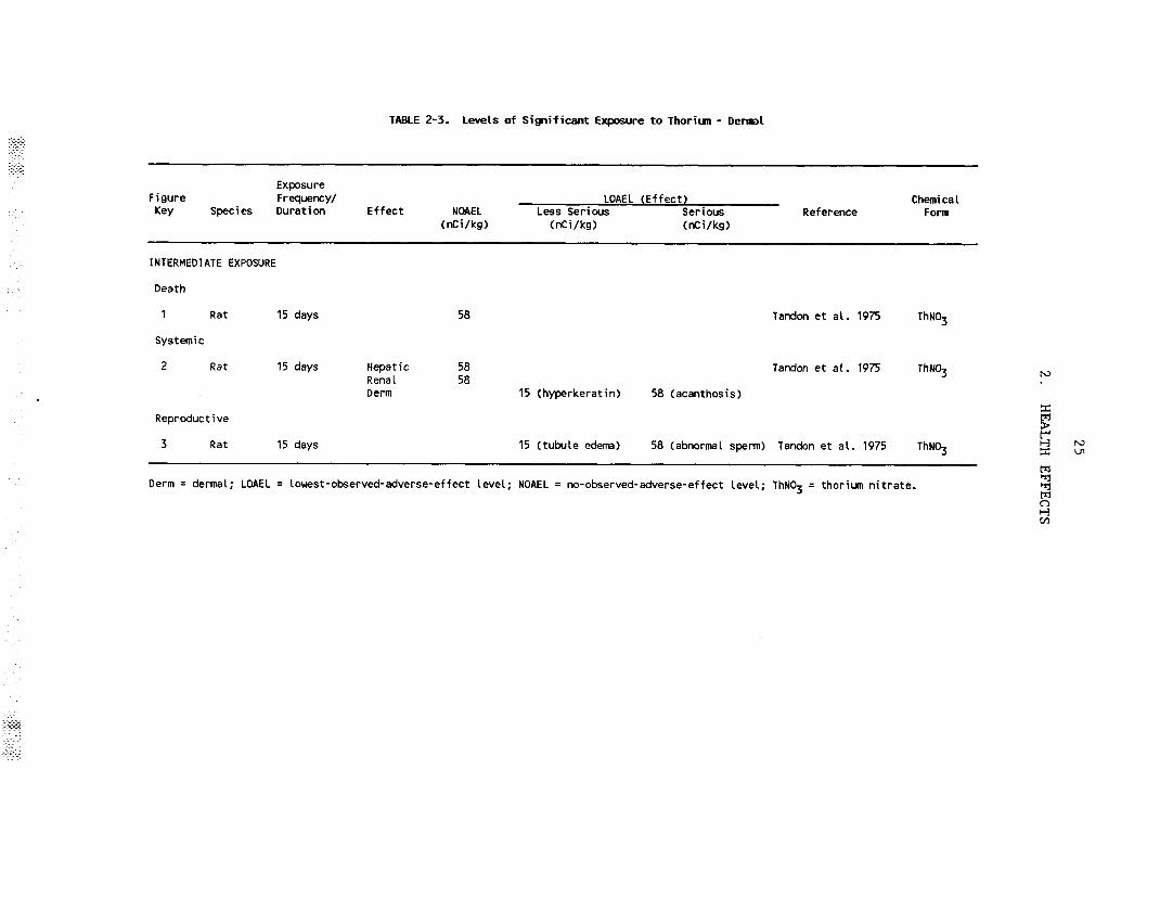
Tandon et al. (1975) reported no lethality in rats following dermal application of 529 mg thorium/kg body weight (58 nCi/kg = 2146 Bq/kg), daily for 15 days, to the lateroabdominal and scrotal skin. Prior to treatment, the hair was clipped. The area remained uncovered for the duration of treatment. The thorium was administered as thorium nitrate. This NOAEL value is reported in Table 2-3.
2.2.3.2 Systemic Effects
Hepatic Effects. No studies were located regarding hepatic effects in humans after dermal exposure to thorium.
Tandon et al. (1975) reported no histopathological effects on the liver of rats following dermal application of 529 mg thorium/kg body weight/day (58 nCi/kg/day - 2146 Bq/kg/day) for 15 days to the lateroabdominal and scrotal areas. Prior to treatment, the hair was clipped. The area remained uncovered for the duration of treatment. The thorium was administered as thorium nitrate. This NOAEL value for hepatic effects in rats is reported in Table 23.
Renal Effects. No studies were located regarding renal effects in humans after dermal exposure to thorium.
26
2. HEALTH EFFECTS
Tandon et al. (1975) reported no histopathological effects on the kidneys of rats following dermal application of 529 mg thorium/kg body weight/day (58 nCi/kg/day = 2146 Bq/kg/day) for 15 days to the lateroabdominal and scrotal areas. Prior to treatment, the hair was clipped. The area remained uncovered for the duration of treatment. The thorium was administered as thorium nitrate. This NOAEL value for renal effects in rats is reported in Table 2-3.
Dermal/Ocular Effects. No studies were located regarding dermal/ocular effects in humans after dermal exposure to thorium.
Tandon et al. (1975) applied daily dermal applications of 132.5 mg thorium/kg body weight/day (15 nCi/kg/day = 555 Bq/kg/day), 265 mg thorium/kg body weight/day (29 nCi/kg/day = 1073 Bq/kg/day), or 529 mg thorium/kg body weight/day (58 nCi/kg/day = 2146 Bq/kg/day) to the lateroabdominal and scrotal areas of rats for 15 days. The thorium was administered to skin (hair was clipped) as thorium nitrate, and the area remained uncovered for the duration of treatment. Mild hyperkeratinization of the lateroabdominal skin was found at all exposure levels. At the highest exposure level, mild acanthosis and thickening of the epithelial lining of the lateroabdominal skin were seen. At this level, mild acanthosis, swollen collagen fibers, and foamy dermis were found in the scrotal skin. The value of 15 nCi/kg/day is a less serious LOAEL, and the exposure level of 58 nCi/kg/day is a serious LOAEL for dermal effects in the rat. These values are reported in Table 2-3.
No studies were located regarding the following health effects in humans or animals after dermal exposure to thorium.
2.2.3.3 Immunological Effects
2.2.3.4 Neurological Effects
2.2.3.5 Developmental Effects
2.2.3.6 Reproductive Effects
No studies were located regarding reproductive effects in humans after dermal exposure to thorium.
Tandon et al. (1975) applied daily dermal applications of 132.5 mg thorium/kg body weight/day (15 nCi/kg/day = 555 Bq/kg/day), 265 mg thorium/kg body weight/day (29 nCi/kg/day = 1073 Bq/kg/day), or 529 mg thorium/kg body weight/day (58 nCi/kg/day = 2146 Bq/kg/day) to the lateroabdominal and scrotal skin of rats for 15 days. The thorium was administered to skin (hair was clipped) as thorium nitrate, ,and the area remained uncovered for the duration of treatment. Mild edema of the seminiferous tubules and the interstitium was seen at all exposure levels. At the highest exposure level, some desquamation of sperm and giant
27
2. HEALTH EFFECTS
spermatid-type cells were found. The percentage of sperm affected by thorium treatment was not reported. It was not clear whether these reproductive changes were due to chemical or radiation effects, although they were more likely the result of chemical toxicity because of the short time required to produce the effects. The level of 15 nCi/kg/day is a less serious LOAEL and 58 nCi/kg/day is a serious LOAEL for the reproductive effects of thorium in rats. These values are reported in Table 2-3.
2.2.3.7 Genotoxic Effects
No studies were located regarding genotoxic effects in humans or animals after dermal exposure to thorium.
2.2.3.8 Cancer
No studies were located regarding carcinogenic effects in humans or animals after dermal exposure to thorium.
2.2.4 Other Routes of Exposure
Most of, the literature deals with the carcinogenic effects of thorium as a result of intravenous injection of Thorotrast, a colloid consisting of approximately 25% thorium-232 dioxide and stabilized with dextran. Thorotrast was used as a radiographic contrast medium between the years 1928 and 1955. It was estimated that 50,000-100,000 patients worldwide received Thorotrast (Harrist et al. 1979; Isner et al. 1978). Generally, l0-75 mL of Thorotrast was injected and toxic effects were found at all exposure -levels.It has been reported that the thorium-232 in Thorotrast has an activity of 24.2 nCi/mL (Steinstrasser 1981); therefore, the injected amounts of l-75 mL correspond to 3.5-26 nCi/kg body weight (129-962 Bq/kg). The toxic effects include formation 4-6 years after exposure of "Thorotrastomas," granulomas at the site of injection resulting from the extravasation of the injected Thorotrast (Frank 1980; Grampa 1971). Blood disorders (hemolytic and aplastic anemia, myelofibrosis, and leukemia) appeared 20 years following injection, and hemangiosarcoma of the liver was found 25-30 years post-exposure (Frank 1980). A relationship was found between the amount of the Thorotrast injected and the incidence of liver tumors (cholangiocarcinoma, angiosarcoma, hepatic cellular carcinoma) (Wesch et al. 1983; Van Kaick et al. 1983). A decrease in the time to tumors was also found with increased injected volume of Thorotrast (Van Kaick et al. 1983). The use of Thorotrast ceased when the potential toxic effects were recognized.
2.2.4.1 Death
No studies were located regarding acute lethal effects in humans after other routes of exposure to thorium. Death from various types of cancer, however, was found 20-30 years after intravenous injection of Thorotrast.
28
2. HEALTH EFFECTS
After a period of 15 months, no increased mortality was seen from a single intravenous injection of 0.5 mL of Thorotrast (403 nCi/kg = 14909 Bq/kg) in mice (Guimaraes et al. 1955).
2.2.4.2 Systemic Effects
Respiratory Effects. No studies were located regarding respiratory effects in humans after other routes of exposure to thorium. No degenerative changes in the pulmonary parenchyma were found, but 7/20 mice that died 15 months after intravenous injection of Thorotrast (Guimaraes et al. 1955) and 8/20 mice that were sacrificed 5-12 months after injection of Thorotrast (Guimaraes and Lamerton 1956) had lung adenomas. There was no significant difference in survival between the treated and control animals in either study. In a few cases, an association between the presence of Thorotrast deposits in the lungs and the proliferation of bronchioles and alveoli was found.
Cardiovascular Effects. Myocardial infarction, severe coronary luminal narrowing, and internal alteration of the carotid artery were found in two patients injected 21-30 years before with an unreported amount of Thorotrast (Isner et al. 1978). The authors concluded that the vascular effects were the result of chronic alpha irradiation. The patients were injected in the carotid artery, and thorotrastoma (see Other Systemic Effects, below) was found in both patients.
Lipchik et al. (1972) reported no significant acute changes in cardiac output, pulse rate, pressure or left ventricle volumes, or clotting time in dogs injected intravenously with up to 1 mL of Thorotrast/kg (1.9 nCi/kg = 70 Bq/kg.
Eleven months after intratracheal and intraperitoneal injection of thorium dioxide in rats, a sharp and persistent fall in blood pressure was found (Syao-Shan 1970). The fall in blood pressure could not be directly attributed to the chemical or radiological effects of thorium.
Hematological Effects. Aplastic anemia, leukemia (erythroleukemia, acute myelogenous leukemia), myelofibrosis, and splenic cirrhosis were among the hematological effects commonly found in patients after injection of Thorotrast (Dejgaard et al. 1984; Kamiyama et al. 1988; Kato et al. 1983; Rao et al. 1986; Summers and Chung 1986; Van Kaick et al. 1983). The appearance of the leukemia commonly occurred 20 years after injection (see Section 2.2.4.8).
No studies were located regarding hematological effects in animals after other routes of exposure to thorium.
Hepatic Effects. Severe cirrhosis of the liver was one of the primary systemic effects seen following injection of Thorotrast in humans (Baxter

29
2. HEALTH EFFECTS
et al. 1980a,b; Faber 1979; Kato and Kido 1987; Kato et al. 1983; Mori et al. 1979, 1983a,b; Rao et al. 1986; Van Kaick et al. 1983). Cases of fibrosis, veno-occlusive disease, and blood-filled cavities were also found in the livers of Thorotrast patients (da Silva Horta 1967a; Dejgaard et al. 1984). The latency period for the appearance of the cirrhosis was not clear, but was probably comparable to the latency period for liver tumors (25-30 years) since the two effects were often found together.
Degenerative liver changes (necrosis, fibrosis, cirrhosis) were found in mice and rats treated with a single dose of Thorotrast and allowed to survive up to 15 months after treatment (Guimaraes et al. 1955; Guimaraes and Lamerton 1956; Wegener et al. 1983). The authors of the mouse study (Guimaraes et al. 1955; Guimaraes and Lamerton, 1956) concluded that radiation was responsible for the cellular proliferation that led to the degeneration and hepatic tumors.
Following intravenous injection of O-2.8 µCi/kg (104,000 Bq/kg) thorium-227 in a solution of citric acid-sodium citrate buffer in dogs, an increase in serum alkaline phosphatase measurements and hypoalbuminemia and hyperglobulinemia were observed (Stevens et al. 1967). No effects on the levels of serum glutamic pyruvic transaminase (SGPT) or serum glutamic oxaloacetic transaminase (SGOT) were found.
Renal Effects. No studies were located regarding renal effects in humans or animals after other routes of exposure to thorium, but tumors of the kidney have been reported after intravenous administration of thorium in humans (see Section 2.2.4.8).
Other Systemic Effects. Localized fibrosis infiltrated with macrophages was often found surrounding deposits of Thorotrast at the point of intravenous injection. These granulomas were termed Thorotrastoma and resulted from fibroblastic proliferation due to the extravascular deposition of Thorotrast (Coorey 1983; Stanley and Calcaterra 1981; Stougaard et al. 1984; Wustrow et al. 1988). Histologically, the Thorotrastoma consisted of dense, hyalinized connective tissue with Thorotrast found both free and in the cytoplasm of macrophages (Grampa 1971). The Thorotrastoma most commonly occurred in the neck after a cerebral angiography and appeared 4-6 years after intravenous injection (Frank 1980).
2.2.4.3 Immunological Effects
Fibrosis of the lymph nodes, which occluded the lymph vessels, and of the spleen were found in patients injected intravenously with unknown quantities of Thorotrast (da Silva Horta 1967a; Wegener et al. 1976; Wegener and Wesch 1979). No malignancies were found in the lymph nodes, but hemangioendothelioma of the spleen was reported in 2/14 patients examined by da Silva Horta (1967a).

30
2. HEALTH EFFECTS
No degenerative changes were observed in the spleen of mice injected intravenously with Thorotrast, but one animal had a malignant hemangioendothelioma (Guimaraes et al. 1955). Michael and Murray (1970) found a suppression in immune response following administration of Thorotrast to mice. The suppression was found to appear sooner (within 1 hour after treatment) and last for a longer period of time (up to 3 days) when Thorotrast was administered intraperitoneally rather than intravenously. Thorotrast was found to affect lymphoid cells involved in antibody formation, as well as the blockade of phagocytic cells in certain organs of the reticuloendothelial system (Michael and Murray 1970).
No studies were located regarding the following health effects in humans or animals after other routes of exposure to thorium:
2.2.4.4 Neurological Effects
2.2.4.5 Developmental Effects
2.2.4.6 Reproductive Effects
2.2.4.7 Genotoxic Effects
The intravenous injection of Thorotrast resulted in radiation-induced chromosomal aberrations in patients (Fischer et al. 1967; Kemmer 1979; Kemmer et al. 1971, 1979; Sadamori et al. 1987; Sasaki et al. 1987). A positive correlation was found between the chromosomal aberration rate and the administered amount of Thorotrast (Buckton and Langlands 1973; Fischer et al. 1967; Kemmer et al. 1971, 1973).
No studies were located regarding the genotoxic effects in animals after intravenous injection or other routes of exposure to thorium.
2.2.4.8 Cancer
The primary effects of intravenously injected Thorotrast in humans are liver tumors (cholangiocarcinoma, angiosarcoma, hepatic cellular carcinoma) and blood disorders (aplastic anemia, erythroleukemia, acute myelogenous leukemia) (BEIR IV 1988; Ito et al. 1988; Kamiyama et al. 1988; Kato and Kido 1987; Kojirs et al. 1985; Levy et al. 1986; Van Kaick et al. 1983; Yamada et al. 1983). Two groups of former Thorotrast patients were examined (one group was 93 autopsy cases from the "Annual of Pathological Autopsy Cases" in Japan and the other was a group of 78 autopsy cases from the Japanese literature). Cholangiocarcinoma was found in 55-58%, angiosarcoma was found in 24-25%, and hepatocellular carcinoma was found in 17-21% of these cases (Yamada et al. 1983). The mean latency period for all tumor types was between 25 and 30 years (Frank 1980). The mean value of absorbed dose to the liver was calculated to be 876 rads for hepatocellular carcinoma and 1053 rads for cholangiocarcinoma (Mori et al. 1983b). It was determined that angiosarcoma developed later (33.5-year mean latency period) than
31
2. HEALTH EFFECTS
cholangiocarcinoma (27.8 years) (Yamada et al., 1983). Blood-filled cavities in the tumors were common (Visfeldt and Poulsen 1972). Kato et al. (1983) reported that malignant hepatic tumors accounted for 63% of all Thorotrast-related deaths, and as the dose rate increased (<15 to ≥45 rad/year;<0.15 to ≥0.45 Gy/year), the severity of the liver effects increased and the latency period decreased. A dose-effect relationship was found between the amount of Thorotrast injected and the incidence of liver tumors in humans (Van Kaick et al. 1983). The radiation dose to the liver from l-10 mL injected was 10 rad/year (0.10 Gy/year), 11-20 mL was 18 rad/year (0.18 Gy/year), and >20 mL was 30 rad/year (0.30 Gy/year).
The latency period for leukemia was about 20 years, which was 5-10 years shorter than the liver tumors (Frank 1980). The primary forms of leukemia found were erythroleukemia and acute myelogenous leukemia (da Motta et al. 1979; Kamiyama et al. 1988; Mori et al. 1983b). Kamiyama etal. (1988) reported that the damage from Thorotrast may have been due to effects on the hematopoietic stem cell level.
Fifteen cases of bone tumors resulting from intravenous Thorotrast injection have been reported (9 of which were osteosarcoma). The mean latency period was 26 years and the latency period and injected amount of Thorotrast were inversely related (Harrist et al. 1979). The mean dose rate to bone was 16 rads/year (0.16 Gy/year) per 25 mL of injected Thorotrast (Van Kaick et al. 1983).
Tumors of the kidneys, spleen, and pancreas have also been reported (Christensen et al. 1983; Guimaraes et al. 1955; Kauzlaric et al. 1987; Levy et al. 1986; Mori et al. 1979; Van Kaick et al. 1983; Westin et al. 1973). Christensen et al. (1983) determined that transitional cell carcinoma of the kidneys have a significantly longer latency period (35.8 years; p<0.005) compared to carcinoma of other histological types (27.6 years). A few cases of meningioma and gliosarcoma were found (da Silva Horta 1967b; Kyle et al. 1963; Sussman et al. 1980; Wargotz et al. 1988).
The literature suggests that the toxic effects of Thorotrast are due to the alpha radiation effects of thorium and not to the chemical effects of thorium or of the.colloid (Faber 1973; BEIR IV 1988; Taylor et al. 1986; Wesch et al. 1983). Wesch et al. (1983) injected Thorotrast enriched with thorium-230 into rats and found a linear relationship between radiation level and tumor incidence. At a constant radioactive level, an increase in the injected colloidal volume had little influence on the number of liver tumors, but resulted in a decrease in tumor appearance time and, therefore, a decrease in lifespan. The larger colloidal volume may result in a more diffuse organ dose and a less "hot spot" distribution, Injection of the nonradioactive colloid resulted in no appreciable incidence of liver tumors. It is not known whether the colloidal particles induce the liver tumors when given in combination with the radioactive thorium, or if the colloid only accelerates the expression of the radiation-induced tumors. However,
32
2. HEALTH EFFECTS
studies in mice reported by Taylor et al. (1986) suggest that the induction of liver cancer can be accounted for by the radiation alone.
Thorotrast studies in rats found a positive correlation between the administered amount and the number of liver and splenic tumors (Johansen 1967; Wesch et al. 1983).
2.3 TOXICOKINETICS
2.3.1 Absorption
2.3.1.1 Inhalation Exposure
The absorption of thorium from the lungs is dependent upon the chemical nature of the isotope and the size of the aerosol particle (Boecker 1963; Boecker et al. 1963; Moores et al. 1980; Newton et al. 1981; Sunta et al. 1987; Syao-Shan 1970a). Increasing the particle size (>2 µm) increases deposition in the respiratory tract of mice, but decreases deposition in the alveolar region. A linear relationship was found between aerosol dosage of thorium-232 and the amount deposited in the alveolar region (Moores et al. 1980). Approximately twice as much thorium-234 is absorbed from the lungs of rats exposed to soluble thorium citrate (33%) compared to soluble thorium chloride (19%) (Boecker et al. 1963). However, following the initial difference in absorption, thorium shows the same distribution and excretion pattern, regardless of absorbed compound. Syao-Shan (1970b) determined that 1.5-5.0% of the administered amount to rats is absorbed from the lungs 1 day after intratracheal administration of insoluble thorium-232 dioxide. Deposited thorium dioxide tends to remain in the lungs for long periods of time; 68-73% of thorium-232 dioxide remained in the lungs after 1 day, while 15-30% remained after 21 months. Thorium is removed primarily by ciliary clearance and is excreted in the feces (Wrenn et al. 1981). ICRP (ICRP 1979) assumes a total of 5% absorption of inhaled thorium.is transferred to the blood. However, the solubilities of the thorium compounds appear to be an important biological factor, as evidenced by differences in toxicity: LD5Os after 30 days following intraperitoneal injection in mice were 370.8 mg thorium/kg for soluble thorium-232 nitrate and 589.1 mg thorium/kg for soluble thorium-232 chloride, while 2000 mg thorium/kg for insoluble thorium-232 dioxide resulted in deaths of only 2/18 mice (Syao-Shan 1970b).
Lung levels of thorium (230 and 232) in workers occupationally exposed to thorium (miners and millers) are significantly higher than those not occupationally exposed (Gilbert et al. 1985; Singh et al. 1981; Vocaturo et al. 1983; Wrenn et al. 1985). In a review of the epidemiological evidence, Wrenn et al. (1981) concluded that the major route of exposure was inhalation. Though intake of thorium through the air may account for less than 1% of the total intake, absorption through the lungs accounts for approximately 2/3 of the ultimate uptake in the body. This is due primarily
33
2. HEALTH EFFECTS
to the low gastrointestinal absorption rate (0.02%) in humans (Maletskos et al. 1969; Sullivan et al. 1983).
2.3.1.2 Oral Exposure
ICRP has recommended a human gastrointestinal absorption value of 0.02% for all forms of thorium (ICRP 1979). In a recent review of the literature by Johnson and Lamothe (1989), a human gastrointestinal absorption value of 0.1 to 1% was calculated. Absorption of thorium in the form of thorium nitrate is 40-fold higher in neonatal rats (l.l-1.2%) (Sullivan et al.1980b, 1983) than in adult rats (0.028-0.5%) (Sullivan et al. 1980a; Traikovich 1970). Absorption of thorium in adult mice was 0.065% (Sullivan et al. 1983). These data suggest that infants may be a susceptible population for exposure. In other studies of actinide elements (including thorium), little variation in gastrointestinal absorption was found between rats, guinea pigs, or dogs. Solubility factors and particle size were found to be the determinants of absorption (Sullivan 1980a). The absorption of various forms and isotopes of thorium in rats was compared by Pavlovskaia (1973). It was found that the rate of absorption of thorium-EDTA by the gastrointestinal tract was 60 times greater than that of thorium dioxide. Thorium nitrate had a 4 times greater absorption rate than thorium dioxide, and the absorption rate of thorium chloride was 10 or 20 times greater than thorium dioxide, depending on concentration. The absorption differences are attributable to different solubilities of the various chemical forms.
2.3.1.3 Dermal Exposure
No studies were located regarding the rate and extent of absorption of thorium following dermal exposure of humans or animals. Absorption of thorium through the skin of animals can be inferred, however, because testicular effects were seen in rats following application of thorium nitrate directly to the lateroabdominal and scrotal skin (Tandon et al. 1975).
2.3.2 Distribution
2.3.2.1 Inhalation Exposure
The median concentrations of thorium-232, thorium-230, and thorium-228 in bone and various soft tissues of autopsy samples of a control population from Grand Junction, CO, and Washington, DC are presented in Table 2-4 (Ibrahim et al. 1983; Wrenn et al. 1981; Singh et al. 1983). The maximum concentration of all three thorium isotopes was found in the tracheobronchial lymph nodes, with lungs and bones containing the next highest activity of thorium isotopes. The high activity in the lymph nodes implies that some of the thorium is cleared from the lungs by the lymphatic system and deposited in the lymph nodes (Mausner 1982; Wrenn et al. 1981). One possible explanation for the higher activity of thorium-228 than
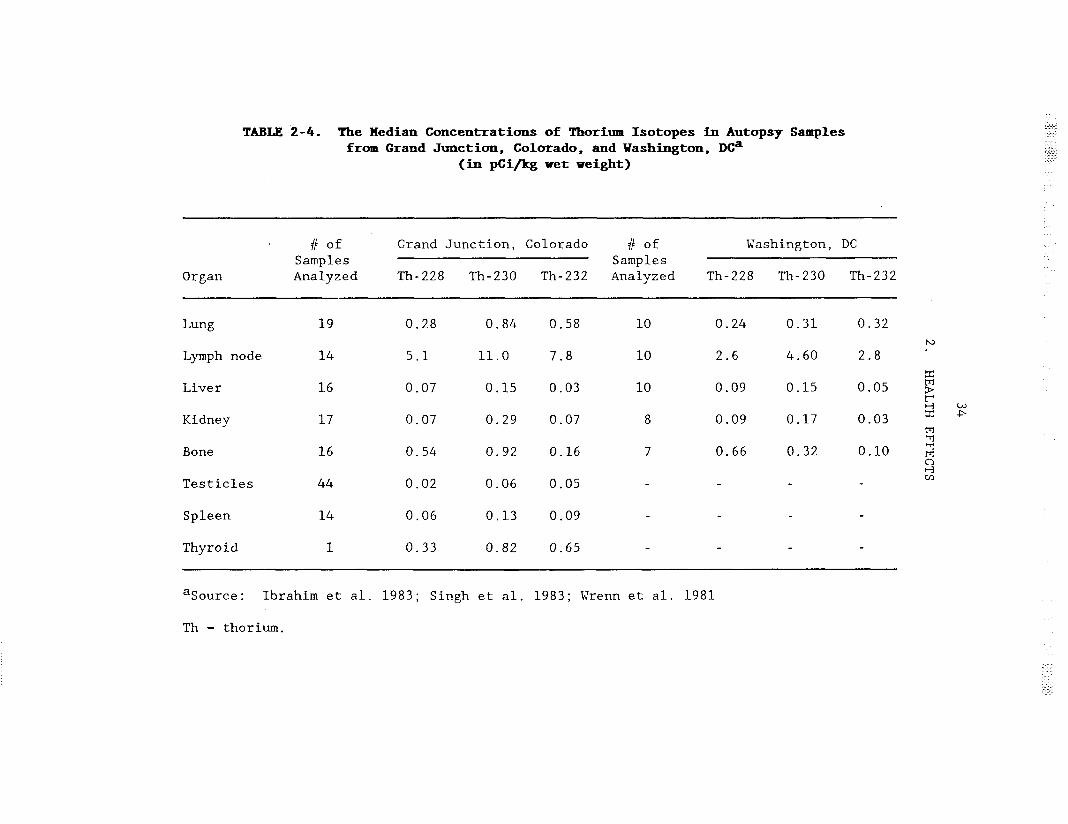

35
2. HEALTH EFFECTS
thorium-232 in bone is that a major portion of the thorium-228 may be from intake of radium-228 (radium appears to be absorbed from the gastrointestinal tract to a greater extent than thorium); radium-228 concentrates in bones and decays to thorium-228 (Wrenn et al. 1981). Studies in mice have shown that thorium-227, injected intraperitoneally, distributes directly to the bone (Miiller et al. 1978); therefore, there may be other explanations for the higher levels of thorium-228 than thorium-232 in the bone. More thorium-232 is retained in the lungs and lymph nodes than thorium-230, suggesting that the solubilization of thorium-230 may be faster than that of thorium-232 in the lungs. This may be due to thorium230 being inhaled in smaller particles than thorium-232. Consequently, thorium-230 is removed more quickly from the lungs and is transported to bone. The low content of thorium in the reticuloendothelial system (liver, spleen, bone marrow) is in contrast to the distribution following intravenous injection of thorotrast (Th02 colloid), where the vast majority of the thorium is taken up by the macrophages of the reticuloendothelial system.
The dose rates to various organs in humans from environmental thorium were estimated to be: 2.2-4.5, 0.41-0.44, 0.19-0.23, 0.057-0.071, and 0.071-0.072 mrad/year in the lymph nodes, bone, lungs, liver, and kidneys, respectively (Wrenn et al. 1981). The dose rates to organs tended to be higher in subjects living in the vicinity of uranium mine tailings, and the dose rates to the organs in miners were even higher (4.8-10.5 mrad/year in the lymph nodes and 1.2-1.5 mrad/year in the lungs) (Wrenn et al. 1981).
2.3.2.2 Oral Exposure
Autopsy data of persons environmentally exposed to thorium indicated that pulmonary lymph nodes contained the highest levels of thorium (mean 53.4 µg/kg), followed by the lungs (mean of 5.4 µg/kg, ranging from 1.5-16 µg/kg) and bones (mean of 0.55 µg/kg, ranging from 0.2-9.0 µg/kg) (Sunta et al. 1987). This study estimated that the daily intake of thorium through food, water, and inhalation was 2.29 µg /day, with the majority from food and water ingestion (2.27 µg/kg). However, it was determined that, since absorption through the gastrointestinal tract is so low (0.02%), twothirds of the body burden of thorium results from inhalation exposure.
Neonatal rats retained 50% of the absorbed amount of thorium (1.1% of the administered amount) in the skeleton (Sullivan et al. 1983). In the same study, adult mice retained 75% of the absorbed amount of thorium (0.065% of the administered amount) in the skeleton. Traikovich (1970) found that about 75% of the absorbed amount (0.5% of the administered amount) of thorium-232 nitrate was located in the bones of rats.
2.3.2.3 Dermal Exposure
No studies were located regarding the rate and extent of distribution of thorium following dermal exposure of humans or animals.

36
2. HEALTH EFFECTS
2.3.2.4 Other Routes of Exposure
The majority of thorium studies concern the injection of colloidal thorium-232 dioxide (Thorotrast) into patients as a radiographic contrast medium. Approximately 97% of intravenously injected Thorotrast is taken up by the reticuloendothelial system (RES) and distributed to the liver (59X%), spleen (29%), and bone marrow (9%) (BEIR IV 1988; Kaul and Muth 1978; Kaul and Noffz 1978; Parr et al. 1968; Wegener et al. 1976). Thorium is also deposited in the lymph nodes throughout the body after being transported from the liver and the spleen via the lymph ducts (Wegener et al. 1976). The distribution is inhomogeneous in all tissues and organs since thorium, which is complexed with transferrin in the serum (Peter and Lehmann 1981), is taken up by the macrophages of the RES (Hallegot and Galle 1988; Odegaard et al. 1978). Thorotrast tends to remain in the RES, but some of the radium-228 and radium-224, produced by decay of their parent nuclides, escapes from Thorotrast deposits, possibly as a result of the recoil energy created from decay, and migrates to bone (Kaul and Noffz 1978; Parr et al. 1968). The dose rate to the organs of the RES is dependent upon the nonuniform deposition of Thorotrast aggregates (clumping of the colloid within the organ), the self-absorption of alpha particles in the aggregate itself (alpha particles are absorbed by the aggregate and not by the surrounding tissue), and the characteristic metabolic behavior of thorium daughters (Kato et al. 1979; Kaul and Noffz 1978). Kaul and Noffz (1978) determined that, as the concentration of Thorotrast increases in an organ, alpha self-absorption is increased so that the effective alpha dose to the tissue may be reduced. Kato et al. (1979) found that the value for selfabsorption in fibrous tissue was higher than for nonfibrous tissue and was dose dependent. Mean annual radiation doses from the intravenous injection of 30 mL of Thorotrast were: 30 rads/year in liver, 80 rads/year in spleen, 10 rads/year in red bone marrow, 4.5 rads/year in lungs, and 15 rads/year in the cells on the bone surface. The dose to compact bone was 3.3 rads/year and the dose to cancellous bone was 4.8 rads/year (Kaul and Muth 1978). Due to the uneven distribution of thorium within the colloid, however, these mean annual doses must be considered estimates. The fact that toxic effects rarely appeared in the spleen following Thorotrast injection regardless of the high radiation dose was unexplained, but implies that the liver is more susceptible than the spleen to the effects of radiation and/or Thorotrast. Mays (1978) determined the dose rate to the endosteum (the sensitive cells for the induction of bone sarcoma may lie within 10 µm of bone surfaces) to be about 16 rad/year (7 rad/year from radium-224 [5.1], thorium-228 [1.5], and radium228 [0.4] translocated from Thorotrast to calcified bone and 9 rad/year from Thorotrast on bone surfaces [5.9] and in red marrow [3.1]). Kaul and Noffz (1978) estimated that the alpha dose 30 years after injection of 25 mL of Thorotrast would be: 750 rad in liver, 2100 rad in spleen, 270 rad in red bone marrow, 18 rad in total calcified bone, 13 rad in the kidneys, and 60-620 rad in various parts of the lungs.

37
2. HEALTH EFFECTS
The distribution pattern of intravenously-injected Thorotrast in animals is similar to the pattern in humans; most of the Thorotrast is taken up by the RES (Guimaraes et al. 1955; McNeil1 et al. 1973; Reidel et al. 1979). Reidel et al. (1979) determined that the average percent distribution of Thorotrast in the liver was within one order of magnitude in mice, rats, rabbits, dogs, and humans. The amount of thorium in the spleen of all species, except mice, was clearly below that in humans. Only 50% of the thorium in rats was retained in the liver and spleen, while approximately 85% was retained in humans. Direct comparison of the species is difficult, since the data were taken from other authors and analyzed by Reidel et al. (1979). The study concluded that the biological behavior of colloids was similar in humans and animals. Kaul and Heyder (1972) reported an extremely low rate of clearance of the colloid form from the blood about 1 hour after intravenous injection in rabbits. Subsequently, an increase in the rate of disappearance from the blood of the colloid form (biological half-life of 90 minutes) and of the soluble form (biological half-life of 75 minutes) was found. After 3, 6, or 12 hours, 23, 45, or 60% of the injected amount, respectively, was located in the liver.
Maletskos et al. (1969) found that, following intravenous injection in humans, thorium-234 citrate generally was retained in the skeleton and soft tissues rather than in the RES, as found with Thorotrast. A similar distribution pattern was found in dogs injected intravenously with thorium228 citrate (Stover et al. 1960). Intravenous exposure studies in rats and guinea pigs, however, showed a distribution of thorium-234 sulfate similar to Thorotrast: 60-68% in the liver, 3-7% in the spleen, 0.4-l% in the kidneys, and about 10% in the remaining carcass, including bone (Scott et al. 1952). Peter-Witt and Volf (1985) determined that the mass of thorium-234 intravenously injected (carrier-free) in rats dictated the pattern of distribution. A "critical" concentration of thorium in the
-7 -6extracellular space was found to be between 10 and 10 ; above this concentration thorium hydrolizes, becomes colloidal, and distributes primarily to organs of the reticuloendothelial system; below this concentration, thorium is distributed primarily to bone. The exposure levels in the human and animal studies cannot be compared since the concentration injected was not reported in the human study.
2.3.3 Metabolism
Transferrin plays a major role in the transport and cellular uptake of thorium (Peter and Lehmann 1981). Thorium can be displaced from transferrin by an excess of iron, but it is not known whether thorium and iron bind to the same sites on the transferrin molecule. It has also been determined that thorotrast (Th02 colloid) blocks the uptake of labelled protein by the RES in female rabbits and in both male and female rats (Hyman and Paldino 1967). The mechanism of the blockade is not clear. Sex differences were found in rabbits but not in rats. The particle size of the

38
2. HEALTH EFFECTS
Thorotrast colloid influences its effect on the uptake of protein; only particles larger than 1 µm will interfere with uptake of protein by the RES.
2.3.4 Excretion
2.3.4.1 Inhalation Exposure
After inhalation exposure, the primary route of excretion is in the feces following ciliary clearance from the lungs to the gastrointestinal tract (Wrenn et al. 1981). Fecal excretion may account for as much as 97% of total excretion (Fisher et al. 1983). Higher levels of thorium-230 were excreted in the feces by active crushermen (uranium mill workers exposed to uranium ore dust in the crusher building) compared to retired workers or controls (Fisher et al. 1983). Levels of thorium-230 in the urine were comparable to those of retired workers, and the levels in both were significantly greater than controls.
The biological half-lives of thorium-232 and thorium-230 in the lungs of subjects living in the vicinity of uranium mine tailings (Grand Junction, CO) were 5.3 and 1.4 years, respectively. The biological half-lives for subjects in a non-mine area (Washington, DC) were 2.6 and 1.0 years for thorium-232 and thorium-230, respectively (Wrenn et al. 1981). Since biological half-lives in humans should be the same regardless of where people live, the differences at the two locations may reflect the duration of exposure, the time between exposure and sampling, or the inhalation of larger particle size dust in Grand Junction compared to Washington, DC. The 232 isotope from nature apparently is retained in the lungs longer than the 230 isotope.
In a subject who had accidentally inhaled thorium-228 dioxide (alpha emitter, radioactive half-life of 1.9 years), the biological half-life for long-term clearance of thorium-228 from the body was at least 14 years as a result of skeletal deposition (Newton et al. 1981). The early lung clearance of thorium-228 was found to be on the order of approximately 50 days, thereby designating thorium dioxide a class W compound (biological half-life in weeks) as opposed to the class Y (biological half-life in years) designation recommended by ICRP (ICRP 1979). Davis (1985), however, concluded that both thorium-232 nitrate and thorium-232 dioxide were class Y compounds by determining the solubility in simulated lung fluid. The near equilibrium of thorium-230, uranium-234, and uranium-238 in the lungs of former uranium miners suggests that the elimination rates of these nuclides are similar (Singh et al. 1987; Wrenn et al. 1985). In dogs, the thorium230/uranium-234 ratio increases with time, suggesting that uranium is cleared faster than thorium from dog lungs (Singh et al. 1986). The effective half-life of inhaled thorium-227 nitrate (radioactive half-life of 18.7 days and biological half-life of about 20 days) in the lungs of rats was found to be about 10 days (Müller et al. 1975). Pavlovskaia et al. (1974a) determined that the excretion of intratracheally-administered thorium-228 (as thorium dioxide or thorium chloride) in the feces occurred

39
2. HEALTH EFFECTS
in two phases in the rat: in the first phase, up to 60% of the thorium-228 contained in the body was eliminated, and in the second phase, the rate of thorium-228 excretion in the feces averaged 0.25% of the body burden daily.
2.3.4.2 Oral Exposure
It was determined in several species of animals (mice, rats, rabbits) that more than 95% of the ingested amount is excreted in the feces within several days (approximately 2-4 days) (Patrick and Cross 1948; Scott et al. 1952; Sollmann and Brown 1907). Sollmann and Brown (1907) concluded that, since very little thorium was excreted in the feces following intravenous or intramuscular injection, and since very little thorium was excreted in the urine following ingestion, appreciable amounts of thorium were neither absorbed nor excreted from the gastrointestinal tract.
2.3.4.3 Dermal Exposure
No studies were located regarding the rate and extent of excretion of thorium following dermal exposure of humans or animals.
2.3.4.4 Other Routes of Exposure
A very small percentage of injected thorium-232 dioxide (Thorotrast) in humans was excreted (more in the feces than urine) (Kaul and Muth 1978; Molla 1975). Jee et al. (1967) found that a patient excreted 0.7% of the injected amount of Thorotrast in the 17 days between injection and the death of the patient (mode of excretion not reported). Kemmer (1979) determined that the amount of thoron (radon-220) exhaled by the lungs in humans correlated to the amount of Thorotrast intravenously injected. The thoron (radon-220) correlated with a "radium-224 equivalent value."
In contrast to the thorium from Thorotrast (a thorium dioxide and dextran suspension) after intravenous injection, a higher percentage of thorium from more soluble thorium compounds is excreted. Following intravenous injection of thorium-234 citrate in humans, there is a relatively rapid but small (7%) amount of excretion within the first 20 days. A urine/feces ratio of 12 for male subjects and 24 for female subjects was determined. About 93% of the injected thorium-234 was retained at 100 days after injection, with a biological half-time of more than 5 years (Maletskos et al. 1969).
Less than 5% of thorium was excreted in the urine up to 42 days after intravenous injection of thorium-234 sulfate in rats and guinea pigs (Scott et al. 1952). After intravenous injection, the amount of thorium excreted in the feces was 0.7-24.5% of the level administered for 14-42 days in rats, 0.6 and 14.6% for 2 and 5 days in guinea pigs, and 0.9% for 7 days in rabbits. In dogs injected with thorium-228 citrate, urinary excretion dominated initially, but after 2.5 years, the fecal to urinary ratio approximated 1.0 (Stover 1981; Stover et al. 1960). Thomas et al. (1963)

40
2. HEALTH EFFECTS
reported the excretion of thorium citrate administered as thorium-234 tracer plus thorium-232 carrier in rats. No differences were found in the rate and route of excretion following various routes of administration (intravenous, intraperitoneal, intratracheal, and intramuscular). In the first 2 days, 25-30% of the thorium was excreted. Most of the thorium was excreted in the feces and not in the urine. At a high exposure level, the feces/urine ratio was 45 and at a low level, it was 1.6. This indicates that at the high level, thorium was hydrolyzed, became insoluble, was taken up by the RES and quickly cleared from the blood. The higher fecal levels of thorium in the high exposure level animals suggest greater biliary excretion.
2.4 RELEVANCE TO PUBLIC HEALTH
Very little data exist on health effects due to inhalation, oral, or dermal exposure of thorium in humans or animals. The existing health effects data suggest that thorium may pose a potential health threat to an exposed population. Some evidence of respiratory disease and increased incidence of pancreatic, lung, and hematopoietic cancers in humans was found following inhalation exposure (Archer et al. 1973; Polednak et al. 1983; Stehney et al. 1980). These effects were seen in thorium workers exposed to many toxic agents, so that the effects cannot be attributed directly to thorium exposure, but a causal effect cannot be discounted. High incidences of malignancies found in patients injected intravenously with colloidal thorium (Thorotrast) demonstrates the carcinogenic potential of thorium. Subchronic animal studies have shown pneumocirrhosis and increased incidences of lung cancer following inhalation exposure (Likhachev et al. 1973a). No studies regarding the health effects to humans from oral or dermal exposure were reported in the literature. Animal studies of oral exposure to thorium showed death at high exposure levels, but no other systemic effects were observed (Patrick and Cross 1948). Animal pharmacokinetic data suggest that, while soluble forms are absorbed to a greater extent than insoluble forms, no chemical form of thorium is absorbed appreciably from the gastrointestinal tract (Pavlovskaia 1973; Sullivan et al. 1980a, 1980b, 1983). Dermally-administered thorium nitrate in animals showed effects on the skin, the testes, and sperm morphology when administered directly on the scrotum, but no other systemic effects were observed (Tandon et al. 1975).
The chemical form of thorium (soluble or insoluble) inhaled,,ingested, or injected determines the absorption distribution and excretion of thorium into the body and, consequently, the toxic effects. Thirty-three percent of inhaled thorium citrate and 19% of inhaled thorium chloride was absorbed from the lungs of rats (Boecker et al. 1963), while 1.5-5.0% of insoluble thorium dioxide was absorbed following intratracheal administration in rats (Syao-Shan 1970b). Following ingestion of thorium, soluble chemical forms were absorbed 4 (thorium nitrate), l0-20 (thorium chloride) or 60 (thorium-EDTA) times greater than insoluble thorium dioxide (Pavlovskaia 1973). The fact that the LD5Os of these thorium compounds increase with

41
2. HEALTH EFFECTS
solubility in a similar pattern, thorium dioxide <<< thorium nitrate < thorium chloride (Syao-Shan, 1970b), indicates that solubility and acute toxicity are closely related. The less soluble forms of thorium, however, are of greater radiological concern because they remain in the body for long periods of time. Thorium dioxide is retained in the body for long periods of time after inhalation (Newton et al., 1981>, and the long-term radiation effects from thorium dioxide, as evidenced by the induction of cancer by intravenously injected Thorotrast, are apparently of greater concern than from the more soluble (and readily excreted) forms of thorium.
The organ distribution of the thorium isotopes was similar in humans and dogs, with the skeleton having the highest levels of thorium (Singh et al. 1988). In both humans and dogs, the level of thorium-232 > thorium230 > thorium-228 in the lungs. The average skeletal activity of thorium228 was 30-50 times greater than thorium-232 in dogs, but the difference was only 4- to 5-fold in humans. The higher levels of thorium-228 in dogs compared to humans may be due to a higher intake of radium-228 in their diet (Singh et al. 1988). Radium-228 will translocate to bones and decay to thorium-228. In this study, dogs had been exposed in the food, water, and air to simulate human exposure. Wrenn et al. (1983) reported that thorium230, uranium-234, and uranium-238 were in close equilibrium in the lungs of humans, while they were in disequilibrium in the lungs of dogs (thorium230/uranium-234 ratio averaged 6.3). The data suggest that, under these experimental conditions, uranium is cleared faster than thorium from dog lungs (Wrenn et al. 1983). These species differences have been proven not to be due to radiochemical methods (Singh et al. 1986), but may be due to time factors or to a particle size difference, as the experimental animals and the uranium miners inhaled aerosols of different composition and size.
The vast majority of the human thorium data deal with the effects of Thorotrast (colloidal thorium-232 dioxide) administered intravenously to patients as a radiologic contrast medium. The literature suggests that the toxic effects of Thorotrast are due to the alpha radiation effects of thorium and not to the chemical effects of thorium or of the colloid (BEIR IV 1988; Taylor et al. 1986). Cirrhosis of the liver, hepatic tumors, and blood dyscrasias were the most common effects of intravenously injected Thorotrast. Because Thorotrast is no longer used in this capacity, the introduction of new health risks from injection of Thorotrast is not considered a prob.lem. Although the pharmacokinetic behavior of intravenously injected Thorotrast in the body is vastly different from the behavior of inhaled or ingested thorium, and the injected Thorotrast was sometimes enriched with a higher proportion of more radioactive isotopes of thorium (e.g., thorium-230 or thorium-228) than is generally found in the environment, the effects from Thorotrast in patients suggest that thorium could be a potential carcinogen (BEIR IV 1988).
The main issue regarding thorium is its potential radiological effect. Since thorium is an alpha-emitting bone-seeker, the small amount of thorium that enters the body migrates to bone surfaces. The carcinogenic potential
42
2. HEALTH EFFECTS
of thorium is most likely a result of its radiological properties. Different isotopes of thorium have different radioactive half-lives: thorium-232 (natural thorium) is a long-lived alpha emitter (half-life of 1.4x10¹⁰ years), thorium-230 is a relatively long-lived alpha-emitter that is a member of the uranium decay series (half-life of 8x10⁴ years), thorium228 is a short-lived alpha-emitter that is a member of the thorium series (half-life of 1.9 years), and thorium-234 is a short-lived beta-emitter that is a member of the uranium series (half-life of 24.1 days).
For short-term experiments, thorium-232 is considered radiologically inert since its half-life is so long. Therefore, the chemical toxicity of thorium was tested using this isotope. The low chemical toxicity of thorium was evidenced by the lack of initial systemic effects in patients injected with Thorotrast and in occupationally exposed workers. Animal studies also showed low toxicity (Guimaraes et al. 1955; Patrick and Cross 1948). Natural thorium (thorium-232) is toxic only after a latency period of 20-30 years, when the radiological effects are manifested.
The radiological effects of thorium were examined by testing isotopes with shorter radioactive half-lives than thorium-232. No increased mortality was found in mice injected intravenously with 0.5 mL Thorotrast (3660 mg thorium-232/kg) (Guimaraes et al. 1955), or in dogs after intraarterial injection of thorium nitrate (476 mg thorium-232/kg), but the LD50 for intravenously-injected thorium-230 in rats was 42.7 mg thorium/kg (Boone et al. 1958). The toxic effects of thorium were attributed to radiological and not chemical effects (Boone et al. 1958).
The removal of thorium from the body has been achieved by the use of chelating agents, primarily ethylenediaminetetraacetic acid (EDTA) and diethylenetriaminepentaacetic acid (DTPA) (Fried and Schubert 1961; Peter-Witt and Volf 1984, 1985; Young and Tebrock 1958). In animals, DTPA was about 10 times as effective at removing thorium from the body as EDTA, and Ca-DTPA was more effective than Zn-DTPA (Fried and Schubert 1961; Peter-Witt and Volf 1985). The total percentage of thorium removed from the body using chelation therapy was not reported. Thorium administered in the monomeric form (single molecule) was cleared more effectively from the body than when given in the polymeric form (colloidal). The polymeric form was deposited primarily in the liver and the level was only lowered when massive, nearlethal amounts of. chelating agent were given (Fried and Schubert 1961). The chelating agents were most effective shortly after thorium dosing (Young and Tebrock, 1957).
Death. No deaths in humans resulting from acute inhalation, oral, or dermal exposure to thorium have been reported. Deaths from various forms of cancers have been observed (liver tumors, leukemia, bone tumors), however, in patients injected intravenously with Thorotrast (Thorotrast was enriched with more active forms of thorium than are environmentally available) (BEIR IV 1988). The toxicity of thorium depends on the specific isotope (dose rate effect: isotopes with shorter radioactive half-lives and higher energy

43
2. HEALTH EFFECTS
are more toxic after intravenous injection [Boone et al. 19581) and the chemical form of thorium (total dose effect: soluble compounds are more toxic than insoluble compounds after intraperitoneal injection [Syao-Shan 1970b]). Intravenous and intraperitoneal injections, however, are not normal routes of environmental exposure. No deaths were reported in animals following inhalation exposure, and high exposure levels were necessary to produce death in animals following oral exposure, since gastrointestinal absorption is very small (0.02%). The acute exposure levels required to cause death in humans are not known and cannot be predicted based on the animal data.
Systemic Effects. There is evidence in the literature of an increase in respiratory disease (SMR=1.31) (Polednak et al. 1983) and hepatic effects (increase in serum levels of aspartate aminotransferase, globulin, and total bilirubin) (Farid and Conibear 1983) in individuals occupationally exposed to thorium. The individuals, however, were occupationally exposed to other toxic substances (uranium dust) as well as other sources of radioactivity, so that these effects cannot be attributed directly to thorium. Both studies suggest that the observed effects may have resulted from radiological toxicity. Progressive cirrhosis of the lungs was observed in rats subchronically exposed by inhalation to thorium dioxide (Likhachev et al. 1973a). The severity of the lung cirrhosis was related directly to the radiation dose and treatment, but the exact radiation exposure level was not reported. Effects on hematological parameters, suggestive of radiological toxicity, were found in dogs subchronically exposed by inhalation to various chemical forms of thorium (Hall et al. 1951). No other systemic effects were observed in animals exposed by inhalation, and no systemic effects were found in orally or dermally exposed animals.
Following intravenous injection of Thorotrast, cirrhosis of the liver was the primary systemic effect in humans and animals. Hematological disorders (aplastic anemia, leukemia, myelofibrosis, and splenic cirrhosis), cardiovascular effects (myocardial infarction, severe coronary luminal narrowing and internal alteration of the carotid artery), and Thorotrastoma (localized fibrosis surrounding deposits of Thorotrast) were also found in patients injected with Thorotrast. The effects of Thorotrast were a result of the radiological toxicity of thorium.
The existing data indicate that, in humans, respiratory and hepatic effects result from inhalation exposure and that the liver, hematopoietic system and cardiovascular system are the target organs following intravenous injection of Thorotrast. Studies in animals have not reported effects in these tissues, with the exception of the liver, further supporting the concern that the humans were exposed to other toxic agents, thereby preventing an accurate assessment of the systemic toxicity of thorium.
Immunological Effects. Studies in humans or animals were not located regarding the immunological effects of thorium following oral and dermal exposure. No histopathological effects were found in the lymph nodes of
44
2. HEALTH EFFECTS
animals exposed by inhalation to thorium dioxide (Hodge et al. 1960). Following injection of Thorotrast in humans, fibrosis of the lymph nodes and the spleen were observed (da Silva Horta 1967a; Wegener and Wesch 1979; Wegener et al. 1976). A suppression of immune response was found in mice following intravenous and intraperitoneal injection of Thorotrast (Michael and Murray 1970). Thorotrast affected lymphoid cells involved in antibody formation and blockaded phagocytic cells in organs of the RES. In the absence of data via relevant routes of exposure, the immunotoxic potential of thorium cannot be fully assessed.
Neurological Effects. There have been no human or animal studies specifically designed to determine the neurological effects of thorium. Neurological effects, such as narcosis, ataxia, or cholinergic signs, have not been reported in any of the inhalation or oral studies in animals. It is not known whether humans would experience major neurological deficits from exposure to thorium.
Developmental Effects. There have been no human or animal studies regarding the developmental effects of thorium following any relevant route of exposure (inhalation, oral, dermal). Since information regarding developmental effects in animals are not known, conclusions regarding the potential for developmental effects in humans are not known.
Reproductive Effects. No studies were located regarding reproductive effects in humans or animals following inhalation or oral exposure to thorium. When thorium was applied directly to the scrotum of rats, mild edema of the seminiferous tubules and the interstitium and desquamation of sperm and giant spermatid-type cells were observed (Tandon et al. 1975). Since information is limited to one study in one species and one sex, and since no information on the effect of thorium on reproductive function was located, conclusions regarding the potential for humans to develop these effects are not known.
Genotoxic Effects. Chromosomal aberrations have been found in the lymphocytes of Thorotrast patients and occupationally exposed workers (Fischer et al. 1967; Hoegerman and Cummins 1983; Kemmer et al. 1971, 1979). A study by Nishioka (1975) screened thorium chloride (0.05 M) as a potential mutagen by determining whether it inhibited bacterial growth. Since bacterial growth was not inhibited, thorium was not further tested for mutagenicity. Thorium chloride (10%) was shown to have no effect on the survival of Klebsiella oxvtoca or Klebsiella pneumoniae (Wong 1988). Based on the limited human data, thorium appears to be a genotoxic agent.
Cancer. Increased incidences of lung, pancreatic, and hematopoietic cancers were observed in individuals occupationally exposed to thorium (Archer et al. 1973; Polednak et al. 1983; Stehney et al. 1980). The individuals were occupationally exposed to other toxic substances (uranium dust) as well as other sources of radioactivity; hence, the higher incidences of cancer cannot be attributed directly to thorium. The follow

45
2. HEALTH EFFECTS
up period in the studies ranged from 17-35 years, and the latency periods of the various types of cancer were not reported. In rats chronically exposed by inhalation to thorium as thorium dioxide, lung tumors were found that correlated with the total radioactivity in the lungs (Likhachev 1976; Likhachev et al. 1973b). The exact amount of administered radioactivity was not reported. No studies in humans were located regarding oral or dermal exposure to thorium. No malignancies were reported following oral or dermal exposure of rats to thorium, but these studies were of relatively short duration (oral study, 4 months; dermal study, 15 days) and were not designed to detect an increase in the incidence of neoplasms.
Following intravenous injection of Thorotrast in humans and animals, various malignancies were found, primarily liver cancers (latency period of 25-30 years), leukemia (latency period of 20 years), and bone cancers (latency period of about 26 years). Short-lived daughter products of thorium also resulted in the induction of bone sarcoma because of their short radioactive half-lives. Intravenous injection of thorium-228 resulted in dose-dependent induction of bone sarcoma in dogs (Lloyd et al. 1985; Mays et al. 1987; Stover 1981; Wrenn et al. 1986). At the highest administered level, the animals died from systemic radiological effects (e.g., radiation induced blood dyscrasia and nephritis) before the bone sarcoma could develop (Stover 1981; Taylor et al. 1966). A relationship was found between the amount of thorium-227 (half-life of 18.7 days) injected intraperitoneally and the incidence of bone sarcoma in mice (Luz et al. 1985; Miiller et al. 1978).
2.5 BIOMARKERS OF EXPOSURE AND EFFECT
Biomarkers are broadly defined as indicators signaling events in biologic systems or samples. They have been classified as markers of exposure, markers of effect, and markers of susceptibility (NAS/NRC, 1989).
A biomarker of exposure is a xenobiotic substance or its metabolite(s) or the product of an interaction between a xenobiotic agent and some target molecule or cell that is measured within a compartment of an organism (NAS/NRC 1989). The preferred biomarkers of exposure are generally the substance itself or substance-specific metabolites in readily obtainable body fluid or excreta. However, several factors can confound the use and interpretation of-biomarkers of exposure. The body burden of a substance may be the result of exposures from more than one source. The substance being measured may be a metabolite of another xenobiotic (e.g., high urinary levels of phenol can result from exposure to several different aromatic compounds). Depending on the properties of the substance (e.g., biologic half-life) and environmental conditions (e.g., duration and route of exposure), the substance and all of its metabolites may have left the body by the time biologic samples can be taken. It may be difficult to identify individuals exposed to hazardous substances that are commonly found in body tissues and fluids (e.g., essential mineral nutrients such as copper, zinc

46
2. HEALTH EFFECTS
and selenium). Biomarkers of exposure to thorium are discussed in Section 2.5.1.
Biomarkers of effect are defined as any measurable biochemical, physiologic, or other alteration within an organism that, depending on magnitude, can be recognized as an established or potential health impairment or disease (NAS/NRC 1989). This definition encompasses biochemical or cellular signals of tissue dysfunction (e.g., increased liver enzyme activity or pathologic changes in female genital epithelial cells), as well as physiologic signs of dysfunction such as increased blood pressure or decreased lung capacity. Note that these markers are often not substance specific. They also may not be directly adverse, but can indicate potential health impairment (e.g., DNA adducts). Biomarkers of effects caused by thorium are discussed in Section 2.5.2.
A biomarker of susceptibility is an indicator of an inherent or acquired limitation of an organism's ability to respond to the challenge of exposure to a specific xenobiotic. It can be an intrinsic genetic or other characteristic or a preexisting disease that results in an increase in absorbed dose, biologically effective dose, or target tissue response. If biomarkers of susceptibility exist, they are discussed in Section 2.7, "POPULATIONS THAT ARE UNUSUALLY SUSCEPTIBLE."
2.5.1 Biomarkers Used to Identify or Quantify Exposure to Thorium
Exposure to thorium can be determined by measurement of radioactive thorium and/or daughters in the feces, urine, and expired air. The primary route of excretion of thorium is in the feces following either inhalation or oral exposure. Fecal excretion is essentially complete in a matter of several days (Patrick and Cross 1948; Scott et al. 1952; Sollman and Brown 1907; Wrenn et al. 1981). The measurement of external gamma rays emitted from thorium daughters present in the subject's body and of thoron in the expired air many years following exposure can be used to estimate the body burden of thorius'(Conibear 1983).
No tissue concentrations in humans were found that correlated with health effects, but about 20 pCi was found in the lungs of an exposed worker suffering from lung fibrosis. However, it was not clear if the fibrosis was due to thorium or to rare-earth-containing fumes and dusts (Vocaturo et al. 1983).
Blood levels of thorium following oral exposure of humans to simulated radium dial paint demonstrated that approximately 0.02% of the ingested amount was absorbed by the gastrointestinal tract (Maletskos et al. 1969). This study was the basis for the ICRP (1979) recommendation of an oral absorption factor of 0.02% for thorium.

47
2. HEALTH EFFECTS
2.5.2 Biomarkers Used to Characterize Effects Caused by Thorium
Occupational and experimental studies have shown that the lung, liver, and hematopoietic system are the target organ systems following inhalation exposure to thorium. No relationship was found, however, between the measured body burden of thorium in exposed workers and complete blood count parameters (e.g., hemoglobin, red and white blood cell) (Conibear 1983). Target organs systems have not been identified for oral or dermal exposure to thorium. For further information on the health effects of thorium, see Section 2.2 of Chapter 2.
2.6 INTERACTIONS WITH OTHER CHEMICALS
Thorium is commonly found in combination with other actinide elements, with organic and inorganic chemicals, and with acids and bases during occupational exposure. The health effects of occupational exposures to thorium on humans, therefore, cannot necessarily be attributed to thorium. The daughter products of thorium have unique properties that also add to the radiological toxicity of thorium. For further information, see the toxicological profiles on uranium, radon, and radium.
The injection of tetracycline either before or simultaneously with injection of thorium-228 markedly reduced the deposition of thorium-228 in rat bone to about 60% of control values (Taylor et al. 1971). The effects of tetracycline injection following injection of thorium-228 were not reported. Studies with a similar actinide element, plutonium, suggest that a thorium-tetracycline complex may be formed, which is excreted rapidly through the kidneys. Similarly, chelating agents such as EDTA and DTPA can remove some thorium from the body (see Section 2.4).
2.7 POPUIATIONS THAT ARE UNUSUALLY SUSCEPTIBLE
Neonatal animals have been found to absorb 20-40 times more thorium through the gastrointestinal tract than adult animals (Sullivan et al. 1980a, 1980b, 1983) indicating that children may be more susceptible to both the chemical and radiological effects of thorium than adults.
2.8 ADEQUACY OF THE DATABASE
Section 104(i)(5) of CERCLA, directs the Administrator of ATSDR (in consultation with the Administrator of EPA and agencies and programs of the Public Health Service) to assess whether adequate information on the health effects of thorium is available. Where adequate information is not available, ATSDR, in conjunction with the National Toxicology Program (NTP), is required to assure the initiation of a program of research designed to determine the health effects (and techniques for developing methods to determine such health effects) of thorium.
48
2. HEALTH EFFECTS
The following categories of possible data needs have been identified by a joint team of scientists from ATSDR, NTP, and EPA. They are defined as substance-specific informational needs that, if met would reduce or eliminate the uncertainties of human health assessment. In the future, the identified data needs will be evaluated and prioritized, and a substancespecific research agenda will be proposed.
2.8.1 Existing Information on Health Effects of Thorium
The existing data on health effects of inhalation, oral, and dermal exposure of humans and animals to thorium are summarized in Figure 2-3. The purpose of this figure is to illustrate the existing information concerning the health effects of thorium. Each dot in the figure indicates that one or more studies provide information associated with that particular effect. The dot does not imply anything about the quality of the study or studies. Gaps in this figure should not be interpreted as "data needs" information.
The effects of thorium in humans have been well studied following intravenous exposure, but not following any relevant routes of exposure (inhalation, oral, dermal). Several case studies exist on the effects on thorium millers, especially on the genotoxic and carcinogenic effects of the agent. No studies on the effects of pure thorium on humans have been found by any route of exposure (the thorium miners were exposed to many toxic agents in addition to thorium, and it is impossible to directly attribute effects to thorium). The effects of thorium in animals following intravenous injection have been well studied, but very few studies have been done by a relevant route of exposure. Figure 2-3 reflects the few studies regarding oral and dermal exposure of animals to thorium located in the existing literature.
2.8.2 Identification of Data Needs
Acute-Duration Exposure. No studies were located regarding the effects of thorium in humans following acute exposure by any relevant route. Intravenous injection of thorium as an x-ray contrast medium into people resulted in death from various malignancies 20-30 years following injection. Animals studies were limited to determining dose levels resulting in death following inhalation and oral exposure, and in dermal and reproductive effects following dermal administration to the lateroabdominal and scrotal skin, Inhalation-based pharmacokinetic data indicate that the lymph nodes, lungs, and bone may be the target organs of thorium toxicity. Oral pharmacokinetic data indicate that bone may be the target organ of toxicity following ingestion of thorium. The acute toxicity of thorium in animals has also been tested by routes of exposure (intravenous, intraperitoneal, intratracheal) that are difficult to interpret, and it would be useful to compare these toxic levels to toxic levels found after administration by a relevant route (inhalation, oral, dermal). Knowledge about the acute toxicity of thorium is important because people living near hazardous waste sites might be exposed for brief periods.
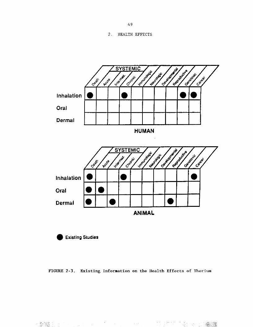
50
2. HEALTH EFFECTS
Intermediate-Duration Exposure. No studies were located regarding the effects of thorium in humans following intermediate-duration exposure by any route of exposure. Two animal studies were reported: one inhalation study in rats showing lung damage and one oral study in mice resulting in death. Intermediate-duration dermal studies in animals were not located. The lungs appear to be the target organs following intermediate-duration inhalation exposure to thorium. Oral pharmacokinetic data indicate that bone may be the target organ of toxicity following ingestion of thorium. More extensive studies by all relevant routes (inhalation, oral, dermal) would be useful in assessing both the chemical and radiological toxicity of thorium. Intermediate-duration toxicity information is important because people living near hazardous waste sites might be exposed for corresponding time periods.
Chronic-Duration Exposure and Cancer. Several studies have been reported regarding the toxic effects on workers occupationally exposed to thorium or monazite sand found in refinery dust. In these studies, effects on the lungs and chromosomes and an increased cancer incidence were reported. Because the workers were exposed to many toxic agents, however, effects cannot be attributed directly to thorium. Epidemiology studies investigating workers exposed primarily to thorium (e.g., during the production of gas lamp mantles) would be useful. No human studies were located regarding chronic oral or dermal exposure. Many studies on longterm radiological effects of thorium have been reported following intravenous injection of thorium dioxide in the form of Thorotrast in both humans and animals. Effects reported in these studies included cirrhosis of the liver, hematological disorders, and various malignancies. Studies have shown that the lungs and the hematological system are the target organ systems for thorium toxicity. Oral pharmacokinetic data indicate that bone may be the target organ of toxicity following ingestion of thorium. Chronic studies by relevant routes of exposure, inhalation and oral, are important because people living near hazardous waste sites might be exposed to thorium for years.
Studies in workers occupationally exposed to thorium have reported an increase in the incidence of pancreatic, lung and hematopoietic cancers. These effects were observed in workers exposed to many toxic agents, so they cannot be attribu.ted directly to thorium. Intermediate duration inhalation exposure of rats to thorium dioxide resulted in lung tumors. No data were located regarding the carcinogenic effects of oral or dermal thorium exposure in humans or animals. Further chronic exposure studies by all relevant routes of exposure (inhalation, oral, dermal) using wider exposure level ranges and a number of species of animals may be useful in assessing the carcinogenic potential of thorium in humans.
Genotoxicity. Occupationally exposed workers as well as patients injected with Thorotrast show chromosomal abnormalities in their lymphocytes. No in vivo genotoxicity studies in animals or in vitro studies

51
2. HFALTH EFFECTS
in bacterial strains were located, but these studies may be useful in determining whether thorium has the potential to cause gene mutations in addition to chromosomal abnormalities.
Reproductive Toxicity. No studies were located regarding the reproductive effects of thorium in humans following exposure by any route. Neither inhalation nor oral reproduction studies in animals were located. Pharmacokinetic data following inhalation or oral exposure were not located to allow the prediction of possible reproductive effects. One dermal rat study found testicular effects after administration directly onto the scrotal skin. Additional inhalation, oral, and dermal reproduction studies and multigenerational studies would be helpful in assessing the potential risk to humans.
Developmental Toxicity. No studies were located regarding the developmental effects of thorium in humans or animals following exposure by any route. Also, pharmacokinetic data do not exist that may predict whether thorium crosses the placental barrier. Further developmental studies in animals by all relevant routes of exposure may clarify the potential developmental effects of thorium in humans.
Immunotoxicity. No studies were located regarding the immunological effects of thorium in humans or animals following any relevant route of exposure (inhalation, oral, dermal). One report, however, showed intraperitoneal and intravenous injection of thorium dioxide in mice resulted in a suppression of the immune response. Studies on the immunotoxic effects of thorium, both histopathological and effects on the immune response, by all relevant routes of exposure in animals may determine the potential immunotoxic effects in humans.
Neurotoxicity. No studies were located regarding the neurological effects of thorium in humans or animals following exposure by any route. Other metals, such as lead, however, have been shown to have more severe neurological effects on children than adults; therefore, it is possible that children may be more susceptible than adults to the effects of thorium. Studies on the neurological effects of thorium, both histopathological and effects on behavior by all relevant routes of exposure in animals, may determine the potential neurological effects in humans.
Epidemiological and Human Dosimetry Studies. Epidemiology studies have investigated the relationship between long-term exposure to thorium and systemic effects, genotoxic effects, and cancer in humans. The authors of these studies found increases in respiratory disease and certain types of cancer (lung, pancreatic, hematopoietic) in exposed thorium workers, but the findings were not definitive. The existing epidemiological studies are often weakened by not sufficiently accounting for smoking habits or exposure to other chemicals and by relying too heavily on the accuracy of death certificates. Increased incidences of chromosomal abnormalities were found in exposed workers (approximately 4% dicentric in controls vs. 20% in
52
2. HEALTH EFFECTS
exposed workers) as well as in patients injected intravenously with colloidal thorium dioxide (Thorotrast) (l-25 dicentric per 100 cells). The occupational studies focus primarily on adult males. It would be useful to study groups that include women, children, and neonates that have been exposed to greater than normal levels of thorium to determine their level of susceptibility. Epidemiology studies investigating workers exposed primarily to thorium (e.g., during the production of gas lamp mantles) would also be useful. Further studies assessing the cause/effect relationship between thorium exposure and human health effects would be helpful in monitoring individuals living near a hazardous waste site.
Biomarkers of Exposure and Effect. The major route of excretion of inhaled or ingested thorium is in the feces. Exposure to thorium can be determined by measurement of thorium and/or its daughters in the feces, urine, blood, or expired air. The body burden of thorium may be estimated by the measurement of external gamma rays emitted from thorium daughters in the body. Further studies correlating thorium exposure with thorium and/or thorium daughters in the urine, feces, blood, and expired air would be helpful in more accurately quantifying thorium exposure.
No relationship was found between the measured body burden of thorium and complete blood count parameters (e.g., hemoglobin, red and white blood cells) in humans occupationally exposed to thorium (see Section 2.2.1.2). Further studies may reveal thorium-specific biomarkers that may alert health professionals to thorium exposure before toxicological effects occur.
Absorption, Distribution, Metabolism and Excretion. The absorption of thorium from the lungs and the gastrointestinal tract and the tissue distribution of thorium have been studied in both humans and animals. Inhalation was found to be the major route of exposure with gastrointestinal absorption being very low (see Section 2.3.1). The data in humans correlate well with the animal data. The excretion of systemic thorium in humans has not been extensively studied, especially the partition between feces and urine, and work in this area in both humans and animals would be helpful. No studies were located regarding the pharmacokinetics in humans or animals following dermal exposure to thorium. Studies on the dermal route of exposure may be helpful in determining whether thorium is a human health hazard by this route.

53
2. HEALTH EFFECTS
2.8.3. On-going Studies
No on-going studies were located relating to the toxicity of thorium in humans or animals.


55
3. CHEMICAL AND PHYSICAL INFORMATIONS
3.1 CHEMICAL IDENTITY
Data pertaining to the chemical identity (that is, the common terms or smbols used for the identification of the element) of thorium are listed in Table 3-1.
3.2 PHYSICAL AND CHEMICAL PROPERTIES
The physical and chemical properties of elemental thorium and a few rpresentative water soluble and insoluble thorium componds are presented in Table 3-2. Water soluble thorium compounds include the chloride, fluoride, nitrate, and sulfate salts (Weast 1983). These compounds dissolve fairly readily in water. Soluble thorium compounds, as a class, have greater bioavailability than the insoluble thorium compounds. Water Insoluble thorium compounds include the dioxide, carbonate, hydroxide, oxalate, and phosphate salts. Thorium carbonate is soluble in concentrated sodium carbonate (West 1983). Thorium metal and several of its compounds are commercially available. No general specifications for commercially prepared thorium metal or compounds have been established. Manufactures prepare thorium products according to contractual specifications (Hedrick 1985)
Thorium is a metallic element of the actinide series. It exists in several isotopic forms. The isotope thorium-232 is a naturally occuring element that is radioactive. Ti decays through the emission of a series of alpha and beta particles, gamma radiation, and the formation of daughter products, finally yielding the stable isotope of lead, lead-208. The decay series of thorium-232, together with that of uranium-238 and uranium-235, are shown in Figure 3-1. It can be seen from Figure 3-1 that the isotopes thorium-234 and thorium-230 are produced during the decay of naturally occuring uranium-238, the isotope thorium-228 diring the decay of thorium-232, and the isotopes thorium-231 and thorium-227 during the decay of naturally occuring uranium-235. Of these naturally produced isotopes of thorium, only thorium-232, thorium-230, and thorium-228 have long enough half-lives to be environmentally significant. More than 99.99% of natural thorium is thorium-232; the rest is thorium-230 and thorium-228.
Including artifically produced isotopes, there are 12 isotopes of Thorium with atomic masses ranging from 223 to 234. All are radioactive and Decay with the emission of alpha or beta particles and/or gamma radiation (West 1983). The percent occurrence and the energies of the major alpha and beta particles emitted by these isotopes are shown in Table 3-3. In general, the alpha particles are more intensely ionizing and less penetrating than the beta particles. The gamma radiation is the most penetrating of the three, but it has the least ionizing intensity. Alpha particles do not penetrate external skin to a sufficient depth to produce biological damage due to the protective effect of the epidermis. However, alpha particles emitted from thorium deposited in the lung are able to penetrate lung tissue and produce adverse biological damage since the

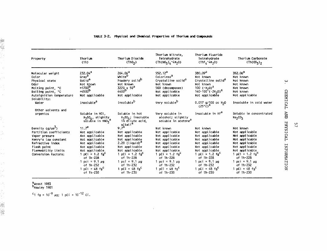


61
3. CHEMICAL AND PHYSICAL INFORMATION
protective coating of the lung tissue is very thin. In turn, beta particles are able to penetrate the skin to a sufficient depth to cause biological effects in the skin just below the epidermis. Likewise, they penetrate lung tissues to a greater depth. Gamma rays can generally pass through all tissue and interact with tissue at any depth.
Alpha particles give up all of their energy in a very short distance and, hence, produce ionization. Beta particles produce less dense ionization, and gamma rays produce less yet. In general, the severity of biological effects of exposures to ionizing radiations is proportional to the density of the ionization produced by their passage through tissue.
Finely divided thorium metal is pyrophoric in air, and thorium ribbon burns in air to give the oxide. The metal also reacts vigorously with hydrogen, nitrogen, the halogens, and sulfur. Thorium compounds are stable in +4 oxidation state (Katzin 1983). Details of thorium chemistry are given by Katzin (1983).

63
4. PRODUCTION, IMPORT, USE, AND DISPOSAL
4.1 PRODUCTION
The principal source of thorium is monazite (phosphate of rare earth metals, usually with thorium), a mineral produced as a by-product of mineral sands mined for titanium and zirconium. Thorium compounds are extracted from monazite by acid and alkali treatment processes (Hedrick 1985). Associated Minerals, a subsidiary of the Australian-owned firm Associated Minerals Consolidated Ltd., was the only commercial operation in the United States to produce purified monazite in 1987. This company produced monazite as a by-product of mineral sands mined for titanium and zirconium minerals at Green Cove Springs, FL. The monazite produced in the United States was exported. Thorium products used domestically were obtained from imported material, existing company stocks, and thorium nitrate previously released from the National Defense Stockpile (Hedrick 1987). In 1984, the mine production capacity for thorium in the United States was 20 metric tons of thorium oxide equivalent (Hedrick 1985). Actual mine production data have not been released over the years to avoid disclosure of proprietary information. Nevertheless, the domestic mine production volume of monazite or other thorium ore is expected to be approximately the same as the United States export volume. The principal processors of thorium-containing ores in the United States during 1987 were W.R Grace & Co. in Chattanooga, TN, and Rhone-Poulenc Inc. in Freeport, TX (Hedrick 1987). United States companies that have thorium processing and fabricating capacities are listed in Table 4-1.
4.2 IMPORT
Imports of thorium into the United States in metric tons of thorium oxide equivalent were 45.8 in 1983, 45.4 in 1984, 69.3 in 1985, 19.7 in 1986, and 30.7 in 1987. Additionally, concentrated monazite containing 350550 tons of Th02 has been imported annually (Hedrick 1987). Imports of thorium by the United States may decrease as a result of increased costs of processing thorium. These increased costs are primarily due to increasing concerns about the radiological risks of handling, storing, and disposing of thorium, thereby encouraging the search for nonradioactive substitutes (Hedrick 1987). Exports of thorium metal, waste, and scrap from the United States in metric tons of thorium oxide equivalent were 1.1 in 1983, 1.0 in 1984, 1.6 in 1985, 17.0 in 1986, and 20.4 in 1987 (Hedrick 1987).
4.3 USE
Thorium can be used as fuel in the generation of nuclear energy. However, there is currently only one plant in the United States that is using thorium for the production of energy (Hedrick 1987). In 1983, 3 metric tons of thorium oxide equivalent were used for energy uses in the United States (Hedrick 1985). Nonenergy uses accounted for almost all of the thorium used in the United States during 1987. The 1987 use pattern for thorium was as follows: refractory applications (57%); lamp mantles (18%); aerospace alloys (15%); welding electrodes (5%); nuclear weapon


65
4. PRODUCTION, IMPORT, USE, AND DISPOSAL
production; and other applications including ceramics and special use lighting (5%). Specific applications include production of investment molds for casting high-temperature metals and alloys, crucibles and alloys of special shapes for use in high-temperature vacuum or oxidizing furnaces. Other special applications include production of core-retention beds used in nuclear reactors to contain and possibly diffuse heat generated by accidental core meltdown; magnesium-thorium alloys for strategic aircraft such as military jet fighters and bombers; mantles for incandescent lanterns such as those used on camping trips; thoriated tungsten electrodes used to join stainless steels and other alloys which require controlled weld applications; special lighting such as airport runway lighting; computer memory components; photoconductive film; and target material for x-rays (Hedrick 1985). Natural thorium is also used in ceramic tableware glaze and in flints for lighters (UNSCEAR 1977). Domestic nonenergy thorium consumption was estimated to be 39.4 metric tons of thorium oxide equivalent in 1987, a decrease of 33 metric tons from 1986 usage. The drop in consumption was primarily the result of reduced demand for thorium oxide in high-temperature refractory molds, because suitable substitutes had been developed (Hedrick 1987).
4.4 DISPOSAL
Disposal of radioactive wastes is a serious environmental problem for which there is, as yet, no completely satisfactory solution. Intensive research is being conducted by both government and industry for the disposal of this type of waste. Small amounts of low-level wastes containing radioisotopes can be diluted with an inert material sufficiently to reduce its activity to an acceptable level for further storage or disposal. At one nuclear waste disposal site, high-level reactor wastes are stored in concrete tanks lined with steel which are buried under a foot of concrete and 5-6 feet of soil. Use of compressed alumina (corundum) containers has been recommended, since this material remains impervious to water indefinitely. The Department of Energy has recommended disposal in deep geologic formations. Disposal in salt formations is being considered since they are self-sealing and free from water (Hawley 1981). The Department of Defense Authorization Act, 1987 (Public Law 99-661) authorized 4536 kg (10,000 pounds) of thorium nitrate for disposal in fiscal year 1987. Further information regarding the amount of thorium disposed of in the United States was not located. Regulations established by the Environmental Protection Agency regarding release limits which apply to the storage and disposal of spent nuclear fuel, high-level radioactive wastes, and transuranic radioactive wastes can be found in 40 CFR 191 and 40 CFR 192.


67
5. POTENTIAL FOR HUMAN EXPOSURE
5.1 OVERVIEW
Thorium is ubiquitous in our environment. Release of thorium to the atmosphere can occur both from natural and anthropogenic sources, and emissions from the latter sources can produce locally elevated atmospheric levels of thorium over the background. Windblown terrestrial dust and volcanic eruptions are two important natural sources of thorium in the air (Fruchter et al. 1980; Kuroda et al. 1987). Uranium and thorium mining, milling and processing, tin processing, phosphate rock processing and phosphate fertilizer production, and coal fired utilities and industrial boilers are the primary anthropogenic sources of thorium in the atmosphere (Hu and Kandaiya 1985; McNabb et al. 1979; Nakoaka et al. 1984; Sill 1977). The major industrial releases of thorium to surface waters are effluent discharges from uranium and thorium mining, milling and processing, tin processing, phosphate rock processing and phosphate fertilizer production facilities (Hart et al. 1986; McKee et al. 1987; Moffett and Tellier 1978; Platford and Joshi 1988). The primary sources of thorium at the Superfund sites are perhaps from the processing and extraction of thorium, uranium, and radium from ores and concentrates (EPA 1988a). At this time, elevated levels (higher than background) of thorium have been found at 16 out of 1177 National Priority List (NPL) hazardous waste sites in the United States (VIEW Database 1989). The frequency of these sites within the United States can be seen in Figure 5-l.
Data regarding the fate and transport of thorium in the air are limited. Wet and dry deposition are expected to be mechanisms for removal of atmospheric thorium. The rate of deposition will depend on the meteorological conditions, the particle size and density, and the chemical form of thorium particles. Although atmospheric residence times for thorium and compounds were not located, judging from residence times of other metals (e.g., lead) and their compounds, they are likely to be a few days. Thorium particles with small aerodynamic diameters (<10 micron aerodynamic diameter) will travel long distances from their sources of emission. In water, thorium will be present in suspended matters and sediment and the concentration of soluble thorium will be low (Platford and Joshi 1987). Sediment resuspension and mixing may control the transport of particlesorbed thorium in water. The concentration of dissolved thorium in some waters may increase due to formation of soluble complexes with carbonate, humic materials, or other ligands in the water (LaFlamme and Murray 1987). Thorium has been found to show significant bioconcentration in lower trophic animals in water, but the bioconcentration factors decrease as the trophic level of aquatic animals increases (Poston 1982; Fisher et al. 1987). The fate and mobility of thorium in soil will be governed by the same principles as in water, In most cases, thorium will remain strongly sorbed to soil and its mobility will be very slow (Torstenfelt 1986). However, leaching into groundwater is possible in some soils with low sorption capacity and the ability to form soluble complexes. The plant/soil transfer ratio for thorium is less than 0.01 (Garten 1978), indicating that it will not bioconcentrate in plants from soil. However, plants grown at the edge of


69
5. POTENTIAL FOR HUMAN EXPOSURE
impoundments of uranium tailings containing elevated levels of thorium had a plant/soil concentration ratio of about 3 (Ibrahim and Whicker 1988).
The atmospheric mass concentration of thorium ranged from 0.2-1.0 ng/m3, with a mean value of 0.3 ng/m3 in air samples collected from 250 sites in the United States (Lambert and Wilshire 1979). In another study, the mean activity concentrations of thorium-228, thorium-230, and thorium232 in New York City air were 36 aCi/m3 (aCi = l0-l8 Ci), 36 aCi/m3, and 37 aCi/m3, respectively (Wrenn et al. 1981). The average population-weighted concentrations of thorium-232 and thorium-230 in United States community water supplies derived both from surface and groundwater were less than 0.01 pCi/L and less than 0.04 pCi/L, respectively (Cothern 1987; Cothern et al. 1986). The maximum concentration of thorium-232 in several fruits, vegetables and other type of foods from New York City was reported to be less than 0.01 pCi/g (Fisenne et al. 1987). The daily intakes of thorium230 and thorium-232 for residents of New York City were estimated to be 0.17 and 0.11 pCi, respectively. Significant exposure to thorium requires special exposure scenarios (Fisenne et al. 1987). People who consume foods grown in high background areas, reside in homes with high thorium background levels, or live near radioactive waste disposal sites may be exposed to higher than normal background levels of thorium. Workers in uranium, thorium, tin, and phosphate mining, milling, and processing industries, and gas mantle manufacture may also be exposed to higher than normal background levels of thorium (Bulman 1976; Hannibal 1982; Hu et al. 1984; Kotrappa et al. 1976; Metzger et al. 1980).
5.2 RELEASES TO THE ENVIRONMENT
5.2.1 Air
Releases of thorium to the atmosphere can occur from both natural and anthropogenic sources. The release of thorium in volcanic ash containing as much as 0.116 pCi/g (1.06 µg/g) of thorium-232 was reported by Fruchter et al. (1980). Increased concentrations of thorium in rain water following a volcanic eruption have also been observed (Kuroda et al. 1987). Since the average level of thorium in soil is about 6 µg/g of thorium (Harmsen and De Haan 1980), windblown terrestrial dust is also a likely natural source of thorium in the atmosphere. Since coal contains 0.5-7.3 µg/g thorium (Nakaoka et al. 1984), burning of coal for power generation produces thorium in the fly ash and is a manmade source of this chemical in the atmosphere. The amount of thorium in the fly ash from coal-burning power plants depends on the nature of coal burned and the emission control devices of the plant, but concentrations usually range from 4.5-37 µg/g (Abel et al. 1984; Coles et al. 1979; Tadmor 1986; Weissman et al. 1983). However, the concentrations of all natural radioactive isotopes in (including thorium isotopes) the stack effluents from coal-fired power plants are usually much lower than those from the natural background concentrations of these radionuclides (Nakaoka et al. 1984; Roeck et al. 1987). Similarly, fly ash

70
5. POTENTIAL FOR HUMAN EXPOSURE
from oil- and peat-fired power plants can also be atmospheric sources of thorium (Mustonen and Jantunen 1985).
Thorium-230 has been detected in air dust from uranium ore processing and mill tailings. These concentrations of thorium-230 (a decay product of uranium-238) may be particularly high in ore crushing areas (Sill 1977). Similarly, processing of thorium ores is expected to be an atmospheric source of thorium. Elevated levels of thoron (thoron or radon-220 originating from thorium-232) daughters, such as bismuth-212 and polonium216, were present at a former thorium and rare-earth extraction facility waste site, although the concentrations of thorium in air particulate samples were not significant (Jensen et al. 1984). Since phosphate ores usually contain thorium-230, phosphate-ore processing plants are also atmospheric sources of thorium-230 (Metzger et al. 1980; McNabb et al. 1979). The by-products obtained during processing of tin ores usually contain thorium-232. Therefore, tin processing industries are sources of atmospheric thorium-232 emissions (Hu et al. 1981, 1984; Hu and Kandaiya 1985).
EPA (1984) estimated that about 0.2 Ci of thorium-230 is annually emitted into the air from uranium mill facilities, coal-fired utilities and industrial boilers, phosphate rock processing and wet-process fertilizer production facilities, and other mineral extraction and processing facilities. About 0.084 Ci of thorium-234 from uranium fuel cycle facilities and 0.0003 Ci of thorium-232 from underground uranium mines are emitted into the atmosphere annually (EPA 1984).
5.2.2 Water
The acidic leaching of uranium tailing piles in certain areas is a source of thorium-230 in surface water and groundwater (Moffett and Tellier 1978; Platford and Joshi 1988). The contamination of surface waters and benthic organisms by thorium-230 (a decay product of uranium-238) from uranium mining and milling operations and from radium and uranium recovery plants has been reported (Hart et al. 1986; McKee et al. 1987). Similarly, effluents from thorium mining, milling, and recovery plants are expected to be sources of thorium in water. Other industrial processes that are expected to be sources of thorium contamination into water are phosphorus and phosphate fertilizer production and processing of some tin ores. Since both phosphate rocks and the tailings from tin ore processing contain thorium mainly as thorium-230 and thorium-232, respectively (see Section 5.2.1), discharges of processed or unprocessed effluents and leaching from tailing piles can be sources of thorium in water. Leaching from landfill sites containing uranium and thorium may result in the contamination of surface water and groundwater with thorium (Cottrell et al. 1981).

71
5. POTENTIAL FOR HUMAN EXPOSURE
5.2.3 Soil
Thorium occurs naturally in the earth's crust at an average lithospheric concentration of 8-12 µg/g (ppm). The typical concentration range of naturally-occurring thorium in soil is 2-12 µg/g, with an average value of 6 µg/g (Harmsen and De Haan 1980). Manmade sources of thorium contamination in soil are mining, milling and processing operations and uranium, thorium, tin and phosphate fertilizer production (Chong et al. 1985; Hu and Kandaiya 1985; Joshi 1987; McNabb et al. 1979; Sill 1977). The two principal processes that can contaminate soil from these industries are precipitation of airborne dusts and land disposal of uranium or thoriumcontaining wastes.
According to EPA (1988a), the primary sources of thorium at the Superfund sites are processing and extraction of thorium, uranium and radium from ores or ore-concentrates. The following radioactive waste Superfund sites have been found to contain one or more isotopes of thorium (VIEW 1989): Shpack and adjacent landfills, Norton, MA; Maywood Chemical Co., Sears Property, Maywood, NJ; W.R.Grace and Co., Wayne, NJ; West Chicago Sewage Treatment Plant, W. Chicago, IL; Reed-Keppler Park, West Chicago, IL; Kerr-McGee (Residential Areas), W. Chicago, IL; Kress Creek and the West Branch of the DuPage River, W. Chicago, IL; United Nuclear Corp., Church Rock, NM; Homestake Mining Co., Milan, NM; Kearsarge Metallurgical Corp., Conway, NH; Naval Air Engineering Center, Lakehurst, NJ: Teledyne Wah Chang, Albany, OR; Woodland Route 72 Dump, Woodland Township, NJ; Weldon Spring Quarry, St. Charles City, MO; Monticello Radioactivity-Contaminated Properties, Monticello, UT; Uravan Uranium Project, Montrose City, CO. Disposal of incandescent lights and lanterns containing thorium-232 will be an additional source of thorium at waste disposal sites.
5.3 ENVIRONMETAL FATE
Thorium occurs in nature in four isotopic forms, thorium-228, thorium230, thorium-232, and thorium-234. Of these, thorium-228 is the decay product of naturally-occurring thorium-232, and both thorium-234 and thorium-230 are decay products of natural uranium-238. To assess the environmental fate of thorium, these isotopes of thorium with the exception of thorium-234 which has short half-life (24.1 days), should be considered.
5.3.1 Transport and Partitioning
Data regarding the transport and partitioning of thorium in the atmosphere are limited. Release of atmospheric thorium from mining, milling, and processing operations of thorium will mainly consist of thorium-232 particulate matter. Emissions from mining, milling, and processing of uranium and the airblown dust from uranium tailing piles wil contribute to the presence of thorium-230 as an atmospheric particulate aerosol. The aerodynamic diameters of both thorium-230 and thorium-232 in atmospheric aerosols are greater than 2.5 µm. The aerodynamic diameter of
72
5. POTENTIAL FOR HUMAN EXPOSURE
thorium-228, however, is less than 1.6 pm (Hirose and Sugimura 1987) and may travel longer distances than both thorium-230 and thorium-232. Like other particulate matter in the atmosphere, thorium will be transported from the atmosphere to soil and water by wet and dry deposition.
The deposition of thorium through snow and rain water has been observed (Jiang and Kuroda 1987). Dry deposition of thorium through impaction and gravitational settling has also been observed (see Section 5.2.3). The atmospheric residence time of thorium depends on the aerodynamic diameter of the particles. Those with small diameters are likely to be transported longer distances. For example, high thorium-228/thorium-232 activity ratios observed in surface air of the Western North Pacific Ocean are thought to be due to long distance transport of small particles of thorium-228 (Hirose and Sugimura 1987).
The dry deposition velocity of lead-212, a thoron (thoron or radon-220 itself originating from thorium-232) decay product has been reported to be in the range 0.03-0.6 cm/set (Bigu 1985; Rangarajan et al. 1986). These low deposition velocities indicate that the thoron daughter, stable lead, may have a long residence time in the atmosphere with respect to dry deposition.
Thorium discharged as Th02 into surface waters from mining, milling, and processing will be present as suspended particles or sediments in water because of the low solubility of thorium in water (Platford and Joshi 1986). Other soluble thorium ions will hydrolyze at pH above 5 forming Th(OH)4 precipitate or hydroxy complexes, e.g., Th(OH)2+2, Th2(0H)2
+6, Th3(OH)5+7
(Bodek et al. 1988; Hunter et al. 1988; Milic and Suranji 1982). The hydroxy complexes will be adsorbed by particulate matter in water, e.g., goethite (alpha-FeOOH), with the result that most of the thorium will be present in suspended matter or sediment, and the concentration of soluble thorium in water will be low (Hunter et al. 1988; Sheppard 1980). The adsorption of thorium to suspended particles or sediment in water depends un the particle size, and the adsorption and subsequent removal from aqueous phase is expected to be higher for finer grained particles (Carpenter et al. 1987). The residence times for thorium with respect to removal by adsorption onto particles were reported to be shorter in nearshore waters than in deeper waters, probably because of the availability of more adsorbents (particulate matter). The residence time may vary from 1 day to 70 days (Cochran 1984). The scavenging rate varied seasonally and was inversely related to the sediment resuspension rate. Therefore, the removal rate was found to be dependent on both sediment resuspension rate and the concentration of iron and manganese compounds (good adsorption properties) in water (Cochran 1984).
The transport of thorium in water is principally controlled by the particle flux in the water, i.e., most of the thorium will be carried in the particle-sorbed state (Santschi 1984), and sediment resuspension and mixing may control the transport of particle-sorbed thorium in water (Santschi
73
5. POTENTIAL FOR HUMAN EXPOSURE
et al. 1983). Although the concentration of dissolved thorium is low in most waters, its value could be higher in some waters. For example, the concentration of dissolved thorium in an alkaline lake was up to 4.9 dpm/L (2.21 pCi/L) compared to about 1.3x10⁻⁵ dpm/L (O.59x10-5 pCi/L) in sea water (LaFlanune and Murray 1987). The dissolved thorium concentration can increase by the formation of soluble complexes. The anions or ligands likely to form complexes with thorium in natural water are C03
-2 and humic materials, although some of the thorium-citrate complexes may be stable at pH above 5 (LaFlamme and Murray 1987; Miekeley and Kuchler 1987; Platford and Joshi 1986; Raymond et al. 1987; Simpson et al. 1984).
The transport of thorium from water to aquatic species has been reported. The bioconcentration factor (concentration in dry organism/concentration in water) (dry weight basis) in algae may be as high as 975x10+4, but the maximum value in zooplankton (calanoids and cyclopoids) may be 2x10⁴ (Fisher et al. 1987). Fisher et al. (1987) suggested that sinking plankton and their debris may account for the sedimentation of most of the thorium from oceanic surface waters. The highest observed thorium bioconcentration factor in the whole body of rainbow trout (Salmo gairdneri) was 465 (Poston 1982). The succeedingly lower bioconcentration factors in higher trophic animals indicates that thorium will not biomagnify in the aquatic environment. It was also noted that the majority of thorium body burden in fish is in the gastrointestinal tract (Poston 1982).
The mobility of thorium in soil will be governed by the same principles as in water. In most soil, thorium will remain strongly sorbed onto soil and the mobility will be very slow (Torstenfelt 1986). The presence of ions or ligands (C03
-2, humic matter) that can form soluble complexes with thorium should increase its mobility in soil. The contamination of groundwater through the transport of thorium from soil to groundwater will not occur in most soils, except soils that have low sorption characteristics and have the capability to form soluble complexes. Chelating agents produced by certain microorganisms (Pseudomonas aeruginosa) present in soils may enhance the dissolution of thorium in soils (Premuzic et al. 1985).
The transport of atmospherically deposited thorium from soil to plants is low. The soil to plant transfer coefficients (concentration in dry plant to concentration in dry soil) were estimated to be 10-4 to 7x10⁻³ by Garten (1978) and 0.6x10⁻⁴ for thorium-232 by Linsalata et al. (1989). The root systems of grasses and weeds adsorb thorium from the soil but the transport of thorium from the root to the aboveground parts of the plant is not very extensive, as indicated by l00-fold higher concentrations of all three isotopes (thorium-228, thorium-230, and thorium-232) in the root than in the aboveground parts of the plant (Taskayev et al. 1986). However, Ibrahim and Whicker (1988) showed that under certain conditions, vegetation can accumulate thorium-230, as indicated by the plant/soil concentration ratio (dry weight) of 1.9-2.9 for mixed grasses, mixed forbs and sagebrush plants

74
5. POTENTIAL FOR HUMAN EXPOSURE
grown at the edge of uranium tailings impoundments. Vegetation concentration ratios for thorium-232 (a concentration ratio of about 0.1) and thorium-228 (a maximum concentration ratio of about 0.4) were lower than that of thorium-230. It was postulated that the acidity and wet conditions at this site enhanced the solubility of thorium in soil and that the difference in solubility was responsible for the difference in plant uptake of the three thorium isotopes (Ibrahim and Whicker 1988). However, it is possible that the observed difference in the uptake of the three isotopes by plants is due to a difference in the chemical compounds formed by the isotopes, making one more leachable than the other (therefore more available for uptake) under the prevailing local conditions (also see Section 5.6).
5.3.2 Transformation and Degradation
5.3.2.1 Air
Thorium may change from one chemical species to another in the atmosphere (such as Th02 to Th(S04)2) as a result of chemical reactions, but nothing definitive is known about the atmospheric chemical reactions of thorium. The chemical forms in which thorium may reside in the atmosphere are also not known, but it is likely to be present mostly as Th02.
5.3.2.2 Water
The principal abiotic processes that may transform thorium compounds in water are complexation by anions/organic ligands and hydroxylation. The increase in the mobility of thorium through the formation of soluble
-2complexes with C03 , humic materials, and other anions or ligands and the
decrease in the mobility due to formation of Th(OH)4 or anionic thoriumhydroxide complexes were discussed in Section 5.3.1.2. In a model experiment with seawater at pH 8.2 and freshwater at pH 6 and pH 9, it was estimated that almost 100% of the thorium resides as hydroxo complexes (Boniforti 1987);
5.3.2.3 Soil
No published data were located referencing biotic transformation of thorium in soil. Abiotic transformation processes that can convert immobile thorium in soil into mobile forms through the formation of complexes were discussed in Section 5.3.1.3.
5.4 LEVELS MONITORED OR ESTIMATED IN THE ENVIRONMENT
5.4.1 Air
The level of thorium in air have not been measured as frequently as it has for uranium. The concentration of thorium in the atmosphere of the South Pole measured in 1970 ranged between 18 and 83 fg/m3, with a mean value of 59 fg/m3 (1 fg = l0-l5 g). The origin of thorium in the polar
75
5. POTENTIAL FOR HUMAN EXPOSURE
atmosphere was speculated to be either crustal weathering or the ocean water (Zoller et al. 1974). The thorium level in the air of Algonquin Park, Ontario, Canada was reported to be 7.1 pm3 (Sheppard 1980). The level of thorium measured in 1969 in East Chicago, IN, a heavily polluted industrial area, was 1.3 ng/m3 compared to a value of 0.27 ng/m3 at a rural location in Michigan (Niles, MI) (Dams et al. 1970). The air particulate samples collected from 250 sites in the United States by the National Air Surveillance Network (NASN) of EPA during 1975 and 1976 were analyzed for thorium-232 by neutron activation analysis. The measured concentrations at 250 urban and nonurban sites in the United States ranged from 0.2-1.0 ng/m3 , with a mean concentration of 0.3 ng/m3 (Lambert and Wilshire 1979). The mean concentrations of thorium-228, thorium-230 and thorium-232 in New York City air (sample collected on the roof above the 14th floor) were 36 aCi/m3
(aCi = 10-l8 Ci), 36 aCi/m3, and 37 aCi/m3, respectively (Wrenn et al. 1981).
The air concentrations of thorium and other airborne radioactivity near a former thorium and rare-earth extraction facility in the United States were measured. The maximum radioactivity due to all three isotopes of thorium at a site about 450 feet from the primary waste pile was 0.66 fCi/m3. Although the background thorium radioactivity was not reported, the total radioactivity at a site about 4000 feet south of the waste pile was about 3.5 times lower than a site 450 feet from the pile (Jensen et al. 1984).
The concentration of thorium in rainwater over Fayeteville, AR, ranged from 2.8-123 fCi/L for thorium-228, 1.7-123 fCi/L for thorium-230, and 0.8118 fCi/L for thorium-232. The peak values in thorium concentrations correlated well with the 1980 eruption of Mount St. Helen and the 1982 eruption of El Chichon (Jiang et al. 1986; Jiang and Kuroda 1987; Salaymeh and Kuroda 1987).
The natural decay of uranium-238 and thorium-232 will produce radon-222 and thoron (radon-220). The indoor air levels of radon (radon-222) and thoron (radon-220) daughters arising from some building materials and the soil, have been reported by several authors. It was generally believed that the effective dose equivalent from radon-220 (thoron) daughters (originating from thorium-232) might average about one-fifth of that due to radon-222 daughters (originating from uranium-238) in the temperate regions (Schery 1985). However, more recent measurements at varied indoor locations within the United States and Germany have shown that the potential alpha energy concentrations from radon-220 daughters may be as high as 60% of that originating from radon-222. It has also been shown that the concentrations of thoron (radon-220) and radon-222 daughters in the indoor air are dependent on the air exchange rate in the dwellings and that the indoor concentrations are about 3-4 times higher than the outdoor concentrations (Keller and Folkerts 1984; Schery 1985).
76
5. POTENTIAL FOR HUMAN EXPOSURE
5.4.2 Water
Compared to uranium, relatively less information was located on the levels of thorium in natural waters. The concentrations of dissolved thorium in water with high pH (more than 8) are expected to be very low, and the concentration may increase with the decrease of pH (Harmsen and De Haan 1980). Cothern et al. (1986) reported thorium-232 concentrations rarely exceed 0.1 pCi/L in natural waters, but that the concentrations of thorium230, a progeny of uranium-238, may be as high as 0.4 pCi/L. In a natural surface water in Austria, the concentration of thorium (isotope undefined) was reported to be 1.24-2.90 µg/L (Harmsen and De Haan 1980). The concentration of thorium (isotope undefined but probably thorium-230) in water under low pH conditions which may occur from the leaching of uranium tailings may be as high as 38 mg/L (Harmsen and De Haan 1980). The individual concentrations of thorium-228, -230, and -232 in an area of Great Bear Lake in Canada contaminated with mine wastes (silver and uranium mines) were less than 0.5 pCi/L (Moore and Sutherland 1981). The concentrations of thorium-228, -230, and -232 in a highly alkaline (pH of about 10) lake (Mono Lake) in California have been reported to be as high as 1.02, 1.41, and 0.7 pCi/L, respectively (Anderson et al. 1982; Simpson et al. 1982).
The concentrations of thorium in seawater at various depths and locations have been reported by several authors. Because of the very low concentrations of thorium and the differences in location and the varying characteristics of the water, the reported results are different. The concentration of total thorium in seawater ranges from 4x10⁻⁵ to less than 0.5 µg/kg (Greenberg and Kinston 1982; Sheppard 1980) and the world average concentration in seawater is 0.05 µg/L (Harmsen and De Haan 1980). The concentrations of the individual isotopes thorium-232, thorium-230, and thorium-228 in seawater have been reported to range from 0.00023-0.032, 0.014-0.72, and 0.023-3.153 fCi/L, respectively (Anderson et al. 1982; Hirose 1988; Huh and Bacon 1985; Livingston and Cochran 1987; Simpson et al. 1982). The concentrations of thorium in sediments are much higher than in seawater. In several sediments, concentrations of thorium-232, thorium-230, and thorium-228 ranged from 0.52-1.96, 1.01-30.77, and 0.36-1.93 pCi/g, respectively (Huh et al. 1987; Yang et al. 1986).
Thorium has also been detected in groundwaters. In groundwater in Austria, concentrations ranged from 0.5-2.90 µg/L (Harmsen and De Haan 1980). Briny groundwater from a well in Palo Duro Basin, WA, contained 0.009, 0.1, and 0.59 pCi/L of thorium-232, thorium-230, and thorium-228, respectively (Laul et al. 1987). In a California well, thorium-230 was detected at a concentration as high as 1.3 pCi/L (Aieta et al. 1987). The average population-weighted concentrations of thorium-232 and thorium-230 in United States community water supplies derived from both surface water and groundwater are less than 0.01 pCi/L and less than 0.04 pCi/L, respectively (Cothern 1987; Cothern et al. 1986).
77
5. POTENTIAL FOR HUMAN EXPOSURE
5.4.3 Soil
The typical concentration range of thorium in soil is 2-12 µg/g (ppm), with an average value of 6 µg/g (Harmsen and De Haan 1980). The thorium content of soil normally increases with an increase in clay content of soil (Harmsen and De Haan 1980). The thorium contents in most soils from the Superfund sites listed in Section 5.2.3 were above background levels. The soil concentrations of thorium-232 at the Reed-Keppler Park, W. Chicago, IL, site and the Kerr-McGee Residential areas in W. Chicago, IL, were 11,000 and 16,000 pCi/g, respectively (EPA 1988a). Soils near processing and milling operations, and concentrations of uranium and thorium ores, phosphate ores, and tin ores may contain thorium at concentrations higher than the background levels. Higher concentrations of thorium in soils near uranium ore crushing facilities have been reported (Jensen et al. 1984; Sill 1977).
5.4.4 Other Media
Because concentrations of thorium in foods are very low, very few data exist. The thorium-232 content in fresh fruits, vegetables, and tea was determined (in pCi/g), and the values are listed in Table 5-l. Vegetables grown in an area of high natural activity in Brazil had the following concentrations of thorium (µg/g in dry sample) (Linsalata et al. 1987): brown beans, 0.011; potato, 0.0019; zucchini, 0.011; corn, 0.0022; carrot, 0.0074; and sweet potato, 0.0027. These authors did not observe rapid transport of thorium-232 from soil to the edible parts of the plants.
The concentrations of thorium in both hard and soft tissues of humans have been determined by a few authors. The concentration of thorium-232 in the blood of normal populations (not occupationally or otherwise known to be exposed to levels higher than background level of thorium) in the United Kingdom was 2.42 µg/L. The thorium-232 level in the urine of the same population was below the detection limit of 0.001 µg/L, although the concentration in the urine of exposed workers ranged from less than 0.001-2.24 µg/L. The highest value (2.24 µg/L) was found in a worker in the thorium nitrate gas mantle industry (Bulman 1976; Clifton et al. 1971).
The thorium-232 concentration in rib bones from several control humans from the United States ranged from less than 0.1-72 ng/g (ppb) and were found to increase-with age (Lucas et al. 1970). A similar increase in thorium concentration with age was seen in bones (primarily vertebral wedges) of a Colorado population (Wrenn et al. 1981). The level of thorium232 in rib bones of individuals in the United Kingdom not occupationally exposed to thorium ranged from 0.8-163.8 ng/g, with a mean value of 28.7 ng/g in dry ash (Clifton et al. 1971). The concentration of thorium in the fibula of a Thorotrast patient was reported to be 2.0 µg/g (ppm) (Edgington 1967). Singh et al. (1985) reported more recent measurements of isotopic concentrations of thorium in different human bones from the general population of Colorado and Pennsylvania. These values are shown in Table 5-2. The authors concluded that the concentrations of thorium-230 in ribs
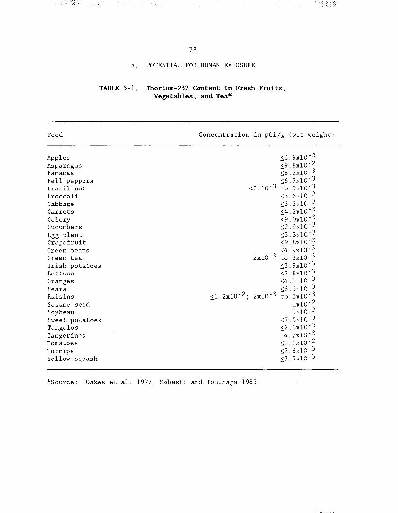


80
5. POTENTIAL FOR HUMAN EXPOSURE
of the Colorado population were significantly higher (statistically), probably because of exposure to uranium tailings, than those from the Pennsylvania population.
The levels of thorium in the tissues of a hard-rock miner, a uranium miner, and the levels in two uranium millers (thorium-230 is a decay product of uranium-238, and thorium-238 and thorium-232 are impurities in uranium) were compared with the levels in the 50th percentile for the general population (Singh et al. 1987; Wrenn et al. 1981). These data are given in Table 5-3. The levels of thorium-230 in the hard-rock miner were about 10 times higher than the median levels in most tissues of the general population. In the case of the uranium miner and millers, the values were more than two orders of magnitude higher than the median tissues levels in the general population.
Wrenn et al. (1981) determined the median concentrations of thorium228, thorium-230, and thorium-232 in the lungs of smokers and nonsmokers; the respective values were 0.22, 0.56, and 0.43 pCi/kg for smokers and 0.37, 0.84, and 0.60 pCi/kg for nonsmokers. The investigators concluded that cigarette smoking had no effect relative to increasing the concentration of thorium isotopes in lungs.
5.5 GENERAL POPULATION AND OCCUPATIONAL EXPOSURE
The general population will be exposed to thorium through the inhalation of air and ingestion of food and drinking water containing trace amounts of the chemical. Because the concentration of thorium is normally very low in air, drinking water, and foods (see Section 5.4), few studies were located that determined the daily human intake of thorium. According to Cothern (1987), the estimated daily intakes of thorium-230 in the United States population through inhalation of air and ingestion of drinking water are 0.0007 and less than 0.06 pCi, respectively. The corresponding values for thorium-232 are 0.0007 and less than 0.02 pCi. Cothern (1987) assumed that the intake from food would be negligible. Based on these values, the total daily intakes of thorium-230 and thorium-232 are expected to be less than 0.06 and less than 0.02 pCi, respectively. However, other authors estimated the contribution of food to the total human thorium intake may not be negligible and may be the most significant. Based on a survey of the levels of thorium in air, water, and food, Fisenne et al. (1987) estimated the daily intake of thorium-230 and thorium-232 by New York City residents. The daily dietary, water, and inhalation intake of thorium-230 was estimated to be 0.164, 0.005, and 0.0003 pCi, respectively, giving a total daily intake of 0.17 pCi. The corresponding estimated values for thorium-232 are 0.110, 0.002, and 0.0002 pCi, with a total daily intake being 0.112 pCi. From the measured values of thorium in feces and the assumed values for uptake and elimination rates, Linsalata et al. (1985) estimated a daily ingestion intake of thorium-232 for New York residents to be about 0.08 pCi or 0.7 µg. This value is considerably smaller than the value estimated by Fisenne et al. (1987). The value from Linsalata et al. (1985) is again
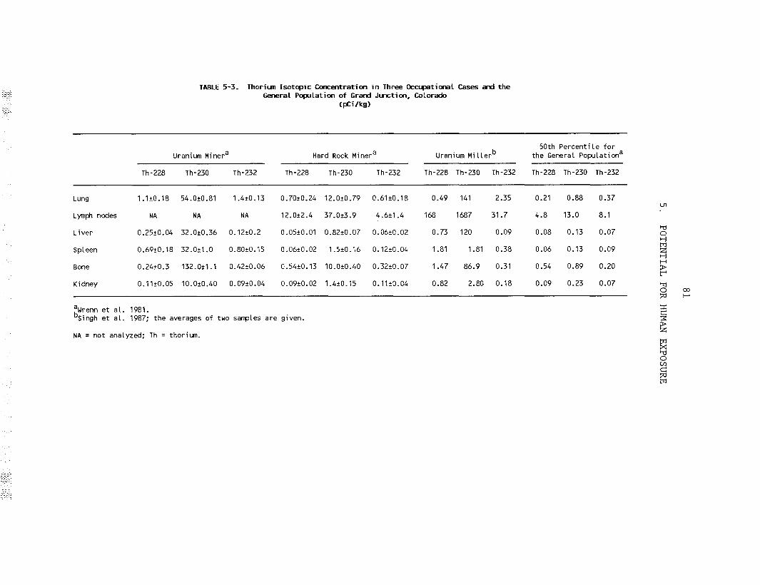

82
5. POTENTIAL FOR HUMAN EXPOSURE
considerably smaller than the daily dietary, water, and inhalation intakes of 2.24, 0.02, and 0.02 µg, respectively, as estimated for residents of Bombay, India (Dang et al. 1986). It can be concluded from the above discussion that the total intake of thorium by the United States population may vary depending on the thorium content in the consumed food and that no firm United States average thorium intake value is yet available. The importance of the intake of thorium from foods is overshadowed by the relative absorption of thorium by lung compared with its uptake by gut (see Chapter 2).
Occupational exposures to higher levels of thorium isotopes occur primarily to workers in uranium, thorium, tin, and phosphate mining, milling, and processing industries, radium dial workers, and gas lantern mantle workers. From the measurement of airborne thorium concentrations in workplaces of the uranium and thorium industry, it was concluded that radioactive dust, particularly from crushing areas, represents an important route of exposure (Hannibal 1982; Kotrappa et al. 1976). It has also been reported that exposure of workers in the fertilizer industry to natural radioactivity may increase by 100% over normal background (Metzger et al. 1980). Measuring external gamma radiation dosages to a person working 8 hours/day has shown that monazite and xenotime storage rooms of Amang upgrading plants (tin processing) on the west coast of Malaysia exhibited exposure rates exceeding the ICRP recommended maximum value of 5 rem/year (Hu et al. 1984). From the radioactivity released by a burning gas mantle (contains thorium), it was concluded that the user would be at minimal risk unless the person was in a small unventilated room (Leutzelschwab and Googins 1984). However, workers in the gas mantle manufacturing industry are expected to be exposed to higher concentrations of radioactivity than the normal population.
Workers are exposed to higher levels of thorium and other radionuclides in certain thorium industries, as indicated by the measured exhaled breath and tissue levels of these chemicals. The significantly higher level of radon-220 (a decay product of thorium-232) in the exhaled breath of some thorium plant workers (Mayya et al. 1986) is indirect evidence of higher thorium intakes. Similarly, other authors have found higher tissue and body fluid levels (compared to background) of thorium in workers in the thoriumprocessing industry (Clifton et al. 1971; Mausner 1982; Twitty and Boback 1970), workers in the radium dial industry (Keane et al. 1986), in uranium mill crushermen (Fisher et al. 1983), and in uranium and hard rock miners and uranium millers (Singh et al. 1987; Wrenn et al. 1981).
Thorium-doped glass is also used in the production of some camera lenses (Waligorski et al. 1985). A relatively recent measurement has shown that the external dose rate from exposure to a camera lens can be 10 times higher (as high as 9.25 mrem/hour at the front glass surface of the lens) than previously reported (Waligorski et al. 1985). Therefore, professional photographers and workers in the thorium-doped photographic lens
83
5. POTENTIAL FOR HUMAN EXPOSURE
manufacturing industry may be at slightly higher risk of exposure to thorium and its daughter products from inhalation and/or external radiation.
5.6 POPULATIONS WITH POTENTIALLY HIGH EXPOSURE
The three groups of the general population that have the potential of exposure to thorium and its decay products at levels higher than background are people who consume large amounts of foods grown in high background areas, people who reside in homes built with high thoron (radon-220)emitting building materials and constructed on soil with high background levels of thorium, and people who live near radioactive waste disposal sites. Linsalata et al. (1987) analyzed vegetables grown in two areas near Sao Paulo, Brazil, that contained high natural radioactivity, and found the thorium is not bioaccumulated in the vegetables but maintained a mean concentration ratio (concentration in dry vegetable/concentration in dry soil) of 10-4. Root vegetables (e.g., carrots and potatoes) showed lower concentration ratios than zucchini and beans. Therefore, it can be concluded that vegetables grown in these soils would contain more thorium than vegetables grown in soil with normal background levels.
Linsalata et al. (1985) 1 a so estimated that the intake of thorium by populations residing in these parts of Brazil was 6-10 times higher than the population in New York City, as indicated by the analysis of human bones from the two areas. The concentration of thorium in human bones was found to be 100 times higher in high background monazite areas in India than in areas with normal thorium concentration in soils (Pillai and Matkar 1987).
The building construction materials that contain higher levels of thorium-232 are granite, clay bricks and certain kinds of concrete blocks and gypsum, particularly the materials in which waste products from uranium mining and milling industry are used (Beretka and Mathew 1985; Ettenhuber and Lehmann 1986; Hamilton 1971). Ettenhuber and Lehmann (1986) reported that the indoor gamma radiation dose equivalent in buildings made from bricks and concrete is mainly due to radon-222 (originating from uranium) and radon-220 (originating from thorium-232), and can be over 7 times higher than outdoors.
The effect of soil on the level of thorium and its decay products in indoor air has been discussed by Gunning and Scott (1982). Homes near the Elliot Lake (Canada) uranium mines were suspected to contain higher than normal levels of thoron (radon-220) and its daughters, because of higher levels of thorium in the surface soil and building materials used in the town. The ratio of the concentration of decay products of thoron (radon220) to radon-222 found in these homes was 0.3. Therefore, the concentrations of thoron in decay products originating from thorium-232 inside the homes were lower than radon-222 decay products originating from uranium-238, and the levels were insignificant compared with the remedial action limit of 20 mWL (1 WL is the concentration of short-lived radon decay
84
5. POTENTIAL FOR HUMAN EXPOSURE
5products that will result in 1.3x10 MeV of potential alpha energy per liter of air) (Gunning and Scott 1982).
The concentrations of thorium-232 in soil from several residential lots near the Kerr-McGee ore processing facilities in W. Chicago, IL, have been determined to be up to 16,000 pCi/g (EPA 1988a). Therefore, homes built on such lots or homes that are close to other radioactive disposal sites may be sources of higher thorium exposure.
Both cigarette tobacco and its smoke contain thorium (Munita and Mazzilli 1986; Neton and Ibrahim 1978) (see Section 5.2.1). However, the effect of cigarette smoking on potential thorium exposure remains unclear. Joyet (1971) analyzed the lungs of 10 autopsied smokers and two nonsmokers. In 5 of 10 smokers, the lungs contained significantly higher levels of thorium than the nonsmokers, and the thorium levels in the residual five were not significantly different from the nonsmokers. Limited data suggest that cigarette smoking has no effect on the concentration of thorium isotopes in the lungs (Wrenn et al. 1981).
5.7 ADEQUACY OF THE DATABASE
Section 104(i)(5) of CERCLA, directs the Administrator of ATSDR (in consultation with the Administrator of EPA and agencies and programs of the Public Health Service) to assess whether adequate information on the health effects of thorium is available. Where adequate information is not available, ATSDR, in conjunction with the NTP, is required to assure the initiation of a program of research designed to determine the health effects (and techniques for developing methods to determine such health effects) of thorium.
The following categories of possible data needs have been identified by a joint team of scientists from ATSDR, NTP, and EPA. They are defined as substance-specific. informational needs that, if met would reduce or eliminate the uncertainties of human health assessment. In the future, the identified data needs will be evaluated and prioritized, and a substance-specific research agenda will be proposed.
5.7.1 Identification of Data Needs
Physical and Chemical Properties. Some of the physical and chemical properties (i.e., Kow, Koc and Henry's law constant) that are often used in the estimation of environmental fate of organic compounds are not useful or relevant for most inorganic compounds including thorium and its compounds, Relevant data concerning the physical and chemical properties, such as solubility, stability, and oxidation-reduction potential of thorium salts and complexes have been located in the existing literature.Production, Use, Release, and Disposal. In the absence of experimental or estimated population exposure data, information concerning

85
5. POTENTIAL FOR HUMAN EXPOSURE
production volume, uses, release, and disposal are sometimes useful indicators of potential population exposure. For example, if the production volume of a chemical is high, it is likely that the release of the chemical in the workplace and in the environment will be high. The exposure of population groups to a certain substance is dependent on its use pattern. The frequency of general population exposure will be high for substances that have widespread uses in homes. The production volumes and their past and future trends of the commercially important thorium compounds are known. The use pattern of thorium and compounds is well described in the literature. It is also known that occupational groups are most susceptible to thorium exposure. Data regarding the amounts of thorium disposed in the past, the present rates of disposal, and future disposal trends in the United States were not located. These data would be helpful in determining the potential for and extent of general population exposure to thorium. The current disposal and storage methods for thorium or its byproducts must be efficient in order to meet the NRC and EPA guidelines and regulations regarding their release into the accessible environment and exposure of the general population.
According to the Emergency Planning and Community Right-to-Know Act are of 1986 (EPCRTKA), (§313), (Pub. L. 99-499, Title III, §313), industries required to submit release information to the EPA. The Toxics Release Inventory (TRI), which contains release information for 1987, became available in May of 1989. This database will be updated yearly and shou provide a more reliable estimate of industrial production and emission. Id
Environmental Fate. It can be concluded from the transport characteristics that surface water sediment will be the repository for atmospheric and aquatic thorium. Normally, thorium compounds will not transport long distances in soil. They will persist in sediment and soil. There is a lack of data on the fate and transport of thorium and its compounds in air. Data regarding measured particulate size and deposition velocity (that determines gravitational settling rates), and knowledge of the chemical forms and the lifetime of the particles in air would be useful.
Bioavailability from Environmental Media. The absorption and distribution of thorium as a result of inhalation and ingestion exposures have been discussed in Sections 2.3.1 and 2.3.2. However, quantitative data relating physical/chemical properties, such as particle size, chemical form of thorium, and degree of adsorption with the bioavailability of thorium in inhaled air particles and inhaled and/or ingested soil particles are lacking. Such studies would be useful in assessing potential thorium toxicity to people living near a hazardous waste site.
Food Chain Bioaccumulation. Information about bioaccumulation in fish and food exists, as does information on the levels of thorium in various foods. Existing data in the literature indicate that thorium does not biomagnify in predators due to consumption of contaminated prey organisms.

86
5. POTENTIAL FOR HUMAN EXPOSURE
Exposure Levels in Environmental Media. Because of the paucity of data on the levels of thorium in air, water, and food, there are conflicting reports on the importance of each medium to the total human dietary intake of this substance. Data on the levels of thorium in foods grown in contaminated areas, particularly in the vicinity of hazardous waste sites, are limited, and further development of these data will be useful. There is also a lack of air monitoring data around hazardous waste sites. Exposure Levels in Humans. Although some data on the levels of thorium in human tissues exists, neither consensus values of the background levels for thorium in human tissues nor its levels in tissues of populations residing in the vicinity of hazardous waste sites were located. Conflicting data also exist regarding the level of thorium in the lungs of smokers and nonsmokers. Further research would be useful to provide conclusive evidence regarding the effect of cigarette smoking on thorium content in the lung. In addition, there are no reliable data on urinary and fecal excretion of thorium in general populations in the United States. The skeleton is the main organ for the accumulation of thorium, yet there are also no reliable data on macro and micro distribution of thorium in human bone necessary to quantify its body burden.
Exposure Registries. No exposure registries for thorium were located. This compound is not currently one of the compounds for which a subregistry has been established in the National Exposure Registry. The compound will be considered in the future when chemical selection is made for subregistries to be established. The information that is amassed in the National Exposure Registry facilitates the epidemiological research needed to assess adverse health outcomes that may be related to exposure to this compound.
5.7.2 On-going Studies
According to the Federal Research in Progress Database, Perry and Tsao at the Lawrence Berkeley Laboratory are studying the chemical species and transport of thorium in soil. In other on-going projects, Krey et al. at Environmental Measurements Laboratory in New York are studying the daily intake of thorium, and McInroy et al. at Los Alamos National Laboratory are studying the tissue levels of thorium in the general population and occupationally exposed individuals.
87
6. ANALYTICAL METHODS
The purpose of this chapter is to describe the analytical methods available for detecting and/or measuring and monitoring thorium in environmental media and in biological samples. The intent is not to provide an exhaustive list of analytical methods that could be used to detect and quantify thorium. Rather, the intention is to identify well-established methods that are used as the standard methods of analysis. Many of the analytical methods used to detect thorium in environmental samples are the methods approved by federal agencies such as EPA and the National Institute for Occupational Safety and Health (NIOSH). Other methods presented in this chapter are those that are approved by a trade association such as the Association of Official Analytical Chemists (AOAC) and the American Public Health Association (APHA). Additionally, analytical methods are included that refine previously used methods to obtain lower detection limits, and/or to improve accuracy and precision.
6.1 BIOLOGICAL MATERIALS
Some of the methods commonly used for the determination of thorium in biological materials are given in Table 6-1. The calorimetric methods are not capable of isotope-specific determination of thorium isotopes. Alpha spectrometric and neutron activation analysis are useful in the quantification of isotope-specific thorium and thorium-232, respectively, and have better sensitivities than calorimetric methods. Alpha spectrometry is the commonly used isotope-specific analysis for the determination of thorium-232 and the thorium-230 derived from the decay of uranium-238 (Wrenn et al. 1981). Standard reference materials (SRMs) containing thorium in human liver (SRM-4352) and human lung (SRM-4351) necessary for the determination of absolute recovery in a given sample are available from the National Institute of Standards and Technology (Inn 1987).
In vitro monitoring methods for the analysis of thorium in urine, feces, hair, and nails have been used to show that none of these biological media is a good indicator of thorium uptake, and hence thorium exposure in the human. In vivo monitoring with large NaI detectors are probably good methods for determining thorium lung burdens. In one method, thoron (radon220) is determined in exhaled air as a measure of thorium lung burden. The exhaled air is passed to a delay chamber where the positively-charged decay products of thoron (e.g., polonium-216 and lead 212) are collected electrostatically and the collection electrode is measured in an alpha scintillation counter. The method has the required sensitivity to be used as an indicator of thorium uptake. However, because of lack of information regarding the thoron escape rate from the thorium particles in the lungs, the method is not accurate for indicating lung uptake of thorium (Davis 1985). Several authors have measured the levels of exhaled thoron or its decay products in human breath (Keane and Brewster 1983; Mayya et al. 1986).
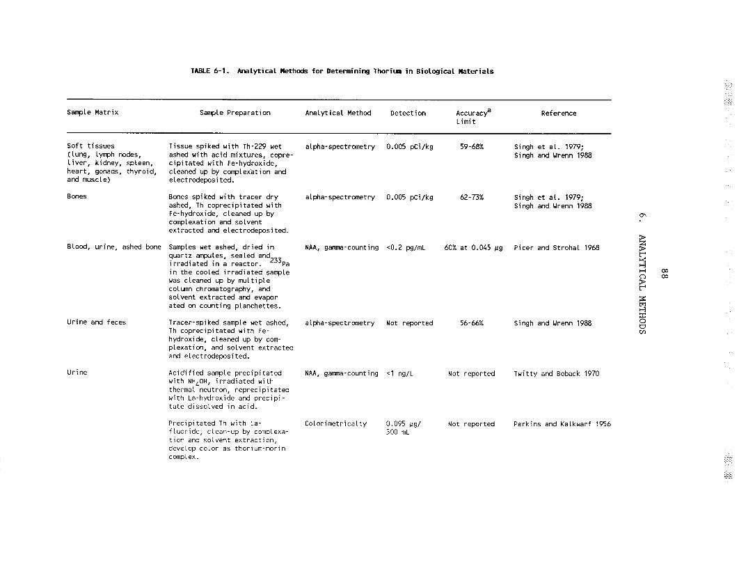
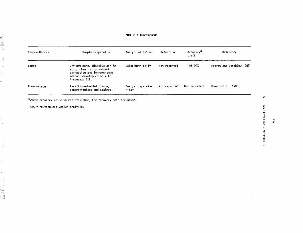

90
6. ANALYTICAL METHODS
6.2 ENVIRONMENTAL SAMPLES
Some of the commonly used methods for the determination of thorium in environmental samples are shown in Table 6-2. Standard reference materials (SRMs) for thorium in river and freshwater lake sediment (SRM-4350B and SRM4354), soils (SRM-4355 and SRM-4353), coal (SRM-1632), and fly ash (SRM 1633) are available from the National Institute of Standards and Technology (formerly National Bureau of Standards) (Inn 1987; Ondov et al. 1975). Neither calorimetric nor atomic absorption/emission methods are suitable for the determination of thorium-specific isotopes; these methods are also not sensitive enough for the quantification of trace amounts of thorium, e.g., in seawater. The filtration of particulate phases by inert polypropylene fiber filter and adsorption of solution phase thorium onto Mn02-coated fiber or preconcentration of thorium on XAD-2 resin by adsorption of thorium-Xylenol Orange complexes and quantitation by alpha spectrometry or neutron activation analysis are two of the better methods for the quantification of low levels of thorium in water (Hirose 1988; Huh and Bacon 1985; Livingston and Cochran 1987). The isotope dilution-mass spectrometric method provides the most accurate and sensitive thorium quantification (Arden and Gale 1974) but is rarely used because of the specialized nature and the cost of the analytical technique. The beta counting of thorium deposited on counting discs is useful for the determination of thorium-234 derived from uranium238 (Velten and Jacobs 1982). The direct gamma radiation counting with a Ge planer detector has been used for the quantification of thorium-228 in grass samples (Joshi 1987). The recoveries of thorium from soil and sediment samples are usually poor (Singh and Wrenn 1988) and special attention should be given to sample treatment during their analysis.
6.3 ADEQUACY OF THE DATABASE
Section 104(i)(5) of CERCLA, directs the Administrator of ATSDR (in consultation with the Administrator of EPA and agencies and programs of the Public Health Service) to assess whether adequate information on the health effects of thorium is available. Where adequate information is not available, ATSDR, in conjunction with the NTP, is required to assure the initiation of a program of research designed to determine the health effects (and techniques for developing methods to determine such health effects) of thorium.
The following categories of possible data needs have been identified by a joint team of scientists from ATSDR, NTP, and EPA. They are defined as substance-specific informational needs that, if met, would reduce or eliminate the uncertainties of human health assessment. In the future, the identified data needs will be evaluated and prioritized, and a substance-specific research agenda will be proposed.

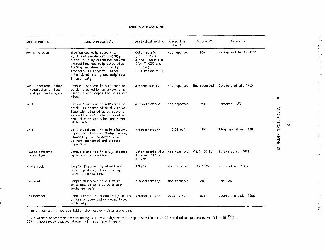

93
6. ANALYTICAL METHODS
6.3.1 Identification of Data Needs
Methods for Determining Biomarkers of Exposure and Effect. A few authors have found elevated levels of thorium in tissues of thorium workers and these studies have been discussed in Sections 2.6 and 5.4.4. However, there are no data in the literature that correlate the concentrations of thorium in any human tissue or body fluid with its level of exposure. If a biomarker for thorium in human tissue or fluid were available, the level of the biomarker in a tissue could be used as an indicator of exposure to thorium. Analytical methods with satisfactory sensitivity are available to determine the levels of thorium in most human tissues and body fluids of exposed and background population, but the recovery of thorium by these methods needs further refinement.
No studies were located that identified biomarkers specific to thorium-induced disease states. If a biomarker for thorium-induced effect in humans were found, this effect could also be used as an indicator of exposure to thorium. Therefore, there is a need to develop biomarkers that will serve as indicators of exposure and effect from exposure to uranium. It may be necessary to develop analytical methods of satisfactory sensitivity and precision for the quantification of thorium-induced effects in humans.
Methods for Determining Parent Compounds and Degradation Products in Environmental Media. Analytical methods with required sensitivity and precision are available for the quantification of thorium in most environmental samples. However, some of the more sensitive analytical methods have not always been used for the determination of thorium concentrations in drinking water and food. Knowledge of the levels of thorium compounds in environmental media can be used to indicate human exposure to thorium through inhalation of air and ingestion of drinking water and foods containing these compounds. The concentration of thorium is usually very low in drinking water and food and the more sensitive methods may not always have been used for quantification. Because of this, there is controversy in the literature about the relative importance of drinking water and food in contributing to the total dietary daily intake of thorium. It will be helpful to reevaluate the concentrations of thorium in drinking water and food by using the more sensitive analytical methods.
In the environment, thorium and its compounds do not degrade or mineralize like many organic compounds, but instead speciate into different chemical compounds and form radioactive decay products. Analytical methods for the quantification of radioactive decay products, such as radium, radon, polonium and lead are available. However, the decay products of thorium are rarely analyzed in environmental samples. Since radon-220 (thoron, a decay product of thorium-232) is a gas, determination of thoron decay products in some environmental samples may be simpler, and their concentrations may be used as an indirect measure of the parent compound in the environment if a secular equilibrium is reached between thorium-232 and all its decay
94
6. ANALYTICAL METHODS
products. There are few analytical methods that will allow quantification of the speciation products formed as a result of environmental interactions of thorium (e.g., formation of complex). A knowledge of the environmental transformation processes of thorium and the compounds formed as a result is important in the understanding of their transport in environmental media. For example, in aquatic media, formation of soluble complexes will increase thorium mobility, whereas formation of insoluble species will enhance its incorporation into the sediment and limit its mobility.
6.3.2 On-going Studies
Under the sponsorship of the National Science Foundation and in collaboration with scientists in New Zealand, Burnett of Florida State University is attempting to develop a chemical separation technique for uranium, thorium, and their daughter products (Federal Research in Progress 1990). Other than this research, no studies are in progress for improving the method for the quantification of thorium and daughter products in biological and environmental samples.
95
7. REGULATIONS AND ADVISORIES
International and national regulations and guidelines pertinent to human exposure to thorium and its radiations are summarized in Table 7-l. Recommendations for radiation protection for people in the general population as a result of exposure to radiation in the environment are found in the Federal Radiation Guidance (FRC 1960) and ICRP No. 26 (ICRP 1977). National guidelines for occupational radiation protection are found in the "Federal Radiation Protection Guidance for Occupational Exposure" (EPA 1987). This guidance for occupational exposure supercedes recommendations of the Federal Radiation Council for occupational exposure (FRC 1960). The new guidance presents general principles for the radiation protection of workers and specifies the numerical primary guides for limiting occupational exposure. These recommendations are consistent with the ICRP (ICRP 1977).
The basic philosophy of radiation protection is the concept of ALARA (As Low As Reasonably Achievable). As a rule, all exposure should be kept as low as reasonably achievable and the regulations and guidelines are meant to give an upper limit to exposure. Based on the primary guides (EPA 1987), guides for Annual Limits on Intake (ALIs) and Derived Air Concentrations (DACs) have been calculated (EPA 1988b). The AL1 is defined as "that activity of a radionuclide which, if inhaled or ingested by Reference Man (ICRP 1975), will result in a dose equal to the most limiting primary guide for committed dose" (EPA 1988b; ICRP 1979) (see Appendix B). The DAC is defined as "the concentration of radionuclide in air which, if breathed by Reference Man (ICRP 1975) for a work-year, would result in the intake of one AL1 (EPA 1988b). The ALIs and DACs refer to occupational situations but may be converted to apply to exposure of persons in the general population by application of conversion factors (Table 7-l).

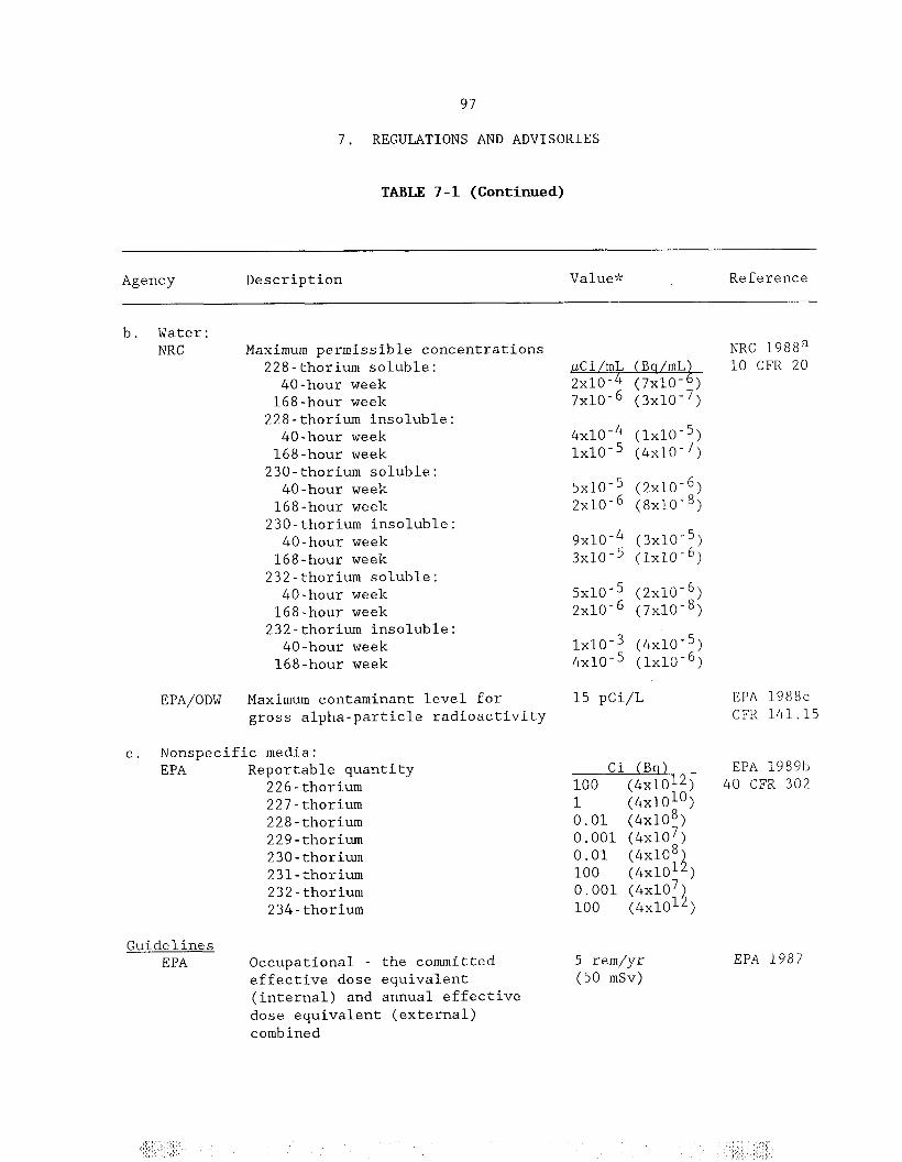
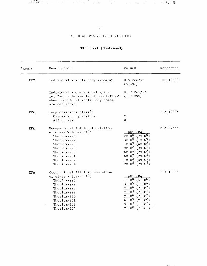


____________________________
101
8. REFERENCES
Abbatt JD. 1979. History of the use and toxicity of thorotrast. Environ Res 18:6-12.
*Abel KJ, Young JA, Rancitelli LA. 1984. Atmospheric chemistry of inorganic emissions. In: Randerson D, ed. Atmospheric science and power production. Oak Ridge, TN: The Technical Information Center, Office of Scientific and Technical Information, U.S. Department of Energy, 423-465.
Adelglass JM, Samara M, Cantor JO, et al. 1980. Thorotrast induced multiple carcinomatosis of the frontal sinus. Bull NY Acad Med 56:453-457.
*Aieta EM, Singley JE, Trussell AR, et al. 1987. Radionuclides in drinking water: An overview. Am Water Works Assoc J 79:144-152.
*Albert R, Klevin P, Fresco J, et al. 1955. Industrial hygiene and medical survey of a thorium refinery. Arch Ind Health 11:234-242.
*Anderson RF, Bacon MP, Brewer PG. 1982. Elevated concentrations of actinides in Mono Lake (California, USA). Science 216:514-516.
*APHA/AWWA/WPCF (American Public Health Association/American Water Work Association/Water Pollution Control Federation). 1985. Standard methods for the examination of water and wastewater. 16th ed. Washington, DC: American Public Health Association, 162-164.
*Archer VE, Wagoner JK, Lundin FE. 1973. Cancer mortality among uranium mill workers. J Occup Med 15:11-14.
*Arden JW, Gale NH. 1974. Separation of trace amounts of uranium and thorium and their determination by mass spectrometric isotope dilution. Anal Chem 46:687-691.
*Arnold AG, Oelbaum MH. 1980. Thorotrast administration followed by myelofibrosis. Postgrad Med J 56:124-127.
*ASTM. 1986. 1986 annual book of ASTM standards, Vol 11.01 (Section 11). Method D2333-80: Test method for thorium in water. American Society for Testing of Materials, 714-717.
*ATSDR. 1990a. Toxicological profile for thorium. U.S. Department of Health and Human Services. Public Health Service. Agency for Toxic Substances and Disease Registry. Atlanta, GA.
*Cited in text
102
8. REFERENCES
*ATSDR. 1990b. Toxicological profile for radium. U.S. Department of Health and Human Services. Public Health Service. Agency for Toxic Substances and Disease Registry. Atlanta, GA.
*ATSDR. 199oc. Toxicological profile for radon. U.S. Department of Health and Human Services. Public Health Service. Agency for Toxic Substances and Disease Registry. Atlanta, GA.
*ATSDR. 1990d. Toxicological profile for uranium. U.S. Department of Health and Human Services. Public Health Service. Agency for Toxic Substances and Disease Registry. Atlanta, GA.
Ballou JE, Hursh JB. 1972. The measurement of thoron in the breath of dogs administered inhales to injected Th02. Health Phys 22:155-159.
*Barnes D, Bellin J, DeRosa C, et al. 1987. Reference dose (RfD): description and use in health risk assessments. Volume I, Appendix A: Integrated risk information system supportive documentation. Washington, DC: U.S. Environmental Protection Agency, Office of Health and Environmental Assessment. EPA/600/8-86/032a.
Baserga R, Yokoo H, Henegar GC. 1960. Thorotrast induced cancer in man. Cancer 13:1021-1031.
YcBaxter PJ, Anthony PP, Macsween RNM, et al. 1980a. Angiosarcoma of the liver: Annual occurrence and aetiology in Great Britain. Br J Ind Med 37:213-221.
*Baxter PJ, Langlands AO, Anthony PP, et al. 1980b. Angiosarcoma of the liver: A marker tumour for the late effects of Thorotrast in Great Britain Br J Cancer 41:446-453.
Behari JR, Tandon SK, Mathur AK, et al. 1975. Histological and biochemical alterations in rat's liver and kidney induced by thorium nitrate. Arh Hig Rada Toksikol 26:267-274.
*BEIR III. 1980. The effects on populations of exposure to low levels of ionizing radiation. Committee on the Biological Effects of Ionizing Radiations, National Research Council. Washington, DC: National Academy Press.
*BEIR IV. 1988. Health risks of radon and other internally deposited alpha-emitters. Committee on the Biological Effects of Ionizing Radiations, National Research Council. Washington, DC: National Academy Press, 245-275.
*BEIR V. 1990. Health effects of exposure to low levels of ionizing radiation. Committee on the Biological Effects of Ionizing Radiations, National Research Council. Washington, DC: National Academy Press.

103
8. REFERENCES
*Beretka J, Mathew PJ. 1985. Natural radioactivity of Australian building materials, industrial wastes and by-products. Health Phys 48:87-95.
*Bernabee RP. 1983. A rapid method for the determination of americium, curium, plutonium and thorium in biological and environmental samples. Health Phys 44:688-692.
*Bigu J. 1985. Radon daughter and thoron daughter deposition velocity and unattached fraction under laboratory-controlled conditions and in underground uranium mines. J Aerosol Sci 16:157-165.
*Bodek I, Lyman WJ, Reehl RF, et al., eds. 1988. Environmental inorganic chemistry: Properties, processes and estimation methods. SETAC Special Publication Series, New York, NY: Pergamon Press, 9.2-19.
*Boecker BB. 1963. Thorium distribution and excretion studies. Am Ind Hyg Assoc J 24:155-163.
*Boecker BB, Thomas RG, Scott JK. 1963. Thorium distribution and excretion studies. II. General patterns following inhalation and the effect of the size of the inhaled dose. Health Phys 9:165-176.
*Boniforti R. 1987. Lanthanides, uranium, and thorium as possible simulators of the behavior of transuranics in the aquatic environment. Sci Total Environ 64:181-189.
*Boone IU, Rogers BS, White DC, et al. 1958. Toxicity excretion and tissue distribution of ionium (Th-230) in rats. Am Ind Hyg Assoc J 19:285-295.
*Bowen JH, Woodward BH, Mossler JA, et al. 1980. Energy dispersive x-ray detection of thorium dioxide. Arch Path01 Lab Med 104:459-461. Brody H, Cullen M. 1957. Carcinoma of the breast seventeen years after mammography with thorotrast. Surgery 42:600-606.
Brown WM, Buckton KE, Langlands AO, et al. 1967. The identification of lymphocyte clones with chromosome structural aberrations in irradiated men and women. Int J Radiat Biol 13:155-169..
Brues AM. 1979. The long term follow up radium dial painters and thorium workers. Med Bases Radiat Accident Prep:441-450.
*Buckton KE, Langlands AO. 1973. The Edinburgh Thorotrast series - report of a cytogenetic study. In: 3rd International Meeting: Toxicity of Thorotrast, April 25-27, 1973. RISO Report No. 294, 114-125.
*Bulman RA. 1976. Concentration of actinides in the food chain. Gov't Rep Announce, Issue 17803. NRPB.R44, 11-12.

104
8. REFERENCES
Burnett WC. n.d. US-New Zealand cooperative research into the development and application of a new method for separation of uranium-series isotopes. National Science Foundation, Division of International Programs, Washington, DC.
Cantor JO, Adelglass J, Cerreta JM, et al. 1981. Collision tumor of the frontal sinus: Evidence of prior intrasinus instillation of thorotrast. Laryngoscope 91:798-803.
*Carpenter R, Beasley TM, Zahnie D, et al. 1987. Cycling of fallout (plutonium, americium-241, cesium-137) and natural (uranium, thorium, lead210) radionuclides in Washington continental slope sediments. Geochim Cosmochim Acta 51:1897-1921.
CAS. 1990. Chemical Abstract Service. February 14, 1989.
*Chong CS, Chong HY, Fun HK, et al. 1985. Gamma radioactivity level in Sn slag dumps. Health Phys 49:1008-1010.
*Christensen P, Madsen MR, Jensen OM. 1983. Latency of Thorotrast-induced renal tumours. Survey of the literature and a case report. Stand J Urol Nephrol 17:127-130.
*Clifton RJ, Farrow M, Hamilton EI. 1971. Measurements of Th-232 in normal and industrially exposed humans. Ann Occup Hyg 14:303-308.
*Cochran JK. 1984. The fates of uranium and thorium decay series nuclides in the estuarine environment. Estuary Filter, 179-220.
*Coles DG, Ragaini RC, Ondov JM, et al. 1979. Chemical studies of a stack fly ash from a coal-fired power plant. Environ Sci Technol 13:455-459.
*Conibear SA. 1983. Long term health effects of thorium compounds on exposed workers: The complete blood count. Health Phys 44(Suppl 1):231-237.
*Coorey KD. 1983. Thorotrast granuloma of the neck with local complications. Med J Aust 2:580-581.
*Costa-Ribeiro C, Barcinski MA, Figueiredo N, et al. 1975. Radiobiological aspects and radiation levels associated with the milling of monazite sand. Health Phys 28:225-231.
*Cothern CR. 1987. Development of regulations for radionuclides in drinking water. In: Graves B ed. Conference: NWWA, April 7-19, Somerset, NJ. Michigan: Lewis Publishers, l-11.
*Cothern CR, Lappenbusch WL, Michel J. 1986. Drinking water contribution to natural background radiation. Health Phys 50:33-47.

105
8. REFERENCES
*Cottrell WD, Haywood, FF, Witt, et al. 1981. Radiological survey of the Shpack Landfill, Norton, Massachusetts. Report DOE/EV-ORNL-5799, 0005/31. Oak Ridge, TN: Oak Ridge National Laboratory.
Dahlgren S. 1961. Thorotrast tumors: A review of the literature and report of two cases. Acta Pathol Microbial Stand 53:147.
*Dams R, Robbins JA, Rahn KA, et al. 1970. Nondestructive neutron activation analysis of air pollution particles. Anal Chem 42:861-867.
*da Motta LC, da Silva Horta J, Tavares MH. 1979. Prospective epidemiological study of Thorotrast exposed patients in Portugal. Environ Res 18:152-172.
*Dang HS, Jaiswal DD, Sunta CM. 1986. Daily intake of thorium by an Indian urban population. Sci Total Environ 57:73-77.
*da Silva Horta J. 1956. Late lesions in man caused by colloidal thorium dioxide (Thorotrast). Arch Path01 62:403-418.
*da Silva Horta J. 1967a. Late effects of Thorotrast on the liver and spleen, and their efferent lymph nodes. Ann NY Acad Sci 145:675-699.
*da Silva Horta J. 1967b. Effects of colloidal thorium dioxide extravasates in the subcutaneous tissues of the cervical region in man. Ann NY Acad Sci 145:776.
da Silva Horta J, Moura NJ. 1979. Movement of Thorotrast aggregates in the bone marrow. Environ Res 18:184-191.
*da Silva Horta J, Cayolla Da Motta L, Tavares MH. 1974. Thorium dioxide effects in man. Epidemiological clinical and pathological studies (Experience in Portugal). Environ Res 8:131-159.
*Davis MW. 1985. Radiological significance of thorium processing in manufacturing. Report. INFO-0150.
*Dejgaard A, Krogsgaard K, Jacobsen M. 1984. Veno-occlusive disease and peliosis of the liver after Thorotrast administration. Virchows Archiv Path01 Anat 403:87-94.
Dougherty JH, Rosenblatt LS. 1971. Long term hematological effects of internal emitters in beagles. Radiation Res 48:319-331.
Dougherty TF, Stover BJ, Dougherty JH, et al. 1962. Studies of the biological effects of Ra-226 Pu-239 Ra-228 Th-228 and Sr-90 in adult beagles. Rad Res 17:625-681.

106
8. REFERENCES
*Downs WL, Scott KJ, Maynard EA, et al. 1959. Studies on the toxicity of thorium nitrate. Report UR-561, Contract W-7401-eng-49. Atomic Energy Project, University of Rochester, Rochester, NY, l-35.
Dudley RA. 1978. Bone irradiation in Thorotrast cases: Results of measurements at IAEA. Health Phys 35: 103-112.
*Early P, Razzak M, Sodee D. 1979. Nuclear medicine technology, 2nd ed. St. Louis: C.V. Mosby Company.
*Edgington DN. 1967. The estimation of thorium and uranium at the submicrogram level in bone by neutron activation. Int J Appl Radiat Isot 18:11-18.
*Eichholz G. 1982. Environmental aspects of nuclear power. Ann Arbor, MI: Ann Arbor Science.
Einck-Rosskamp P, Rajewsky B, Stahlhofen W. 1969. Cross placental passage of radium-228 and its daughters after intravenous injection of colloidal thorium. Aec Symp Ser 17:121-135.
Ellis LC, Berliner DL. 1967. The effects of ionizing radiations on endocrine cells. Radiation Res 32:520-537.
*EPA. 1980. U.S. Environmental Protecton Agency. Guidelines and methodology used in the preparation of health effect assessment chapters of the consent decree water criteria documents. Federal Register 45:7934779357.
*EPA. 1984. Background information document (integrated risk assessment). Final rules for radionuclides. Vol I. U.S. Environmental Protecton Agency, Office of Radiation Programs. Washington, DC. EPA 520/1-84-022-l.
*EPA. 1987. U.S. Environmental Protecton Agency. Radiation protection guidance to federal agencies for occupational exposure; approval of Environmental Protection Agency recommendations. Federal Register 52:2823-2834.
*EPA. 1988a. Technological approaches to the cleanup of radiologically contaminated superfund sites. U.S. Environmental Protecton Agency. Office of Research and Development. Washington, DC. EPA 540/2-88-002.
*EPA. 1988b. Limiting values of radionuclide intake and air concentration and dose conversion factors for inhalation, submersion, and ingestion. Federal Guidance Report No. 11. Washington, DC: U.S. Environmental Protection Agency, Office of Radiation Programs. EPA-520/l-88-020.

107
8. REFERENCES
*EPA. 1988c. Maximum contaminant levels for radium-226, radium-228, and gross alpha-particle radioactivity in community water systems. 40 CFR 141.15 Federal Register, 533.
*EPA. 1989a. Interim Methods for Development of Inhalation References Doses. U.S. Environmental Protection Agency, Office of Health and Environmental Assessment. Washington, DC. EPA 600/8-88-066F.
*EPA. 1989b. Reportable Quantity Adjustment-Radionuclides; Final Rules. Federal Register 54~22524-22543.
*Ettenhuber E, Lehmann R. 1986. The collective dose equivalent due to the naturally occurring radionuclides in building materials in the German Democratic Republic. Part I: External exposure. Health Phys 50:49-56.
*Faber M. 1973. Effect of Thorotrast enriched with thorium-230. Dan. AEC., Res Establ Risoe, Risoe Rep 294: 294-302.
*Faber M. 1979. 28 years of continuous follow-up of patients injected with Thorotrast for cerebral angiography. Environ Res 18:37-43.
*Falk H, Telles NC, Ishak KG. 1979. Epidemiology of Thorotrast induced hepatic angiosarcoma in the USA. Environ Res 18:67-73.
*Farid I, Conibear SA. 1983. Hepatic function in previously exposed thorium refinery workers as compared to normal controls from the health and nutrition survey. Health Phys 44(Suppl 1):221-230.
*Federal Research in Progress. 1990. Databank. July 24, 1990.
*Filer TD. 1970. Fluorometric determination of submicrogram quantities of thorium. Anal Chem 42:1265-1267.
*Fischer P, Golob E, Kunze-Muehl E. 1967. Chromosomal aberrations in thorium dioxide patients. Ann NY Acad Sci 145:759-766.
*Fisenne IM, Perry PM, Decker KM, et al. 1987. The daily intake of 234,235,238U 228,230,232Th, and 226,228Ra by New York City residents. Health Phys 53:357-363.
*Fisher DR, Jackson PO, Brodaczynski GG, et al. 1983. Level of uranium-234 uranium-238 and thorium 230 in excreta of uranium mill crushermen. Health Phys 45:617-630.
*Fisher NS, Teyssie JL, Krishnaswami S, et al. 1987. Accumulation of thorium, lead, uranium, and radium in marine phytoplankton and its geochemical significance. Limnol Oceanogr 32:131-142.

108
8. REFERENCES
Fishman EK, Gayler BW, Kashima HK, et al. 1984. Computed tomography in the evaluation of cervical Thorotrast granuloma. J Comput Assist Tomogr 8:224228.
*Frank AL. 1980. Diseases associated with exposure to metals-thorium. In: Last JMM-R, ed. Public health and preventive medicine. 11th ed. New York, NY: Appleton-Century-Crofts, 681-682.
*FRC. 1960. Federal Radiation Council. Radiation protection guidance for federal agencies. Federal Register 60:4402-4403.
*Fried JF, Schubert J. 1961. Effect of chelating agent administration on the removal of monomeric and polymeric thorium. Rad Res 15:227-235.
*Fruchter JS, Robertson DE, Evans JC, et al. 1980. Mount St. Helen's ash from the 18 May 1980 eruption: Chemical, physical, mineralogical, and biological properties. Science 29:1116-1125.
Gaidova ES, Syao-Shan Y. 1970. Morphological changes in the lungs and other organs in rats on intratracheal administration of thorium dioxide. In: Letavet, Kurlyandskaya, eds. The toxicology of radioactive substances, Vol. 4. New York: Pergamon Press, 66-82.
*Garten CT Jr. 1978. A review of parameter values used to assess the transport of plutonium, uranium and thorium in terrestrial food chains. Environ Res 17:437-452.
*Gilbert GE, Casella VR, Bishop CT, et al. 1985. Isotopes of uranium and thorium, lead-210, and polonium-210 in the lungs of coal miners of Appalachia and the lungs and livers of residents of central Ohio. Energy Res Abstr 11:4090.
Gilman JPW, Ruckerbauer GM. 1962. Metal carcinogenesis I: Observations on the carcinogenicity of a refinery dust, cobalt oxide and colloidal thorium dioxide. Cancer Res 22:152-156.
*Golchert NW, Iwami FS, Sedlet J. 1980. Determination of actinides in soil. In: Lyon WS, ed. Radioelement analysis: progress and problems. 23rd conference: Analytical Chemistry in Energy Technology, Gatlinburg, TN, Ott 9-11, 1979. Ann Arbor, MI: Ann Arbor Science Publishers, Inc., 215-222.
Goren AD, Harley N, Eisenbud L, et al. 1980. Clinical and radiobiologic features of Thorotrast induced carcinoma of the maxillary sinus. Oral Surg Oral Med Oral Path01 49:237-242.
Gossner W. 1982. Thorium II: Biological effects of thorium. In: Galle P, Masse R, eds. Radionuclide Metab Toxic Proc Symp. Masson Publ., 273-280.

109
8. REFERENCES
*Grampa G. 1967. Liver distribution of colloidal thorium dioxide and development of liver epithelial tumors in rats. Ann NY Acad Sci 145:738.
*Grampa G. 1971. Radiation injury with particular reference to Thorotrast. Path01 Ann 6:147-169.
*Greenberg RR, Kinston HM. 1982. Simultaneous determination of 12 trace elements in estuarine and sea water using pre-irradiation chromatography. In: 1981 International Conference: Modern Trends in Activation Analysis, Part 3, Toronto, Ont, Canada, June 15-16, 1981. J Radioanal Chem 71:147168.
Gridgeman NT. 1971. Methods of assay of the relative toxicity of certain bone seeking radionuclides. Radiat Res 48:291-302.
*Guimaraes JP, Lamerton LF. 1956. Further experimental observations on the late effects of Thorotrast administration. Br J Cancer 10:527.
*Guimaraes JP, Lamerton LF, Christensen WR. 1955. The late effects of Thorotrast administration: A review and experimental study. Br J Cancer 9:253.
*Gunning C, Scott AG. 1982. Radon and thoron daughters in housing. Health Phys 42:527-528.
*Hall RH, Stroud CA, Scott JK, et al. 1951. Acute toxicity of inhaled thorium compounds. Atomic Energy Project, University of Rochester, Rochester, NY. Report No. UR-190.
*Hallegot PH, Galle P. 1988. Microanalytical study of thorium 232 deposits in bone marrow and liver. Rad Environ Biophys 27:67-78.
*Hamilton EI. 1971. The relative radioactivity of building materials. Alll Ind Hyg Assoc J. 32:398-403.
+Hannibal L. 1982. On the radiological significance of inhaled uranium and thorium ore dust. Health Phys 42: 367-371.
*Harley NH, Pasternack BS. 1979. Potential carcinogenic effects of actinides in the environment. Health Phys 37:291-300.
*Harmsen K, De Haan FAM. 1980. Occurrence and behaviour of uranium and thorium in soil and water. Neth J Agric Sci 28:40-62.
*Harrist TJ, Schiller AL, Trelstad RL, et al. 1979. Thorotrast-associated sarcoma of bone: A case report and review of the literature. Cancer 44:2049-2058.

110
8. REFERENCES
*Hart DR, McKee PM, Burt AJ, et al. 1986. Benthic community and sediment quality assessment of Port Hope Harbor, Lake Ontario (Canada). J Great Lakes Res 12:206-220.
*Hawley GG, ed. 1981. The condensed chemical dictionary. 10th ed. New York, NY: Van Nostrand Reinhold, 881, 1021-1022.
*Hedrick JB. 1985. Thorium. Preprint from Bulletin 675: Mineral facts and problems, 1985 ed. Washington, DC: Bureau of Mines, U.S. Department of the Interior.
*Hedrick JB. 1987. Thorium. Preprint from the 1987 Bureau of Mines mineral yearbook. Washington, DC: Bureau of Mines, U.S. Department of the Interior.
Hemphill FM, Hesselgren RD. 1971. Thorotrast quantities and whole body counts. Health Phys 21:85-89.
*Hendee W. 1973. Radioactive isotopes in biological research. New York, NY: John Wiley and Sons.
*Hirose K. 1988. Determination of thorium isotopes in seawater by using preconcentration of thorium-X0 complexes on XAD-2 resin. J Radioanal Nucl Chem-Lett 127:199-207.
*Hirose K, Sugimura Y. 1987. Thorium isotopes in the surface air of the western north Pacific Ocean. J Environ Radioact 5:459-475.
*Hobbs C, McClellan RO. 1986. Radiation and radioactive materials. In: Doull J, et al., eds. Casarett and Doull's Toxicology, 3rd ed. New York, NY: Macmillan Publishing Co., Inc., 497-530.
*Hodge HC, Maynard EA, Leach LJ. 1960. The chemical toxicity of thorium dioxide following 'inhalation by laboratory animals. University of Rochester Atomic Energy Project. Report UR-562. University of Rochester, Rochester, NY, l-33.
*Hoegerman SF, Cummins HT. 1983. Chromosome damage in peripheral lymphocytes from American thorium workers. Health Phys 44(Suppl 1):365-371.
*HSDB. 1990. Hazardous Substances Data Bank. National Library of Medicine, Washington, DC. February 14, 1989.
*Hu SJ, Chong CS, Subas S. 1981. U-238 and Th-232 in Cassiterites samples and Amang by-products. Health Phys 40:248-250.
*Hu SJ, Koo WK, Tan KL. 1984. Radioactivity associated with Amang upgrading plants. Health Phys 46:452-455.

111
8. REFERENCES
*Hu SJ, Kandaiya S. 1985. Radium-226 and 232Th concentration in Amang. Health Phys 49:1003-1007.
*Huh C-A, Bacon MP. 1985. Determination of thorium concentration in seawater by neutron activation analysis. Anal Chem 57:2138-2142.
*Huh C-A, Zahnle DL, Small LF, et al. 1987. Budgets and behaviors of uranium and thorium series isotopes in Santa Monica basin (California, USA) sediments. Geochim Cosmochim Acta 51:1743-1754.
*Hunter KA, Hawke DJ, Choo LK. 1988. Equilibrium adsorption of thorium by metal-oxides in marine electrolytes. Geochim Cosmochim Acta 52:627-636.
*Hyman C, Paldino RL. 1967. Influence of sex hormones and tissues on colloidal thorium dioxide distribution and effects in rabbits. Ann NY Acad Sci 145:576-584.
*Ibrahim SA, Whicker FW. 1988. Comparative uptake of U and Th by native plants at a U production site. Health Phys 54:413-419.
*Ibrahim SA, Wrenn ME, Singh NP, et al. 1983. Thorium concentration in human tissues from two U.S. populations. Health Phys 44(Suppl 1):213-220.
*ICRP. 1966. International Commission on Radiological Protection. ICRP task group on lung dynamics, depositions and retention models for internal dosimetry of the human respiratory tract. Health Phys 21:173-207.
*ICRP. 1975. International Commission on Radiological Protection. Report of the Task Group on Reference Man. ICRP Publication 23. New York: Pergamon Press.
*ICRP. 1977. International Commission on Radiological Protection. Recommendations of the International Commission on Radiological Protection. ICRP Publication No. 26. Vol. 1. No. 3. Oxford: Pergamon Press.
*ICRP. 1979. International Commission on Radiological Protection. Limits for intakes of radionuclides by workers. ICRP Publication 30. New York: Pergamon Press, 31, 32, 37, 100-101.
*ICRP. 1979. International Commission on Radiological Protection. Limits for intakes of radionuclides by workers. ICRP Publication 20. Vol 3. No. l-4. Oxford: Pergamon Press.
*ICRP. 1984. International Commission on Radiological Protection. A compilation of the major concepts and quantities in use by ICRP. ICRP Publication 42. Oxford: Pergamon Press.
ICRU. 1980. International Commission on Radiation Units and Measurements. ICRU Report No. 33. Washington, DC.
112
8. REFERENCES
*Inn KGW. 1987. The National Bureau of Standards freshwater lake sediment environmental-level radioactivity standard reference material. J Radioanal Nucl Chem 115:91-112.
Ishihara T, Kohno S, Hayta I, et al. 1978. Nine year cytogenetic follow up of a patient injected with Thorotrast. Hum Genet 42:99-108.
*Isner JM, Sazama KJ, Roberts WC. 1978. Vascular complications of thorium dioxide. Am J Med 67:1069-1074.
*Ito Y, Kojiro M, Nakashima T, et al. 1988. Pathomorphologic characteristics of 102 cases of Thorotrast-related hepatocellular carcinoma, cholangiocarcinoma, and hepatic angiosarcoma. Cancer 62:1153-1162.
Jadon A, Mathur R. 1983. Gametogenic count and histo pathological effect of thorium nitrate and uranyl nitrate on mice testes. Andrologia 15:40-43.
*James A. 1987. A reconsideration of cells at risk and other key factors in radon daughter dosimetry. In: Hopke P, ed. Radon and its decay products: Occurrence, properties and health effects. ACS Symposium Series 331. Washington, DC: American Chemical Society, 400-418.
*James A, Roy M. 1988. Dosimetric lung models. In: Gerber G, et al., ed. Age-related Factors in Radionuclide Metabolism and Dosimetry. Boston: Martinus Nijhoff Publishers, 95-108.
*Jee WS, Dockum NL, Mica1 RS, et al. 1967. Distribution of thorium daughters in bone. Ann NY Acad Sci 145:660-673.
*Jensen L, Regan G, Goranson S, et al. 1984. Ambient monitoring of airborne radioactivity near a former thorium processing plant. Health Phys 46:1021-1033.
*Jiang FS, Kuroda PK. 1987. Variation of the isotopic composition of thorium in the atmosphere. Radiochim Acta 42123-28.
*Jiang FS, Lee SC, Bakhtiar SN, et al. 1986. Determination of thorium, uranium and plutonium isotopes in atmospheric samples. J Radioanal Nucl them 100:65-72.
*Johansen C. 1967. Tumors in rabbits after injections of various amounts of thorium dioxide. NY Acad Sci 145:724
*Johannson L. 1982. Oral intake of radionuclides in the population: A review of biological factors of relevance for assessment of absorbed dose at long term waste storage. FOA Report C40161-W4. DE 83-704553.

113
8. REFERENCES
Johannsson W, Perrault G, Savoie L, et al. 1968. Action of various metallic chlorides on calcaemia and phosphataemia. Br 3 Pharamcol Chemother 33:91-97.
*Johnson JR, Lamothe ES. 1989. A review of the dietary uptake of Th. Health Phys 56:165-168.
*Joshi SR. 1987. Nondestructive determination of selected uranium and thorium-series radionuclides in biological samples. Health Phys 53:417-420.
*Joyet G. 1971. The thorium-series in cigarettes and lungs of smokers. Experientia 27:85-89.
Kamboj MP, Kar AB. 1964. Antitesticular effect of metallic and rare earth salts. J Repro Fert 7:21-28.
*Kamijo A, Okabe K, Hirose T. 1979. Thorium dioxide granuloma of the neck with resultant fatal hemorrhage. Arch Otolaryngol 105:45-47.
*Kamiyama R, Ishikawa Y, Hatakeyama S, et al. 1988. Clinicopathological study of hematological disorders after Thorotrast administration in Japan. Blut 56:153-160.
Kaneko M, Kamiya Y, Hoshino M, et al. 1981. Identification and mapping of Thorotrast in tissue by analytical electron microscopy. Health Phys 41:677-680.
*Kate I, Kido C. 1987. Increased risk of death in Thorotrast-exposed patients during the late follow-up period. Jpn J Cancer Res 78:1187-1192.
*Kate H, Schull W. 1982. Studies of the mortality of A-bomb survivors. Report 7, Part 1: Cancer mortality among atomic bomb survivors, 1950-78. Radiat Res 90:395-432.
*Kate Y, Mori T, Kumatori T. 1979. Thorotrast dosimetric study in Japan. Environ Res 18:32-36.
*Kate I, Mori T, Kumatori T. 1983. Estimated absorbed dose in tissues and radiation effects in Japanese Thorotrast patients. Health Phys 44(Suppl 1):273-279.
Katzin LI. 1983. Thorium and thorium compounds. In: Grayson M, Eckroth D, eds. Kirk-Othmer encyclopedia of chemical technology, vol. 22, 3rd ed. New York, NY: John Wiley and Sons, 989-1002.
*Kaul A, Heyder J. 1972. Clearance of thorium-232 and daughters from the blood. In: Symposium: Assessment Radioactive Contam Man, 469-481.
114
8. REFERENCES
*Kaul A, Muth H. 1978. Thorotrast kinetics and radiation dose. Results from studies in Thorotrast patients and from animal experiments. Radiat Environ Biophys 15:241-259.
*Kaul A, Noffz W. 1978. Tissue dose in Thorotrast patients. Health Phys 35:113-121.
Kaul A, Foll U, Haase VA, et al. 1979. Microdistribution of Thorotrast and dose to cellular structures. Env Research 18:13-22.
*Kauzlaric D, Barmeir E, Luscieti P, et al. 1987. Renal carcinoma after retrograde pyelography with Thorotrast. Am J Radio1 148:897-898.
*Keane AT, Brewster DR. 1983. Calibration of a decay-product collection and counting apparatus for the determination of exhaled thoron. Health Phys 45:801-805.
*Keane AT, Lucas HF, Markun F, et al. 1986. The estimation and potential radiobiological significance of the intake of radium-228 by early radium dial workers in Illinois. Health Phys 51:313-328.
*Keller G, Folkerts KH. 1984. Radon-222 concentrations and decay-product equilibrium in dwellings and in the open air. Health Phys 47:385-398.
*Kemmer W. 1979. A field study in 156 Thorotrast cases results of biophysical measurements and clinical examinations. Environ Res 18:147-151.
*Kemmer W, Muth H, Tranekjer F. 1971. Dose dependence of the chromosome aberration rate in Thorotrast patients. Biophysik 7:342-351.
*Kemmer W, Muth H, Trandkjer F, et al. 1973. Chromosome aberrations caused by Thorotrast. In: 3rd International Meeting: Toxicity of Thorotrast, April 25-27, 1973. RISO Report No. 294, 104-113.
*Kemmer W, Steinstraesser A, Muth H. 1979. Chromosome aberrations as a biological dosimeter in Thorotrast patients: Dosimetric problems. Environ Res 18:178-183.
*Kobashi A, Tominaga T. 1985. Radium-228-thorium-228 dating of plant samples. Int J Appl Radiat Isot 36:547-554.
*Kojiro M, Nakashima T, Ito Y, et al. 1985. Thorium dioxide-related angiosarcoma of the liver. Pathomorphologic study of 29 autopsy cases. Arch Path01 Lab Med 109:853-857.
*Korte N, Kollenbach M, Donivan S. 1983. The determination of uranium, thorium, yttrium, zirconium and hafnium in zircon. Analyt Chim Acta 146:267-270.

115
8. REFERENCES
*Kotrappa P, Bhanti DP, Menon VB, et al. 1976. Assessment of airborne hazards in the thorium processing industry. Am Ind Hyg Assoc J 37:613-616.
Krey P, Fiseene I, Perry P, et al. n.d. Natural radioactivity in the biosphere. Environmental Measurements Laboratory, New York, NY.
Kurlyandskaya EB. 1970. Certain aspects of the toxicology of insoluble compounds of thorium-232 and uranium-238. In: Letavet, Kurlyandskaya, eds. The toxicology of radioactive substances, Vol. 4. New York: Pergamon Press, l-8.
*Kuroda PK, Barbod T, Bakhtiar SN. 1987. Effect of the eruptions of Mount St. Helens and El Chichon on the ratios of thorium and uranium isotopes in rain. J Radioanal Nucl Chem 111:137-146.
*Kyle RH, Oler A, Lasser EC, Rosomoff HL. 1963. Meningioma induced by thorium dioxide. New Eng J Med 368:80-82.
*LaFlamme BD, Murray JW. 1987. Solid/solution interaction: The effect of carbonate alkalinity on adsorbed thorium. Geochim Cosmochim Acta 51:243250.
*Lambert JPF, Wilshire FW. 1979. Neutron activation analysis for simultaneous determination of trace elements in ambient air collected on glass-fiber filters. Anal Chem 51:1346-1350.
*Laul JC, Smith MR, Thomas CW, et al. 1987. Analysis of natural radionuclides from uranium and thorium series in briney groundwaters. J Radioanal Nucl Chem llO:lOl-112.
*Lauria DC, Godoy JM. 1988. A sequential analytical method for the determination of uranium-238, thorium-232, thorium-230,thorium-228, radium228, and radium-226 in environmental samples. Sci Total Environ 70:83-99.
*Levowitz BS, Hughes RE, Alford TC. 1963. Treatment of thorium dioxide granulomas of the neck. New Eng J Med 268:340-342.
*Levy DW, Rindsberg S, Friedman AC, et al. 1986. Thorotrast-induced hepatosplenic neoplasia: CT identification. Am J Radio1 146:997-1004.
*Likhachev YuP. 1976. Pathological anatomy, etiology and pathogenesis of remote sequelae of radiation dust exposure. Arkh Patol 38:18-26. [Russian]
*Likhachev YuP, Lyarskii PP, Elovskaya LT. 1973a. Morphological changes in the lungs with chronic inhalation of thorium dioxide. Med Radio1 18:35-41. [Russian]
*Likhachev YuP, Lyarskii PP, Elovskaya LT. 1973b. Pulmonary neoplasms in rats in chronic inhalation of thorium dioxide. Vopr Onkol 19:47-54.

116
8. REFERENCES
*Linsalata P, Franca EP, Eisenbud M. 1985. Determination of the human intake of thorium and the light rare earth elements from high and typical natural radiation environments. Trace Subst Environ Health 19:257-263.
Linsalata P, Eisenbud M, Franca EP. 1986. Ingestion estimates of thorium and the light rare earth elements based on measurements of human feces. Health Phys 50:163-167.
*Linsalata P, Penna Franca E, Sachett I, et al. 1987. Radium, thorium, and the light rare earth elements in soils and vegetables grown in an area of high natural radioactivity. DOE Symp Ser 59:131-146.
*Linsalata P, Morse RS, Ford H, et al. 1989. An assessment of soil-toplant concentration ratios for some natural analogues of the transuranic elements. Health Phys 56:33-46.
*Lipchik EO, Russell SB, Raphael MJ, et al. 1972. Effect of Thorotrast on the canine heart. Br J Radio1 45:142-145.
*Livingston HD, Cochran JK. 1987. Determination of transuranic and thorium isotopes in ocean water in solution and in filterable particles. J Radioanal Nucl Chem 115:299-308.
Lloyd RD, Jones CW, Mays CW, et al. 1984. Thorium-228 retention and dosimetry in beagles. Radiat Res 98:614-628.
*Lloyd RD, Wrenn ME, Taylor GN, et al. 1985. Toxicity of 228Ra and 228Th relative to 226Ra for bone sarcoma induction in beagles. Strahlentherapie [Sonderb] 80:65-69.
*Lucas HF Jr., Edgington DN, Markun F. 1970. Natural thorium in human bone. Health Phys 19:739-742.
Lucas HF Jr, Markun F, May H, et al. 1972. Thorium daughters in the spleen of a Thorotrast case. Health Phys 23:575-576.
*Luetzelschwab JW, Googins SW. 1984. Radioactivity released from burning gas lantern mantles. Health Phys 46:873-881.
*Lung RJ, Harding RL, Herceg SJ, et al. 1978. Long-term survival with Thorotrast cervical granuloma. J Surg Oncol 10:171-177.
Luz A, Linzner U, Miiller WA, et al. 1979. Osteosarcoma induction by simultaneous incorporation of 227Th and 227Ac. Biological implications of radionuclides released from nuclear industries, Vol. 1. International Atomic Energy Agency, Vienna, 141-151.

117
8. REFERENCES
*Luz A, Miiller WA, Schaffer E, et al. 1985. The sens mice of different ages for osteosarcoma induction with Strahlentherapie [Sonderb] 80:178-182. itivity of female NMRI 227Thorium.
*Maletskos DJ, Keane AT, Telles NC, et al. 1969. Retention and absorption of Ra-224 and Th-234 and some dosimetric considerations of Ra-224 in human beings. In: May C, ed. Delayed effects of bone-seeking radionuclides. Salt Lake City, UT: University of Utah Press, 29-49.
*Mausner LF. 1982. Inhalation exposures at a thorium refinery. Health Phys 42:231-236.
*Mays CW. 1978. Endosteal dose to Thorotrast patients. Health Phys 35:123-125.
*Mays CW. 1988. Alpha-particle-induced cancer in humans. Health Phys 55:637-655.
*Mays CW, Dougherty TF. 1972. Progress in the beagle studies at the University of Utah. Health Phys 22:793-801.
*Mays CW, Lloyd RD, Taylor GN, et al. 1987. Cancer incidence and lifespan vs. alpha-particle dose in beagles. Health Phys 52:617-624.
*Mayya YS, Prasad SK, Nambiar PPVJ, et al. 1986. Measurement of 220Rn in exhaled breath of Th plant workers. Health Phys 51:737-744.
McClinton LT, Schubert J. 1948. The toxicity of some zirconium and thorium salts in rats. J Pharmacol Exp Ther 94:1-6.
McInroy JF. n.d. Measurements of plutonium and metals in man. Los Alamos National Laboratory, Los Alamos, NM.
*McKee PM, Snodgrass WJ, Hart DR, et al. 1987. Sedimentation rates and sediment core profiles of uranium-238 and thorium-232 decay chain radionuclides in a lake affected by uranium mining and milling. Can J Fish Aquat Sci 44:390-398.
*McNabb GJ, Kirk JA, Thompson JL. 1979. Radio nuclides from phosphate ore processing plants: The environmental impact after 30 years of operation. Health Phys 34:585-587.
*McNeil1 KG, Molla MA, Harrison JE. 1973. Distribution of thorium series nuclides in beagles after intraspinal Thorotrast injection. Health Phys 24:403-409.
Mettler F. 1985. Medical effects of ionizing radiation. New York, NY: Grune and Stratton.

118
8. REFERENCES
*Metzger R, McKlveen JW, Jenkins R, et al. 1980. Specific activity of uranium and thorium in marketable rock phosphate as a function of particle size. Health Phys 39:69-76.
*Michael TH, Murray C. 1970. The effects of a blockading agent on the primary immune response. J Immunol 105:411-415.
*Miekeley N, Kuchler IL. 1987. Interactions between thorium and humic compounds in surface waters. Inorg Chim Acta 140:315-319.
*Milic, NB, Suranji TM. 1982. Hydrolysis of the thorium(IV) ion in sodium nitrate medium. Can J Chem 60:1298-1303.
*Moffett D, Tellier M. 1978. Radiological investigations of an abandoned uranium tailings area. J Environ Qua1 7:310-314.
*Molla MAR. 1975. Radionuclides in the autopsy samples from Thorotrast patients. Health Phys 28:295-297.
Molla MAR, Harrison JE, McNeil1 KG. 1976. Doses to the cauda equina due to Thorotrast myelography. Health Phys 31:109-114.
Monath TP, Borden EC. 1971. Effects of Thorotrast on humoral antibody viral multiplication and interferon during infection with St. Louis encephalitis virus in mice. J Infect Dis 123:297-300.
*Moore JW, Sutherland DJ. 1981. Distribution of heavy metals and radionuclides in sediments, water, and fish in an area of Great Bear Lake contaminated with mine wastes. Arch Environ Contam Toxic01 10:329-338.
*Moores SR, Black A, Lambert BE, et al. 1980. Deposition of thorium and plutonium oxides in the respiratory tract of the mouse. DOE Symp Ser 53:103-118.
*Mori T, Kato Y, Shimamini T, et al. 1979. Statistical analysis of Japanese Thorotrast-administered autopsy cases. Environ Res 18:231.
*Mori T, Kato Y, Kumatori T, et al. 1983a. Epidemiological follow-up study of Japanese Thorotrast cases--1980. Health Phys 44(Suppl 1):261.-272.
*Mori T, Kato Y, Aoki N, et al. 1983b. Statistical analysis of Japanese Thorotrast-administered autopsy cases--1980. Health Phys 44(Suppl 1):281292.
*Mϋller WA, Gijssner W, Hug 0, et al. 1978. Late effects after incorporation of the short-lived alpha-emitters radium-224 and thorium-227 in mice. Health Phys 35:33-55.

119
8. REFERENCES
Miiller WA. 1971. Studies on short lived internal emitters in mice and rats. Int J Radiat Bio 20:233-243.
*M•ller WA, Nenot JC, Daburon ML, et al. 1975. Metabolic and dosimeter studies after inhalation of Th-227 in rats with regard to the risk of lung and bone tumors. Radiation Env Biophysics 11:309-318.
*M•ller WA, Luz A, Schaffer EH, et al. 1983. The role of time-factor and RBE for the induction of osteosarcomas by incorporated short-lived boneseekers. Health Phys 44(Suppl 1):203-212.
*Munita CS, Mazzilli BP. 1986. Determination of trace elements in Brazilian cigarette tobacco by neutron activation analysis. J Radioanal Nucl Chem 108:217-228.
*Mustonen R, Jantunen M. 1985. Radioactivity of size fractionated fly-ash emissions from a peat- and oil-fired power plant. Health Phys 49:1251-1260.
*Nakaoka A, Fukushima M, Takagi S. 1984. Environmental effects of natural radionuclides from coal-fired power plants. Health Phys 47:407-416.
*NAS/NRC. 1989. Biologic markers in reproducible toxicology. National Academy of Sciences/National Research Council. Washington, DC: National Academy Press, 15-35.
*NCRP 1971. National Council on Radiation Protection and Measurements. Basic radiation protection criteria. NCRP Report No. 39. Washington, DC.
*NCRP. 1985. A handbook of radioactivity measurements procedures. 2nd ed. Bethesda, MD: National Council on Radiation Protection and Measurements. NCRP Report No. 58.
*Neton JW, Ibrahim SA. 1978. Alpha emitting radio nuclides in cigarette tobacco. Health Phys 35:922.
*Newton D, Rundo J, Eakins JD. 1981. Long-term retention of thorium-228 following accidental intake. Health Phys 40:291-298.
Nielsen M, Albrechtsen R, Johnson NJ, et al. 1979. Carcinoma of the parotid gland following sialography with Thorotrast. Acta Otolaryngol 88:462-467.
*Nishioka H. 1975. Mutagenic activities of metal compounds in bacteria. Mutat Res Sect Environ Mutagen Relat Subj 31:185-190.
*NRC. 1988. Nuclear Regulatory Commission. Maximum permissible concentrations for uranium and thorium. 10 CFR 20 151:4115 and 151:4117.
120
8. REFERENCES
*Oakes TW, Shank KE, Easterly CE, et al. 1977. Concentrations of radionuclides and selected stable elements in fruits and vegetables. Trace Subst Environ Health 11:123-132.
*Odegaard A, Ophus EM, Larsen AM. 1978. Identification of thorium dioxide in human liver cells by electron microscopic x-ray microanalysis. J Clin Path01 31:893-896.
*Ondov JM, Zoller WH, Olmez I, et al. 1975. Elemental concentrations in the National Bureau of Standards' environmental coal and fly ash standard reference materials. Anal Chem 47:1102-1109.
*Otake M, Schull W. 1984. Mental retardation in children exposed in utero to the atomic bombs: A reassessment. Technical Report RERF TR l-83, Radiation Effects Research Foundation, Japan.
*Parr RM, Lucas HF Jr., Griem ML. 1968. Metabolism of 232Th decay series radionuclides in man and other animals following intravascular administration of Thorotrast. ANL-7615. U.S. AEC Argonne Nat1 Lab, 97115.
*Parreira F, De Moura MC. 1979. Morphological study of the bone marrow in patients injected with thorium oxide. Environ Res 18:61-64.
*Patrick SJ, Cross EM. 1948. Some effects of the administration of thorium nitrate to mice. Can J Res 26:303-316.
*Pavlovskaia NA. 1973. Basic patterns of the metabolism of Th232 and its decay products in the body. Gig Sanit 11:55-58. [Russian]
*Pavlovskaia NA, Makeeva LG, Orlianskaia RL. 1969. Distribution of thorium in rat bones with various routes of administration and different chemical compounds of thorium. Gig Sanit 34:27-31.
*Pavlovskaia NA, Makeeva LG, Zel'tser MR. 1974a. Excretion of radionuclides of the thorium-232 (thorium-228, radium-224, lead-212) series from a rats body during the uptake of thorium compounds in respiratory organs. Gig Sanit 9:42-45. [Russian]
*Pavlovskaia NA, Provotorov AV, Makeeva LG. 1974b. Behavior of thorium-228 and its daughter isotopes in the body of rats when administered perorally. Gig Tr Prof Zabol 5:36-39. [Russian]
*Percival DR, Martin DB. 1974. Sequential determination of radium-226, radium-228, actinium-227, and thorium isotopes in environmental and process waste samples. Anal Chem 46:1742-1749.
*Perkins RW, Kalkwarf DR. 1956. Determination of thorium in urine. Anal Chem 28:1989-1993.
121
8. REFERENCES
Perry DL, Tsao L. n.d. Thorium species on basalt surfaces. Lawrence Berkeley Laboratory, Berkeley, CA.
*Peter E, Lehmann M. 1981. Interaction of thorium with blood serum proteins in vivo. Int J Radiat Biol Relat Stud Phys Chem 40:445-450.
*Peter-Witt E, Volf V. 1984. Efficacy of different diethylenetriaminepentaacetic acid treatment schedules for removal of thorium-234 from simulated wounds in rats. Int J Radiat Biol Relat Stud Phys Chem Med 45:45-50.
*Peter-Witt E, Wolf V. 1985. Comparison of diethylenetriaminepentaacetic acid calcium chelate and diethylenetriaminepentaacetic acid zinc chelate in removing thorium-234 from the rat. Health Phys 49:395-404.
Peter-Witt E, Volf V. 1985. The distribution and decorporation of 234Th in the rat as influenced by the mass of thorium administered. Strahlentherapie [Sonderb] 80:183-185.
*Petrow HG, Strehlow CD. 1967. Spectrophotometric determination of thorium in bone ash using arsenazo. 3. Anal Chem 39:265-267.
*Picer M, Strohal P. 1968. Determination of thorium and uranium in biological materials. Anal Chim Acta 40:131-136.
*Pillai KC, Matkar VM. 1987. Determination of plutonium and americium in environmental samples and assessment of thorium in bone samples from normal and high background areas. J Radioanal Nucl Chem 115:217-229.
Planas-Bohne F, Taylor DM, Lamble G, et al. 1982. The localization of metallic radionuclides in abscesses in rats. Br J Radio1 55:289-293.
*Platford RF, Joshi SR. 1986. The chemistry of uranium and related radionuclides in Lake Ontario (USA, Canada) waters. J Radioanal Nucl Chem 106:333-344.
*Platford RF, Joshi SR. 1988. Dose rates to aquatic life near a uranium waste site. Health Phys 54:63-68.
Pleskach SD. 1985. Determination of U and Th in urine by neutron activation. Health Phys 48:303-307.
*Polednak AP, Stehney AF, Lucas HF. 1983. Mortality among male workers at a thorium-processing plant. Health Phys 44(Suppl 1):239-251.
*Popper H, Thomas LB, Telles NC. 1978. Development of hepatic angiosarcoma in man induced by vinyl chloride, Thorotrast, and arsenic. Comparison with cases of unknown etiology. Am J Path01 92:349-376.
122
8. REFERENCES
*Poston TM. 1982. The bio accumulation potential of thorium and uranium in rainbow trout Salmo-nairdneri. Bull Environ Contam Toxicol 28:682-690.
*Premuzic ET, Francis AJ, Lin M, et al. 1985. Induced formation of chelating agents by Pseudomonas aeruginosa grown in presence of thorium and uranium. Arch Environ Contam Toxic01 14:759-768.
*Rangarajan C, Eapen CD, Gopalakrishnan SS. 1986. Measured values of the dry deposition velocities of atmospheric aerosols carrying natural and fallout radionuclides using artificial collectors. Water Air Soil Pollut 27:305-314.
*Rae BK, Brodell GK, Haaga JR, et al. 1986. Visceral CT findings associated with Thorotrast. J Comput Assist Tomogr 10:57-61.
*Raymond DP, Dufield JR, Williams DR. 1987. Complexation of plutonium and thorium in aqueous environments. Inorg Chim Acta 140:309-313.
*Riedel WR, Hirschberg AK, Kaul A, et al. 1979. Comparative investigations on the biokinetics of colloidal thorium, zirconium, and hafnium dioxides in animals. Environ Res 28:127-139.
Riedel W, Dalheimer A, Said M, et al. 1983. Recent results of the German Thorotrast study dose relevant physical and biological properties of Thorotrast equivalent colloids. Health Phys 44:293-298.
*Roeck DR, Reavey TC, Hardin JM. 1987. Partitioning of natural radionuclides in the waste streams of coal-fired utilities. Health Phys 52:311-324.
*RTECS. 1989. Registry of Toxic Effects of Chemical Substances. February 14, 1989.
*Rubin P, Casarett G. 1968. Clinical radiation pathology. Philadelphia: W.B. Sanders Company, 33.
*Ruhmann AG, Berliner DL. 1965. Serum lactic dehydrogenase levels in adult beagle dogs with internally deposited radionuclides. Radiation Res 26:287294.
*Sadamori N, Miyajima J, Okajima S, et al. 1987. Japanese patients with leukaemia following the use of Thorotrast including a patient with marked chromosomal rearrangements. Acta Haematologica 77:11-14.
*Saisho H, Tanaka M, Nakamura K, et al. 1988. Determination of traces of uranium and thorium in microelectronics constituent materials. J Res Nat1 Bureau Stand 93:398-400.
123
8. REFERENCES
*Salaymeh S, Kuroda PK. 1987. Variation of the thorium to uranium ratio in rain: Thorium-230 chronology of the eruptions of Mt. St. Helens and El Chichon volcanoes. J Radioanal Nucl Chem 116:261-269.
*Saldanha MJ, Shah VR, Nambiar PPUJ. 1975. Health Assessment of thorium nitrate plant workers of Indian rare earths. Indian J Ind Med 21:114-123.
*SANSS. 1989. Structure and Nomenclature Search System. February 14, 1989.
*Santschi PH. 1984. Particle flux and trace metal residence time in natural waters. Limnol Oceanogr 29:1100-1108.
*Santschi PH, Li YH, Adler DM, et al. 1983. The relative mobility of natural (thorium, lead and polonium) and fallout (plutonium, americium, cesium) radionuclides in the coastal marine environment: Results from model ecosystems (MERL) and Narragansett Bay. Geochim Cosmochim Acta 47:201-210.
*Sasaki MS, Takatsuji T, Ejima Y, et al. 1987. Chromosome aberration frequency and radiation dose to lymphocytes by alpha-particles from internal deposit of Thorotrast. Radiat Environ Biophys 26:227-238.
*Schery SD. 1985. Measurements of airborne lead-212 and radon-220 at varied indoor locations within the United States. Health Phys 49:1061-1067.
*Scott JK, Neuman WF, Bonner JF. 1952. The distribution and excretion of thorium sulphate. J Pharmacol Experi Therapeu 106:286-290.
Serio CS, Henninb CB, Toohey RE, et al. 1983. Measurement of lymphoblastogenic activity from thorium workers. Int J Radiat Biol 44:251256.
Sheppard MI. 1980. The environmental behavior of uranium and thorium. At Energy Can Ltd [Rep.] AECL, AECL-6795.
*Sill CW. 1977. Simultaneous determination of U-238, U-234, Th-230, Ra226, and PB.210 in uranium ores, dusts, and mill tailings. Health Phys 33:393-404.
*Sill CW, Willis CP. 1962. Fluorometric determination of submicrogram quantities of thorium. Anal Chem 34:954-964.
*Simpson HJ, Trier RM, Toggweiler JR, et al. 1982. Radionuclides in Mono Lake, California, USA. Science 216:512-514.
*Simpson HJ, Trier RM, Li YH, et al. 1984. Field experiment determinations of distribution coefficients of actinide elements in alkaline lake environments. Nucl Waste Geochem, 326-342.
124
8. REFERENCES
Sindelar WF, Costa J, Ketcham AS. 1978. Osteosarcoma associated with Thorotrast administration: Report of two cases and literature review. Cancer 42:2604-2609.
*Singh NP, Wrenn ME. 1988. Determinations of actinides in biological and environmental samples. Sci Total Env 70:187-203.
*Singh, NP, Ibrahim SA, Cohen N, et al. 1979. Simultaneous determination of alpha-emitting radionuclides of thorium plutonium in human tissues including bone. Analytical Chem 51:1978-1981.
*Singh NP, Wrenn ME, Archer VE, et al. 1981. Uranium-238 uranium-234 and thorium-230 in uranium miners' lungs. Radiat Hazards Min: Control, Meas, Med Aspects, Int Conf 236-239.
Singh NP, Wrenn ME, Ibrahim SA, et al. 1982. Thorium concentration in human tissues form two geographic locations of the United States. Nat Rad Env 258-268.
*Singh NP, Wrenn ME, Ibrahim SA. 1983. Plutonium concentration in human tissues: Comparison to thorium. Health Phys 44:469-476.
Singh NP, Bennett D, Saccomanno G, et al. 1985. Concentrations of lead-210 in uranium miners' lungs and its states of equilibria with uranium-238 uranium-234 and thorium-230. Occup Radiat Saf Min 2:503-506.
*Singh NP, Lewis LL, Wrenn ME. 1985. Uranium thorium and plutonium in bones from the general population of the United States. Comm Eur Communities Issue 9250:231-241.
Singh NP, Bennett DB, Wrenn ME, et al. 1986. Concentrations of 210Pb and its states of equilibrium with 238U 234U and 230Th in U miners' lungs. Health Phys 51:501-507.
*Singh NP, Wrenn ME, McInroy JF, et al. 1986. Ratios of 234U, 238U and 230Th in dog lungs exposed to U ore dust: An interlaboratory comparison. Health Phys 50:292-297.
*Singh NP, Bennett DD, Wrenn ME, et al. 1987. Concentrations o.f alphaemitting isotopes of uranium and thorium in uranium miners' and millers' tissues. Health Phys 53:261-266.
*Singh NP, Zimmerman CJ, Taylor GN, et al. 1988. The beagle: An appropriate experimental animal for extrapolating the organ distribution pattern of Th in humans. Health Phys 54:293-299.
*Sollman T, Brown ED. 1907. Pharmacologic investigations on thorium. Am J Physiol 18:426-456.

125
8. REFERENCES
Spiers FW, Beddow AH, 1983. Sites of incidence of osteosarcoma in the long bones of man and beagle. Health Phys 44:49-64.
*Stannard JN. 1988. Radioactivity and health: A history. DOE/RL/01830T59.
Stanley RB Jr. 1983. Thorium-dioxide induced pharyngeal hemorrhage. Am J . Otolaryngol 4:437-441.
*Stanley RB Jr, Calcaterra TC. 1981. Management of cervical thorium dioxide granulomas. Arch Otolaryngol 107:631-634.
*Stehney AF, Polednak AP, Rundo J, et al. 1980. Health status and body radioactivity of former thorium workers. Argonne, IL, Argonne Nat1 Laboratory 80-37:44.
*Steinstrasser A. 1981. Biophysical investigations of the dose-effect relationship in chromosome aberrations of human lymphocytes caused by Thorotrast deposits. Radiat Env Biophys 19:1-15.
*Stevens W, Nabors CJ, Jr, Berliner DL. 1967. A comparison of serum transaminase levels and other serum constituents in dogs burdened with 239Pu, 228-Th, 228-RA and 226-Ra. NY Acad Sci 145:817-829.
*Stougaard M, Praestholm J. Stoier M. 1984. Late effects of perivascular injection of Thorotrast in the neck. J Laryngol Otol 98:1003-1007.
*Stover BJ. 1981. Toxicology of thorium-228 in young adult beagles: Potential relationship to the thorium fuel cycle. In: Wrenn ME, ed. Actinides in Man and Animals: Proc Snowbird Actinide Workshop. R D Press, University of Utah, Salt Lake City, UT, 483-500.
Stover BJ. 1983. Effects of Thorotrast in humans. Health Phys 44:253257.
*Stover BJ, Atherton DR, Keller, et al. 1960. Metabolism of Th-228 decay series in adult beagle dogs. Radiation Res 12:657-671.
Stover BJ, Atherton DR, Buster DS, et al. 1965a. The Th-228 decay series in adult beagles: Ra-224 PB.212 and Bi-212 in selected bones and soft tissues. Radiat Res 26:132-145.
Stover BJ, Atherton Dr, Buster DS, et al. 1965b. The Th-228 decay series in adult beagles: Ra224 Pb212 and Bi-212 in blood and excreta. Radiat Res 26:226-243.
*Sullivan MF. 1980a. Absorption of actinide elements from the gastrointestinal tract of rats, guinea pigs and dogs. Health Phys 38:159-171.
126
8. REFERENCES
*Sullivan MF. 1980b. Absorption of actinide elements from the gastrointestinal tract of neonatal animals. Health Phys 38:173-185.
*Sullivan MF, Miller BM, Ryan JL. 1983. Absorption of thorium and protactinium from the gastrointestinal tract in adult mice and rats and neonatal rats. Health Phys 44:425-428.
*Summers DE, Chung EB. 1986. Thorotrast associated anemia and bone marrcw hypoplasia. J Nat1 Med Assoc 78:1161-1165.
*Sunta CM, Dang HS, Jaiswal DD. 1987. Thorium in man and environment: Uptake and clearance. J Radional Nucl Chem 115:149-158.
*Sussman S, Terbrugge KG, Solt LC, et al. 1980. Thorotrast induced meningioma case report. J Neurosurg 52:834-837.
Syao-Shan Y. 1970. The effect of thorium dioxide on the peripheral blood of rats. In: Lefavet, Kurlyandskaya, eds. The toxicology of radioactive substances, Vol. 4. New York, Pergamon Press, 42-57.
Syao-Shan, Y. 1970. The effect of thorium dioxide on arterial pressure and threshold of stimulation of the nerve muscle apparatus in rats. In: Lefavet, Kurlyandskaya, eds. The toxicology of radioactive substances, Vol. 4. New York, Pergamon Press, 58-65.
*Syao-Shan Y. 1970a. The behavior of thorium dioxide in rats after intratracheal and intraperitoneal administration. In: Lefavet, Kurlyandskaya, eds. The toxicology of radioactive substances, Vol. 4. New York, Pergamon Press, 30-41.
*Syao-Shan Y. 1970b. Comparative toxicity of soluble and insoluble compounds of thorium-232. In: Lefavet, Kurlyandskaya, eds. The toxicology of radioactive substances, Vol. 4. New York, Pergamon Press, 9-19.
*Tadmor J. 1986. Atmospheric release of volatilized species of radioelements from coal fired plants. Health Phys 50:270-273.
*Tandon SK, Dikshith TSS, Behari JR, et al. 1975. Effect of thorium nitrate in male albino rats. Ind Health 13:221-225.
Tandon SK, Mathur AK, Behari JR, et al. 1975. Thorium induced testicular changes in rats. Acta Biol Med Ger 34:1835-1842.
*Taskayev AI, Popova ON, Alexakhin RM, et al. 1986. Root absorption of radon-222 and its transfer into above ground plant organs. Health Phys 50:589-594.

127
8. REFERENCES
*Taylor GN, Jee WSS, Christensen WR, et al. 1966. Thorium-228 induced fractures in beagles. Health Phys 12:889-893.
*Taylor GN, Mays CW, Lloyd RD, et al. 1986. Liver cancer induction by 241Am and Thorotrast in deer mice and grasshopper mice. Strahlen Therapie 8O(Suppl):172-177.
*Taylor DM, Chipperfield AR, James AC. 1971. Effects of tetracycline on the deposition of plutonium and related elements in rat bone. Health Phys 21:197-204.
*Teixeira-Pinto AA, Azevedo e Silva MC. 1979. Chromosome radiation induced aberrations in patients injected with thorium dioxide. Env Res 18:225-230.
*Telles MC, Thomas LB, Popper H, et al. 1979. Evolution of Thorotrast induced hepatic angiosarcomas. Env Res 18:74-87.
*Thomas RG. 1957. The metabolism of thorium-230 administered by intratracheal injection to the rat. Rochester, NY, Univ of Rochester, Atomic Energy Project Repor UR-480, l-26.
Thomas RG. 1968. Transport of relatively insoluble materials from lung to lymph nodes. Health Phys 14:111-117.
*Thomas RG, Lie R, Scott JK. 1963. Thorium distribution and excretion studies I. Patterns following parenteral administration. Health Phys 9:153163.
Thurman GB, Mays CW, Taylor GN, et al. 1971. Growth dynamics of beagle osteosarcomas. Growth 35:119-125.
*Torstenfelt B. 1986. Migration of the actinides, thorium, protactinium, uranium, neptunium, plutonium and americium in clay. Radiochem Acta 39:105112.
*Traikovich M. 1970. Absorption, distribution and excretion of certain soluble compounds of natural thorium. In: Lefavet, Kurlyandskaya, eds. The toxicology of radioactive substances, Vol. 4. New York, Pergamon Press, 20-29.
*Twitty BL, Boback MW. 1970. Rapid determination of thorium in urine by thermal neutron activation analysis. Anal Chim Acta 49:19-24.
*UNSCEAR. 1977. United Nations Scientific Committee on the Effects of Atomic Radiation. Sources and effects of ionizing radiation. United Nations Publication E.77.IX.l. New York, NY: United Nations, 95-96.

128
8. REFERENCES
*UNSCEAR. 1982. United Nations Scientific Committee on the Effects of Atomic Radiation. Ionizing radiation: Sources and biological effects. New York, NY: United Nations.
*UNSCEAR. 1986. United Nations Scientific Committee on the Effects of Atomic Radiation. Genetic and somatic effects of ionizing radiation. New York: United Nations.
*UNSCEAR. 1988. United Nations Scientific Committee on the Effects of Atomic Radiation. Sources, effects and risks of ionization radiation. New York: United Nations.
*Van Kaick G, Lieberman D, Lorenz D, et al. 1983. Recent results on the German Thorotrast study-epidemiological results and dose effect relationships in Thorotrast patients. Health Phys 44:299-306.
Van Kaick G, Wesch H, Luhrs H, et al. 1986. Radiation induced primary liver tumors in Thorotrast patients. Recent Results Cancer Res 100:16-22.
*Velten RJ, Jacobs BJ. 1982. Determination of thorium in drinking water, Method 910. Environmental Monitoring and Support Laboratory, U.S. Environmental Protection Agency, Cincinnati, OH.
*VIEW Database. 1989. Agency for Toxic Substances and Disease Registry (ATSDR), Office of External Affairs, Exposure and Disease Registry Branch, Atlanta, GA. June 20, 1989. (Map based on VIEW Database June 12, 1989).
*Visfeldt J, Poulsen H. 1972. On the histopathology of liver and liver tumors in thorium dioxide patients. Acta Path Micro Stand Sect A:97-108.
*Vocaturo G, Colombo F, Zanoni M, et al. 1983. Human exposure to heavy metals: Rare earth pneumoconiosis in occupational workers. Chest 83:780783.
*Waligorski MPR, Jasinska M, Schwabenthan J. 1985. Enhanced nuclear radiation from camera lenses. Health Phys 49:491-494.
*Wargotz ES, Sidawy MK, Jannotta FS. 1988. Thorotrast associated gliosarcoma including comments on Thorotrast use and review of sequelae with particular reference to lesions of the central nervous system. Cancer 62:58-60.
*Weast RC, ed. 1983. CRC handbook of chemistry and physics. 64th ed. Boca Raton, FL: CRC Press, Inc., B149-B150.
*Wegener K, Wesch H. 1979. Pathological changes of lymph nodes in Thorotrast patients patho anatomical auto radiographical and quantitative investigations. Env Res 18:245-255.

129
8. REFERENCES
Wegener K, Hasenohrl K, Wesch H. 1983. Recent results of the German Thorotrast study pathoanatomical changes in animal experiments and comparison to human Thorotrastosis. Health Phys 44(Suppl 1):307-316.
*Wegener K, Wesch H, Kampmann H. 1976. Investigations into human Thorotrastosis tissue concentrations of thorium-232 and late effects in 13 autopsy cases. Virchows Arch Path Anat Histol 371:131-143.
*Weissman SH, Carpenter RL, Newton GJ. 1983. Respirable aerosols from fluidized bed coal combustion: Element composition of fly ash. Env Sci Tech 17:65-71.
*Welford GA, Sutton DC, Morse RS, et al. 1958. Determination of thorium in urine. Am Ind Hyg Assoc J 19:4640468.
*Wesch H, van Kaick G, Riedel W, et al. 1983. Recent results of the German Thorotrast study statistical evaluation of animal experiments with regard to the nonradiation effects in human Thorotrastosis. Health Phys 44:317-321.
*West CM. 1962. An evaluation of the health physics problems from thorium and its daughters in a thorium purification and fabrication process. Health Phys 8:279-297.
*Westin J, Lanner LO, Weinfeld A, et al. 1973. Renal pelvic carcinoma after Thorotrast pyelography: Case report. Acta Med Stand 194:141-143.
*Wang SH. 1988. Effects of some metallic compounds on Klebsiella. Bull Environ Contam Toxic01 40:525-531.
*Woodliff HJ, Cohen G, Gallon W. 1972. Chromosome aberrations in lymphocytes from patients exposed to thorium dioxide. Med J Australia 2:768-771.
*Wrenn ME, Singh NP, Cohen N, et al. 1981. Thorium in human tissues. NTIS NUREG/CR-1227, l-97.
*Wrenn ME, Singh NP, Saccomanno G. 1983. Uranium and thorium isotopes and their state of equilibria in lungs from uranium miners. Health Phys 44:385 389.
*Wrenn ME, Singh NP, Paschoa AS, et al. 1985. Concentrations of uranium and thorium isotopes in uranium millers' and miners' tissues. NTIS NUREG/CR-4382/GAR, l-47.
*Wrenn ME, Taylor GN, Stevens W, et al. 1986. DOE life-span radiation effects studies in experimental animals at University of Utah, Division of Radiobiology. In: Life-span radiation effects in animals: What can they tell us? Conference: U.S. Department of Energy, 830951.

130
8. REFERENCES
*Wustrow TP, Behbehani AA, Wiebecks B. 1988. Thorotrast-induced oro- and hypopharyngeal fibrosis with recurrent bleeding. J.Cranio-Maxillo-Facial Surg 16:315-319.
*Yamada S, Hosoda S, Tateno H, et al. 1983. Survey of Thorotrast associated liver cancers in Japan. J Nat Cancer Inst 70:31-35.
*Yang HS, Nozaki Y, Sakai H, et al. 1986. Natural and man made radionuclide distributions in Northwest Pacific deep sea sediments: Rates of sedimentation, bioturbation and radium-226 migration. Geochem J 20:29-40.
*Young WN, Tebrock HA. 1958. The treatment of exposure to thorium and uranium with a chelating agent and supportive measures. Ind Med Surg (Annual Meeting) 27:229-232.
*Zoller WH, Gladney ES, Duce RA. 1974. Atmospheric concentrations and sources of trace metals at the South Pole. Science 183:198-200.

131
9. GLOSSARY
Absorbed Dose -- The mean energy imparted to the irradiated medium, per unit mass, by ionizing radiation. Units: gray (Gy), rd.
Absorbed Fraction -- A term used in internal dosimetry. It is that fraction of the photon energy (emitted within a specified volume of material) which is absorbed by the volume. The absorbed fraction depends on the source distribution, the photon energy, and the size, shape and composition of the volume.
Absorption -- The process by which radiation imparts some or all of its energy to any material through which it passes.
Self-Absorption -- Absorption of radiation (emitted by radioactive atoms) by the material in which the atoms are located; in particular, the absorption of radiation within a sample being assayed.
Absorption Coefficient -- Fractional decrease in the intensity of an unscattered beam of x or gamma radiation per unit thickness (linear absorption coefficient), per unit mass (mass absorption coefficient), or per atom (atomic absorption coefficient) of absorber, due to deposition of energy in the absorber. The total absorption coefficient is the sum of individual energy absorption processes. (See Compton Effect, Photoelectric Effect, and Pair Production.)
Linear Absorption Coefficient -- A factor expressing the fraction of a beam of x or gamma radiation absorbed in a unit thickness of material. In the expression I=Ioe
-µx, Io is the initial intensity, I the intensity of the beam after passage through a thickness of the material x, and µ is the linear absorption coefficient.
Mass Absorption Coefficient -- The linear absorption coefficient per cm divided by the density of the absorber in grams per cubic centimeter. It is frequently expressed as µ/p, where µ is the linear absorption coefficient and p the absorber density.
Absorption Ratio, Differential -- Ratio of concentration of a nuclide in a given organ or tissue to the concentration that would be obtained if the same administered quantity of this nuclide were uniformly distributed throughout the body.
Activation -- The process of inducing radioactivity by irradiation. Activity -- The number of nuclear transformations occurring in a given quantity of material per unit time. (See Curie.)
Activity Median Aerodynamic Diameter (AMAD) -- The diameter of a unitdensity sphere with the same terminal settling velocity in air as that of the aerosol particulate whose activity is the median for the entire aerosol.
132
9. GLOSSARY
Acute Exposure -- Exposure to a chemical for a duration of 14 days or less,as specified in the toxicological profiles.
Acute Radiation Syndrome -- The symptoms which taken together characterize a person suffering from the effects of intense radiation. The effects occur within hours or weeks.
Adsorption Coefficient (KOC) -- The ratio of the amount of a chemical adsorbed per unit weight of organic carbon in the soil or sediment to the concentration of the chemical in solution at equilibrium.
Adsorption Ratio (Kd) -- The amount of a chemical adsorbed by a sediment or soil (i.e., the solid phase) divided by the amount of chemical in the solution phase, which is in equilibrium with the solid phase, at a fixed solid/solution ratio. It is generally expressed in micrograms of chemical sorbed per gram of soil or sediment.
Alpha Particle -- A charged particle emitted from the nucleus of an atom. An alpha particle has a mass charge equal in magnitude to that of a helium nucleus; i.e., two protons and two neutrons and has a charge of +2.
Annihilation (Electron) -- An interaction between a positive and a negative electron in which they both disappear; their energy, including rest energy, being converted into electromagnetic radiation (called annihilation radiation) with two 0.51 Mev gamma photons emitted at an angle of 180° to each other.
Atomic Mass -- The mass of a neutral atom of a nuclide, usually expressed in terms of "atomic mass units." The "atomic mass unit" is one-twelfth the mass of one neutral atom of carbon-12; equivalent to 1.6604x10⁻²⁴ gm. (Symbol: u)
Atomic Number -- The number of protons in the nucleus of a neutral atom of a nuclide. The "effective atomic number" is calculated from the composition and atomic numbers of a compound or mixture. An element of this atomic number would interact with photons in the same way as the compound or mixture. (Symbol: Z)
Atomic Weight -- The weighted mean of the masses of the neutral atoms of an element expressed in atomic mass units.
Auger Effect -- The emission of an electron from the extranuclear portion of an excited atom when the atom undergoes a transition to a less excited state.

133
9. GLOSSARY
Background Radiation -- Radiation arising from radioactive material other than that under consideration. Background radiation due to cosmic rays and natural radioactivity is always present. There may also be background radiation due to the presence of radioactive substances in building materials.
Becquerel (Bq) -- International System of Units unit of activity and equals one transformation (disintegration) per second. (See Units.)
Beta Particle -- Charged particle emitted from the nucleus of an atom. A beta particle has a mass and charge equal in magnitude to that of the electron. The charge may be either +l or -1.
Biologic Effectiveness of Radiation -- (See Relative Biological Effectiveness.)
Bone Seeker -- Any compound or ion which migrates in the body preferentially into bone.
Branching -- The occurrence of two or more modes by which a radionuclide can undergo radioactive decay. For example, radium C can undergo α or β- decay, 64Cu or undergo β-,β+, or electron capture decay. An individual atom of a nuclide exhibiting branching disintegrates by one mode only. The fraction disintegrating by a particular mode is the "branching fraction" for that mode. The "branching ratio" is the ratio of two specified branching fractions (also called multiple disintegration).
Bremsstrahlung -- The production of electromagnetic radiation (photons) by the negative acceleration that a fast, charged particle (usually an electron) undergoes from the effect of an electric or magnetic field, for instance, from the field of another charged particle (usually a nucleus).
Cancer Effect Level (CEL) -- The lowest dose of chemical in a study, or group of studies, that produces significant increases in the incidence of cancer (or tumors) between the exposed population and its appropriate control.
Capture, Electron -- A mode of radioactive decay involving the capture of an orbital electron by its nucleus. Capture from a particular electron shell is designated as "K-electron capture," "L-electron capture," etc.
Capture, K-Electron -- Electron capture from the K shell by the nucleus of the atom. Also loosely used to designate any orbital electron capture process.
Carcinogen -- A chemical capable of inducing cancer.
Carcinoma -- Malignant neoplasm composed of epithelial cells, regardless of their derivation.
134
9. GLOSSARY
Cataract -- A clouding of the crystalline lens of the eye which obstructs the passage of light.
Ceiling Value (DL) -- A concentration of a substance that should not be exceeded, even instantaneously.
Chronic Exposure -- Exposure to a chemical for 365 days or more, as specified in the Toxicological Profiles.
Compton Effect -- An attenuation process observed for x or gamma radiation in which an incident photon interacts with an orbital electron of an atom to produce a recoil electron and a scattered photon of energy less than the incident photon.
Containment -- The confinement of radioactive material in such a way that it is prevented from being dispersed into the environment or is released only at a specified rate.
Contamination, Radioactive -- Deposition of radioactive material in any place where it is not desired, particularly where its presence may be harmful.
Cosmic Rays -- High-energy particulate and electromagnetic radiations which originate outside the earth's atmosphere.
Count (Radiation Measurements) -- The external indication of a radiation-measuring device designed to enumerate ionizing events. It may refer to a single detected event to the total number registered in a given period of time. The term often is erroneously used to designate a disintegration, ionizing event, or voltage pulse.
Counter, Geiger-Mueller -- Highly sensitive, gas-filled radiation-measuring device. It operates at voltages sufficiently high to produce avalanche ionization.
Counter, Scintillation -- The combination of phosphor, photmultiplier tube, and associated circuits for counting light emissions produced in the phosphors by ionizing radiation.
Curie -- A unit of activity. One curie equals 3.7x10¹⁰ nuclear transformations per second. (Abbreviated Ci.) Several fractions of the curie are in common usage.
Megacurie -- One million curies. Abbreviated MCi.
Microcurie -- One-millionth of a curie (3.7x10⁴ disintegrations per set) . Abbreviated µCi.
135
9. GLOSSARY
Millicurie -- One-thousandth of a curie (3.7x10⁷ disintegrations per set) . Abbreviated mCi.
Nanocurie -- One-billionth of a curie. Abbreviated nCi.
Picocurie -- One-millionth of a microcurie (3.7x10⁻² disintegrations per second or 2.22 disintegrations per minute). Abbreviated pCi; replaces the term µµc.
Decay, Radioactive -- Transformation of the nucleus of an unstable nuclide by spontaneous emission of charged particles and/or photons.
Decay Chain or Decay Series -- A sequence of radioactive decays (transformations) beginning with one nucleus. The initial nucleus, the parent, decays into a daughter nucleus that differs from the first by whatever particles were emitted during the decay. If further decays take place, the subsequent nuclei are also usually called daughters. Sometimes, to distinguish the sequence, the daughter of the first daughter is called the granddaughter, etc.
Decay Constant -- The fraction of the number of atoms of a radioactive nuclide which decay in unit time. (Symbol λ). (See Disintegration Constant).
Decay Product, Daughter Product -- A new isotope formed as a result of radioactive decay. A nuclide resulting from the radioactive transformation of a radionuclide, formed either directly or as the result of successive transformations in a radioactive series. A decay product (daughter product) may be either radioactive or stable.
Delta Ray -- Energetic or swiftly moving electrons ejected from an atom during the process of ionization. Delta rays cause a track of secondary ionizations along their path.
Developmental Toxicity -- The occurrence of adverse effects on the developing organism that may result from exposure to a chemical prior to conception (either parent), during prenatal development, or postnatally to the time of sexual maturation. Adverse developmental effects may be detected at any point in the lifespan of the organism.
Disintegration Constant -- The fraction of the number of atoms of a radioactive nuclide which decay in unit time; λ is the symbol for decay constant in the equation N=Noe
-λt, where No is the initial number of atoms present, and N is the number of atoms present after some time, t. (See Decay Constant.)
Disintegration, Nuclear -- A spontaneous nuclear transformation (radioactivity) characterized by the emission of energy and/or mass from the nucleus. When large numbers of nuclei are involved, the process is characterized by a definite half-life. (See Transformation, Nuclear.)
136
9. GLOSSARY
Dose -- A general term denoting the quantity of radiation or energy absorbed. For special purposes it must be appropriately qualified. If unqualified, it refers to absorbed dose.
Absorbed Dose -- The energy imparted to matter by ionizing radiation per unit mass of irradiated material at the place of interest. The unit of absorbed dose is the rad. One rad equals 100 ergs per gram. In SI units, the absorbed dose is the gray which is 1 J/kg. (See Rad.)
Cumulative Dose (Radiation) -- The total dose resulting from repeated or continuous exposures to radiation.
Dose Assessment -- An estimate of the radiation dose to an individual or a population group usually by means of predictive modeling techniques, sometimes supplemented by the results of measurement.
Dose Equivalent (DE) -- A quantity used in radiation protection. It expresses all radiations on a common scale for calculating the effective absorbed dose. It is defined as the product of the absorbed dose in rad and certain modifying factors. (The unit of dose equivalent is the rem. In SI units, the dose equivalent is the sievert, which equals 100 rem.)
Dose, Radiation -- The amount of energy imparted to matter by ionizing radiation per unit mass of the matter, usually expressed as the unit rad, or in SI units, 100 rad=l gray (Gy). (See Absorbed Dose.)
Maximum Permissible Dose Equivalent (MPD) -- The greatest dose equivalent that a person or specified part thereof shall be allowed to receive in a given period of time.
Median Lethal Dose (MID) -- Dose of radiation required to kill, within a specified period, 50 percent of the individuals in a large group of animals or organisms. Also called the LD50.
Threshold Dose -- The minimum absorbed dose that will produce a detectable degree of any given effect.
Tissue Dose -- Absorbed dose received by tissue in the region of interest, expressed in rad. (See Dose and Rad.)
Dose, Fractionation -- A method of administering radiation, in which relatively small doses are given daily or at longer intervals.
Dose, Protraction -- A method of administering radiation by delivering it continuously over a relatively long period at a low dose rate.

137
9. GLOSSARY
Dose-distribution Factor -- A factor which accounts for modification of the dose effectiveness in cases in which the radionuclide distribution is nonuniform.
Dose Rate -- Absorbed dose delivered per unit time.
Dosimetry -- Quantification of radiation doses to individuals or populations resulting from specified exposures. Early Effects (of radiation exposure) -Effects which appear within 60 days of an acute exposure.
Electron -- A stable elementary particle having an electric charge equal to ±1.6O210x10-19 C (Coulombs) and a rest mass equal to 9.1091x10⁻³¹ kg. A positron is a positively charged "electron." (See Positron.)
Electron Volt -- A unit of energy equivalent to the energy gained by an electron in passing through a potential difference of one volt. Larger multiple units of the electron volt are frequently used: keV for thousand or kilo electron volts; MeV for million or mega electron volts. eV, 1 eV=1.6x10⁻¹² erg,) (Abbreviated:
Embryotoxicity and Fetotoxicity -- Any toxic effect on the conceptus as a result of prenatal exposure to a chemical; the distinguishing feature between the two terms is the stage of development during which the insult occurred. The terms, as used here, include malformations and variations, altered growth, and in utero death.
Energy -- Capacity for doing work. "Potential energy" is the energy inherent in a mass because of its spatial relation to other masses. "Kinetic energy" is the energy possessed by a mass because of its motion; MKSA unit: kg-m2/sec2 or joules.
Binding Energy -- The energy represented by the difference in mass between the sum of the component parts and the actual mass of the nucleus.
Excitation Energy -- The energy required to change a system from its ground state to an exited state. Each different excited state has a different excitation energy.
Ionizing Energy -- The average energy lost by ionizing radiation in producing an ion pair in a gas. For air, it is about 33.73 eV.
Radiant Energy -- The energy of electromagnetic radiation, such as radio waves, visible light, x and gamma rays.

138
9. GLOSSARY
Enriched Material -- (1) Material in which the relative amount of one or more isotopes of a constituent has been increased. (2) Uranium in which the abundance of the 235U isotope is increased above normal.
EPA Health Advisory -- An estimate of acceptable drinking water levels for a chemical substance based on health effects information. A health advisory is not a legally enforceable federal standard, but serves as technical guidance to assist federal, state, and local officials.
Equilibrium, Radioactive -- In a radioactive series, the state which prevails when the ratios between the activities of two or more successive members of the series remains constant.
Secular Equilibrium -- If a parent element has a very much longer half-life than the daughters (so there is not appreciable change in its amount in the time interval required for later products to attain equilibrium) then, after equilibrium is reached, equal numbers of atoms of all members of the series disintegrate in unit time. This condition is never exactly attained, but is essentially established in such a case as radium and its series to Radium D. The half-life of radium is about 1,600 years; of radon, approximately 3.82 days, and of each of the subsequent members, a few minutes. After about a month, essentially the equilibrium amount of radon is present; then (and for a long time) all members of the series disintegrate the same number of atoms per unit time.
Transient Equilibrium -- If the half-life of the parent is short enough so the quantity present decreases appreciably during the period under consideration, but is still longer than that of successive members of the series, a stage of equilibrium will be reached after which all members of the series decrease in activity exponentially with the period of the parent. An example of this is radon (half-life of approximately 3.82 days) and successive members of the series to Radium D.
Equilibrium, Radiation -- The condition in a radiation field where the energy of the radiations entering a volume equals the energy of the radiations leaving that volume.
Equilibrium Fraction (F) -- In radon-radon daughter equilibrium, the parents and daughters have equal radioactivity, that is, as many decay into a specific nuclide as decay out. However, if fresh radon is continually entering a volume of air or if daughters are lost by processes other than radioactive decay, e.g., plate out or migration out of the volume, a disequilibrium develops. The equilibrium fraction is a measure of the degree of equilibrium/disequilibrium. The working-level definition of radon does not take into account the amount of equilibrium. The equilibrium fraction is used to estimate working levels based on measurement of radon only.

139
9. GLOSSARY
Excitation -- The addition of energy to a system, thereby transferring it from its ground state to an excited state. Excitation of a nucleus, an atom, or a molecule can result from absorption of photons or from inelastic collisions with other particles. The excited state of an atom is a metastable state and will return to ground state by radiation of the excess energy.
Exposure -- A measure of the ionization produced in air by x or gamma radiation. It is the sum of the electrical charges on all ions of one sign produced in air when all electrons liberated by photons in a volume element of air are completely stopped in air, divided by the mass of the air in the volume element. The special unit of exposure is the roentgen.
Fission, Nuclear -- A nuclear transformation characterized by the splitting of a nucleus into at least two other nuclei and the release of a relatively large amount of energy.
Gamma Ray -- Short wavelength electromagnetic radiation of nuclear origin (range of energy from 10 keV to 9 MeV).
Genetic Effect of Radiation -- Inheritable change, chiefly mutations, produced by the absorption of ionizing radiation by germ cells. On the basis of present knowledge these effects are purely additive; there is no recovery.
Gray (Gy) -- SI unit of absorbed dose. One gray equals 100 rad. (See Units.)
Half-Life, Biological -- The time required for the body to eliminate onehalf of any absorbed substance by regular processes of elimination. Approximately the same for both stable and radioactive isotopes of a particular element. This is sometimes referred to as half-time.
Half-Life, Effective -- Time required for a radioactive element in an animal body to be diminished 50% as a result of the combined action of radioactive decay and biological elimination.
Effective half-life: = Biological half-life x Radioactive half-life Biological half-life + Radioactive half-life
Half-life, Radioactive -- Time required for a radioactive substance to lose 50% of its activity by decay. Each radionuclide has a unique half-life.
Immediately Dangerous to Life or Health (IDLH) -- The maximum environmental concentration of a contaminant from which one could escape within 30 minutes without any escape-impairing symptoms or irreversible health effects.

140
9. GLOSSARY
Immunologic Toxicity -- The occurrence of adverse effects on the immune system that may result from exposure to environmental agents such as chemicals.
In Vitro -- Isolated from the living organism and artificially maintained, as in a test tube.
In Vivo -- Occurring within the living organism.
Intensity -- Amount of energy per unit time passing through a unit area perpendicular to the line of propagation at the point in question.
Intermediate Exposure -- Exposure to a chemical for a duration of 15 to 364 days as specified in the Toxicological Profiles.
Internal Conversion -- One of the possible mechanisms of decay from the metastable state (isomeric transition) in which the transition energy is transferred to an orbital electron, causing its ejection from the atom. The ratio of the number of internal conversion electrons to the number of gamma quanta emitted in the de-excitation of the nucleus is called the "conversion ratio."
Ion -- Atomic particle, atom, or chemical radical bearing a net electrical charge, either negative or positive.
Ion Pair -- Two particles of opposite charge, usually referring to the electron and positive atomic or molecular residue resulting after the interaction of ionizing radiation with the orbital electrons of atoms.
Ionization -- The process by which a neutral atom or molecule acquires a positive or negative charge.
Primary Ionization -- (1) In collision theory: the ionization produced by the primary particles as contrasted to the "total ionization" which includes the "secondary ionization" produced by delta rays. (2) In counter tubes: the total ionization produced by incident radiation without gas amplification.
Specific Ionization -- Number of ion pairs per unit length of path of ionizing radiation in a medium; e.g., per centimeter of air or per micrometer of tissue.
Total Ionization -- The total electric charge of one sign on the ions produced by radiation in the process of losing its kinetic energy. For a given gas, the total ionization is closely proportional to the initial ionization and is nearly independent of the nature of the ionizing radiation. It is frequently used as a measure of radiation energy.

141
9. GLOSSARY
Ionization Density -- Number of ion pairs per unit volume.
Ionization Path (Track) -- The trail of ion pairs produced by ionizing radiation in its passage through matter.
Isobars -- Nuclides having the same mass number but different atomic numbers.
Isomers -- Nuclides having the same number of neutrons and protons but capable of existing, for a measurable time, in different quantum states with different energies and radioactive properties. Commonly the isomer of higher energy decays to one with lower energy by the process of isomeric transition.
Isotones -- Nuclides having the same number of neutrons in their nuclei.
Isotopes -- Nuclides having the same number of protons in their nuclei, and hence the same atomic number, but differing in the number of neutrons, and therefore in the mass number. Almost identical chemical properties exist between isotopes of a particular element. The term should not be used as a synonym for nuclide.
Stable Isotope -- A nonradioactive isotope of an element.
Joule -- The unit for work and energy, equal to one newton expended along a distance of one meter (lJ=lNxlm).
Labeled Compound -- A compound consisting, in part, of labeled molecules. That is molecules including radionuclides in their structure. By observations of radioactivity or isotopic composition, this compound or its fragments may be followed through physical, chemical, or biological processes.
Late Effects (of radiation exposure) -- Effects which appear 60 days or more following an acute exposure.
Lethal Concentration(LO) (LC LO) -- The lowest concentration of a chemical in air which has been reported to have caused death in humans or animals.
Lethal Concentration(50) (LC50) -- The calculated concentration of a chemical in air to which exposure for a specific length of time is expected to cause death in 50% of a defined laboratory animal population.
Lethal Dose(LO) (LD LO) -- The lowest dose of a chemical introduced by a route other than inhalation that is expected to have caused death in humans or animals.
Lethal Dose(50) (LD50) -- The dose of a chemical which has been calculated to cause death in 50% of a defined laboratory animal population.

142
9. GLOSSARY
Lethal Time(50) (LT50) -- A calculated period of time within which a specific concentration of a chemical is expected to cause death in 50% of a defined laboratory animal population.
Linear Energy Transfer (LET) -- The average amount of energy transferred locally to the medium per unit of particle track length.
Low-LET -- Radiation characteristic of electrons, x-rays, and gamma rays.
High-LET -- Radiation characteristic of protons or fast neutrons,
Average LET -- is specified to even out the effect of a particle that is slowing down near the end of its path and to allow for the fact that secondary particles from photon or fast-neutron beams are not all of the same energy.
Lowest-Observed-Adverse-Effect Level (LOAEL) -- The lowest dose of chemical in a study, or group of studies, that produces statistically or biologically significant increases in frequency or severity of adverse effects between the exposed population and its appropriate control.
Linear Hypothesis -- The assumption that a dose-effect curve derived from data in the high dose and high dose-rate ranges may be extrapolated through the low dose and low dose range to zero, implying that, theoretically, any amount of radiation will cause some damage.
Malformations -- Permanent structural changes in an organism that may adversely affect survival, development, or function.
Mass Numbers -- The number of nucleons (protons and neutrons) in the nucleus of an atom. (Symbol: A)
Minimal Risk Level -- An estimate of daily human exposure to a chemical that is likely to be without an appreciable risk of deleterious effects (noncancerous) over a specified duration of exposure.
Mutagen -- A substance that causes mutations. A mutation is a change in the genetic material in a body cell. Mutation can lead to birth defects, miscarriages, or cancer.
Neurotoxicity -- The occurrence of adverse effects on the nervous system following exposure to chemical.
Neutrino -- A neutral particle of very small rest mass originally postulated to account for the continuous distribution of energy among particles in the beta-decay process.

143
9. GLOSSARY
No-Observed-Adverse-Effect Level (NOAEL) -- The dose of chemical at which there were no statistically or biologically significant increases in frequency or severity of adverse effects seen between the exposed population and its appropriate control. Effects may be produced at this dose, but they are not considered to be adverse.
Nucleon -- Common name for a constituent particle of the nucleus. Applied to a proton or neutron.
Nuclide -- A species of atom characterized by the constitution of its nucleus. The nuclear constitution is specified by the number of protons (Z), number of neutrons (N), and energy content; or, alternatively, by the atomic number (Z), mass number A=(N+Z), and atomic mass. To be regarded as a distinct nuclide, the atom must be capable of existing for a measurable time. Thus, nuclear isomers are separate nuclides, whereas promptly decaying excited nuclear states and unstable intermediates in nuclear reactions are not so considered.
Octanol-Water Partition Coefficient (Kow) – The equilibrium ratio of the concentrations of a chemical in n-octanol and water, in dilute solution.
Pair Production -- An absorption process for x and gamma radiation in which the incident photon is annihilated in the vicinity of the nucleus of the absorbing atom, with subsequent production of an electron and positron pair. This reaction only occurs for incident photon energies exceeding 1.02 MeV.
Parent -- A radionuclide which, upon disintegration, yields a specified nuclide--either directly or as a later member of a radioactive series.
Photon -- A quantity of electromagnetic energy (E) whose value in joules is the product of its frequency (v) in hertz and Planck constant (h). The equation is: E=hv.
Photoelectric Effect -- An attenuation process observed for x- and gamma-radiation in which an incident photon interacts with an orbital electron of an atom delivering all of its energy to produce a recoil electron, but with no scattered photon.
Positron -- Particle equal in mass to the electron (9.1091x10⁻³¹ kg) and having an equal but positive charge (+1.6O210x10-1g Coulombs). (See Electron).
Potential Ionization -- The potential necessary to separate one electron from an atom, resulting in the formation of an ion pair.
Power, Stopping -- A measure of the effect of a substance upon the kinetic energy of a charged particle passing through it.

144
9. GLOSSARY
Progeny -- The decay products resulting after a series of radioactive decays. Progeny can also be radioactive, and the chain continues until a stable nuclide is formed.
Proton -- Elementary nuclear particle with a positive electric charge equal numerically to the charge of the electron and a rest mass of 1.007277 mass units.
ql* -- The upper-bound estimate of the low-dose slope of the dose-response curve as determined by the multistage procedure. The ql* can be used to calculate an estimate of carcinogenic potency, the incremental excess cancer risk per unit of exposure (usually µg/L for water, mg/kg/day for food, and µg/m3 for air).
Quality -- A term describing the distribution of the energy deposited by a particle along its track; radiations that produce different densities of ionization per unit intensity are said to have different "qualities."
Quality Factor (QF) -- The linear-energy-transfer-dependent factor by which absorbed doses are multiplied to obtain (for radiation protection purposes) a quantity that expresses - on a common scale for all ionizing radiation the effectiveness of the absorbed dose.
Rad -- The unit of absorbed dose equal to 0.01 J/kg in any medium. (See Absorbed Dose.)
Radiation -- (1) The emission and propagation of energy through space or through a material medium in the form of waves; for instance, the emission and propagation of electromagnetic waves, or of sound and elastic waves. (2) The energy propagated through space or through a material medium as waves; for example, energy in the form of electromagnetic waves or of elastic waves. The term radiation or radiant energy, when unqualified, usually refers td electromagnetic radiation. Such radiation commonly is classified, according to frequency, as Hertzian, infra-red, visible (light), ultra-violet, X-ray and gamma ray. (See Photon.) (3) By extension, corpuscular emission, such as alpha and beta radiation, or rays of mixed or unknown type, as cosmic radiation.
Annihilation Radiation -- Photons produced when an electron and a positron unite and cease to exist. The annihilation of a positron-electron pair results in the production of two photons, each of 0.51 MeV energy.
Background Radiation -- Radiation arising from radioactive material other than the one directly under consideration. Background radiation due to cosmic rays and natural radioactivity is always present. There may also be background radiation due to the presence of radioactive substances in other parts of the building, in the building material itself, etc.
145
9. GLOSSARY
Characteristic (Discrete) Radiation -- Radiation originating from an atom after removal of an electron of excitation of the nucleus. The wavelength of the emitted radiation is specific, depending only on the nuclide and particular energy levels involved.
External Radiation -- Radiation from a source outside the body -- the radiation must penetrate the skin.
Internal Radiation -- Radiation from a source within the body (as a result of deposition of radionuclides in body tissues).
Ionizing Radiation -- Any electromagnetic or particulate radiation capable of producing ions, directly or indirectly, in its passage through matter.
Monoenergetic Radiation -- Radiation of a given type (alpha, beta, neutron, gamma, etc.) in which all particles or photons originate with and have the same energy.
Scattered Radiation -- Radiation which during its passage through a substance, has been deviated in direction. It may also have been modified by a decrease in energy.
Secondary Radiation -- Radiation that results from absorption of other radiation in matter. It may be either electromagnetic or particulate.
Radioactivity -- The property of certain nuclides to spontaneously emit particles or gamma radiation or x radiation following orbital electron capture or after undergoing spontaneous fission.
Artificial Radioactivity -- Man-made radioactivity produced by particle bombardment or electromagnetic irradiation, as opposed to natural radioactivity.
Induced Radioactivity -- Radioactivity produced in a substance after bombardment with neutrons or other particles. The resulting activity is "natural radioactivity" if formed by nuclear reactions occurring in nature, and "artificial radioactivity" if the reactions are caused by man.
Natural Radioactivity -- The property of radioactivity exhibited by more than 50 naturally occurring radionuclides.
Radioisotopes -- A radioactive atomic species of an element with the same atomic number and usually identical chemical properties.
Radionuclide -- A radioactive species of an atom characterized by the constitution of its nucleus.

146
9. GLOSSARY
Radiosensitivity -- Relative susceptibility of cells, tissues, organs, organisms, or any living substance to the injurious action of radiation. Radiosensitivity and its antonym, radioresistance, are currently used in a comparative sense, rather than in an absolute one.
Reaction (Nuclear) -- An induced nuclear disintegration, i.e., a process occurring when a nucleus comes in contact with a photon, an elementary particle, or another nucleus. In many cases the reaction can be represented by the symbolic equation: X+a Y+b or, in abbreviated form, X(a,b) Y. X is the target nucleus, a is the incident particle or photon, b is an emitted particle or photon, and Y is the product nucleus.
Reference Dose (RfD) -- An estimate (with uncertainty spanning perhaps an order of magnitude) of the daily exposure of the human population to a potential hazard that is likely to be without risk of deleterious effects during a lifetime. The RfD is operationally derived from the NOAEL (from animal and human studies) by a consistent application of uncertainty factors that reflect various types of data used to estimate RfDs and an additional modifying factor, which is based on a professional judgment of the entire database on the chemical. The RfDs are not applicable to nonthreshold effects such as cancer.
Relative Biological Effectiveness (RBE) -- The RBE is a factor used to compare the biological effectiveness of absorbed radiation doses (i.e., rnd) due to different types of ionizing radiation. More specifically, it is the experimentally determined ratio of an absorbed dose of a radiation in question to the absorbed dose of a reference radiation required to produce an identical biological effect in a particular experimental organism or tissue. NOTE: This term should not be used in radiation protection. (See Quality Factor.)
Rem -- A unit of dose equivalent. The dose equivalent in rem is numerically equal to the absorbed dose in rad multiplied by the quality factor, the distribution factor, and any other necessary modifying factors.
Reportable Quantity (RQ) -- The quantity of a hazardous substance that is considered reportable under CERCIA. Reportable quantities are (1) 1 lb or greater or (2) for selected substances, an amount established by regulation either under CERCLA or under Section 311 of the Clean Water Act. Quantities are measured over a 24-hour period.
Reproductive Toxicity -- The occurrence of adverse effects on the reproductive system that may result from exposure to a chemical. The toxicity may be directed to the reproductive organs and/or the related endocrine system. The manifestation of such toxicity may be noted as alterations in sexual behavior, fertility, pregnancy outcomes, or modifications in other functions that are dependent on the integrity of this system.
147
9. GLOSSARY
Roentgen (R) -- A unit of exposure for photon radiation. One roentgen-4
equals 2.58x10 Coulomb per kilogram of air.
Short-Term Exposure Limit (STEL) -- The maximum concentration to which workers can be exposed continually for up to 15 minutes. No more than four excursions are allowed per day, and there must be at least 60 minutes between exposure periods. The daily TLV-TWA may not be exceeded.
SI Units -- The International System of Units as defined by the General Conference of Weights and Measures in 1960. These units are generally based on the meter/kilogram/second units, with special quantities for radiation including the becquerel, gray, and sievert.
Sickness, Radiation -- (Radiation Therapy): A self-limited syndrome characterized by nausea, vomiting, diarrhea, and psychic depression following exposure to appreciable doses of ionizing radiation, particularly to the abdominal region. Its mechanism is unknown and there is no satisfactory remedy. It usually appears a few hours after irradiation and may subside within a day. It may be sufficiently severe to necessitate interrupting the treatment series or to incapacitate the patient. (General): The syndrome associated with intense acute exposure to ionizing radiations. The rapidity with which symptoms develop is a rough measure OE the level of exposure.
Sievert -- The SI unit of radiation dose equivalent. It is equal to dose in grays times a quality factor times other modifying factors, for example, a distribution factor; 1 sievert equals 100 rem.
Specific Activity -- Total activity of a given nuclide per gram of an element.
Specific Energy -- The actual energy per unit mass deposited per unit volume in a given event. This is a stochastic quantity as opposed to the average value over a large number of instance (i.e., the absorbed dose).
Standard Mortality Ratio (SMR) -- Standard mortality ratio is the ratio of the disease or accident mortality rate in a certain specific population compared with that in a standard population. The ratio is based on 200 for the standard so that an SMR of 100 means that the test population has twice the mortality from that particular cause of death.
Stopping Power -- The average rate of energy loss of a charged particle per unit thickness of a material or per unit mass of material traversed.
Surface-seeking Radionuclide -- A bone-seeking internal emitter that is deposited and remains on the surface for a long period of time. This contrasts with a volume seeker, which deposits more uniformly throughout the bone volume.

148
9. GLOSSARY
Target Organ Toxicity -- This term covers a broad range of adverse effects on target organs or physiological systems (e.g., renal, cardiovascular) extending from those arising through a single limited exposure to those assumed over a lifetime of exposure to a chemical.
Target Theory (Hit Theory) -- A theory explaining some biological effects of radiation on the basis that ionization, occurring in a discrete volume (the target) within the cell, directly causes a lesion which subsequently results in a physiological response to the damage at that location. One, two, or more "hits" (ionizing events within the target) may be necessary to elicit the response.
Teratogen -- A chemical that causes structural defects that affect the development of a fetus.
Threshold Limit Value (TLV) -- An allowable exposure concentration averaged over a normal 8-hour workday or 40-hour workweek.
Toxic Dose (TD50) -- A calculated dose of a chemical, introduced by a route other than inhalation, which is expected to cause a specific toxic effect in 50% of a defined laboratory animal population.
Transformation, Nuclear -- The process by which a nuclide is transformed into a different nuclide by absorbing or emitting a particle.
Transition, Isomeric -- The process by which a nuclide decays to an isomeric nuclide (i.e., one of the same mass number and atomic number) of lower quantum energy. Isomeric transitions, often abbreviated I.T., proceed by gamma ray and/or internal conversion electron emission.
Tritium -- The hydrogen isotopes with one proton and two neutrons in the3
nucleus (Symbol: H or T).
218Unattached Fraction -- That fraction of the radon daughters, usually Po (Radium A), which has not yet attached to a particle. As a free atom, it has a high probability of being retained within the lung and depositing alpha energy when it decays.
Uncertainty Factor (UF) -- A factor used in operationally deriving the RfD from experimental data. UFs are intended to account for (1) the variation in sensitivity among the members of the human population, (2) the uncertainty in extrapolating animal data to the case of human, (3) the uncertainty in extrapolating from data obtained in a study that is of less than lifetime exposure, and (4) the uncertainty in using LOAEL data rather than NOAEL data. Usually each of these factors is set equal to 10.
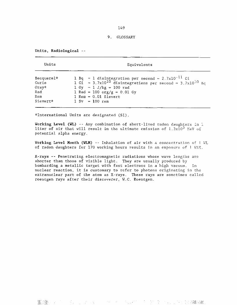


151
APPENDIX A PEER REVIEW
A peer review panel was assembled for thorium. The panel consisted of the following members: Dr. Marvin Goldman, Radiological Sciences and Toxicology, University of California; Dr. Raymond Lloyd, Radiology Division, University of Utah: Dr. Ingeborg Harding-Barlow, private consultant, Palo Alto, California; and Dr. George Angleton, Collaborative Radiology Health Laboratory, Colorado State University. These experts collectively have knowledge of thorium's physical and chemical properties, toxicokinetics, key health end points, mechanisms of action, human and animal exposure, and quantification of risk to humans. All reviewers were selected in conformity with the conditions for peer review specified in the Section 104(i)(13) of the Comprehensive Environmental Response, Compensation, and Liability Act as amended.
A joint panel of scientists from ATSDR and EPA has reviewed the peer reviewers' comments and determined which comments will be included in the profile. A listing of the peer reviewers' comments not incorporated in the profile, with brief explanation of the rationale for their exclusion, exists as part of the administrative record for this compound. A list of databases reviewed and a list of unpublished documents cited are also included in the administrative record.
The citation of the peer review panel should not be understood to imply its approval of the profile's final content. The responsibility for the content of this profile lies with the Agency for Toxic Substances and Disease Registry.


153
APPENDIX B
OVERVIEW OF BASIC RADIATION PHYSICS, CHEMISTRY, AND BIOLOGY
Understanding the basic concepts in radiation physics, chemistry, and biology is important to the evaluation and interpretation of radiationinduced adverse health effects and to the derivation of radiation protectiol principles. This appendix presents a brief overview of the areas of radiation physics, chemistry, and biology and is based to a large extent on the reviews of Mettler and Moseley (1985), Hobbs and McClellan (1986), Eichholz (1982), Hendee (1973), and Early et al. (1979).
B.l RADIONUCLIDES AND RADIOACTIVITY
The substances we call elements are composed of atoms. Atoms in turn are made up of neutrons, protons, and electrons; neutrons and protons in the nucleus and electrons in a cloud of orbits around the nucleus. Nuclide is the general term referring to any nucleus along with its orbital electrons. The nuclide is characterized by the composition of its nucleus and hence by the number of protons and neutrons in the nucleus. All atoms of an element have the same number of protons (this is given by the atomic number) but may have different numbers of neutrons (this is reflected by the atomic mass or atomic weight of the element). Atoms with different atomic mass but the same atomic numbers are referred to as isotopes of an element.
The numerical combination of protons and neutrons in most nuclides is such that the atom is said to be stable; however, if there are too few or too many neutrons, the nucleus of the atom is unstable. Unstable nuclides undergo a process referred to as radioactive transformation in which energy is emitted. These unstable atoms are called radionuclides; their emissions are called ionizing radiation; and the whole property is called radioactivity. Transformation or decay results in the formation of new nuclides some of which may themselves be radionuclides, while others are stable nuclides. This series of transformations is called the decay chain of the radionuclide. The first radionuclide in the chain is called the parent; the subsequent products of the transformation are called progeny, daughters, or decay products.
In general there are two classifications of radioactivity and radionuclides: natural and man-made. Naturally-occurring radionuclides exist in nature and no additional energy is necessary to place them in an unstable state. Natural radioactivity is the property of some naturally occurring, usually heavy elements, that are heavier than lead. Radionuclides, such as radium and uranium, primarily emit alpha particles. Some lighter elements such as carbon-14 and tritium (hydrogen-3) primarily emit beta particles as they transform to a more stable atom. Natural radioactive atoms heavier than lead cannot attain a stable nucleus heavier than lead. Everyone is exposed to background radiation from naturallyoccurring radionuclides throughout life. This background radiation is the major source of radiation exposure to man and arises from several sources.
154
APPENDIX B
The natural background exposures are frequently used as a standard of comparison for exposures to various man-made sources of ionizing radiation.
Man-made radioactive atoms are produced either as a by-product of fission of uranium atoms in a nuclear reactor or by bombarding stable atoms with particles, such as neutrons, directed at the stable atoms with high velocity. These artificially produced radioactive elements usually decay by emission of particles, such as positive or negative beta particles and one or more high energy photons (gamma rays). Unstable (radioactive) atoms of any element can be produced.
Both naturally occurring and man-made radioisotopes find application in medicine, industrial products, and consumer products. Some specific radioisotopes, called fall-out, are still found in the environment as a result of nuclear weapons use or testing.
B.2 RADIOACTIVE DECAY
B.2.1 Principles of Radioactive Decay
The stability of an atom is the result of the balance of the forces of the various components of the nucleus. An atom that is unstable (radionuclide) will release energy (decay) in various ways and transform to stable atoms or to other radioactive species called daughters, often with the release of ionizing radiation. If there are either too many or too few neutrons for a given number of protons, the resulting nucleus may undergo transformation. For some elements, a chain of daughter decay products may be produced until stable atoms are formed. Radionuclides can be characterized by the type and energy of the radiation emitted, the rate of decay, and the mode of decay. The mode of decay indicates how a parent compound undergoes transformation. Radiations considered here are primarily of nuclear origin, i.e., they arise from nuclear excitation, usually caused by the capture of charged or uncharged nucleons by a nucleus, or by the radioactive decay or transformation of an unstable nuclide. The type of radiation may be categorized as charged or uncharged particles (electrons, neutrons, neutrinos, alpha particles, beta particles, protons, and fission products) or electromagnetic radiation (gamma rays and X-rays). Table B.l summarizes the basic characteristics of the more common types of radiation encountered.
B.2.2 Half-Life and Activity
For any given radionuclide, the rate of decay is a first-order process that depends on the number of radioactive atoms present and is characteristic for each radionuclide. The process of decay is a series of random events; temperature, pressure, or chemical combinations do not effect the rate of decay. While it may not be possible to predict exactly which atom is going to undergo transformation at any given time, it is possible
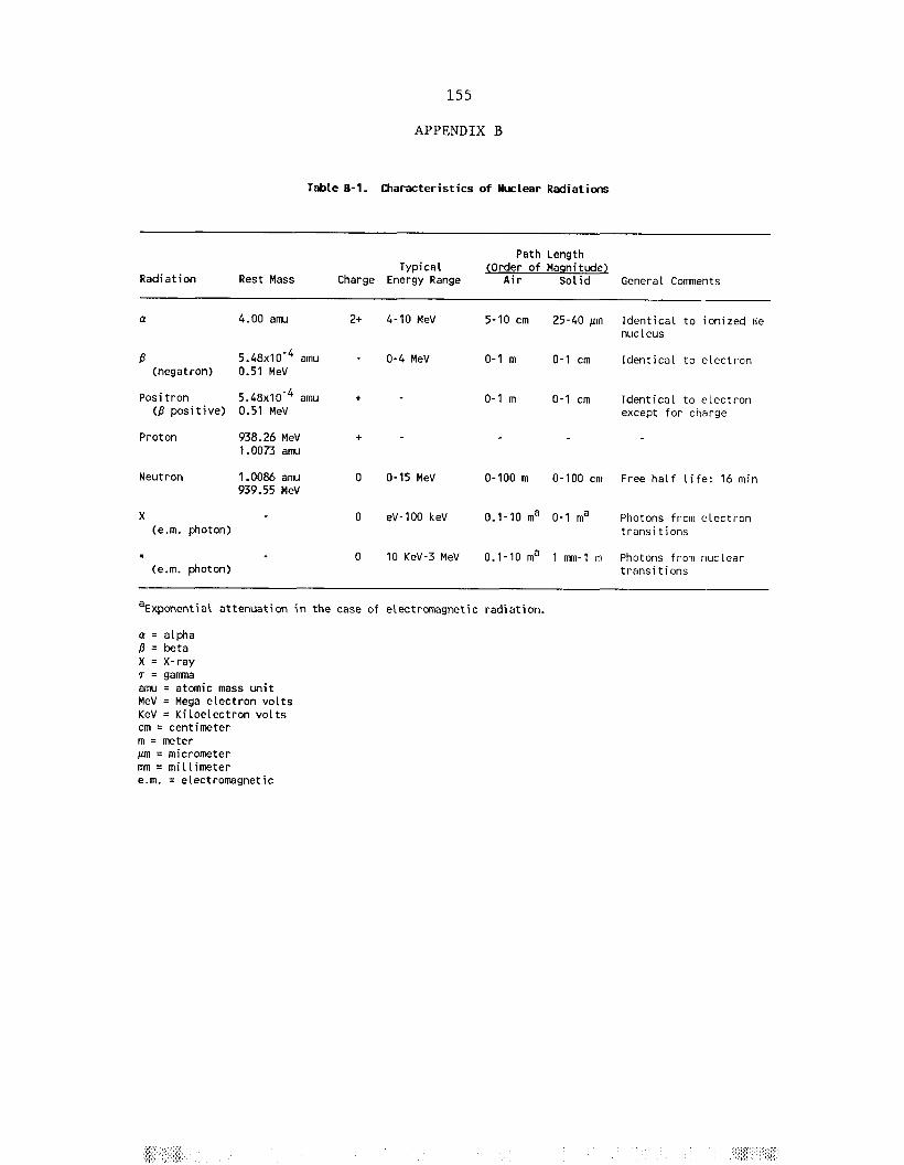

156
APPENDIX B
to predict, on the average, how many atoms will transform during any interval of time.
The source strength is a measure of the rate of emission of radiation. For these radioactive materials it is customary to describe the source strength in terms of the source activity, which is defined as the number of disintegrations (transformations) per unit time occurring in a given quantity of this material. The unit of activity is the curie (Ci) which was originally related to the activity of one gram of radium, but is now defined as:
101 curie (Ci) = 3x710 disintegrations (transformations)/second (dps) or
12.22x10 2 disintegrations (transformations)/minute (dpm).
The SI unit of activity is the becquerel (Bq); 1 Bq = 1 transformation/ second. Since activity is proportional to the number of atoms of the radioactive material, the quantity of any radioactive material is usually expressed in curies, regardless of its purity or concentration. The transformation of radioactive nuclei is a random process, and the rate of transformation is directly proportional to the number of radioactive atoms present. For any pure radioactive substance, the rate of decay is usually described by its radiological half-life, TE, i.e., the time it takes for a specified source material to decay to half its initial activity.
The activity of a radionuclide at time t may be calculated by:
-0A = Aoe ·693t/Trad
where A is the activity in dps, Ao is the activity at time zero, t is the
time at which measured, and Trad is the radiological half-life of the radionuclide. It is apparent that activity exponentially decays with time. The time when the activity of a sample of radioactivity becomes one-half its original value is the radioactive half-life and is expressed in any suitable unit of time.
The specific activity is the radioactivity per unit weight of material. This activity is usually expressed in curies per gram and may be calculated by
8curies/gram = 1.3x10 /(Trad)(atomic weight)
where Trad is the radiological half-life in days.
In the case of radioactive materials contained in living organisms, an additional consideration is made for the reduction in observed activity due to regular processes of elimination of the respective chemical or biochemical substance from the organism. This introduces a rate constant called the biological half-life (Tbiol) which is the time required for
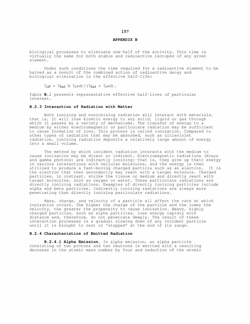
157
APPENDIX B
biological processes to eliminate one-half of the activity. This time is virtually the same for both stable and radioactive isotopes of any given element.
Under such conditions the time required for a radioactive element to be halved as a result of the combined action of radioactive decay and biological elimination is the effective half-life:
Teff = Tbiol X Trad)/(Tbiol + Trad).
Table B.2 presents representative effective half-lives of particular interest.
B.2.3 Interaction of Radiation with Matter
Both ionizing and nonionizing radiation will interact with materials, that is, it will lose kinetic energy to any solid, liquid or gas through which it passes by a variety of mechanisms. The transfer of energy to a medium by either electromagnetic or particulate radiation may be sufficient to cause formation of ions. This process is called ionization. Compared to other types of radiation that may be absorbed, such as ultraviolet radiation, ionizing radiation deposits a relatively large amount of energy into a small volume.
The method by which incident radiation interacts with the medium to cause ionization may be direct or indirect. Electromagnetic radiations (Xrays and gamma photons) are indirectly ionizing; that is, they give up their energy in various interactions with cellular molecules, and the energy is then utilized to produce a fast-moving charged particle such as an electron. It is the electron that then secondarily may react with a target molecule. Charged particles, in contrast, strike the tissue or medium and directly react with target molecules, such as oxygen or water. These particulate radiations are directly ionizing radiations. Examples of directly ionizing particles include alpha and beta particles. Indirectly ionizing radiations are always more penetrating than directly ionizing particulate radiations.
Mass, charge, and velocity of a particle all affect the rate at which ionization occurs. The higher the charge of the particle and the lower the velocity, the greater the propensity to cause ionization. Heavy, highly charged particles, such as alpha particles, lose energy rapidly with distance and, therefore, do not penetrate deeply. The result of these interaction processes is a gradual slowing down of any incident particle until it is brought to rest or "stopped" at the end of its range.
B.2.4 Characteristics of Emitted Radiation
B.2.4.1 Alpha Emission. In alpha emission, an alpha particle consisting of two protons and two neutrons is emitted with a resulting decrease in the atomic mass number by four and reduction of the atomic


159
APPENDIX B
number by two, thereby changing the parent to a different element. The alpha particle is identical to a helium nucleus consisting of two neutrons and two protons. It results from the radioactive decay of some heavy elements such as uranium, plutonium, radium, thorium, and radon. Alpha particles have a large mass as compared to electrons. Decay of alphaemitting radionuclides may result in the emission of several different alp1 particles. A radionuclide has an alpha emission with a discrete alpha energy and characteristic pattern of alpha energy emitted.
The alpha particle has an electrical charge of +2. Because of this double positive charge, alpha particles have great ionizing power, but their large size results in very little penetrating power. In fact, an alpha particle cannot penetrate a sheet of paper. The range of an alpha particle, that is, the distance the charged particle travels from the point of origin to its resting point, is about 4 cm in air, which decreases considerably to a few micrometers in tissue. These properties cause alpha emitters to be hazardous only if there is internal contamination (i.e., if the radionuclidc is ingested, inhaled, or otherwise absorbed).
B.2.4.2. Beta Emission. Nuclei which are excessively neutron rich decay by β-decay. A beta particle (β) is a high-velocity electron ejected from a disintegrating nucleus. The particle may be either a negatively charged electron, termed a negatron (β-) or a positively charged electron, termed a positron (β+). Although the precise definition of "beta emission" refers to both β- and β+, common usage of the term generally applies only to the negative particle, as distinguished from the positron emission, which refers to the β+ particle.
B.2.4.2.1 Beta Negative Emission. Beta particle (β-) emission is another process by which a radionuclide, usually those with a neutron excess, achieves stability. Beta particle emission decreases the number of neutrons by one and increases the number of protons by one, while the atomic mass remains unchanged. This transformation results in the formation of a different element. The energy spectrum of beta particle emission ranges from a certain maximum down to zero with the mean energy of the spectrum being about one-third of the maximum. The range in tissue is much less. Beta negative emitting radionuclides can cause injury to the skin and superficial body tissues but mostly present an internal contamination hazard.
B.2.4.2.2 Positron Emission. In cases in which there are too many protons in the nucleus, positron emission may occur. In this case a proton may be thought of as being converted into a neutron, and a positron (β+) is emitted, accompanied by a neutrino (see glossary). This increases the number of neutrons by one, decreases the number of protons by one, and again leaves the atomic mass unchanged. The gamma radiation resulting from the annihilation (see glossary) of the positron makes all positron emitting isotopes more of an external radiation hazard than pure β emitters of equal energy.

160
APPENDIX B
B.2.4.2.3 Gamma Emission. Radioactive decay by alpha, beta, positron emission or electron capture often leaves some of the energy resulting from these changes in the nucleus. As a result, the nucleus is raised to an excited level. None of these excited nuclei can remain in this high-energy state. Nuclei release this energy returning to ground state or to the lowest possible stable energy level. The energy released is in the form of gamma radiation (high energy photons) and has an energy equal to the change in the energy state of the nucleus. Gamma and X-rays behave similarly but differ in their origin; gamma emissions originate in the nucleus while X-rays originate in the orbital electron structure.
B.3 ESTIMATION OF ENERGY DEPOSITION IN HUMAN TISSUES
Two forms of potential radiation exposures can result -- internal and external. The term exposure denotes physical interaction of the radiation emitted from the radioactive material with cells and tissues of the human body. An exposure can be "acute" or "chronic" depending on how long an individual or organ is exposed to the radiation. Internal exposures occur when radionuclides, which have entered the body (e.g., through the inhalation, ingestion, or dermal pathways), undergo radioactive decay resulting in the deposition of energy to internal organs. External exposures occur when radiation enters the body directly from sources located outside the body, such as radiation emitters from radionuclides on ground surfaces, dissolved in water, or dispersed in the air. In general, external exposures are from material emitting gamma radiation, which readily penetrate the skin and internal organs. Beta and alpha radiation from external sources are far less penetrating and deposit their energy primarily on the skin's outer layer. Consequently, their contribution to the absorbed dose of the total body dose, compared to that deposited by gamma rays, may be negligible.
Characterizing the radiation dose to persons as a result of exposure to radiation is a complex issue. It is difficult to: (1) measure internally the amount of energy actually transferred to an organic material and to correlate any observed effects with this energy deposition; and (2) account for and predict secondary processes, such as collision effects or biologically triggered effects, that are an indirect consequence of the primary interaction event.
B.3.1 Dose Units
B.3.1.1 Roentgen. The roentgen (R) is a unit of exposure related to the amount of ionization caused in air by gamma or x-radiation. One
-4roentgen equals 2.58x10 Coulomb per kilogram of air. In the case of gamma radiation, over the commonly encountered range of photon energy, the energy deposition in tissue for a dose of 1 R is about 0.0096 joules(J)/kg of tissue.

161
APPENDIX B
B.3.1.2 Absorbed Dose and Absorbed Dose Rate. Since different typesof radiation interact differently with any material through which they pass, any attempt to assess their effect on humans or animals should take into account these differences. The absorbed dose is defined as the energy imparted by the incident radiation to a unit mass of the tissue or organ. The unit of absorbed dose is the rad; 1 rad = 100 erg/gram = 0.01 J/kg in any medium. The SI unit is the gray which is equivalent to 100 rad or 1 J/kg. Internal and external exposures from radiation sources are not usually instantaneous but are distributed over extended periods of time. The resulting rate of change of the absorbed dose to a small volume of mass is referred to as the absorbed dose rate in units of rad/unit time.
B.3.1.3 Working Levels and Working Level Months. Working levels are units that have been used to describe the radon decay-product activities in air in terms of potential alpha energy. It is defined as any combination of short-lived radon daughters (through polonium-214) per liter of air that
5will result in the emission of 1.3x10 MeV of alpha energy. An activity concentration of 100 pCi radon-222/L of air, in equilibrium with its daughters, corresponds approximately to a potential alpha-energy concentration of 1 WL. The WL unit can also be used for thoron daughters.
5In this case, 1.3x10 MeV of alpha energy (1 WL) is released by the thoron daughters in equilibrium with 7.5 pCi thoron/L. The potential alpha energy exposure of miners is commonly expressed in the unit Working Level Month (WIJf). One WLM corresponds to exposure to a concentration of 1 WL for the reference period of 170 hours.
B.3.2 Dosimetry Models
Dosimetry models are used to estimate the internally deposited dose from exposure to radioactive substances. The models for internal dosimetry consider the quantity of radionuclides entering the body, the factors affecting their movement or transport through the body, distribution and retention of radionuclides in the body, and the energy deposited in organs and tissues from the radiation that is emitted during spontaneous decay processes. The models for external dosimetry consider only the photon doses to organs of individuals who are immersed in air or are exposed to a contaminated ground surface. The dose pattern for radioactive materials in the body may be strongly influenced by the route of entry of the material. For industrial workers, inhalation of radioactive particles with pulmonary deposition and puncture wounds with subcutaneous deposition have been the most frequent. The general population has been exposed via ingestion and inhalation of low levels of naturally occurring radionuclides as well as man-produced radionuclides from nuclear weapons testing.
B.3.2.1 Ingestion. Ingestion of radioactive materials is most likely to occur from contaminated foodstuffs or water or eventual ingestion of inhaled compounds initially deposited in the lung. Ingestion of radioactive material may result in toxic effects as a result of either absorption of the radionuclide or irradiation of the gastrointestinal tract during passage

162
APPENDIX B
through the tract, or a combination of both. The fraction of a radioactive material absorbed from the gastrointestinal tract is variable, depending on the specific element, the physical and chemical form of the material ingested, and the diet, as well as some other metabolic and physiological factors. The absorption of some elements is influenced by age usually with higher absorption in the very young.
B.3.2.2 Inhalation. The inhalation route of exposure has long been recognized as being of major importance for both nonradioactive and radioactive materials. The deposition of particles within the lung is largely dependent upon the size of the particles being inhaled. After the particle is deposited, the retention will depend upon the physical and chemical properties of the dust and the physiological status of the lung. The retention of the particle in the lung depends on the location of deposition, in addition to the physical and chemical properties of the particles. The converse of pulmonary retention is pulmonary clearance. There are three distinct mechanisms of clearance which operate simultaneously, Ciliary clearance acts only in the upper respiratory tract. The second and third mechanisms act mainly in the deep respiratory tract. These are phagocytosis and absorption. Phagocytosis is the engulfing of foreign bodies by alveolar macrophages and their subsequent removal either up the ciliary "escalator" or by entrance into the lymphatic system. Some inhaled soluble particulates are absorbed into the blood and translocated to other organs and tissues. Dosimetric lung models are reviewed by James (1987) and James and Roy (1987).
B.3.3 Internal Emitters
The absorbed dose from internally deposited radioisotopes is the energy absorbed by the surrounding tissue. For a radioisotope distributed uniformly throughout an infinitely large medium, the concentration of absorbed energy must be equal to the concentration of energy emitted by the isotope. An infinitely large medium may be approximated by a tissue mass whose dimensions exceed the range of the particle. All alpha and most beta radiation will be absorbed in the organ (or tissue) of reference. Gamma emitting isotope emissions are penetrating radiation and a substantial fraction may travel great distances within tissue, leaving the tissue without interacting. The dose to an organ or tissue is a function of the effective retention half-time, the energy released in the tissue, the amount of radioactivity initially introduced, and the mass of the organ or tissue,
B.4 BIOLOGICAL EFFECTS OF RADIATION
When biological material is exposed to ionizing radiation, a chain of cellular events occurs as the ionizing particle passes through the biological material. A number of theories have been proposed to describe the interaction of radiation with biologically important molecules in cells and to explain the resulting damage to biological systems from those interactions. Many factors may modify the response of a living organism to
163
APPENDIX B
a given dose of radiation. Factors related to the exposure include the dose rate, the energy of the radiation, and the temporal pattern of the exposure. Biological considerations include factors such as species, age, sex, and tile portion of the bodjr exposed. Several excellent reviews of the biological effects of radiation have been published, and the reader is referred to these for a more in-depth discussion (Hobbs and McClellan 1986; ICRP 1984; Mettler and Moseley 1985; Rubin and Casarett 1968).
B.4.1 Radiation Effects at the Cellular Level
According to Mettler and Moseley (1985), at acute doses up to 10 rad (100 mGy), single strand breaks in DNA may be produced. These single strand breaks may be repaired rapidly. With doses in the range of 50 to 500 rad (0.5 to 5 Gy), irreparable double-stranded DNA breaks are likely, resulting in cellular reproductive death after one or more divisions of the irradiated parent cell. At large doses of radiation, usually greater than 500 rad (5 GY) t direct cell death before division (interphase death) may occur from the direct interaction of free-radicals with essentially cellular macromolecules. Morphological changes at the cellular level, the severity of which are dose-dependent, may also be observed.
The sensitivity of various cell types varies. According to the Bergoni-Tribondeau law, the sensitivity of cell lines is directly proportional to their mitotic rate and inversely proportional to the degree of differentiation (Mettler and Moseley 1985). Rubin and Casarett (1968) devised a classification system that categorized cells according to type, function, and mitotic activity. The categories range from the most sensitive type, "vegetative intermitotic cells," found in the stem cells of the bone marrow and the gastrointestinal tract, to the least sensitive cell type, "fixed postmitotic cells," found in striated muscles or long-lived neural tissues.
Cellular changes may result in cell death, which if extensive, may produce irreversible damage to an organ or tissue or may result in the of the individual. If the cell recovers, altered metabolism and function may still occur, which may be repaired or may result in the manifestation of clinical symptoms. These changes may also be expressed at a later time as tumors or mutations.
B.4.2 Radiation Effects at the Organ Level
In most organs and tissues the injury and the underlying mechanism for that injury are complex and may involve a combination of events. The extent and severity of this tissue injury are dependent upon the radiosensitivity of the various cell types in that organ system. Rubin and Casarett (1968) describe and schematically display the events following radiation in several organ system types. These include: a rapid renewal system, such as the gastrointestinal mucosa; a slow renewal system, such as the pulmonary epithelium; and a nonrenewal system, such as neural or muscle tissue. In
164
APPENDIX B
the rapid renewal system, organ injury results from the direct destruction of highly radiosensitive cells, such as the stem cells in the bone marrow. Injury may also result from constriction of the microcirculation and from edema and inflammation of the basement membrane (designated as the histohematic barrier - HHB), which may progress to fibrosis. In slow renewal and nonrenewal systems, the radiation may have little effect on the parenchymal cells, but ultimate parenchymal atrophy and death over several months result from HHB fibrosis and occlusion of the microcirculation.
B.4.3 Acute and Chronic Somatic Effects
B.4.3.1 Acute Effects. The result of acute exposure to radiation is commonly referred to as acute radiation syndrome. This effect is seen only after exposures to relatively high doses (>50 rad), which would only be expected to occur in the event of a serious nuclear accident. The four stages of acute radiation syndrome are prodrome, latent stage, manifest illness stage, recovery or death. The initial phase is characterized by nausea, vomiting, malaise and fatigue, increased temperature, and blood changes. The latent stage is similar to an incubation period. Subjective symptoms may subside, but changes may be taking place within the bloodforming organs and elsewhere which will subsequently give rise to the next stage. The manifest illness stage gives rise to symptoms specifically associated with the radiation injury. Among these symptoms are hair loss, fever, infection, hemorrhage, severe diarrhea, prostration, disorientation, and cardiovascular collapse. The symptoms and their severity depend upon the radiation dose received.
B.4.3.2 Delayed Effects. The level of exposure to radioactive pollutants that may be encountered in the environment is expected to be too low to result in the acute effects described above. When one is exposed to radiation in the environment, the amount of radiation absorbed is more likely to produce long-term effects, which manifest themselves years after the original exposure, and may be due to a single large over-exposure or continuing low-level exposure.
Sufficient evidence exists in both human populations and laboratory animals to establish that radiation can cause cancer and that the incidence of cancer increases with increasing radiation dose. Human data are extensive and include epidemiological studies of atomic bomb survivors, many types of radiation-treated patients, underground miners, and radium dial painters. Reports on the survivors of the atomic bomb explosions at Hiroshima and Nagasaki, Japan (with whole-body external radiation doses of 0 to more than 200 rad) indicate that cancer mortality has increased (Kato and Schull 1982). Use of X-rays (at doses of approximately 100 rad) in medical treatment for ankylosing spondylitis or other benign conditions or diagnostic purposes, such as breast conditions, has resulted in excess cancers in irradiated organs (BEIR 1980, 1990; UNSCEAR 1977, 1988). Cancers, such as leukemia, have been observed in children exposed in utero to doses of 0.2 to 20 rad (BEIR, 1980, 1990; UNSCEAR 1977, 1988). Medical

165
APPENDIX B
use of Thorotrast (colloidal thorium dioxide) resulted in increases in the incidence of cancers of the liver, bone, and lung (ATSDR 1990a; BEIR 1980, 1990; UNSCEAR 1977, 1988). Occupational exposure to radiation provides further evidence of the ability of radiation to cause cancer. Numerous studies of underground miners exposed to radon and radon daughters, which are alpha emitters, in uranium and other hard rock mines have demonstrated i-ncreases in lung cancer in exposed workers (ATSDR 1990b). Workers who ingested radium-226 while painting watch dials had an increased incidence of leukemia and bone cancer (ATSDR 199Oc). These studies indicate that depending on radiation dose and the exposure schedule, ionizing radiation can induce cancer in nearly any tissue or organ in the body. Radiationinduced cancers in humans are found to occur in the hemopoietic system, the lung, the thyroid, the liver, the bone, the skin, and other tissues.
Laboratory animal data indicate that ionizing radiation is carcinogenic and mutagenic at relatively high doses usually delivered at high dose rates. However, due to the uncertainty regarding the shape of the dose-response curve, especially at low doses, the commonly held conservative position is that the cancer may occur at dose rates that extend down to doses that could be received from environmental exposures. Estimates of cancer risk are based on the absorbed dose of radiation in an organ or tissue. The cancer risk at a particular dose is the same regardless of the source of the radiation. A comprehensive discussion of radiation-induced cancer is found in BEIR IV (1988), BETR V (1990), and UNSCEAR (1982, 1988).
B.4.4 Genetic Effects
Radiation can induce genetic damage, such as gene mutations or chromosomal aberrations, by causing changes in the structure, number, 0 r genetic content of chromosomes in the nucleus. The evidence for the mutagenicity of radiation is derived from studies in laboratory animals, mostly mice (BEIR 1980, 1988, 1990; UNSCEAR 1982, 1986, 1988). Evidence for genetic effects in humans is derived from tissue cultures of human lymphocytes from persons exposed to ingested or inhaled radionuclidcs (ATSDR) 139Oc, 1990d). Evidence for mutagenesis in human germ cells (cells of the ovaries or testis) is not conclusive (BEIR 1980, 1988, 1990; UNSCEAR 1977,1986, 1988). Chromosome aberrations following radiation exposure have been demonstrated in man andn in experimental animals (BEIR 1980, 1988, 1990; UNSCEAR 1982, 1986, 1988).
B.4.5 Teratogenic Effects
There is evidence that radiation produces teratogcnicity in animals. It appears that the developing fetus is more sensitive to radiation than the mother and is most sensitive to radiation- induced damage during the. early stages of organ development. The type of malformation depends on the stage of development and the cells that are undergoing the most rapid differentiation at the time. Studies of mental retardation in children exposed in utero to radiation from the atonic bomb provide evidence that

166
APPENDIX B
radiation may produce teratogenic effects in human fetuses (Otake and Schull 1984). The damage to the child was found to be related to the dose that the fetus received.
B.5 UNITS IN RADIATION PROTECTION AND REGULATION
B.5.1 Dose Equivalent and Dose Equivalent Rate
Dose equivalent or rem is a special radiation protection quantity that is used to express the absorbed dose in a manner which considers the difference in biological effectiveness of various kinds of ionizing radiation. The ICRU has defined the dose equivalent, H, as the product of the absorbed dose, D, the quality factor, Q, and all other modifying factors, N, at the point of interest in biological tissue. This relationship is expressed as follows:
H=DxQxN.
The quality factor is a dimensionless quantity that depends in part on the stopping power for charged particles, and it accounts for the differences il biological effectiveness found among the types of radiation. By definition it is independent of tissue and biological end point and, therefore, of little use in risk assessment now. Originally Relative Biolotical Effectiveness (RBE) was used rather than Q to define the quantity, rem, which was of use in risk assessment. The generally accepted values for quality factors for various radiation types are provided in Table B.3. The dose equivalent rate is the time rate of change of the dose equivalent to organs and tissues and is expressed as rem/unit time or sievert/unit time.
B.5.2 Relative Biological Effectiveness
The term relative biologic effectiveness (RBE) is used to denote the experimentally determined ratio of the absorbed dose from one radiation type to the absorbed dose of a reference radiation required to produce an identical biologic effect under the same conditions. Gamma rays from cobalt-60 and 200 to 250 KeV X-rays have been used as reference standards. The term RBE has been widely used in experimental radiobiology, and the term quality factor used in calculations of dose equivalents for radiation protection purposes (ICRP 1977; NCRP 1971; UNSCEAR 1982). The generally accepted values for RBE are provided in Table B.4.
B.5.3 Effective Dose Equivalent and Effective Dose Equivalent Rate
The absorbed dose is usually defined as the mean absorbed dose within an organ or tissue. This represents a simplification of the actual problem. Normally when an individual ingests or inhales a radionuclide or is exposed to external radiation that enters the body (gamma), the dose is not uniform throughout the whole body. The simplifying assumption is that the detriment

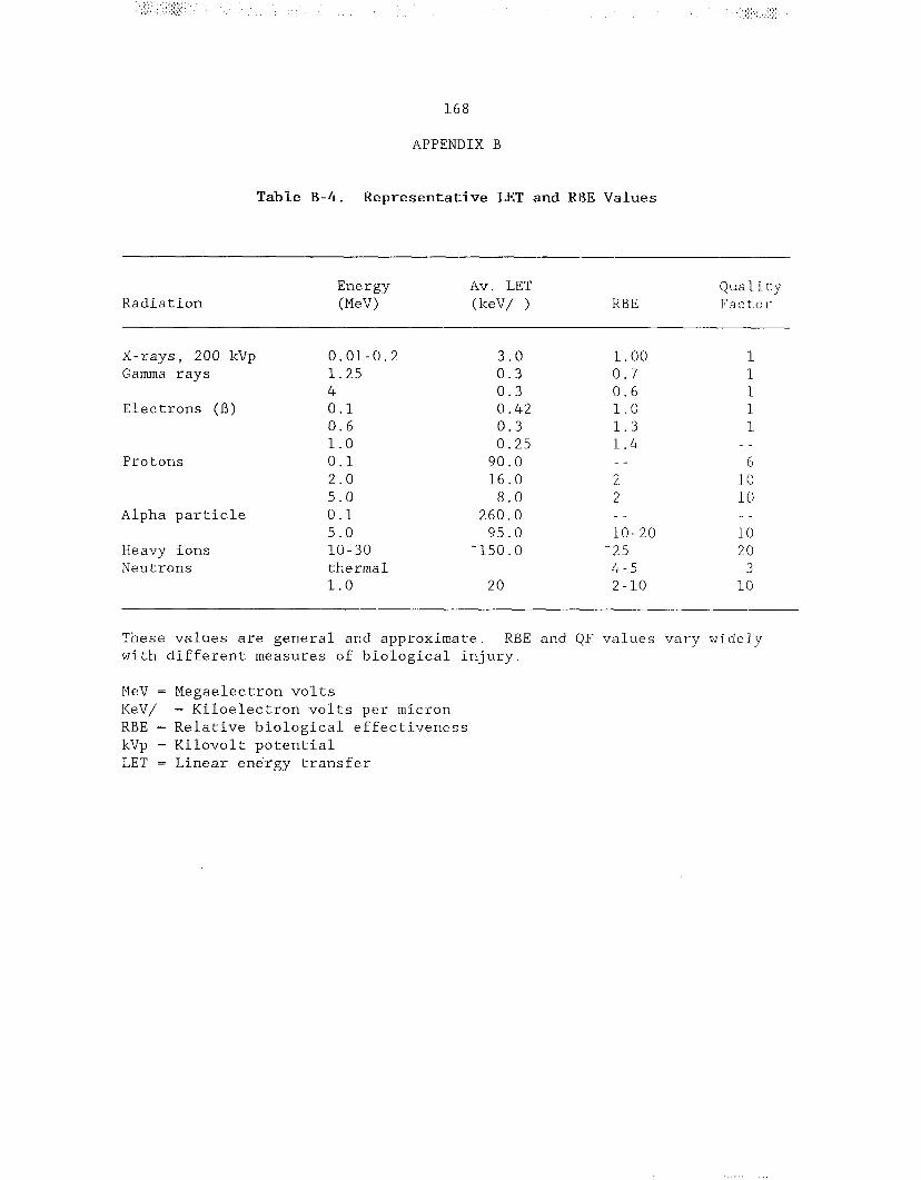
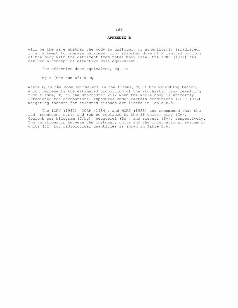
169
APPENDIX B
will be the same whether the body is uniformly or nonuniformly irradiated. In an attempt to compare detriment from absorbed dose of a limited portion of the body with the detriment from total body dose, the ICRP (1977) has derived a concept of effective dose equivalent.
The effective dose equivalent, Hg, is
Hg = (the sum of) Wt Ht
where Ht is the dose equivalent in the tissue, Wt is the weighting factor, which represents the estimated proportion of the stochastic risk resulting from tissue, T, to the stochastic risk when the whole body is uniformly irradiated for occupational exposures under certain conditions (ICRP 1977). Weighting factors for selected tissues are listed in Table B.5.
The ICRU (1980), ICRP (1984), and NCRP (1985) now recommend that the rad, roentgen, curie and rem be replaced by the SI units: gray (Gy), Coulomb per kilogram (C/kg), becquerel (Bq), and sievert (Sv), respectively. The relationship between the customary units and the international system of units (SI) for radiological quantities is shown in Table B.6.


172
APPENDIX B
References
ATSDR. 1990a. Toxicological profile for thorium. U.S. Department of Health and Human Services. Public Health Service. Agency for Toxic Substances and Disease Registry. Atlanta, GA.
ATSDR. 1990b. Toxicological profile for radium. U.S. Department of Health and Human Services. Public Health Service. Agency for Toxic Substances and Disease Registry. Atlanta, GA.
ATSDR. 199oc. Toxicological profile for radon. U.S. Department of Health and Human Services. Public Health Service. Agency for Toxic Substances and Disease Registry. Atlanta, GA.
ATSDR. 1990d. Toxicological profile for uranium. U.S. Department of Health and Human Services. Public Health Service. Agency for Toxic Substances and Disease Registry. Atlanta, GA.
BEIR III. 1980. The effects on populations of exposure to low levels of ionizing radiation. Committee on the Biological Effects of Ionizing Radiations, National Research Council. Washington, DC: National Academy Press.
BEIR IV. 1988. Health risks of radon and other internally deposited alpha emitters. Committee on the Biological Effects of Ionizing Radiations, National Research Council. Washington, DC: National Academy Press.
BEIR V. 1990. Health effects of exposure to low levels of ionizing radiation. Committee on the Biological Effects of Ionizing Radiations, National Research Council. Washington, DC: National Academy Press.
Early P, Razzak M, Sodee D. 1979. Nuclear medicine technology. 2nd ed. St. Louis: C.V. Mosby Company.
Eichholz G. 1982. Environmental aspects of nuclear power. Ann Arbor, MI: Ann Arbor Science.
Hendee W. 1973. Radioactive isotopes in biological research. New York, NY: John Wiley and Sons.
Hobbs C, McClellan R. 1986. Radiation and radioactive materials. In: Doull J, et al., eds. Casarett and Doull's Toxicology. 3rd ed. New York, NY: Macmillan Publishing Co., Inc., 497-530.
ICRP. 1977. International Commission on Radiological Protection. Recommendations of the International Commission on Radiological Protection. ICRP Publication 26. Vol 1. No. 3. Oxford: Pergamon Press.

173
APPENDIX B
ICRP. 1979. International Commission on Radiological Protection. Limits for intakes of radionuclides by workers. ICRP Publication 20. Vol 3. No. l-4. Oxford: Pergamon Press.
ICRP. 1984. International Commission on Radiological Protection. A compilation of the major concepts and quantities in use by ICRP. ICRP Publication 42. Oxford: Pergamon Press.
ICRU. 1980. International Commission on Radiation Units and Measurements.
ICRU Report No. 33. Washington, DC.
James A. 1987. A reconsideration of cells at risk and other key factors in radon daughter dosimetry. In: Hopke P, ed. Radon and its decay products: Occurrence, properties and health effects. ACS Symposium Series 331. Washington, DC: American Chemical Society, 400-418.
James A, Roy M. 1987. Dosimetric lung models. In: Gerber G, et al., ed. Age-related factors in radionuclide metabolism and dosimetry. Boston: Martinus Nijhoff Publishers, 95-108.
Kato H, Schull W. 1982. Studies of the mortality of A-bomb survivors. Report 7 Part 1, Cancer mortality among atomic bomb survivors, 1950-78. Radiat Res 90:395-432.
Mettler F, Moseley R. 1985. Medical effects of ionizing radiation. New York: Grune and Stratton.
NCRP 1971. National Council on Radiation Protection and Measurements. Basic radiation protection criteria. NCRP Report No. 39. Washington, DC.
NCRP. 1985. A handbook of radioactivity measurements procedures. 2nd ed. Bethesda, MD: National Council on Radiation Protection and Measurements. NCRP Report No. 58.
Otake M, Schull W. 1984. Mental retardation in children exposed in utero to the atomic bombs: A reassessment. Technical Report RERF TR l-83, Radiation Effects Research Foundation, Japan.
Rubin P, Casarett G. 1968. Clinical radiation pathology. Philadelphia: W.B. Sanders Company, 33.
UNSCEAR. 1977. United Nations Scientific Committee on the Effects of Atomic Radiation. Sources and effects of ionizing radiation. New York: United Nations.
UNSCEAR. 1982. United Nations Scientific Committee on the Effects of Atomic Radiation. Ionizing radiation: Sources and biological effects. New York: United Nations.

174
APPENDIX B
UNSCEAR. 1986. United Nations Scientific Committee on the Effects of Atomic
Radiation. Genetic and somatic effects of ionizing radiation. New York: United Nations.
UNSCEAR. 1988. United Nations Scientific Committee on the Effects of Atomic Radiation. Sources, effects and risks of ionization radiation. New York: United Nations.