topography orbscan
DESCRIPTION
TOPOGRAPHY ORBSCAN. S.A.A. Mortazavi MD. Associate Professor of Ophthalmology Isfahan University of Medical Sciences. ORBSCAN SYSTEM. Use the principle of projection - PowerPoint PPT PresentationTRANSCRIPT
1
TOPOGRAPHYORBSCAN
S.A.A. Mortazavi MD.Associate Professor of OphthalmologyIsfahan University of Medical Sciences
2/22/2013
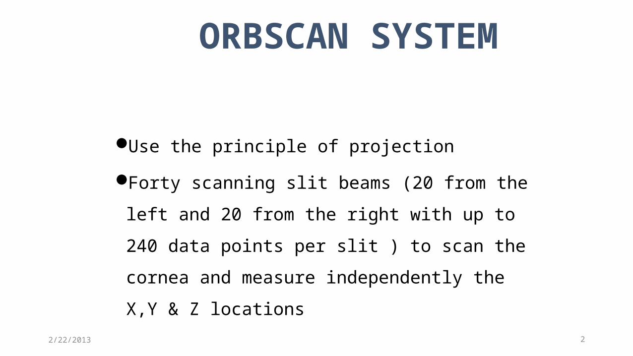
Use the principle of projection
Forty scanning slit beams (20 from the left and
20 from the right with up to 240 data points per
slit ) to scan the cornea and measure
independently the X,Y & Z locations2
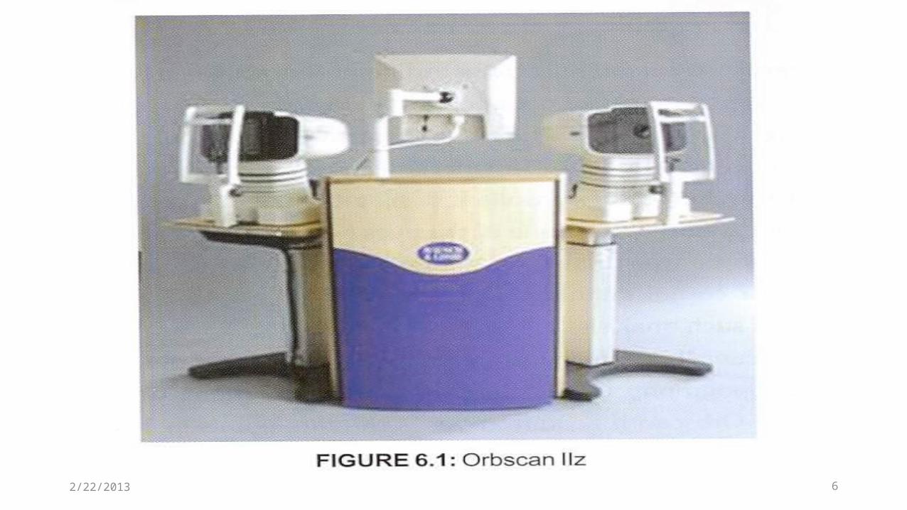
ORBSCAN SYSTEM
2/22/2013
3
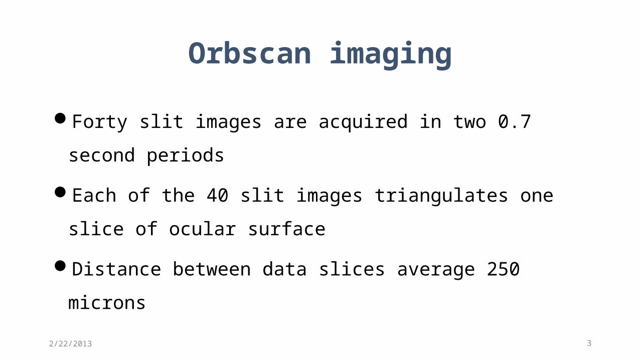
Forty slit images are acquired in two 0.7 second periods
Each of the 40 slit images triangulates one slice of ocular surface
Distance between data slices average 250 microns
Orbscan imaging
2/22/2013
42/22/2013
5
Orbscan I only slit scan topography
Orbscan II the placidodisc added in orbscan I
ORBSCAN
2/22/2013
62/22/2013
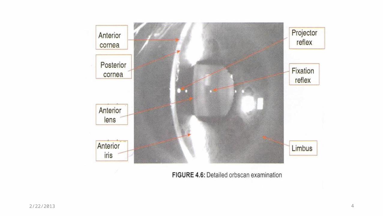
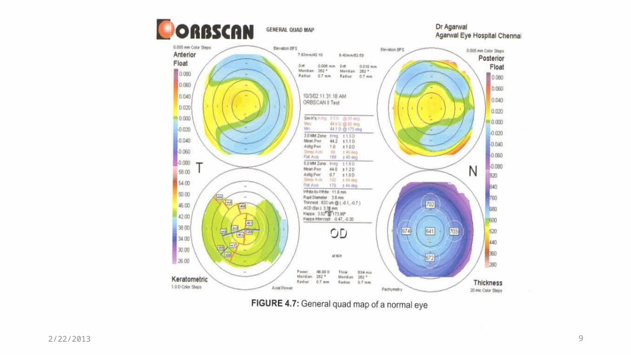
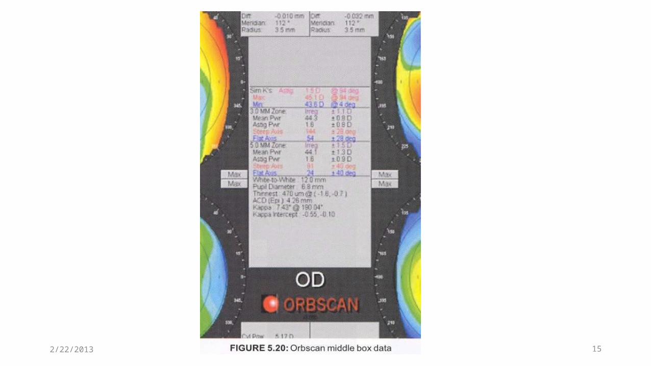
The images used to construct the anterior corneal
surface,posterior corneal surface,anterior iris and
anterior lens surfaces
Data regarding the corneal pachymetry and anterior
chamber depth
7
ORBSCAN
2/22/2013
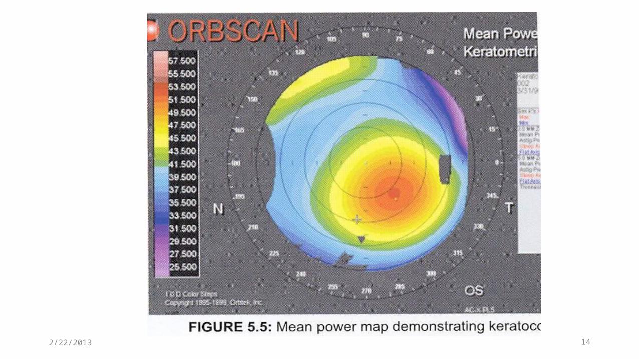
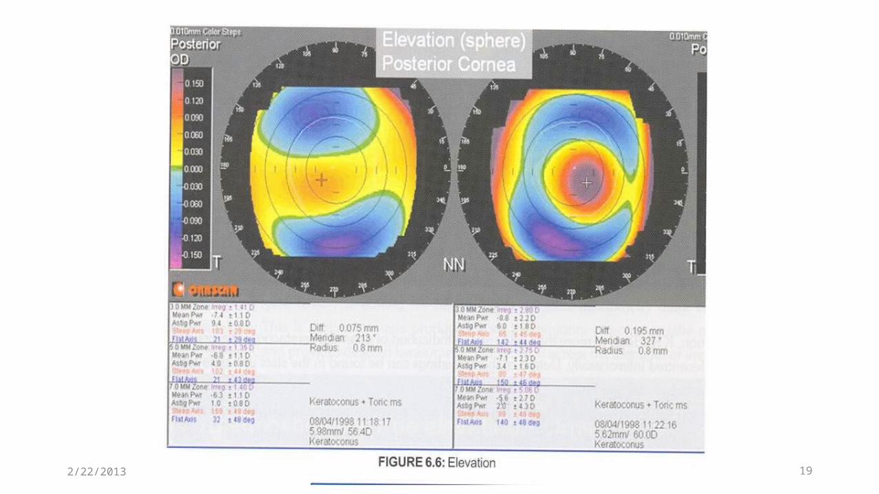
The computer calculates a hypothetical sphere that
matches as close as possible to the actual corneal
shape being measured
Compares the real surface to the hypothetical sphere
showing areas above the surface of the sphere in warm
colours and areas below the surface in cool colours8
BEST FIT SPHERE (BFS)
2/22/2013
92/22/2013
10
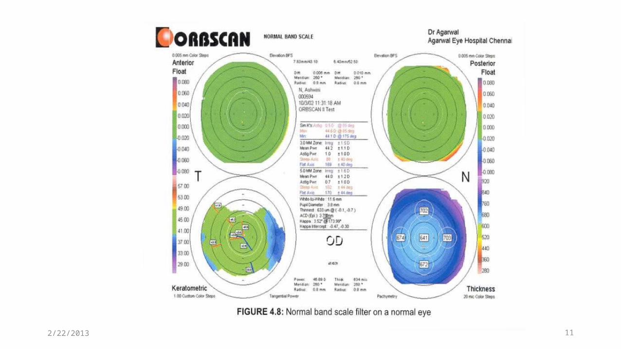
Highlights the abnormal areas in the cornea in orange to red colors
The normal areas are all shown in green
Helpful in generalized screening in preoperative examination
NORMAL BAND SCALE
2/22/2013
112/22/2013
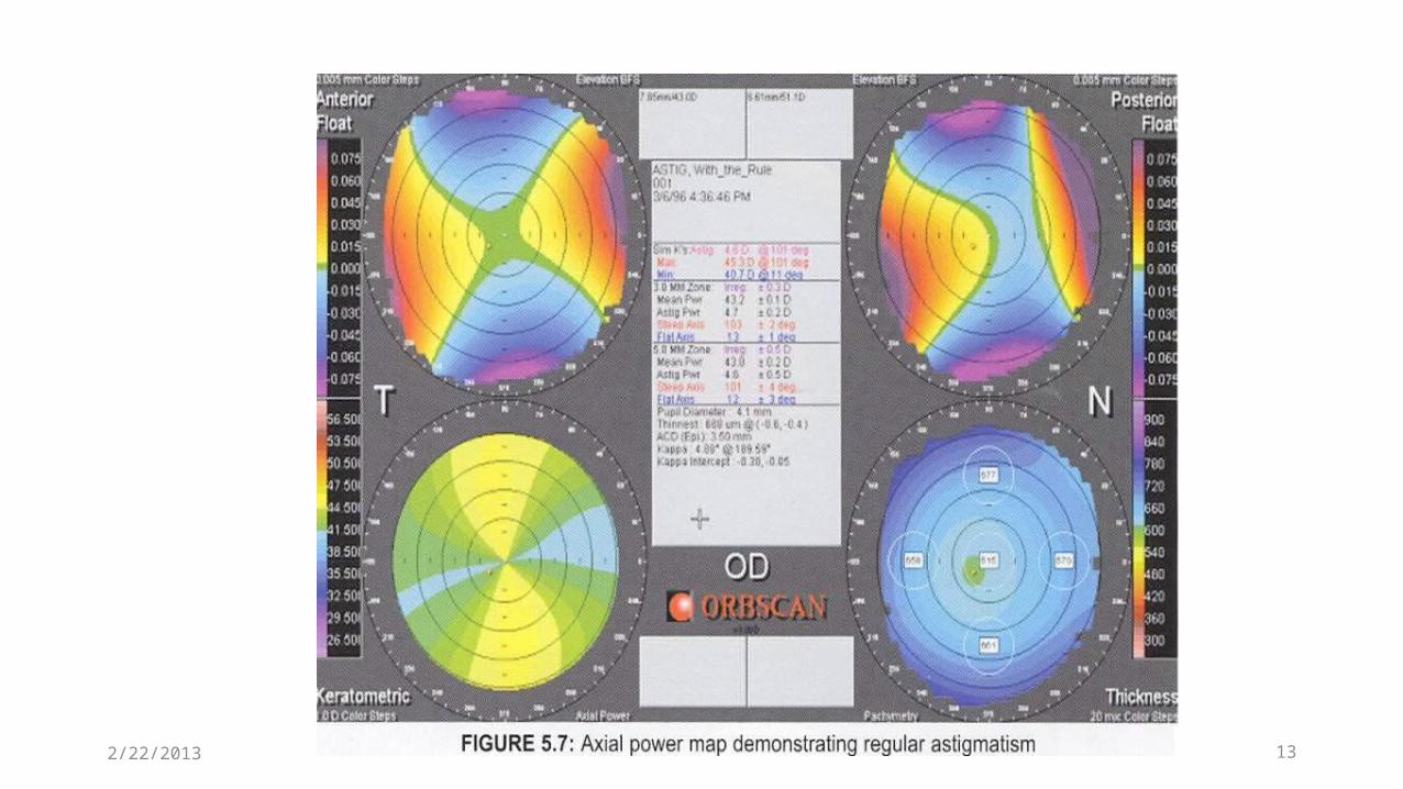
To create a good quality corneal flap in LASIK if
either extremes (too steep or too flat) is the case,
this can lead to surgical flap complications
K readings of more than 48 D are an indication of
potential keratoconus
12
AXIAL MAP
2/22/2013
132/22/2013
142/22/2013
152/22/2013
16
The orbscan measures thickness from the tear film layer to descemet’s membrane and is thicker than that obtained with ultrasound
Adjustment factor (acoustic factor) ,the default setting is 92%
Provides a reading showing the thinnest point of the cornea that may not necessarily be the central reading
PACHYMETRY MAP
2/22/2013
Thinnest point <470 micron
In pathological corneas, thinnest point is
often displaced inferotemporal
Difference of >100 microns from the thinnest
point to the values at 7mm optical zone17
PACHYMETRY MAP
2/22/2013
The green colour is referred as refrence
sphere (at sea level )
The warmer colours are above this level
and the cooler colours are below
18
ELEVATION MAP
2/22/2013
192/22/2013
The difference between the highest and
lowest points is a potential keratoconus
indicator if over 100 microns (Rousch
criteria)
20
ELEVATION DATA
2/22/2013
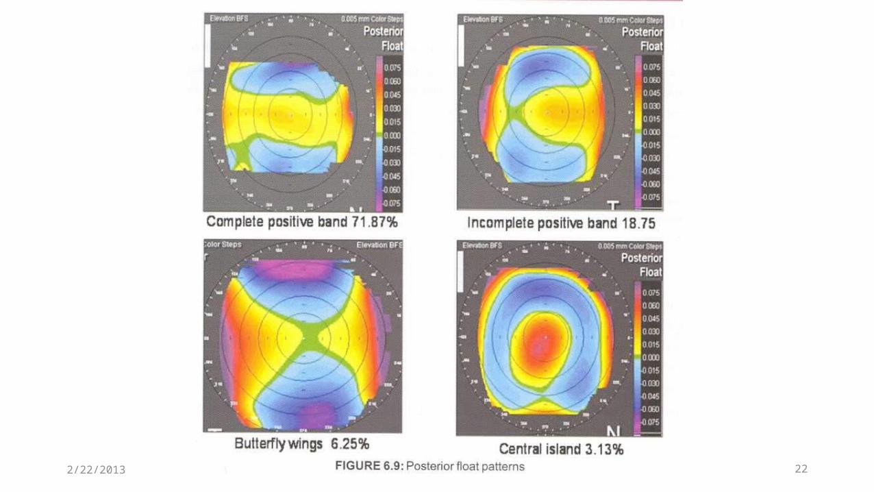
Many surgeons think the first sign of keratoconus appears on the posterior surface of the cornea
3.13% of population screened for laser surgery had posterior ectasia criteria by orbscan , despite having axial topography classified as normal
21
POSTERIOR ELEVATION MAP
2/22/2013
222/22/2013
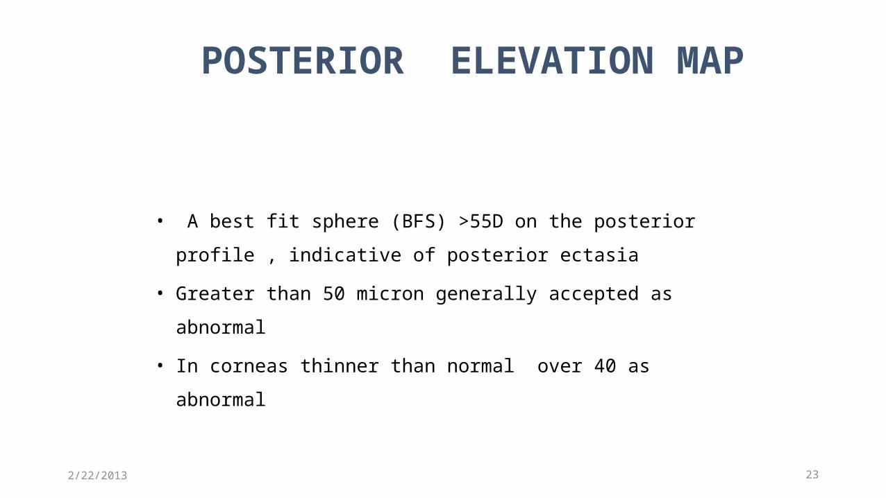
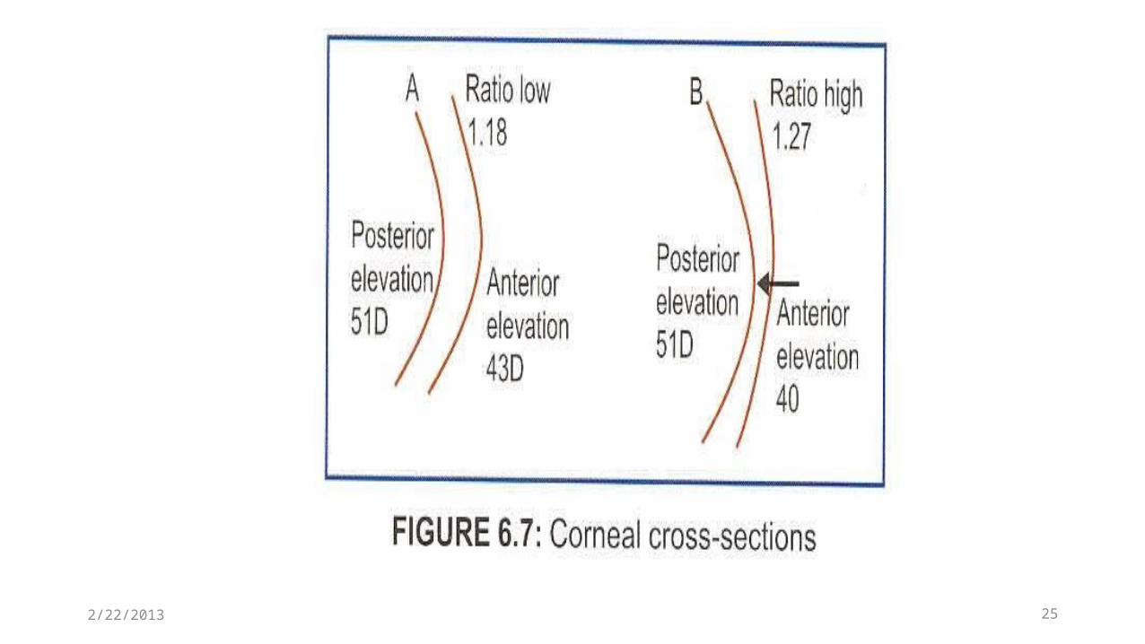
• A best fit sphere (BFS) >55D on the posterior
profile , indicative of posterior ectasia
• Greater than 50 micron generally accepted as
abnormal
• In corneas thinner than normal over 40 as abnormal
23
POSTERIOR ELEVATION MAP
2/22/2013
24
• Post BFS >55D• Difference pachymetry between thinest point & thickest
point in 7 mm zone >100 µ• Diff >45µ• Mean k >47 D • I-S index >1.5D• Distance from corneal apex to thinest point >0.9 mm• Thinest point <470 µ
Orbscan diagnostic parameters for K.C
2/22/2013
252/22/2013
26
• Number of abnormal maps• Posterior float difference >0.050• 3mm & 5mm irregularity• Peripheral thickness changes• Astigmatism variance between eyes• Steep k’s –mean power map
Risks of ectasia indices
2/22/2013
272/22/2013
28
One abnormal map ; perform with caution
Two abnormal map ; with concern
Three abnormal map ;contraindicated
Three step rule
2/22/2013
پو�زش عرض ضمن
باالی حجم در LECTUERبدلیل نمیباشد پذیر امکان اسالیدها ادامهآموزشی مرکز بصری و سمعی واحد به لطفا ادامه به نیاز صورت
تلفن شماره با یا و مراجعه فیض داخلی 03114476010درمانیتماس حاصل نمائید 392
با تشکر