tomography and precession diffraction for 3d structural ... · pdf filetomography and...
TRANSCRIPT
U. Kolb
TU Darmstadt and University of Mainz
Tomography and Precession Diffraction for 3D Structural Analysis of Nanocrystals
M&M Workshop: Exploiting the Diffractive Properties of Electrons for Solving Materials Problems July 2016
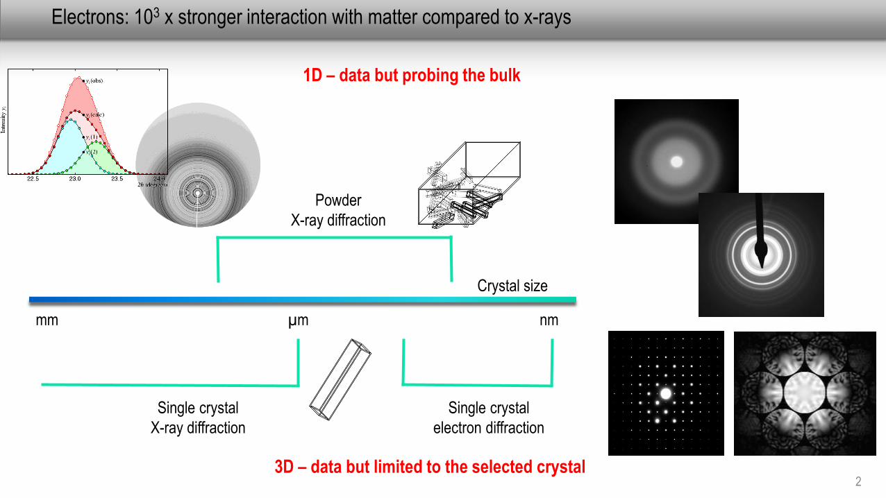
Electrons: 103 x stronger interaction with matter compared to x-rays
2
Crystal size
Single crystal
X-ray diffraction
mm µm nm
Single crystal
electron diffraction
3D – data but limited to the selected crystal
1D – data but probing the bulk
Powder
X-ray diffraction
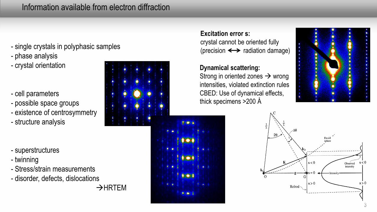
Information available from electron diffraction
3
- single crystals in polyphasic samples
- phase analysis
- crystal orientation
- cell parameters
- possible space groups
- existence of centrosymmetry
- structure analysis
- superstructures
- twinning
- Stress/strain measurements
- disorder, defects, dislocations
HRTEM
Excitation error s:
crystal cannot be oriented fully
(precision radiation damage)
Dynamical scattering:
Strong in oriented zones wrong
intensities, violated extinction rules
CBED: Use of dynamical effects, thick specimens >200 Å
4
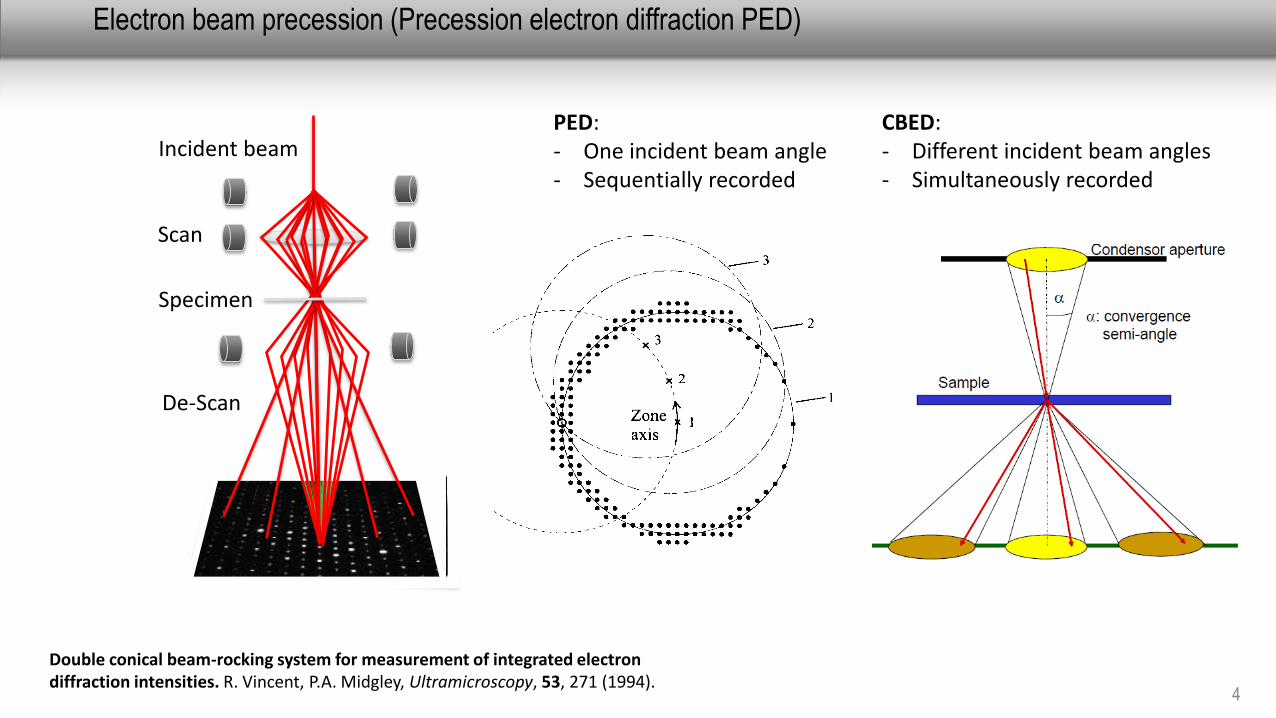
Electron beam precession (Precession electron diffraction PED)
Double conical beam-rocking system for measurement of integrated electron diffraction intensities. R. Vincent, P.A. Midgley, Ultramicroscopy, 53, 271 (1994).
Incident beam
Scan
Specimen
De-Scan
CBED: - Different incident beam angles- Simultaneously recorded
PED: - One incident beam angle- Sequentially recorded
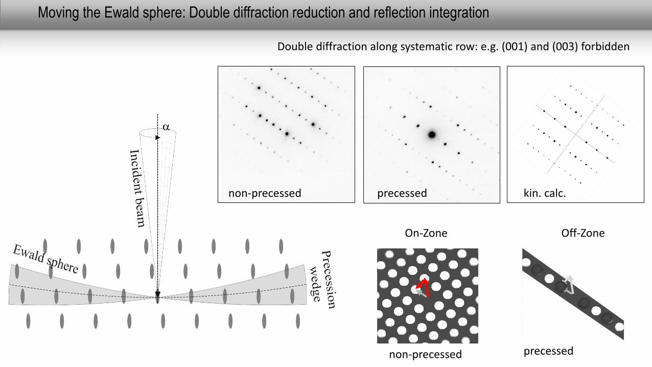
Moving the Ewald sphere: Double diffraction reduction and reflection integration
5
a
non-precessed precessed kin. calc.
Double diffraction along systematic row: e.g. (001) and (003) forbidden
On-Zone
non-precessed
Off-Zone
precessed
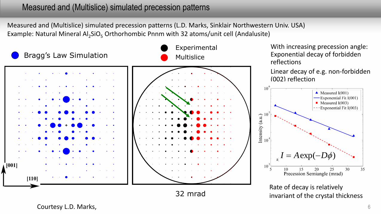
Measured and (Multislice) simulated precession patterns
6
Bragg’s Law Simulation
Precession Off6.5 mrad13 mrad18 mrad24 mrad32 mrad
Experimental
Multislice
Measured and (Multislice) simulated precession patterns (L.D. Marks, Sinklair Northwestern Univ. USA)Example: Natural Mineral Al2SiO5 Orthorhombic Pnnm with 32 atoms/unit cell (Andalusite)
Courtesy L.D. Marks,
With increasing precession angle: Exponential decay of forbidden reflections
Linear decay of e.g. non-forbidden (002) reflection
Rate of decay is relatively invariant of the crystal thickness
)exp( DAI
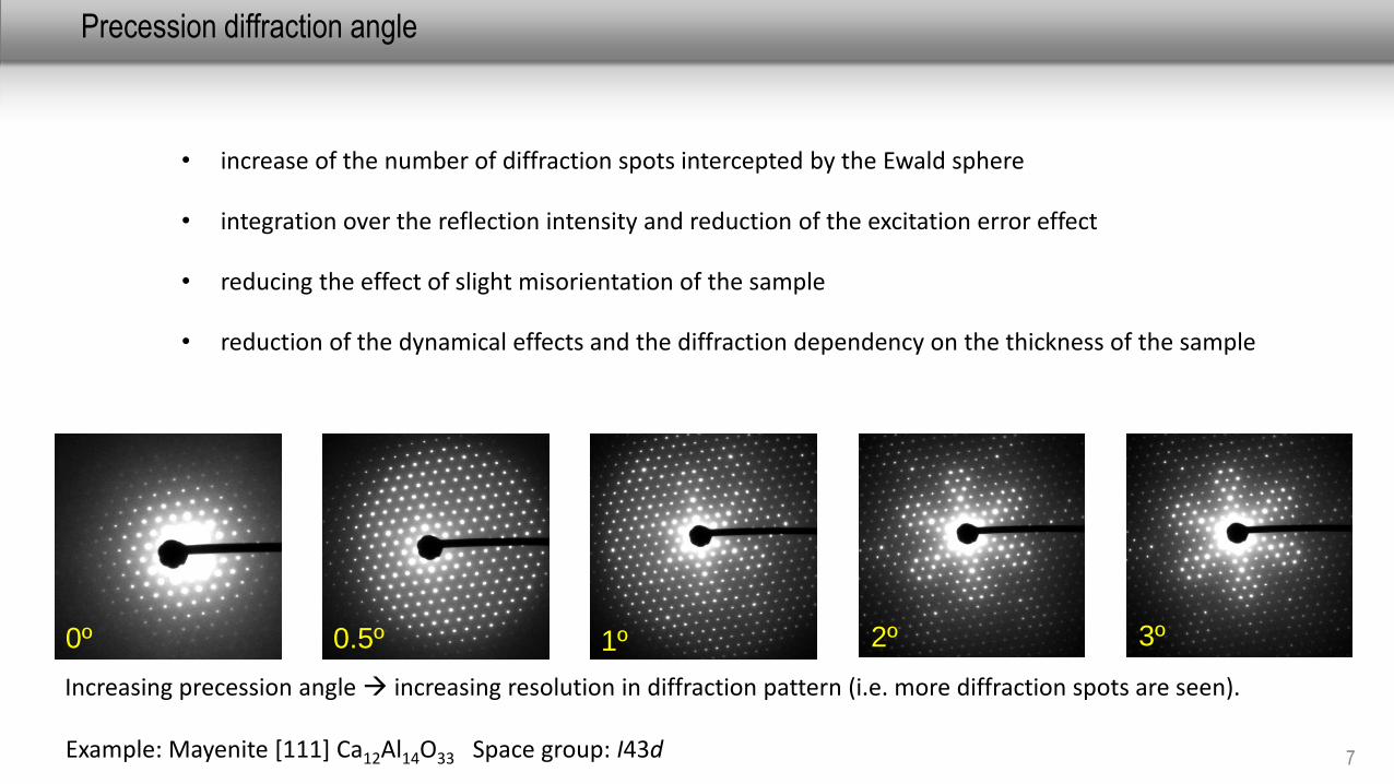
Precession diffraction angle
7
1º 3º2º0º 0.5º
Increasing precession angle increasing resolution in diffraction pattern (i.e. more diffraction spots are seen).
Example: Mayenite [111] Ca12Al14O33 Space group: I43d
• increase of the number of diffraction spots intercepted by the Ewald sphere
• integration over the reflection intensity and reduction of the excitation error effect
• reducing the effect of slight misorientation of the sample
• reduction of the dynamical effects and the diffraction dependency on the thickness of the sample
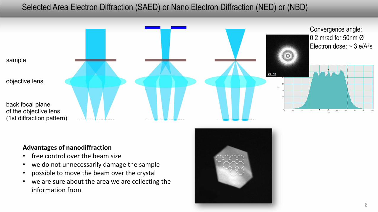
Selected Area Electron Diffraction (SAED) or Nano Electron Diffraction (NED) or (NBD)
8
Advantages of nanodiffraction• free control over the beam size• we do not unnecessarily damage the sample• possible to move the beam over the crystal• we are sure about the area we are collecting the
information from
Convergence angle:
0.2 mrad for 50nm Ø
Electron dose: ~ 3 e/A2s

A-Star Acquisition
9
Orientation map
Non-precessed precessed
Acquisition of precession electron diffraction spot patterns
Phase map 10 nm
Pt particles, Prof. P. Ferreira, J. Ganesh Univ Texas at Austin USA JEOL 2010 FEG (1 nm resolution)
Strain map
Rhee, Y. Du, P. S. Ho, Journal of Applied Physics, 93 (2003) 3926
Mg-Cu-Gd partly recrystallizedmetallic glass with Mg2Cu and Cu2Gd crystalline precipitates
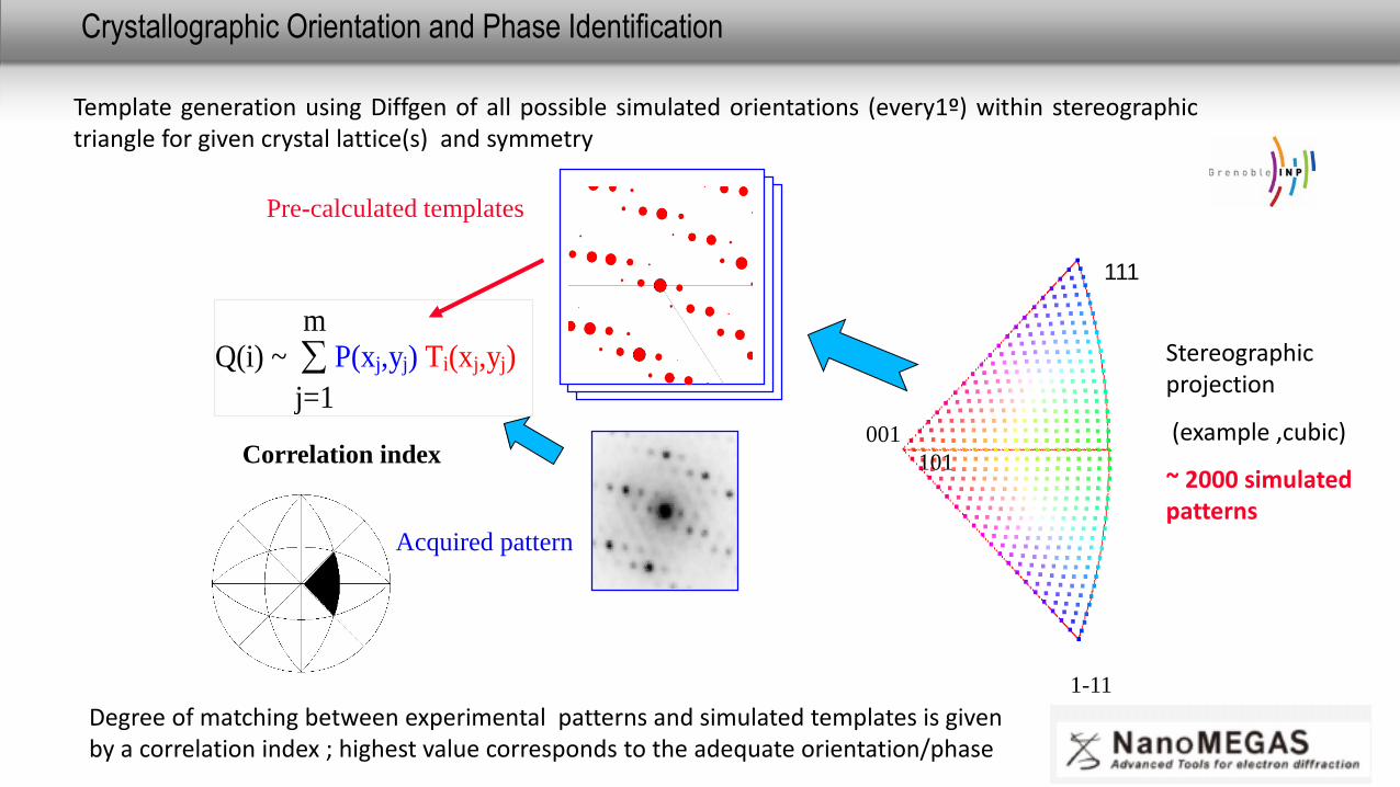
Crystallographic Orientation and Phase Identification
10
Acquired pattern
Correlation index
Q(i) ~ j=1
m P(xj,yj) Ti(xj,yj)
001
101
1-11
Pre-calculated templates
Stereographic projection
(example ,cubic)
~ 2000 simulated patterns
Template generation using Diffgen of all possible simulated orientations (every1º) within stereographictriangle for given crystal lattice(s) and symmetry
Degree of matching between experimental patterns and simulated templates is given by a correlation index ; highest value corresponds to the adequate orientation/phase
111
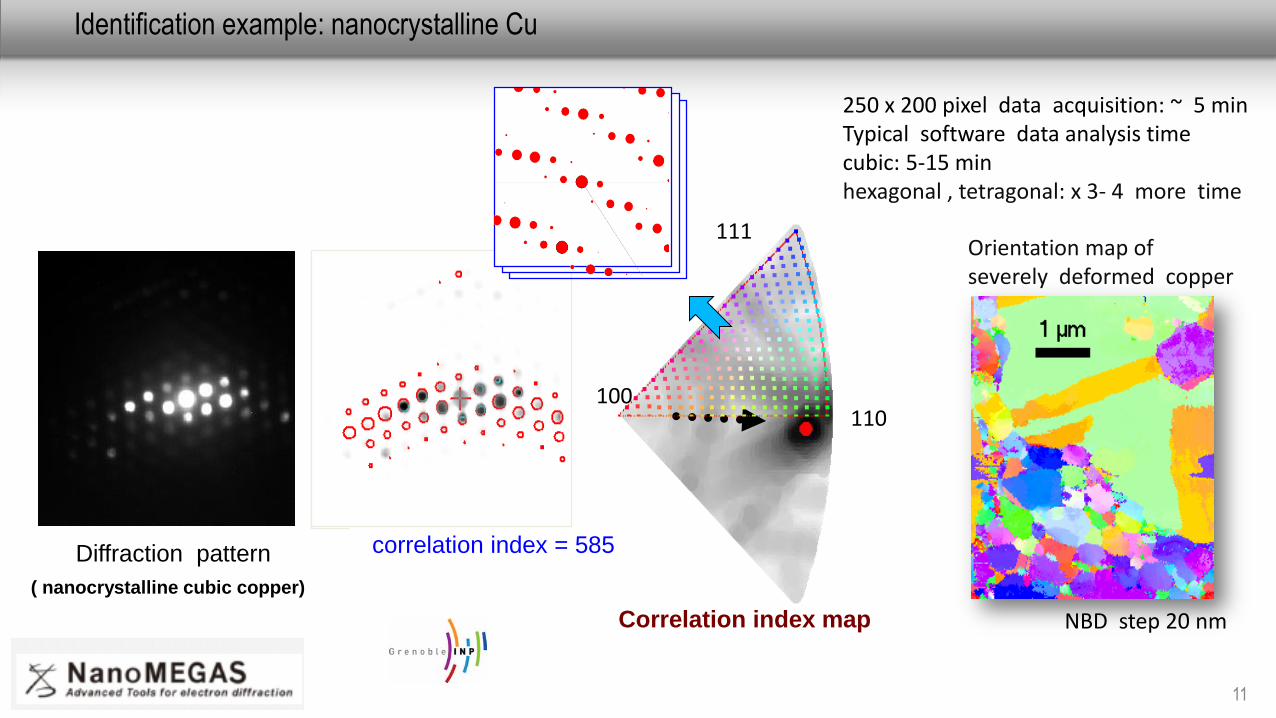
Identification example: nanocrystalline Cu
11
correlation index = 585
Correlation index map
Diffraction pattern
( nanocrystalline cubic copper)
100110
111Orientation map ofseverely deformed copper
NBD step 20 nm
250 x 200 pixel data acquisition: ~ 5 minTypical software data analysis time cubic: 5-15 minhexagonal , tetragonal: x 3- 4 more time
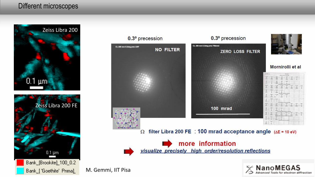
Different microscopes
12
Zeiss Libra 200
Zeiss Libra 200 FE
M. Gemmi, IIT Pisa
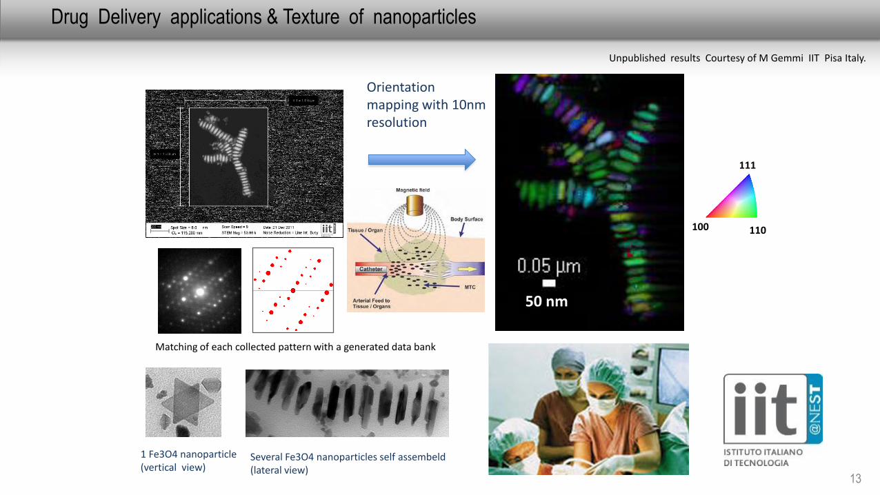
Drug Delivery applications & Texture of nanoparticles
13
Matching of each collected pattern with a generated data bank
100 110
111
1 Fe3O4 nanoparticle(vertical view)
Several Fe3O4 nanoparticles self assembeld(lateral view)
Orientation mapping with 10nm resolution
Unpublished results Courtesy of M Gemmi IIT Pisa Italy.
50 nm
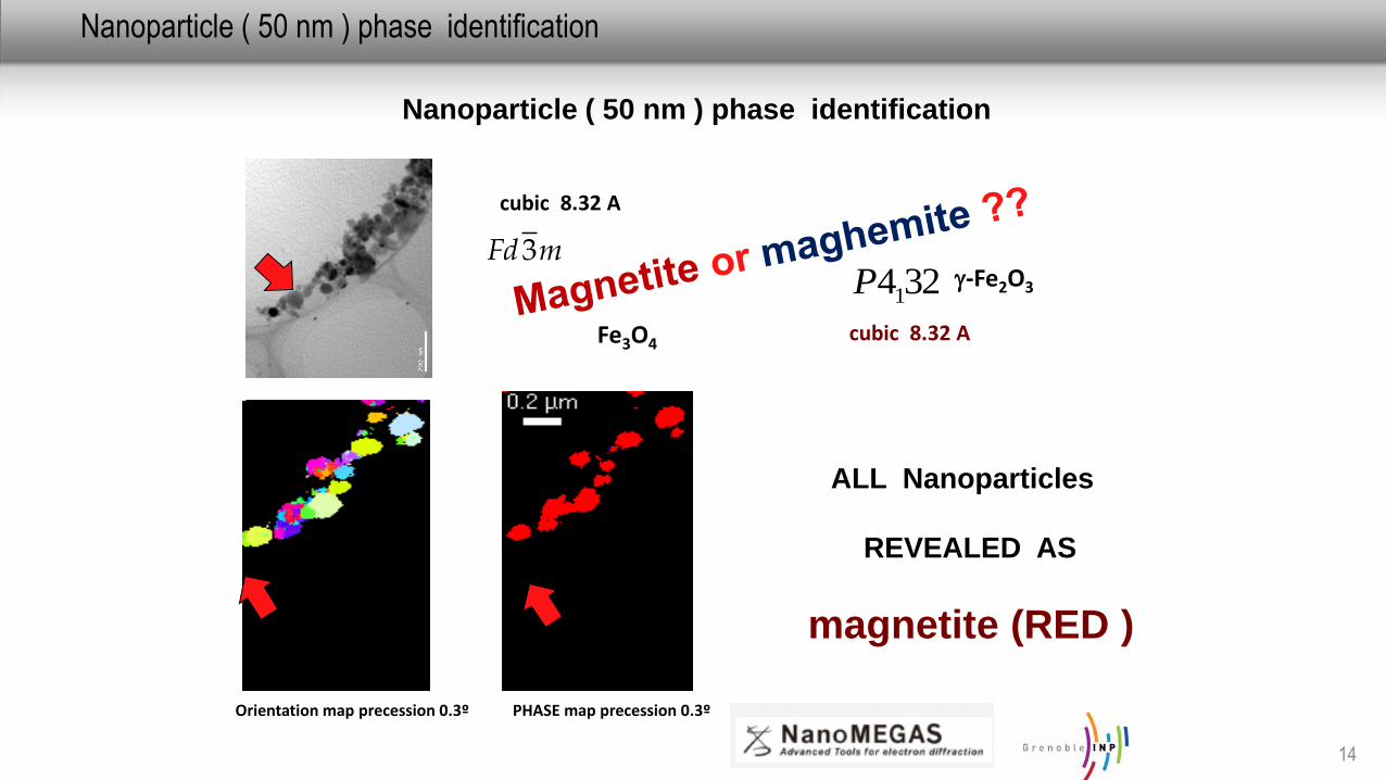
Nanoparticle ( 50 nm ) phase identification
14
Nanoparticle ( 50 nm ) phase identification
Orientation map precession 0.3º PHASE map precession 0.3º
ALL Nanoparticles
REVEALED AS
magnetite (RED )
cubic 8.32 A
cubic 8.32 A
3241PmFd3
-Fe2O3
Fe3O4
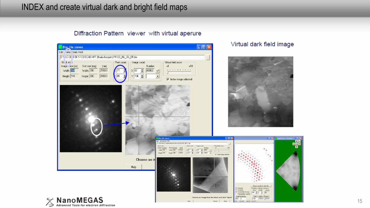
INDEX and create virtual dark and bright field maps
15
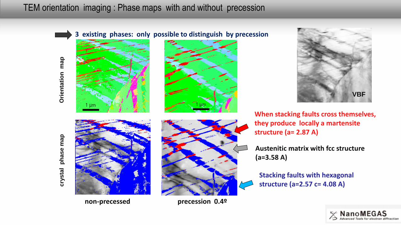
TEM orientation imaging : Phase maps with and without precession
16
cry
sta
lp
ha
se
ma
p
3 existing phases: only possible to distinguish by precession
Ori
en
tati
on
map
non-precessed precession 0.4º
Austenitic matrix with fcc structure (a=3.58 A)
Stacking faults with hexagonal structure (a=2.57 c= 4.08 A)
When stacking faults cross themselves, they produce locally a martensitestructure (a= 2.87 A)
VBF
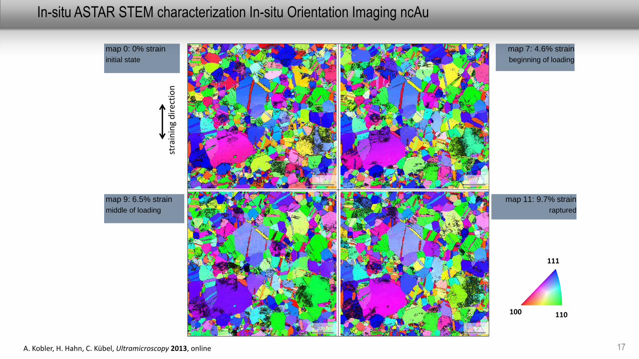
In-situ ASTAR STEM characterization In-situ Orientation Imaging ncAu
17A. Kobler, H. Hahn, C. Kübel, Ultramicroscopy 2013, online
map 0: 0% strain
initial state
stra
inin
g d
irec
tio
n200 nm
200 nm 200 nm
200 nm
map 7: 4.6% strain
beginning of loading
map 11: 9.7% strain
raptured
map 9: 6.5% strain
middle of loading
100 110
111
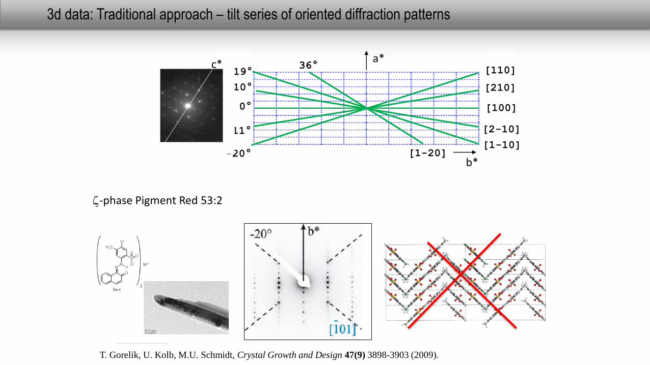
3d data: Traditional approach – tilt series of oriented diffraction patterns
a*
b*-20°
[110]
[210]
-11°
0° [100]
10°
[2-10]
19°
[1-10]
36°
[1-20]
c*
T. Gorelik, U. Kolb, M.U. Schmidt, Crystal Growth and Design 47(9) 3898-3903 (2009).
z-phase Pigment Red 53:2
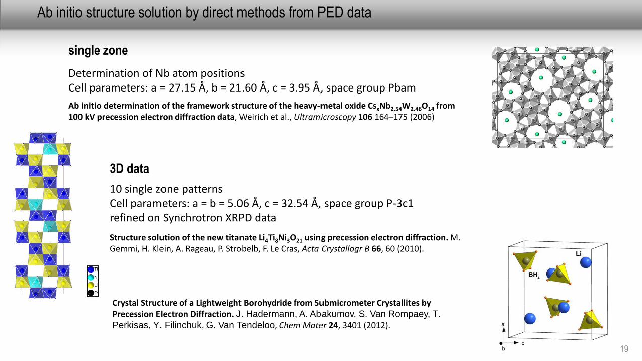
Ab initio structure solution by direct methods from PED data
19
single zone
Determination of Nb atom positionsCell parameters: a = 27.15 Å, b = 21.60 Å, c = 3.95 Å, space group Pbam
Ab initio determination of the framework structure of the heavy-metal oxide CsxNb2.54W2.46O14 from 100 kV precession electron diffraction data, Weirich et al., Ultramicroscopy 106 164–175 (2006)
3D data
10 single zone patternsCell parameters: a = b = 5.06 Å, c = 32.54 Å, space group P-3c1 refined on Synchrotron XRPD data
Crystal Structure of a Lightweight Borohydride from Submicrometer Crystallites by Precession Electron Diffraction. J. Hadermann, A. Abakumov, S. Van Rompaey, T.Perkisas, Y. Filinchuk, G. Van Tendeloo, Chem Mater 24, 3401 (2012).
Structure solution of the new titanate Li4Ti8Ni3O21 using precession electron diffraction. M. Gemmi, H. Klein, A. Rageau, P. Strobelb, F. Le Cras, Acta Crystallogr B 66, 60 (2010).
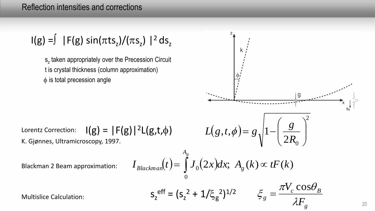
Reflection intensities and corrections
20
sz taken appropriately over the Precession Circuit
t is crystal thickness (column approximation)
is total precession angle
I(g) = |F(g) sin(ptsz)/(psz) |2 dsz
Blackman 2 Beam approximation: )()( ;20
0 ktFkAdxxJtI g
A
Blackman
g
2
021,,
R
ggtgL I(g) = |F(g)|2L(g,t,)Lorentz Correction:
K. Gjønnes, Ultramicroscopy, 1997.
Multislice Calculation:
g
Bcg
F
V
p
cossz
eff = (sz2 + 1/g
2)1/2
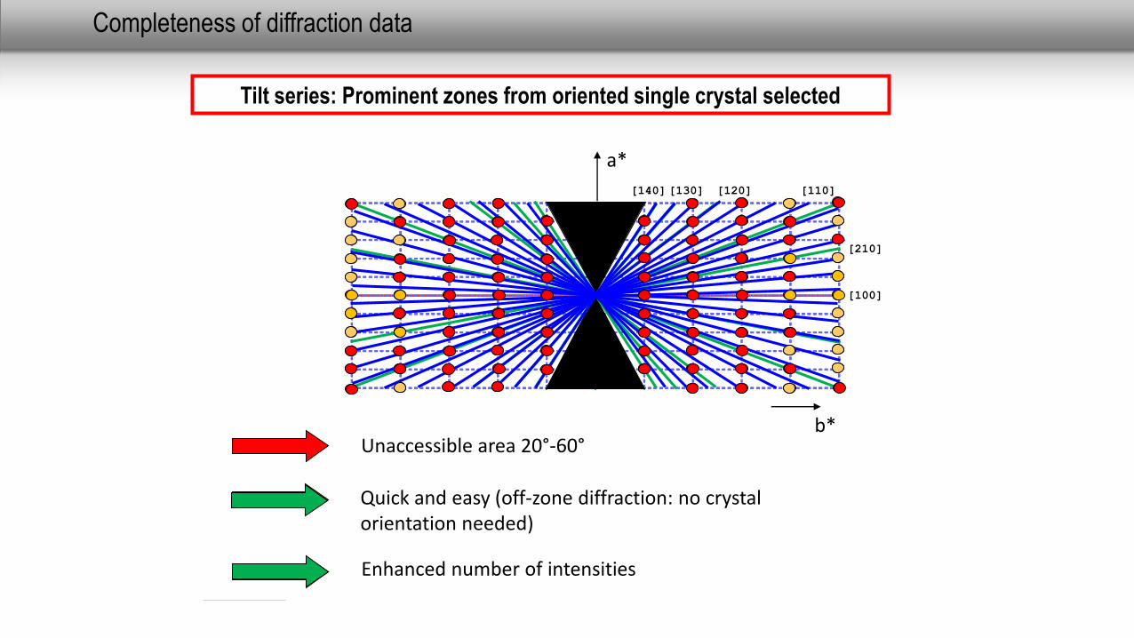
Completeness of diffraction data
Tilt series: Prominent zones from oriented single crystal selected
[100]
a*
b*Unaccessible area 20°-60°
Data of high indexed zones missing
Difficult and time consuming
[140] [120][130]
[210]
[110]
Quick and easy (off-zone diffraction: no crystalorientation needed)
Enhanced number of intensities
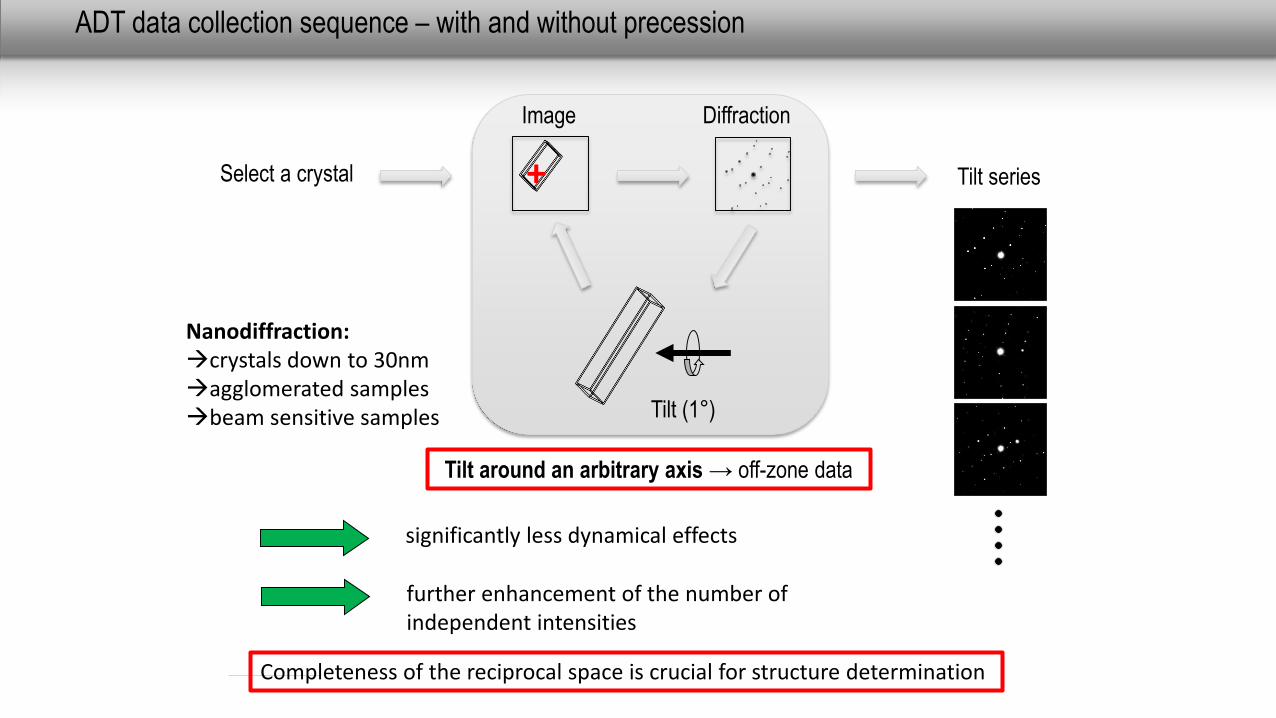
ADT data collection sequence – with and without precession
Diffraction
Select a crystal
Tilt (1°)
Image
+ Tilt series
Tilt around an arbitrary axis → off-zone data
significantly less dynamical effects
Nanodiffraction:crystals down to 30nmagglomerated samplesbeam sensitive samples
further enhancement of the number ofindependent intensities
Completeness of the reciprocal space is crucial for structure determination
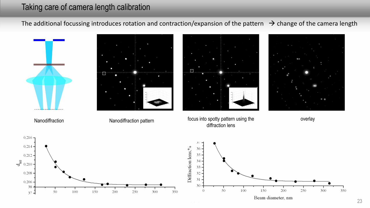
Taking care of camera length calibration
23
focus into spotty pattern using the
diffraction lensNanodiffraction overlay
The additional focussing introduces rotation and contraction/expansion of the pattern change of the camera length
Nanodiffraction pattern
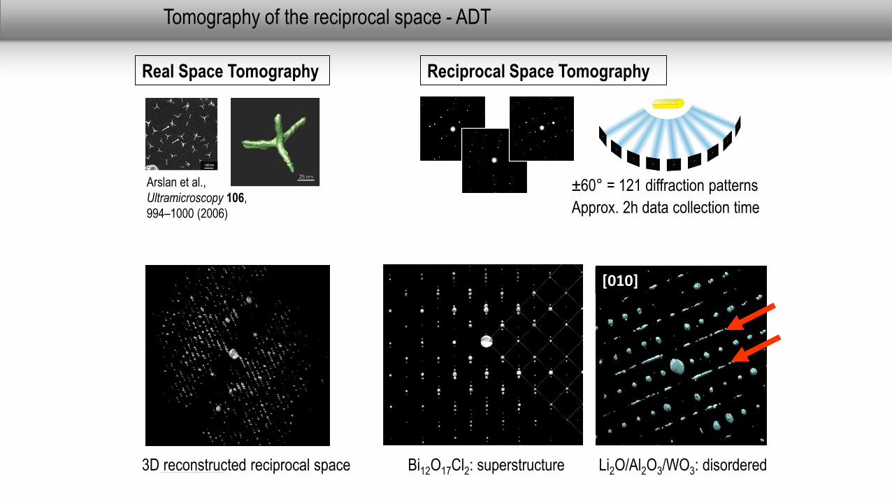
Tomography of the reciprocal space - ADT
Arslan et al.,
Ultramicroscopy 106,
994–1000 (2006)
Real Space Tomography
±60° = 121 diffraction patterns
Approx. 2h data collection time
Reciprocal Space Tomography
3D reconstructed reciprocal space Bi12O17Cl2: superstructure Li2O/Al2O3/WO3: disordered
[010]
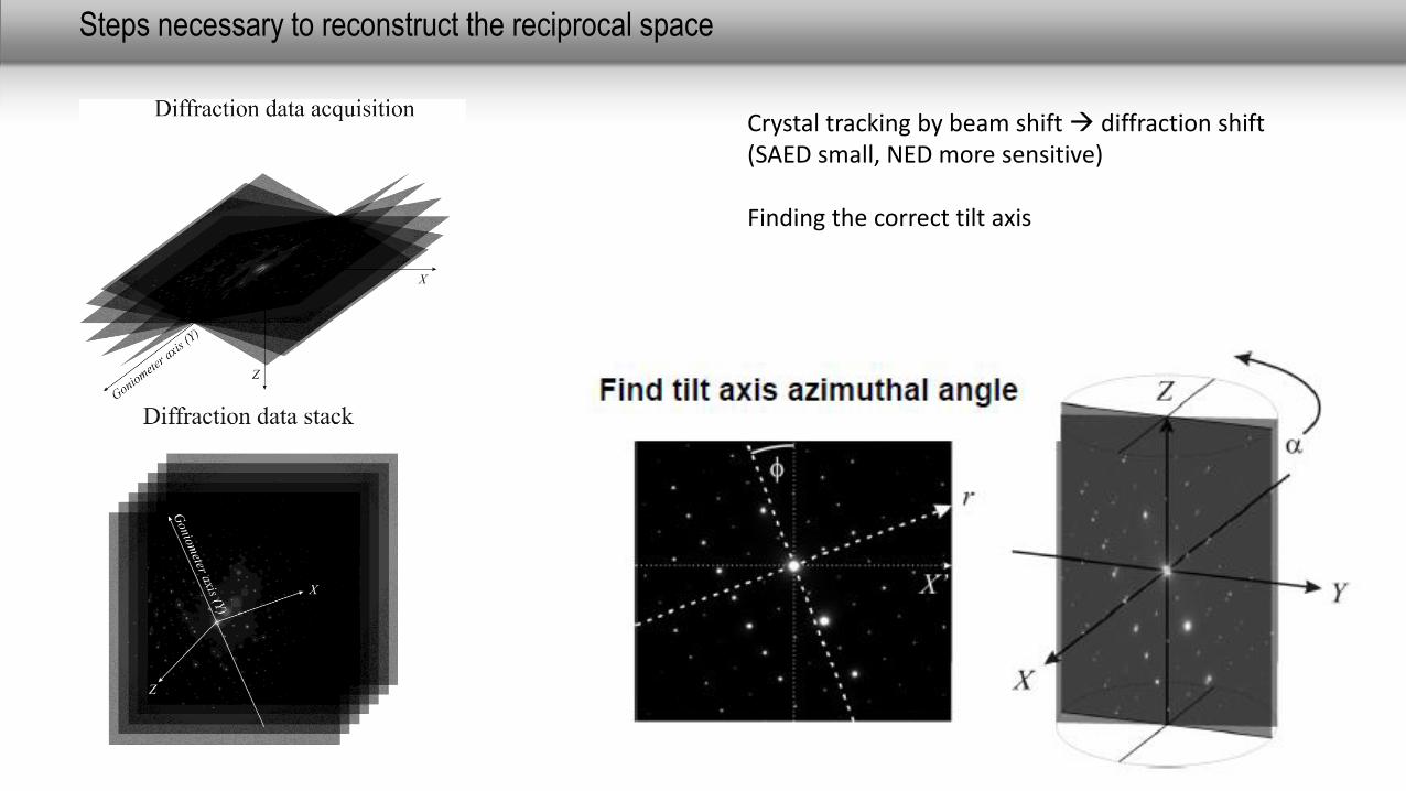
Steps necessary to reconstruct the reciprocal space
Crystal tracking by beam shift diffraction shift(SAED small, NED more sensitive)
Finding the correct tilt axis
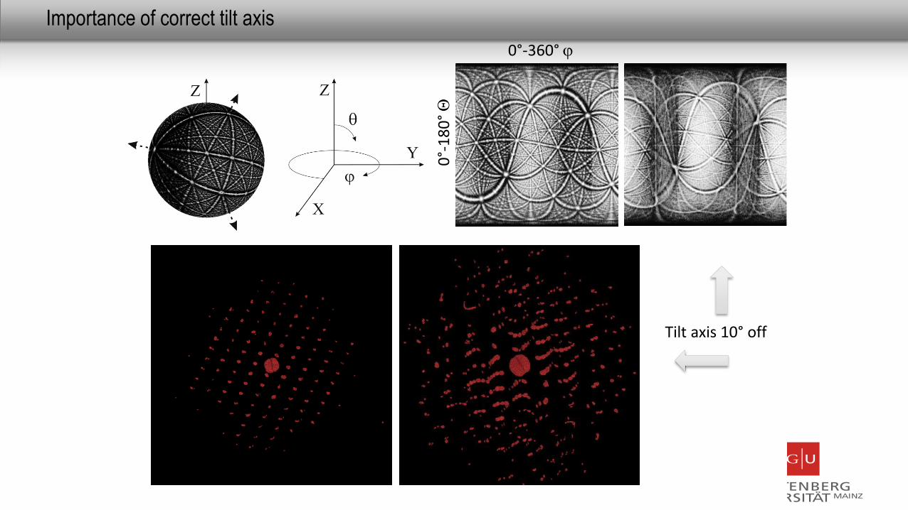
Importance of correct tilt axis
Tilt axis 10° off
0°-360°
0°-
18
0°Q
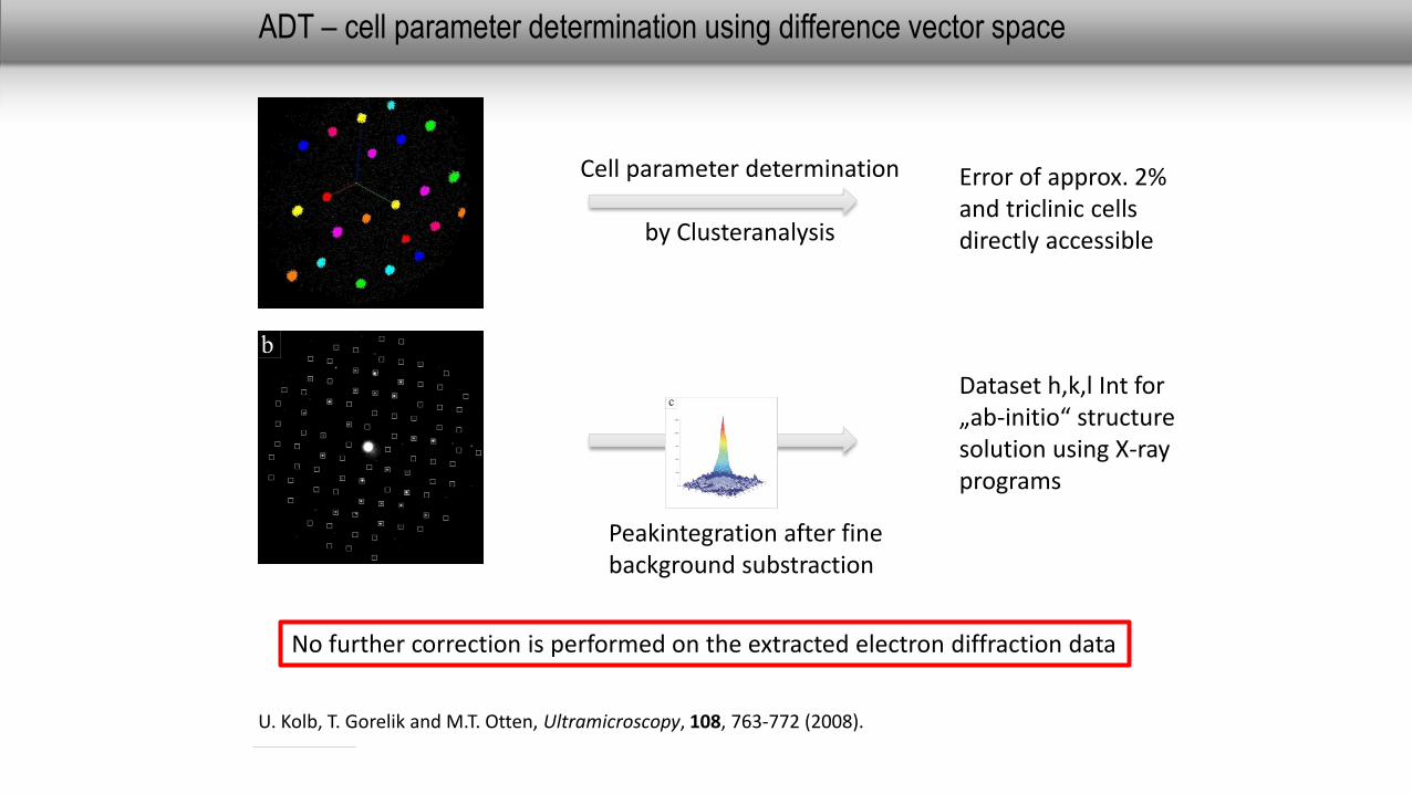
ADT – cell parameter determination using difference vector space
U. Kolb, T. Gorelik and M.T. Otten, Ultramicroscopy, 108, 763-772 (2008).
Error of approx. 2% and triclinic cellsdirectly accessible
Peakintegration after finebackground substraction
Cell parameter determination
by Clusteranalysis
Dataset h,k,l Int for„ab-initio“ structuresolution using X-rayprograms
No further correction is performed on the extracted electron diffraction data
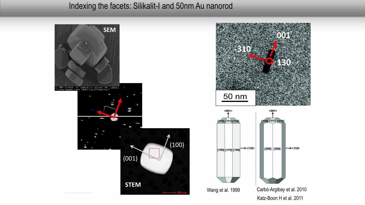
Indexing the facets: Silikalit-I and 50nm Au nanorod
SEM
(100)
(001)
STEM
001
130
-310
Wang et al. 1999 Carbó-Argibay et al. 2010
Katz-Boon H et al. 2011
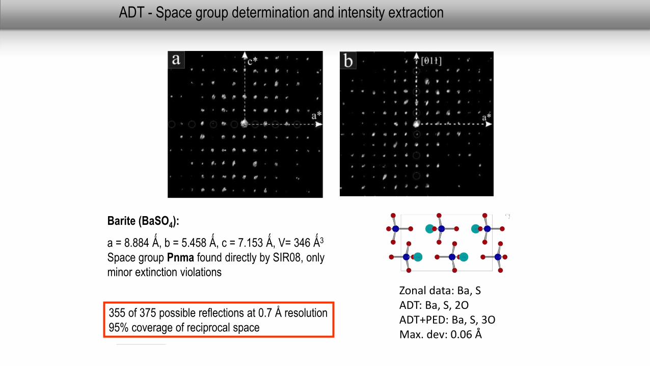
ADT - Space group determination and intensity extraction
355 of 375 possible reflections at 0.7 Å resolution
95% coverage of reciprocal space
Barite (BaSO4):
a = 8.884 Ǻ, b = 5.458 Ǻ, c = 7.153 Ǻ, V= 346 Ǻ3
Space group Pnma found directly by SIR08, only
minor extinction violations
Zonal data: Ba, SADT: Ba, S, 2OADT+PED: Ba, S, 3OMax. dev: 0.06 Å
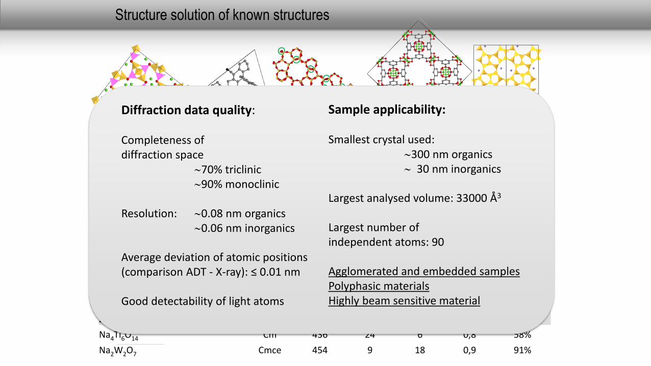
Structure solution of known structures
Space group
N° ind. Refl.
N° ind. Atoms
Data/Param.
Resolution
Completeness
Barite BaSO4 Pnma 355 5 21 0,7 95%
Calcite CaCO3 R-3c 106 3 18 0,7 97%
ZnSb Pbca 106 2 12 1,1 70%
Mullite Al6Si2O13 Pbam 213 5 18 0,7 86%
Natrolite Na2Al2Si3O10*2H2O Fdd2 719 10 18 0,7 92%
10-CNBA C29NH17 P21/c 1871 30 15 1,0 90%
Basolite C6H4CuO5 Fm-3m 384 7 17 1,1 99%
NLO-org C20O3NH15 Pca21 773 24 8 1 88%
IM-5 Si288O576 Cmcm 2170 71 8 1,1 68%
ZSM-5/Silikalit-I Pnma 2288 39 16 1 79%
Na4Ti6O14 Cm 436 24 6 0,8 58%
Na2W2O7 Cmce 454 9 18 0,9 91%
Diffraction data quality:
Completeness ofdiffraction space
70% triclinic90% monoclinic
Resolution: 0.08 nm organics0.06 nm inorganics
Average deviation of atomic positions(comparison ADT - X-ray): ≤ 0.01 nm
Good detectability of light atoms
Sample applicability:
Smallest crystal used:300 nm organics 30 nm inorganics
Largest analysed volume: 33000 Å3
Largest number ofindependent atoms: 90
Agglomerated and embedded samplesPolyphasic materialsHighly beam sensitive material
0°
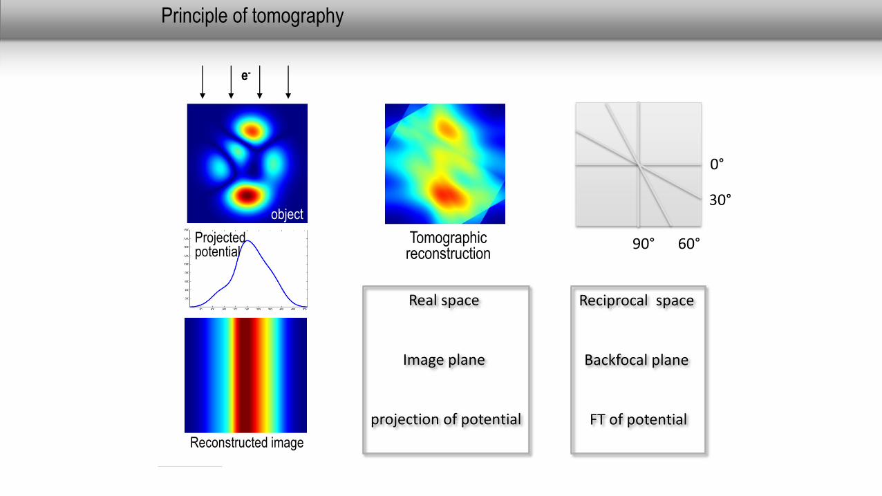
Principle of tomography
object
Projected potential
Reconstructed image
e-
30°
60°
90°
Tomographicreconstruction
0°
30°
60°90°
Real space
Image plane
projection of potential
Reciprocal space
Backfocal plane
FT of potential
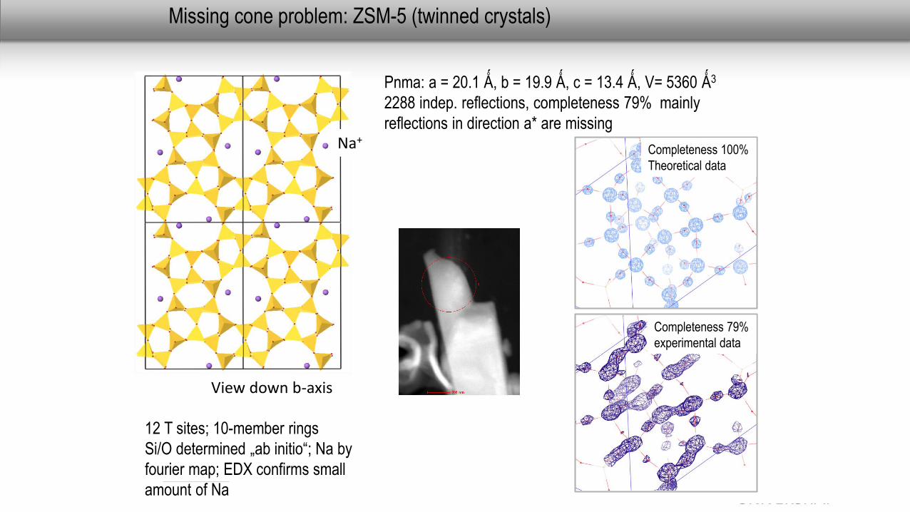
Missing cone problem: ZSM-5 (twinned crystals)
Pnma: a = 20.1 Ǻ, b = 19.9 Ǻ, c = 13.4 Ǻ, V= 5360 Ǻ3
2288 indep. reflections, completeness 79% mainly
reflections in direction a* are missingNa+
View down b-axis
12 T sites; 10-member rings
Si/O determined „ab initio“; Na by
fourier map; EDX confirms small
amount of Na
Completeness 100%
Theoretical data
Completeness 79%
experimental data
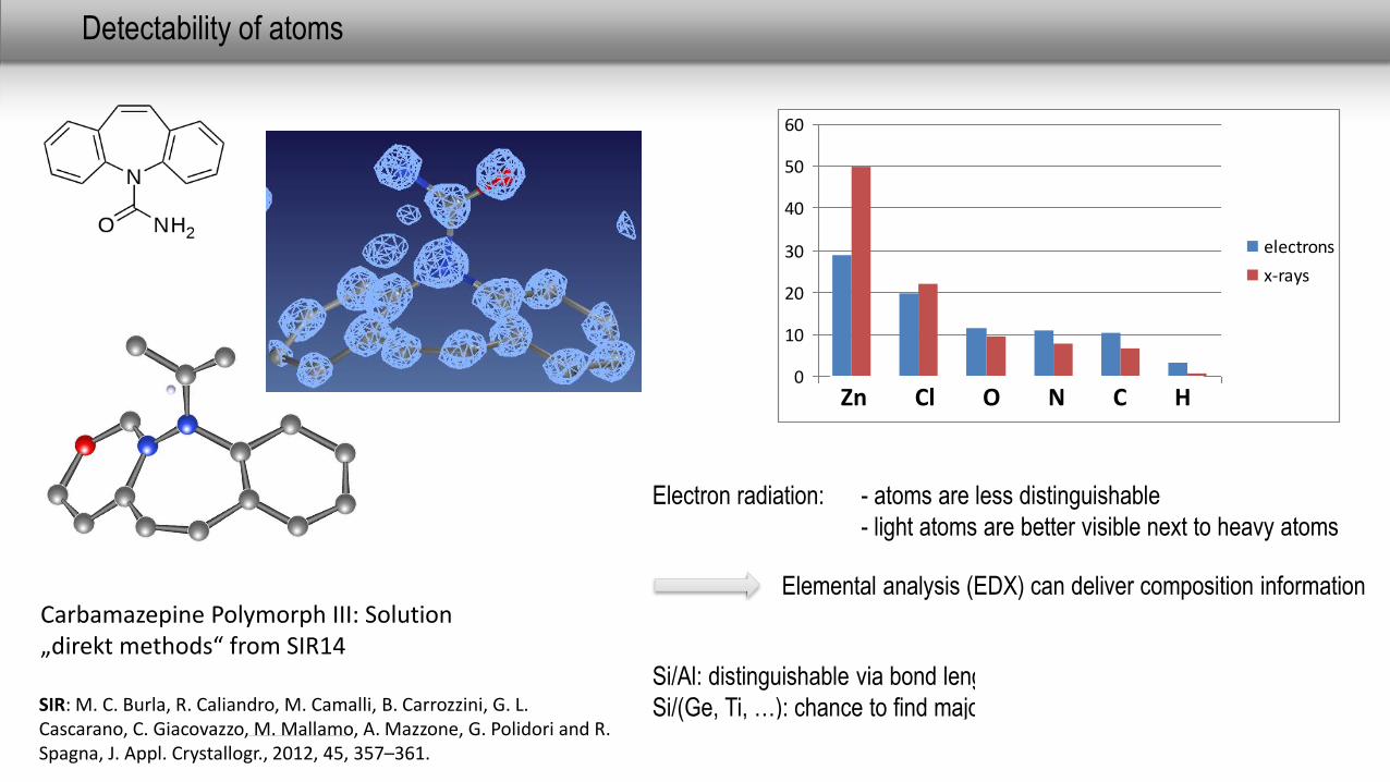
Detectability of atoms
Electron radiation: - atoms are less distinguishable
- light atoms are better visible next to heavy atoms
Elemental analysis (EDX) can deliver composition information
Si/Al: distinguishable via bond length for fully occupied positions
Si/(Ge, Ti, …): chance to find major occupation scheme
0
10
20
30
40
50
60
Zn Cl O N Cl H
electrons
x-rays
Zn Cl O N C H
Carbamazepine Polymorph III: Solution „direkt methods“ from SIR14
SIR: M. C. Burla, R. Caliandro, M. Camalli, B. Carrozzini, G. L. Cascarano, C. Giacovazzo, M. Mallamo, A. Mazzone, G. Polidori and R. Spagna, J. Appl. Crystallogr., 2012, 45, 357–361.
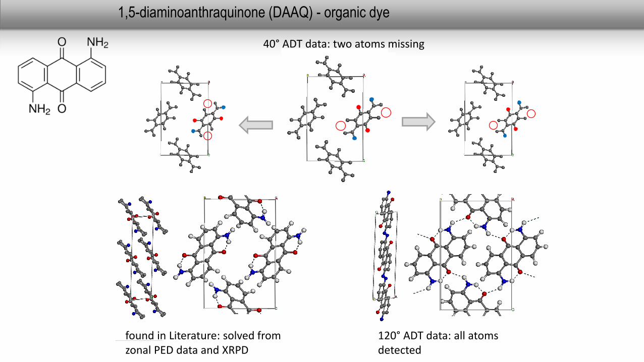
40° ADT data: two atoms missing
found in Literature: solved fromzonal PED data and XRPD
1,5-diaminoanthraquinone (DAAQ) - organic dye
120° ADT data: all atomsdetected
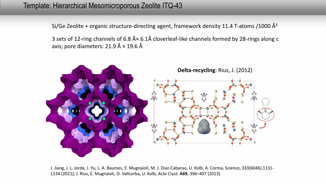
J. Jiang, J. L. Jorda, J. Yu, L. A. Baumes, E. Mugnaioli, M. J. Diaz-Cabanas, U. Kolb, A. Corma, Science, 333(6046),1131-1134 (2011); J. Rius, E. Mugnaioli, O. Vallcorba, U. Kolb, Acta Cryst. A69, 396–407 (2013)
3 sets of 12-ring channels of 6.8 Å× 6.1Å cloverleaf-like channels formed by 28-rings along c axis; pore diameters: 21.9 Å × 19.6 Å
Template: Hierarchical Mesomicroporous Zeolite ITQ-43
Si/Ge Zeolite + organic structure-directing agent, framework density 11.4 T-atoms /1000 Å3
Delta-recycling: Rius, J. (2012)
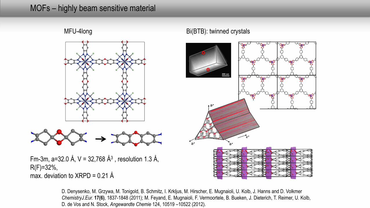
MOFs – highly beam sensitive material
MFU-4long
Fm-3m, a=32.0 Å, V = 32,768 Å3 , resolution 1.3 Å,
R(F)=32%,
max. deviation to XRPD = 0.21 Å
D. Denysenko, M. Grzywa, M. Tonigold, B. Schmitz, I. Krkljus, M. Hirscher, E. Mugnaioli, U. Kolb, J. Hanns and D. Volkmer
ChemistryJ.Eur. 17(6), 1837-1848 (2011); M. Feyand, E. Mugnaioli, F. Vermoortele, B. Bueken, J. Dieterich, T. Reimer, U. Kolb,
D. de Vos and N. Stock, Angewandte Chemie 124, 10519 –10522 (2012).
Bi(BTB): twinned crystals
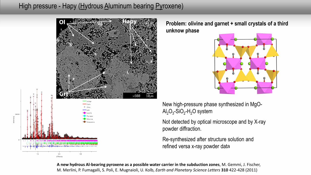
High pressure - Hapy (Hydrous Aluminum bearing Pyroxene)
Problem: olivine and garnet + small crystals of a third
unknow phase
New high-pressure phase synthesized in MgO-
Al2O3-SiO2-H2O system
Not detected by optical microscope and by X-ray
powder diffraction.
Re-synthesized after structure solution and
refined versa x-ray powder data
A new hydrous Al-bearing pyroxene as a possible water carrier in the subduction zones, M. Gemmi, J. Fischer, M. Merlini, P. Fumagalli, S. Poli, E. Mugnaioli, U. Kolb, Earth and Planetary Science Letters 310 422-428 (2011)
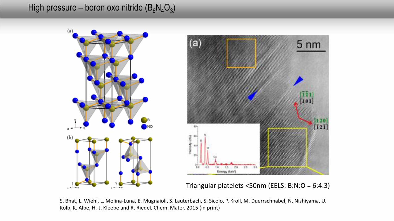
High pressure – boron oxo nitride (B6N4O3)
S. Bhat, L. Wiehl, L. Molina-Luna, E. Mugnaioli, S. Lauterbach, S. Sicolo, P. Kroll, M. Duerrschnabel, N. Nishiyama, U. Kolb, K. Albe, H.-J. Kleebe and R. Riedel, Chem. Mater. 2015 (in print)
Triangular platelets <50nm (EELS: B:N:O = 6:4:3)
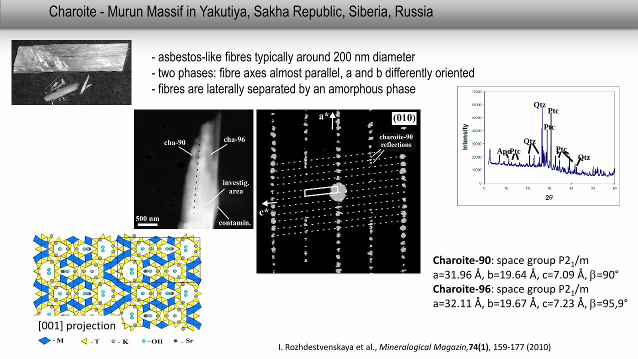
Charoite - Murun Massif in Yakutiya, Sakha Republic, Siberia, Russia
- asbestos-like fibres typically around 200 nm diameter
- two phases: fibre axes almost parallel, a and b differently oriented
- fibres are laterally separated by an amorphous phase
Charoite-90: space group P21/m a=31.96 Å, b=19.64 Å, c=7.09 Å, b=90°Charoite-96: space group P21/m a=32.11 Å, b=19.67 Å, c=7.23 Å, b=95,9°
QtzPtc
Ptc
Qtz
PtcApo PtcQtz
[001] projection
I. Rozhdestvenskaya et al., Mineralogical Magazin,74(1), 159-177 (2010)
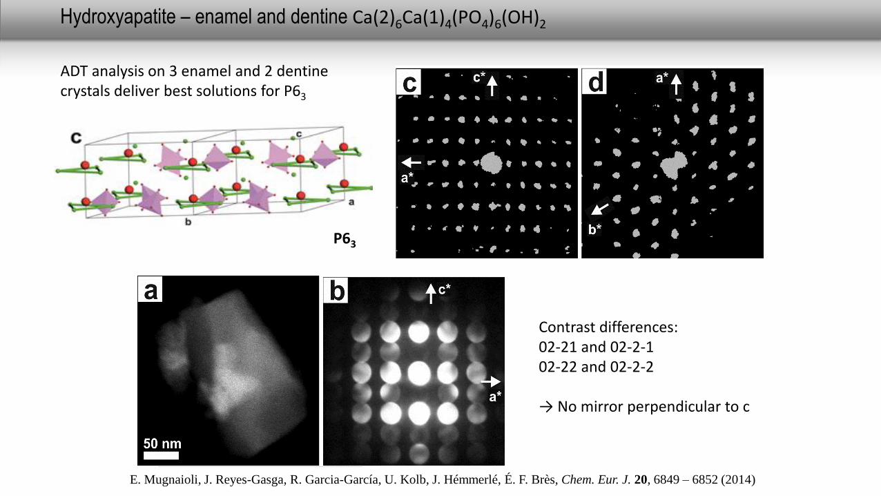
Hydroxyapatite – enamel and dentine Ca(2)6Ca(1)4(PO4)6(OH)2
Contrast differences:02-21 and 02-2-1 02-22 and 02-2-2
→ No mirror perpendicular to c
ADT analysis on 3 enamel and 2 dentinecrystals deliver best solutions for P63
P63
E. Mugnaioli, J. Reyes-Gasga, R. Garcia-García, U. Kolb, J. Hémmerlé, É. F. Brès, Chem. Eur. J. 20, 6849 – 6852 (2014)
Fragmentof platelet
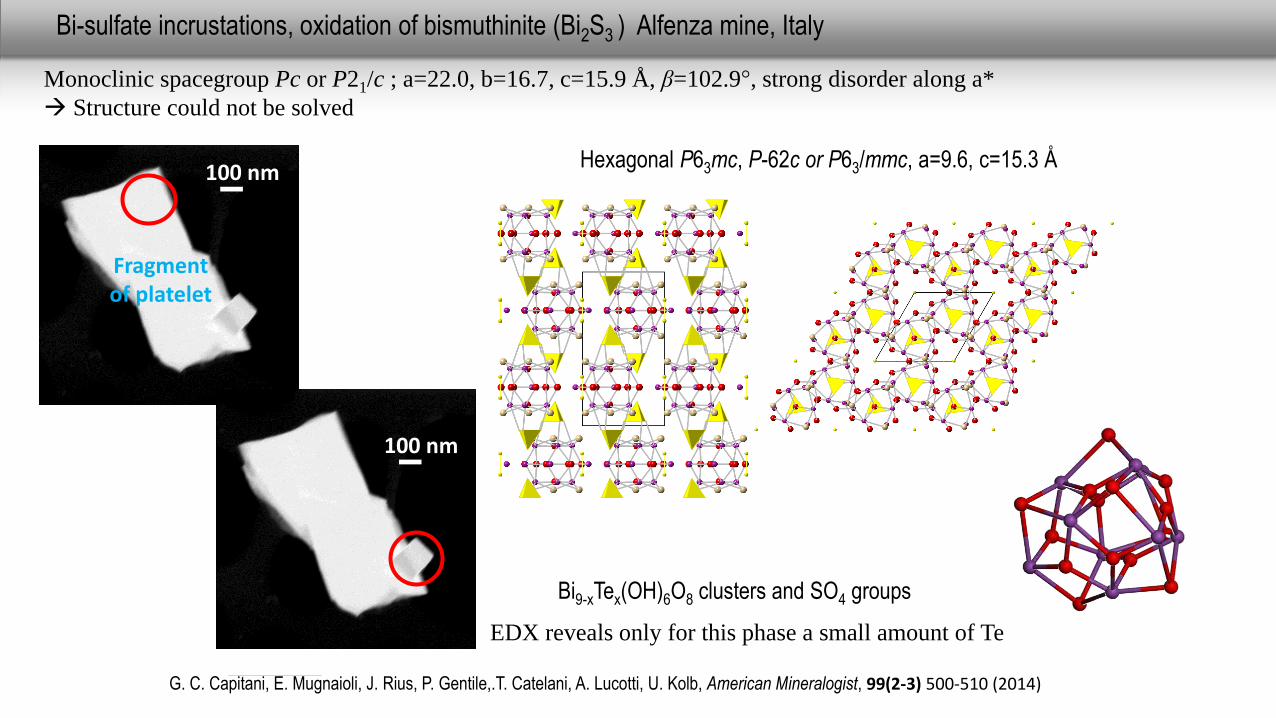
Monoclinic spacegroup Pc or P21/c ; a=22.0, b=16.7, c=15.9 Å, β=102.9°, strong disorder along a*
Structure could not be solved
100 nm
EDX reveals only for this phase a small amount of Te
100 nm
Bi-sulfate incrustations, oxidation of bismuthinite (Bi2S3 ) Alfenza mine, Italy
G. C. Capitani, E. Mugnaioli, J. Rius, P. Gentile,.T. Catelani, A. Lucotti, U. Kolb, American Mineralogist, 99(2-3) 500-510 (2014)
Bi9-xTex(OH)6O8 clusters and SO4 groups
Hexagonal P63mc, P-62c or P63/mmc, a=9.6, c=15.3 Å
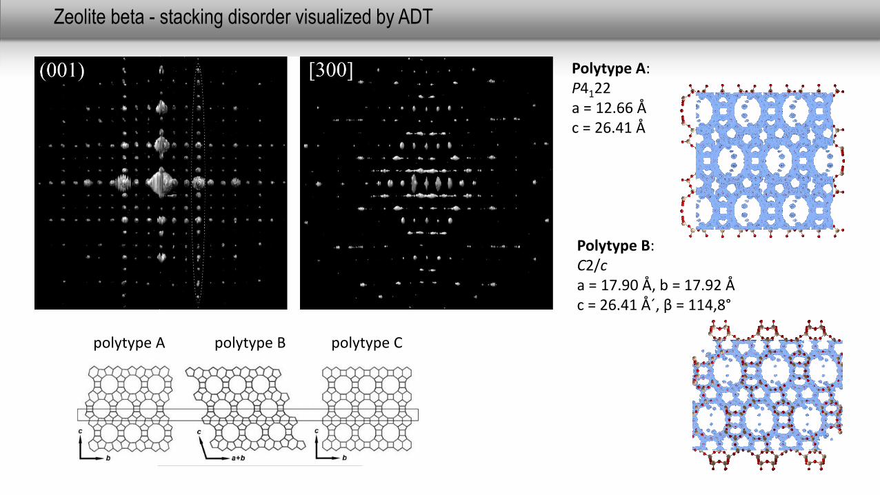
polytype A polytype B polytype C
Zeolite beta - stacking disorder visualized by ADT
Polytype A: P4122a = 12.66 Åc = 26.41 Å
Polytype B: C2/ca = 17.90 Å, b = 17.92 Åc = 26.41 Å´, β = 114,8°
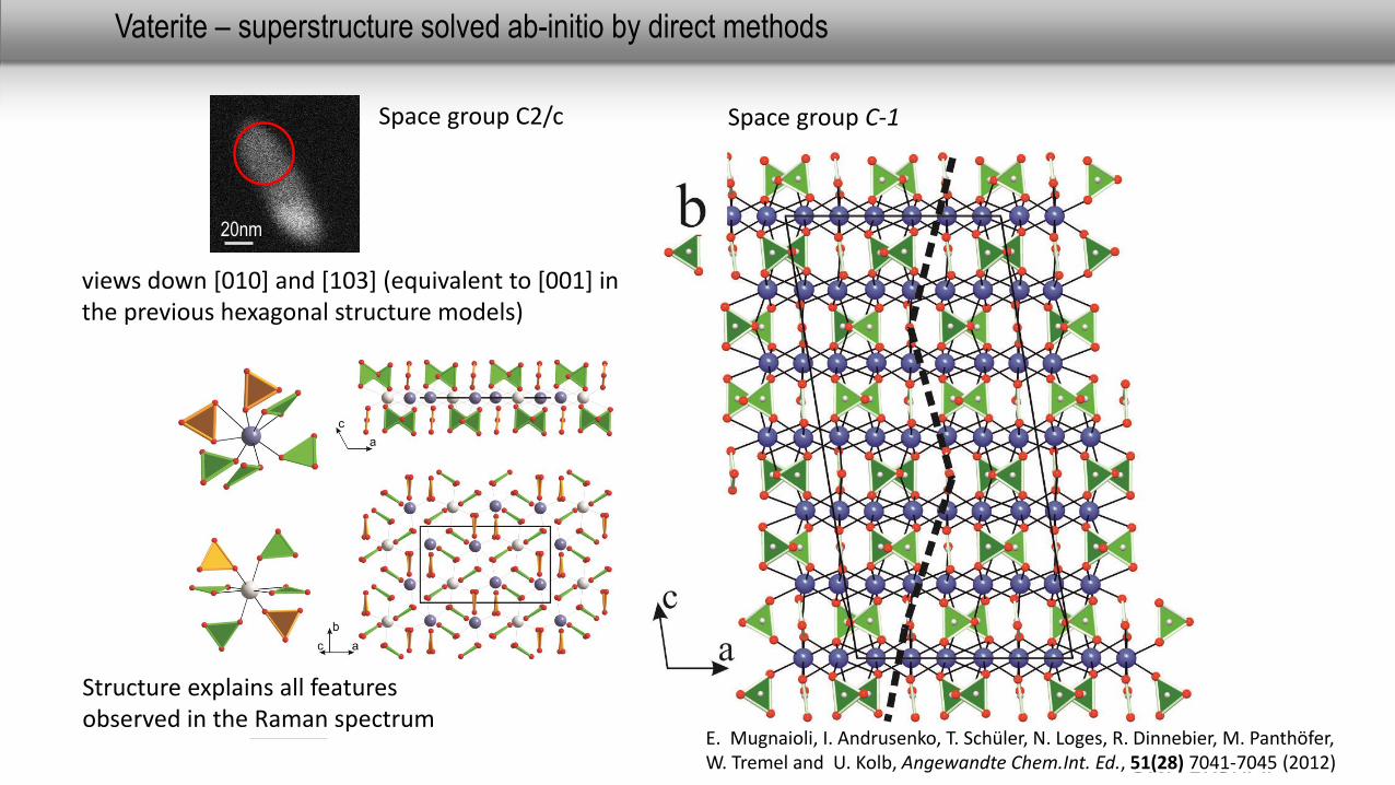
Vaterite – superstructure solved ab-initio by direct methods
views down [010] and [103] (equivalent to [001] in the previous hexagonal structure models)
Space group C2/c
20nm
Structure explains all features observed in the Raman spectrum
Space group C-1
E. Mugnaioli, I. Andrusenko, T. Schüler, N. Loges, R. Dinnebier, M. Panthöfer, W. Tremel and U. Kolb, Angewandte Chem.Int. Ed., 51(28) 7041-7045 (2012)
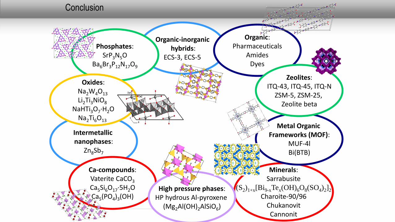
Conclusion
Minerals:Sarrabusite
(S2)1+x[Bi9-xTex(OH)6O8(SO4)2]2
Charoite-90/96ChukanovitCannonit
Intermetallic nanophases:
Zn8Sb7
Ca-compounds:Vaterite CaCO3
Ca5Si6O17·5H2OCa5(PO4)3(OH)
Metal OrganicFrameworks (MOF):
MUF-4lBi(BTB)
High pressure phases:HP hydrous Al-pyroxene
(Mg2Al(OH)2AlSiO6)
Organic-inorganichybrids:
ECS-3, ECS-5
Organic:Pharmaceuticals
AmidesDyes
Zeolites:ITQ-43, ITQ-45, ITQ-N
ZSM-5, ZSM-25, Zeolite beta
Phosphates:SrP3N5O
Ba6Br3P12N17O9
Oxides:Na2W4O13
Li2Ti3NiO8
NaHTi3O7·H2ONa2Ti6O13
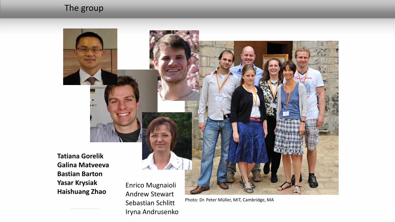
The group
Photo: Dr. Peter Müller, MIT, Cambridge, MA
Tatiana GorelikGalina MatveevaBastian BartonYasar KrysiakHaishuang Zhao
Enrico MugnaioliAndrew Stewart Sebastian SchlittIryna Andrusenko
Acknowledgment
Cooperation:
- Max Otten, FEI, Eindhoven, Netherlands
- Wolfgang Tremel, Universität Mainz, Germany (Vaterit)
- Avelino Corma, Instituto de Tecnologia Quimica, Valencia, Spain (ITQ-43)
- Giovanna Vezzalini, University of Modena and Reggio Emilia and Rossella Arletti, University of Torino, Italy (ZSM-5)
- Ch. Bärlocher, ETH Zürich, Switzerland (IM-5)
- Dirk Volkmer, Dimitry Denysenko, University of Ulm, Germany (MFU-4long)
- Norbert Stock, University of Kiel (CAU), Germany (Bi(BTB))
- Wulf Depmeier, Michael Czank, University of Kiel, Germany andIra Rozhdestvenskaya, University of Moskow, Russia (Charoite)
- Mauro Gemmi, Stefano Poli, University of Milano, Italy (Hapy)
Financial Support:
Stiftung Rheinland-Pfalz InnovationSFB 625SPP 1415
Thank you for your
attention