tom heaps consultant acute physicianacutemedicinebhh.com/files/medical/headache.pdf · and...
TRANSCRIPT
Tom Heaps
Consultant Acute Physician
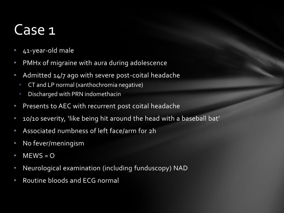
• 41-year-old male
• PMHx of migraine with aura during adolescence
• Admitted 14/7 ago with severe post-coital headache
• CT and LP normal (xanthochromia negative)
• Discharged with PRN indomethacin
• Presents to AEC with recurrent post coital headache
• 10/10 severity, ‘like being hit around the head with a baseball bat’
• Associated numbness of left face/arm for 2h
• No fever/meningism
• MEWS = O
• Neurological examination (including funduscopy) NAD
• Routine bloods and ECG normal
Case 1
Further investigations?
Repeat CT/LP??
Possible Diagnoses?
Case 1 cont.
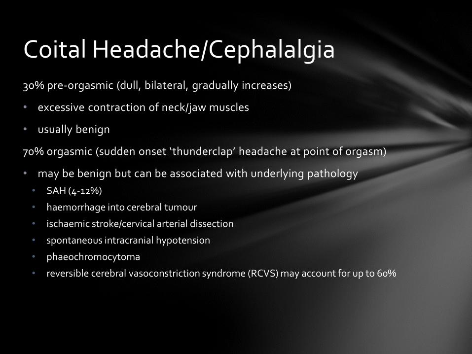
30% pre-orgasmic (dull, bilateral, gradually increases)
• excessive contraction of neck/jaw muscles
• usually benign
70% orgasmic (sudden onset ‘thunderclap’ headache at point of orgasm)
• may be benign but can be associated with underlying pathology
• SAH (4-12%)
• haemorrhage into cerebral tumour
• ischaemic stroke/cervical arterial dissection
• spontaneous intracranial hypotension
• phaeochromocytoma
• reversible cerebral vasoconstriction syndrome (RCVS) may account for up to 60%
Coital Headache/Cephalalgia
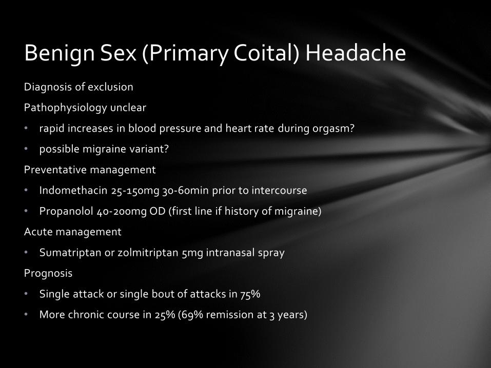
Diagnosis of exclusion
Pathophysiology unclear
• rapid increases in blood pressure and heart rate during orgasm?
• possible migraine variant?
Preventative management
• Indomethacin 25-150mg 30-60min prior to intercourse
• Propanolol 40-200mg OD (first line if history of migraine)
Acute management
• Sumatriptan or zolmitriptan 5mg intranasal spray
Prognosis
• Single attack or single bout of attacks in 75%
• More chronic course in 25% (69% remission at 3 years)
Benign Sex (Primary Coital) Headache
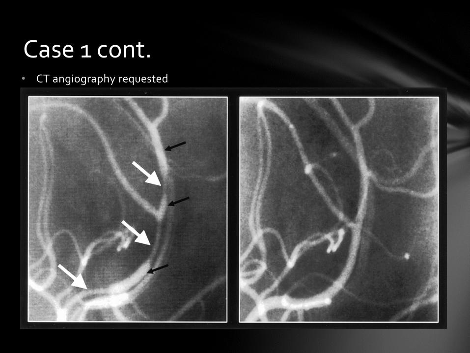
• CT angiography requested
Case 1 cont.
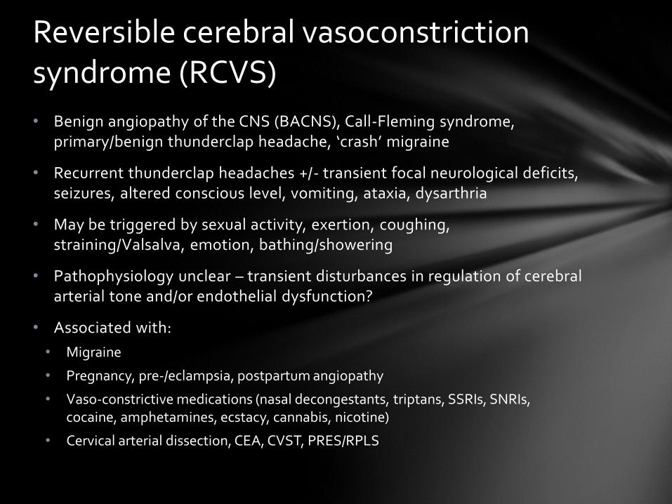
• Benign angiopathy of the CNS (BACNS), Call-Fleming syndrome, primary/benign thunderclap headache, ‘crash’ migraine
• Recurrent thunderclap headaches +/- transient focal neurological deficits, seizures, altered conscious level, vomiting, ataxia, dysarthria
• May be triggered by sexual activity, exertion, coughing, straining/Valsalva, emotion, bathing/showering
• Pathophysiology unclear – transient disturbances in regulation of cerebral arterial tone and/or endothelial dysfunction?
• Associated with:
• Migraine
• Pregnancy, pre-/eclampsia, postpartum angiopathy
• Vaso-constrictive medications (nasal decongestants, triptans, SSRIs, SNRIs, cocaine, amphetamines, ecstacy, cannabis, nicotine)
• Cervical arterial dissection, CEA, CVST, PRES/RPLS
Reversible cerebral vasoconstriction syndrome (RCVS)
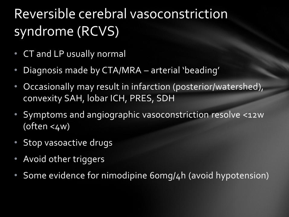
• CT and LP usually normal
• Diagnosis made by CTA/MRA – arterial ‘beading’
• Occasionally may result in infarction (posterior/watershed), convexity SAH, lobar ICH, PRES, SDH
• Symptoms and angiographic vasoconstriction resolve <12w (often <4w)
• Stop vasoactive drugs
• Avoid other triggers
• Some evidence for nimodipine 60mg/4h (avoid hypotension)
Reversible cerebral vasoconstriction syndrome (RCVS)
High severity ‘worst ever’ headache reaching maximal intensity (≥7/10)in <1min
SAH in 11-25%
Other serious pathology in 10-12%
Thunderclap headache
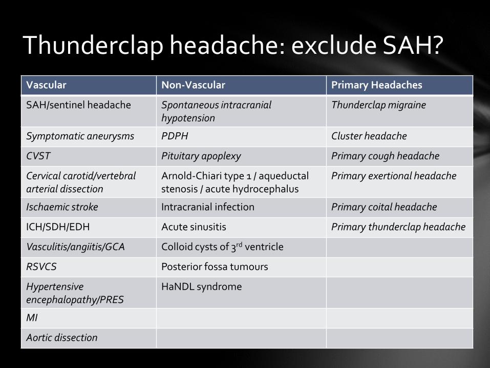
Vascular Non-Vascular Primary Headaches
SAH/sentinel headache Spontaneous intracranial hypotension
Thunderclap migraine
Symptomatic aneurysms PDPH Cluster headache
CVST Pituitary apoplexy Primary cough headache
Cervical carotid/vertebral arterial dissection
Arnold-Chiari type 1 / aqueductal stenosis / acute hydrocephalus
Primary exertional headache
Ischaemic stroke Intracranial infection Primary coital headache
ICH/SDH/EDH Acute sinusitis Primary thunderclap headache
Vasculitis/angiitis/GCA Colloid cysts of 3rd ventricle
RSVCS Posterior fossa tumours
Hypertensive encephalopathy/PRES
HaNDL syndrome
MI
Aortic dissection
Thunderclap headache: exclude SAH?
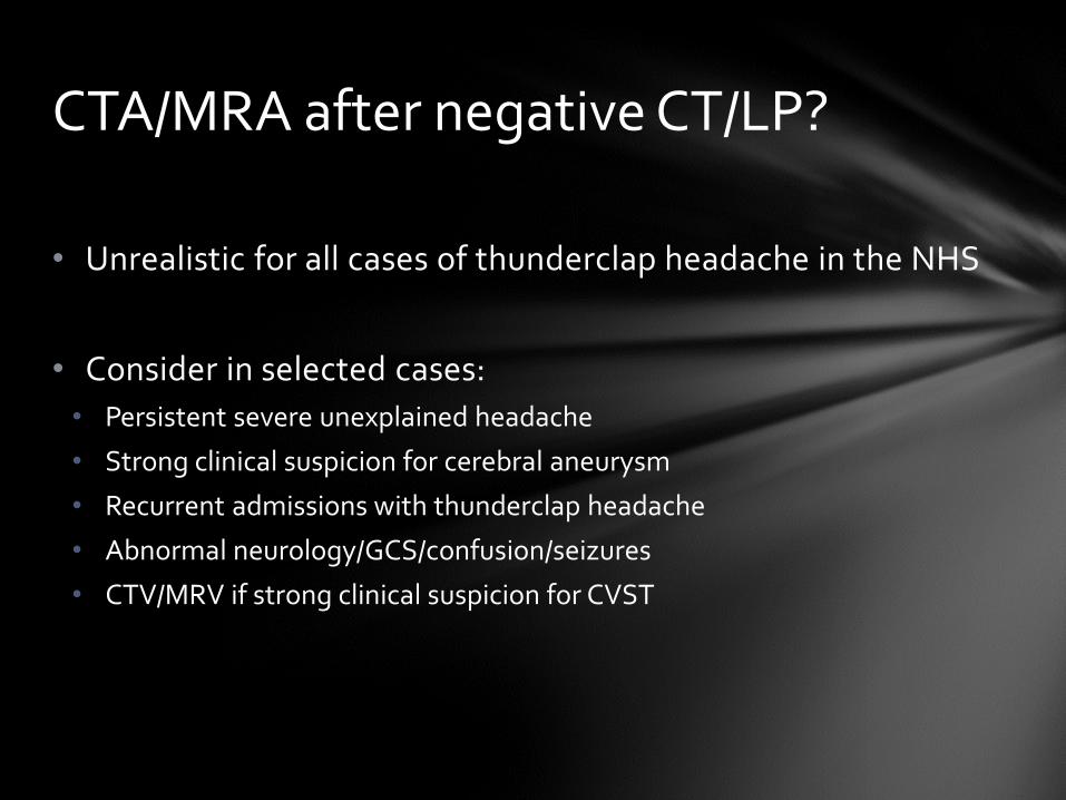
• Unrealistic for all cases of thunderclap headache in the NHS
• Consider in selected cases:
• Persistent severe unexplained headache
• Strong clinical suspicion for cerebral aneurysm
• Recurrent admissions with thunderclap headache
• Abnormal neurology/GCS/confusion/seizures
• CTV/MRV if strong clinical suspicion for CVST
CTA/MRA after negative CT/LP?
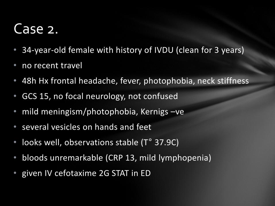
• 34-year-old female with history of IVDU (clean for 3 years)
• no recent travel
• 48h Hx frontal headache, fever, photophobia, neck stiffness
• GCS 15, no focal neurology, not confused
• mild meningism/photophobia, Kernigs –ve
• several vesicles on hands and feet
• looks well, observations stable (T° 37.9C)
• bloods unremarkable (CRP 13, mild lymphopenia)
• given IV cefotaxime 2G STAT in ED
Case 2.

Does he need a CT head prior to LP?
NO – unnecessary CT delays diagnostic LP/makes results more difficult to interpret
Indications for CT prior to LP in suspected meningitis
• age >60
• immunocompromise
• history of CNS disease (mass lesion, stroke, focal infection)
• new onset seizures
• focal neurology
• abnormal GCS
• signs of raised ICP (bradycardia, hypertension, papilloedema)
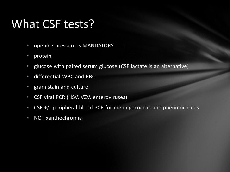
What CSF tests?
• opening pressure is MANDATORY
• protein
• glucose with paired serum glucose (CSF lactate is an alternative)
• differential WBC and RBC
• gram stain and culture
• CSF viral PCR (HSV, VZV, enteroviruses)
• CSF +/- peripheral blood PCR for meningococcus and pneumococcus
• NOT xanthochromia
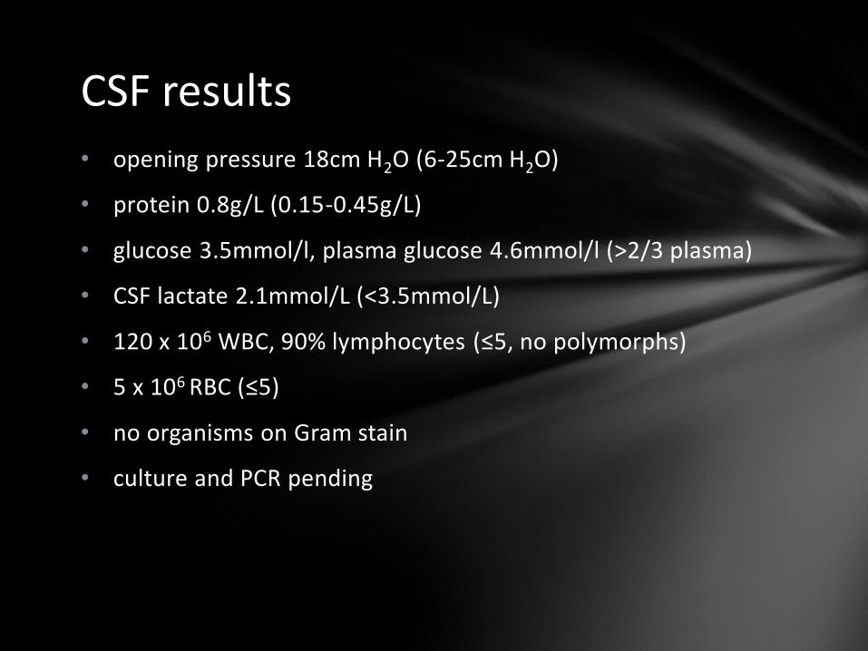
CSF results
• opening pressure 18cm H2O (6-25cm H2O)
• protein 0.8g/L (0.15-0.45g/L)
• glucose 3.5mmol/l, plasma glucose 4.6mmol/l (>2/3 plasma)
• CSF lactate 2.1mmol/L (<3.5mmol/L)
• 120 x 106 WBC, 90% lymphocytes (≤5, no polymorphs)
• 5 x 10⁶ RBC (≤5)
• no organisms on Gram stain
• culture and PCR pending
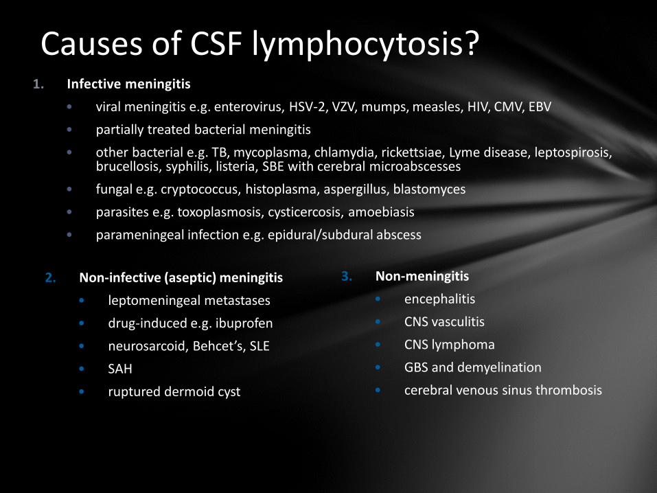
Causes of CSF lymphocytosis? 1. Infective meningitis
• viral meningitis e.g. enterovirus, HSV-2, VZV, mumps, measles, HIV, CMV, EBV
• partially treated bacterial meningitis
• other bacterial e.g. TB, mycoplasma, chlamydia, rickettsiae, Lyme disease, leptospirosis, brucellosis, syphilis, listeria, SBE with cerebral microabscesses
• fungal e.g. cryptococcus, histoplasma, aspergillus, blastomyces
• parasites e.g. toxoplasmosis, cysticercosis, amoebiasis
• parameningeal infection e.g. epidural/subdural abscess
3. Non-meningitis
• encephalitis
• CNS vasculitis
• CNS lymphoma
• GBS and demyelination
• cerebral venous sinus thrombosis
2. Non-infective (aseptic) meningitis
• leptomeningeal metastases
• drug-induced e.g. ibuprofen
• neurosarcoid, Behcet’s, SLE
• SAH
• ruptured dermoid cyst
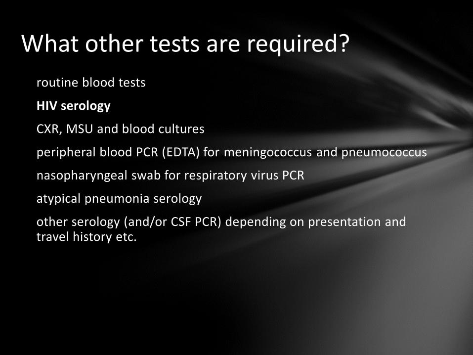
What other tests are required?
routine blood tests
HIV serology
CXR, MSU and blood cultures
peripheral blood PCR (EDTA) for meningococcus and pneumococcus
nasopharyngeal swab for respiratory virus PCR
atypical pneumonia serology
other serology (and/or CSF PCR) depending on presentation and travel history etc.
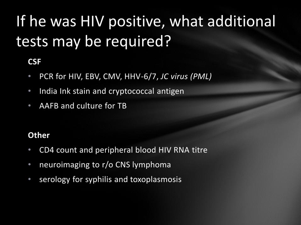
If he was HIV positive, what additional tests may be required?
CSF
• PCR for HIV, EBV, CMV, HHV-6/7, JC virus (PML)
• India Ink stain and cryptococcal antigen
• AAFB and culture for TB
Other
• CD4 count and peripheral blood HIV RNA titre
• neuroimaging to r/o CNS lymphoma
• serology for syphilis and toxoplasmosis
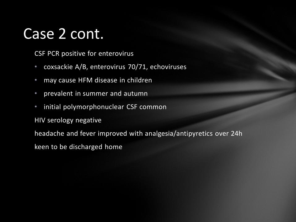
Case 2 cont. CSF PCR positive for enterovirus
• coxsackie A/B, enterovirus 70/71, echoviruses
• may cause HFM disease in children
• prevalent in summer and autumn
• initial polymorphonuclear CSF common
HIV serology negative
headache and fever improved with analgesia/antipyretics over 24h
keen to be discharged home
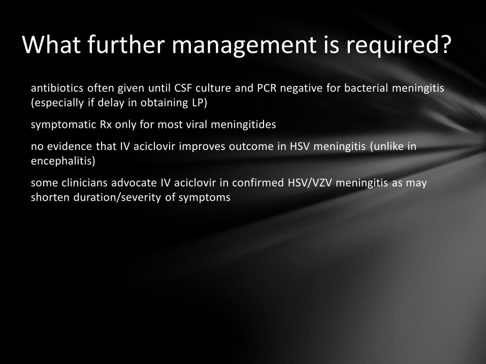
What further management is required?
antibiotics often given until CSF culture and PCR negative for bacterial meningitis (especially if delay in obtaining LP)
symptomatic Rx only for most viral meningitides
no evidence that IV aciclovir improves outcome in HSV meningitis (unlike in encephalitis)
some clinicians advocate IV aciclovir in confirmed HSV/VZV meningitis as may shorten duration/severity of symptoms
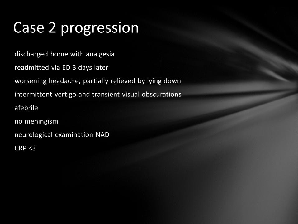
Case 2 progression
discharged home with analgesia
readmitted via ED 3 days later
worsening headache, partially relieved by lying down
intermittent vertigo and transient visual obscurations
afebrile
no meningism
neurological examination NAD
CRP <3
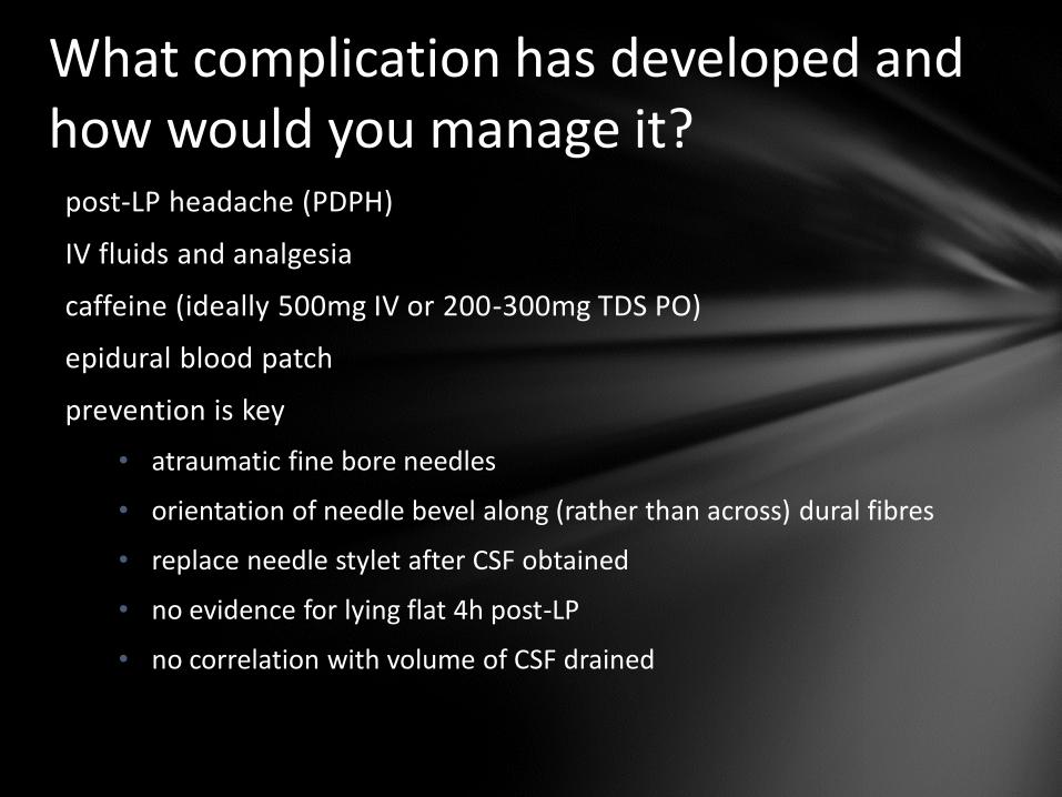
What complication has developed and how would you manage it?
post-LP headache (PDPH)
IV fluids and analgesia
caffeine (ideally 500mg IV or 200-300mg TDS PO)
epidural blood patch
prevention is key
• atraumatic fine bore needles
• orientation of needle bevel along (rather than across) dural fibres
• replace needle stylet after CSF obtained
• no evidence for lying flat 4h post-LP
• no correlation with volume of CSF drained
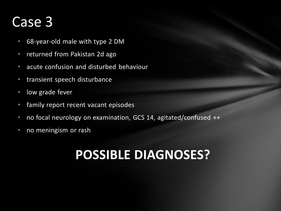
Case 3 • 68-year-old male with type 2 DM
• returned from Pakistan 2d ago
• acute confusion and disturbed behaviour
• transient speech disturbance
• low grade fever
• family report recent vacant episodes
• no focal neurology on examination, GCS 14, agitated/confused ++
• no meningism or rash
POSSIBLE DIAGNOSES?
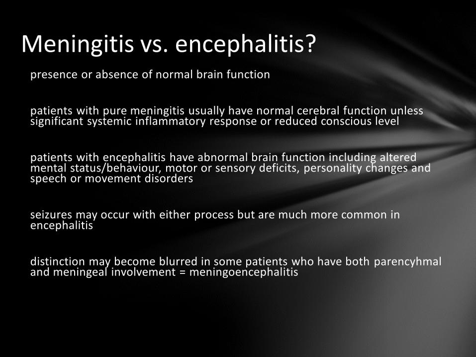
Meningitis vs. encephalitis? presence or absence of normal brain function
patients with pure meningitis usually have normal cerebral function unless significant systemic inflammatory response or reduced conscious level
patients with encephalitis have abnormal brain function including altered mental status/behaviour, motor or sensory deficits, personality changes and speech or movement disorders
seizures may occur with either process but are much more common in encephalitis
distinction may become blurred in some patients who have both parencyhmal and meningeal involvement = meningoencephalitis
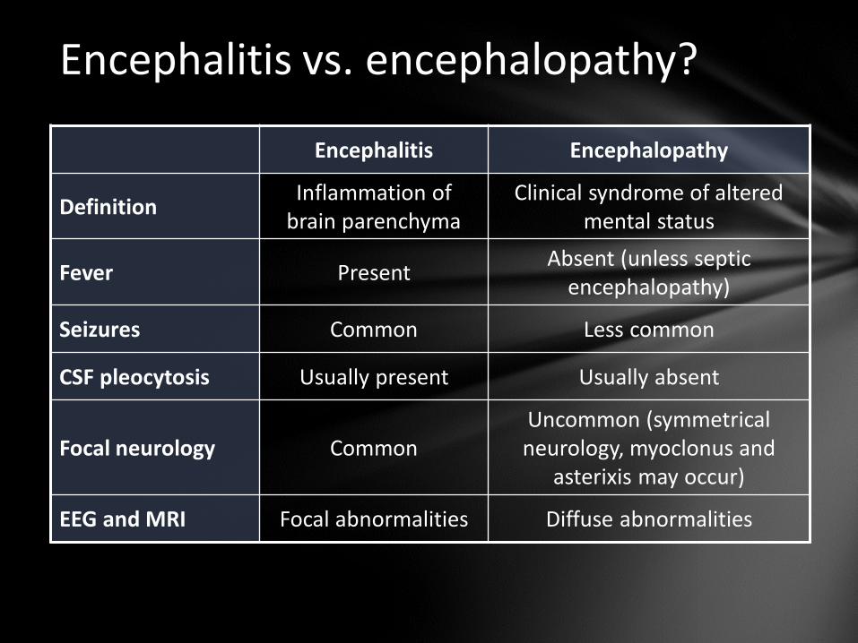
Encephalitis vs. encephalopathy?
Encephalitis Encephalopathy
Definition Inflammation of
brain parenchyma Clinical syndrome of altered
mental status
Fever Present Absent (unless septic
encephalopathy)
Seizures Common Less common
CSF pleocytosis Usually present Usually absent
Focal neurology Common Uncommon (symmetrical neurology, myoclonus and
asterixis may occur)
EEG and MRI Focal abnormalities Diffuse abnormalities
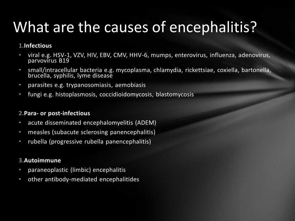
What are the causes of encephalitis? 1.Infectious
• viral e.g. HSV-1, VZV, HIV, EBV, CMV, HHV-6, mumps, enterovirus, influenza, adenovirus, parvovirus B19
• small/intracellular bacteria e.g. mycoplasma, chlamydia, rickettsiae, coxiella, bartonella, brucella, syphilis, lyme disease
• parasites e.g. trypanosomiasis, aemobiasis
• fungi e.g. histoplasmosis, coccidioidomycosis, blastomycosis
2.Para- or post-infectious
• acute disseminated encephalomyelitis (ADEM)
• measles (subacute sclerosing panencephalitis)
• rubella (progressive rubella panencephalitis)
3.Autoimmune
• paraneoplastic (limbic) encephalitis
• other antibody-mediated encephalitides
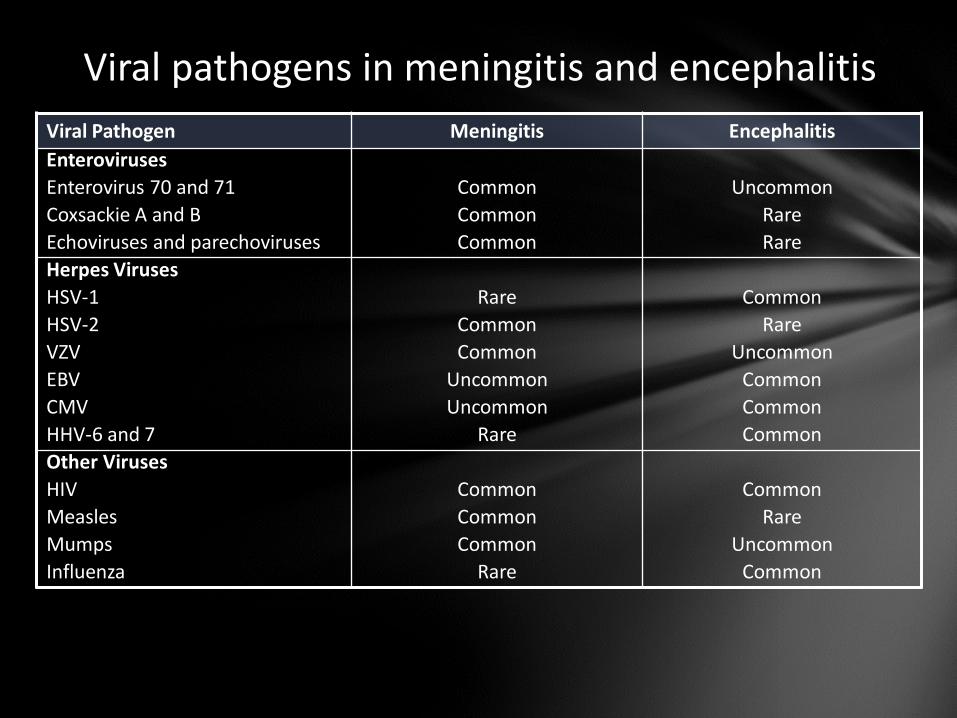
Viral pathogens in meningitis and encephalitis
Viral Pathogen Meningitis Encephalitis
Enteroviruses
Enterovirus 70 and 71 Common Uncommon
Coxsackie A and B Common Rare
Echoviruses and parechoviruses Common Rare
Herpes Viruses
HSV-1 Rare Common
HSV-2 Common Rare
VZV Common Uncommon
EBV Uncommon Common
CMV Uncommon Common
HHV-6 and 7 Rare Common
Other Viruses
HIV Common Common
Measles Common Rare
Mumps Common Uncommon
Influenza Rare Common
How are you going to investigate this patient?
CT prior to LP?
Which CSF tests?
Blood tests?
MRI?
EEG?
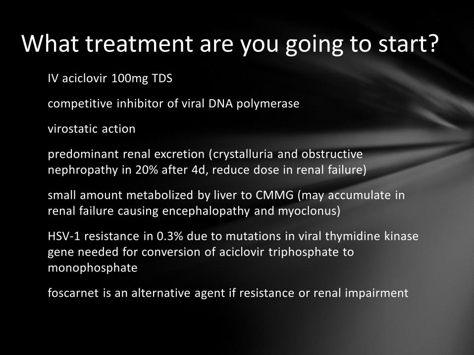
What treatment are you going to start? IV aciclovir 100mg TDS
competitive inhibitor of viral DNA polymerase
virostatic action
predominant renal excretion (crystalluria and obstructive nephropathy in 20% after 4d, reduce dose in renal failure)
small amount metabolized by liver to CMMG (may accumulate in renal failure causing encephalopathy and myoclonus)
HSV-1 resistance in 0.3% due to mutations in viral thymidine kinase gene needed for conversion of aciclovir triphosphate to monophosphate
foscarnet is an alternative agent if resistance or renal impairment
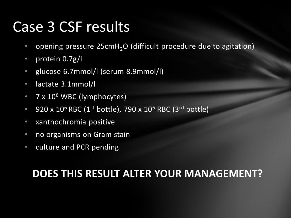
Case 3 CSF results • opening pressure 25cmH2O (difficult procedure due to agitation)
• protein 0.7g/l
• glucose 6.7mmol/l (serum 8.9mmol/l)
• lactate 3.1mmol/l
• 7 x 10⁶ WBC (lymphocytes)
• 920 x 106 RBC (1st bottle), 790 x 106 RBC (3rd bottle)
• xanthochromia positive
• no organisms on Gram stain
• culture and PCR pending
DOES THIS RESULT ALTER YOUR MANAGEMENT?
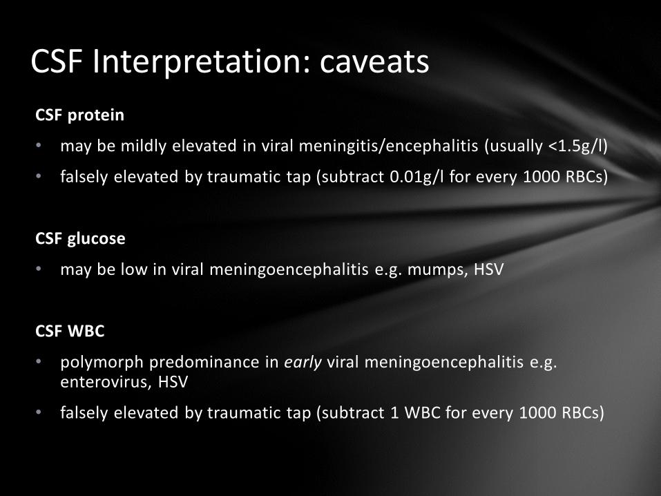
CSF Interpretation: caveats
CSF protein
• may be mildly elevated in viral meningitis/encephalitis (usually <1.5g/l)
• falsely elevated by traumatic tap (subtract 0.01g/l for every 1000 RBCs)
CSF glucose
• may be low in viral meningoencephalitis e.g. mumps, HSV
CSF WBC
• polymorph predominance in early viral meningoencephalitis e.g. enterovirus, HSV
• falsely elevated by traumatic tap (subtract 1 WBC for every 1000 RBCs)
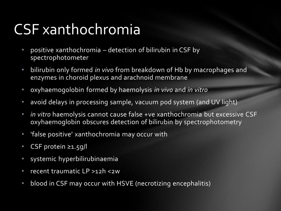
• positive xanthochromia – detection of bilirubin in CSF by spectrophotometer
• bilirubin only formed in vivo from breakdown of Hb by macrophages and enzymes in choroid plexus and arachnoid membrane
• oxyhaemogolobin formed by haemolysis in vivo and in vitro
• avoid delays in processing sample, vacuum pod system (and UV light)
• in vitro haemolysis cannot cause false +ve xanthochromia but excessive CSF oxyhaemoglobin obscures detection of bilirubin by spectrophotometry
• ‘false positive’ xanthochromia may occur with
• CSF protein ≥1.5g/l
• systemic hyperbilirubinaemia
• recent traumatic LP >12h <2w
• blood in CSF may occur with HSVE (necrotizing encephalitis)
CSF xanthochromia
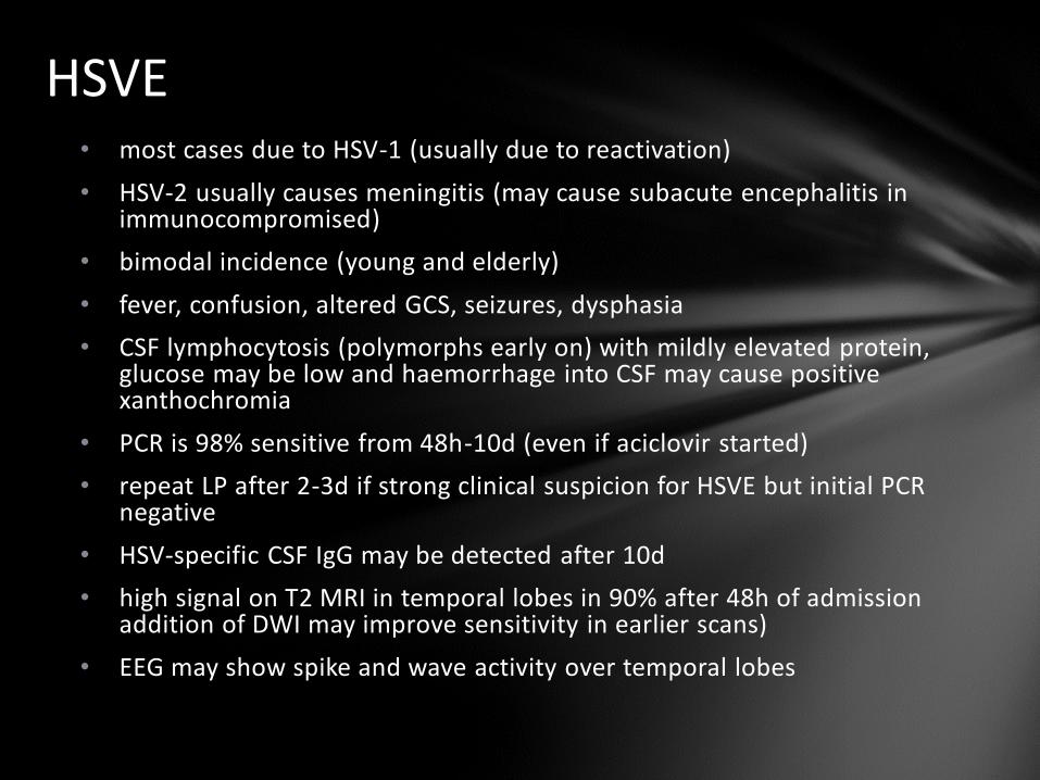
HSVE • most cases due to HSV-1 (usually due to reactivation)
• HSV-2 usually causes meningitis (may cause subacute encephalitis in immunocompromised)
• bimodal incidence (young and elderly)
• fever, confusion, altered GCS, seizures, dysphasia
• CSF lymphocytosis (polymorphs early on) with mildly elevated protein, glucose may be low and haemorrhage into CSF may cause positive xanthochromia
• PCR is 98% sensitive from 48h-10d (even if aciclovir started)
• repeat LP after 2-3d if strong clinical suspicion for HSVE but initial PCR negative
• HSV-specific CSF IgG may be detected after 10d
• high signal on T2 MRI in temporal lobes in 90% after 48h of admission addition of DWI may improve sensitivity in earlier scans)
• EEG may show spike and wave activity over temporal lobes

HSVE • treat with IV aciclovir 100mg/kg TDS for 14d (21d if
immunocompromise)
• reduces mortality from 70% to 20%
• addition of IV dexamethasone 10mg QDS for 4d may improve outcome (ongoing trials)
• repeat LP at end of course of Rx and continue aciclovir for longer if PCR remains positive
• oral aciclovir fails to achieve adequate CSF concentrations ?role for PO valaciclovir if prolonged Rx required
• permanent neuropsychololgical sequelae in 2/3 of survivors (personality changes, cognitive impairment, epilepsy, dysphasia)
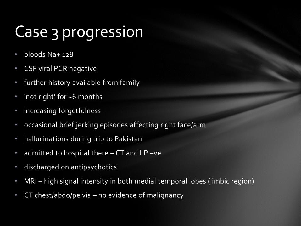
• bloods Na+ 128
• CSF viral PCR negative
• further history available from family
• ‘not right’ for ~6 months
• increasing forgetfulness
• occasional brief jerking episodes affecting right face/arm
• hallucinations during trip to Pakistan
• admitted to hospital there – CT and LP –ve
• discharged on antipsychotics
• MRI – high signal intensity in both medial temporal lobes (limbic region)
• CT chest/abdo/pelvis – no evidence of malignancy
Case 3 progression
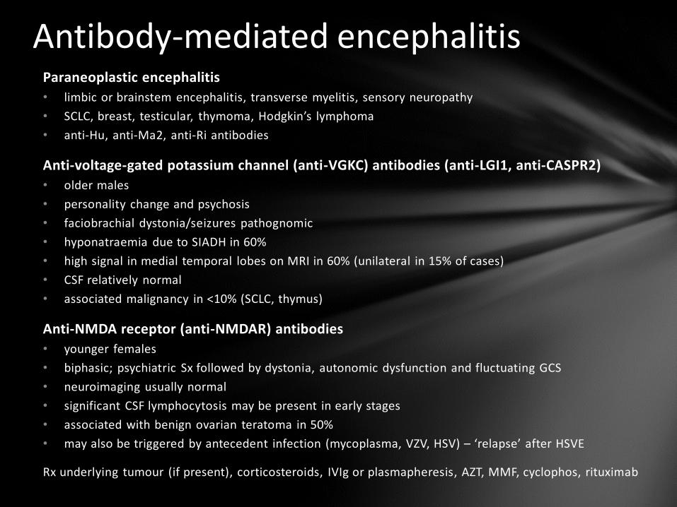
Antibody-mediated encephalitis Paraneoplastic encephalitis
• limbic or brainstem encephalitis, transverse myelitis, sensory neuropathy
• SCLC, breast, testicular, thymoma, Hodgkin’s lymphoma
• anti-Hu, anti-Ma2, anti-Ri antibodies
Anti-voltage-gated potassium channel (anti-VGKC) antibodies (anti-LGI1, anti-CASPR2)
• older males
• personality change and psychosis
• faciobrachial dystonia/seizures pathognomic
• hyponatraemia due to SIADH in 60%
• high signal in medial temporal lobes on MRI in 60% (unilateral in 15% of cases)
• CSF relatively normal
• associated malignancy in <10% (SCLC, thymus)
Anti-NMDA receptor (anti-NMDAR) antibodies
• younger females
• biphasic; psychiatric Sx followed by dystonia, autonomic dysfunction and fluctuating GCS
• neuroimaging usually normal
• significant CSF lymphocytosis may be present in early stages
• associated with benign ovarian teratoma in 50%
• may also be triggered by antecedent infection (mycoplasma, VZV, HSV) – ‘relapse’ after HSVE
Rx underlying tumour (if present), corticosteroids, IVIg or plasmapheresis, AZT, MMF, cyclophos, rituximab
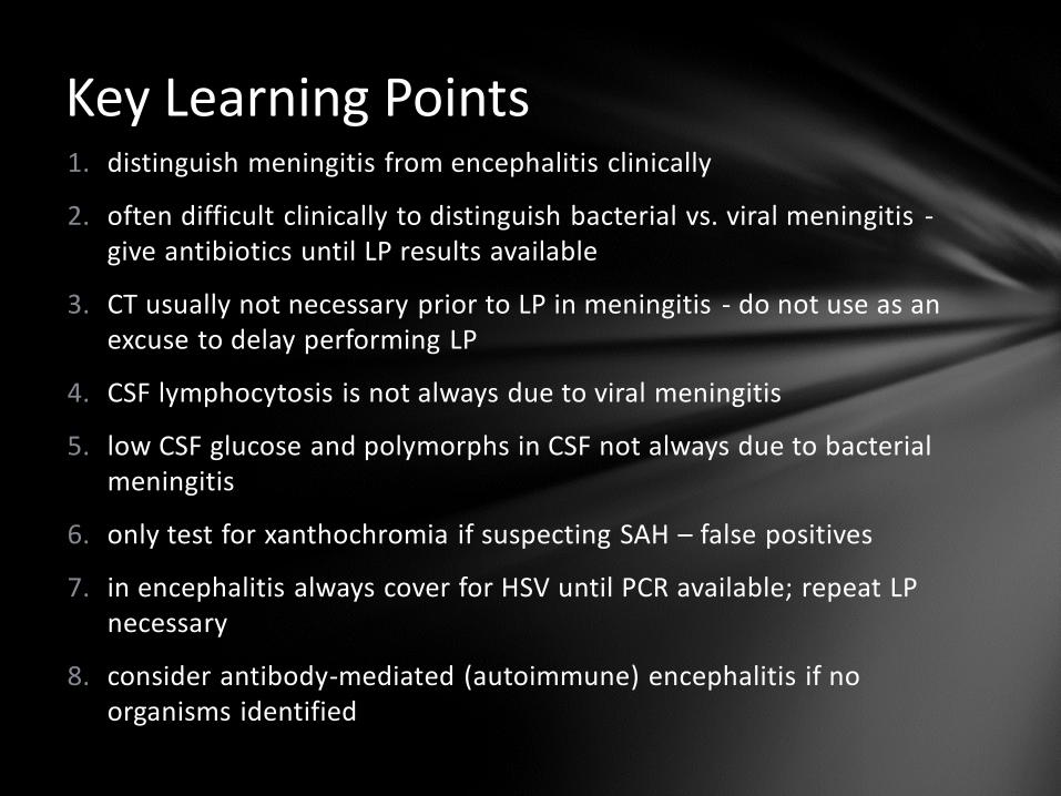
Key Learning Points 1. distinguish meningitis from encephalitis clinically
2. often difficult clinically to distinguish bacterial vs. viral meningitis - give antibiotics until LP results available
3. CT usually not necessary prior to LP in meningitis - do not use as an excuse to delay performing LP
4. CSF lymphocytosis is not always due to viral meningitis
5. low CSF glucose and polymorphs in CSF not always due to bacterial meningitis
6. only test for xanthochromia if suspecting SAH – false positives
7. in encephalitis always cover for HSV until PCR available; repeat LP necessary
8. consider antibody-mediated (autoimmune) encephalitis if no organisms identified