toll-like receptor 9 partially regulates lung inflammation induced
TRANSCRIPT
RESEARCH Open Access
Toll-like receptor 9 partially regulates lunginflammation induced following exposureto chicken barn airDavid Schneberger, Gurpreet Aulakh, Shankaramurthy Channabasappa and Baljit Singh*
Abstract
Background: Exposure to animal barn air is an occupational hazard that causes lung dysfunction in barn workers.Respiratory symptoms experienced by workers are typically associated with endotoxin and TLR4 signalling, butwithin these environments gram negative bacteria constitute only a portion of the total microbial population. Incontrast, unmethylated DNA can be found in all bacteria, some viruses, and mold. We hypothesized that in suchenvironments TLR9, which binds unmethylated DNA, contributes to the overall immune responses in the lung.
Methods: Using a mouse model, wild-type and TLR9−/− mice were exposed to chicken barn air for 1, 5, or 20 days.Blood serum and bronchiolar lavage fluid was tested against a panel of six TLR9-induced cytokines (IL-1β, IL-6, IL-10,IL-12, TNFα, and IFNγ) for changes in expression. Bronchiolar lavage fluid (BAL) was also tested for macrophage aswell as monocyte migration.
Results: There were significant decreases in serum TNFα after a single day exposure in TLR9−/− mice. BALconcentrations of TNFα and IFNγ, as well as TNFα in serum in TLR9−/− mice were also reduced after barn exposurefor 5 days. After 20 days of exposure IFNγ was significantly reduced in lavage of TLR9−/− mice. Myeloperoxidase(MPO) accumulation in the lung was reduced at 20 days of exposure in TLR9−/− mice, as was total lavage cellcounts. However, Masson’s staining revealed no apparent lung histological differences between any of thetreatment groups.
Conclusions: Taken together our data show TLR9 plays a partial role in lung inflammation induced followingexposure to chicken barn air potentially through binding of unmethylated DNA.
Keywords: Barn dust, TLR9, CpG DNA, Macrophages, Neutrophils, BAL
BackgroundWorkers in high-intensity livestock operations have beenrecognized to be at risk for a number of chronic respira-tory problems including bronchitis, rhinitis, chroniccough and phlegm, occupational asthma, and organicdust toxic syndrome to name a few [1–3]. Workers insuch facilities are exposed to a wide variety of agentssuch as ammonia, hydrogen sulfide, dust particles, andlipopolysaccharide (LPS) [3, 4]. Even single exposure tosuch facilities has been shown to elevate a number ofpro-inflammatory cytokines in nasal lavages and serum,
and induce lung inflammation [5–7]. The mechanismsof these complex in vivo pulmonary responses are notfully understood.Endotoxin has been targeted as a critical component
responsible for many of the lung problems seen in ex-posure to barn air [1, 7, 8]. More recent work howeversuggests that within chicken barns, endotoxin-producinggram-negative bacteria comprise only a small fraction ofthe bacterial species, and that these bacteria may be inthe minority as far as total numbers of bacteria present[4]. In contrast, all bacteria and some viruses and moldcontain unmethylated DNA in their genomes, which in-duces a variety of cytokines through binding the TLR9receptor. Many of these cytokines are similar to thoseinduced by endotoxin, which binds TLR4 [9]. Previous
* Correspondence: [email protected] of Veterinary Biomedical Sciences, Western College of VeterinaryMedicine, University of Saskatchewan, 52 Campus Drive, Saskatoon, SK S7N5B4, Canada
© 2016 The Author(s). Open Access This article is distributed under the terms of the Creative Commons Attribution 4.0International License (http://creativecommons.org/licenses/by/4.0/), which permits unrestricted use, distribution, andreproduction in any medium, provided you give appropriate credit to the original author(s) and the source, provide a link tothe Creative Commons license, and indicate if changes were made. The Creative Commons Public Domain Dedication waiver(http://creativecommons.org/publicdomain/zero/1.0/) applies to the data made available in this article, unless otherwise stated.
Schneberger et al. Journal of Occupational Medicine and Toxicology (2016) 11:31 DOI 10.1186/s12995-016-0121-x
work had shown that DNA from barn dust extractscould induce IL-10 and IL-12p40 [10], however this sys-tem exposed isolated peripheral blood mononuclear cellsto purified DNA in vitro. This may not be an optimal re-flection of barn and in vivo conditions, where significantproduction of cytokines may occur in the lung from avariety of cell types. Further, such a system does not ac-count for effects of numerous other components in theair that may synergize with or counter responsesthrough TLR9. Therefore, before we dissect the cellulareffects in in vitro systems, we need in vivo animal modelstudies to understand the lung responses to chickenbarn air.Recently, we delineated expression of TLR9 in mouse
and human lungs using in situ hybridization and lightand electron microscope immunocytochemistry [11].We have also reported TLR9 expression in cattle, horses,dogs and pigs that spend their lives in barns or otheranimal containment facilities [12, 13]. This series ofstudies provided the first data to show in situ expressionof TLR9 in airway epithelium, alveolar septal cells andalveolar macrophages. We used this TLR9 expressiondata to hypothesize that TLR9 promotes in vivo lung in-flammation induced by single or multiple exposures tochicken barn air. We tested this hypothesis by exposingTLR9−/− and wild-type mice to chicken barn air for 1, 5,or 20 days. The data show that TLR9 has a partial rolein lung inflammation induced following exposure tochicken barn air.
MethodsAnimalsThe experimental protocols were approved by the Uni-versity of Saskatchewan Committee on Animal Care As-surance and all experiments conducted according toguidelines of the Canadian Council on Animal Care.Breeding pairs of TLR9-deficient mice (C57BL/6 back-ground) were a gift from Dr. Heather Davis and obtainedfrom Taconic. Knockout status was confirmed by PCRon mouse lung tissue. Mice were raised at the WesternCollege of Veterinary Medicine Animal Care Unit.C57BL/6 (wild type) mice where obtained from the Ani-mal Resource Centre at the University of Saskatchewan.
Experimental exposureMice were transported in sealed cages with vents anddriven to a cage-based chicken barn in the morning andplaced on a shelf approximately 1.8 m (6 ft) off theground. Mice were kept in barn for 8 h and thenreturned to animal care facilities at the University of Sas-katchewan where they were transferred out of theircages for the evening. Each group consisted of 6 animalsper group. Each exposure group was split into two tothree subgroups for transport to the barn to ensure that
variation of barn or transport conditions on a single daywould not account for changes seen in an exposuregroup. Mice were taken to the barn for 1, 5, or 20 days.The 20 day exposure animals were rested for 2 days aftereach cycle of 5 exposures to mimic a 5 day work weekexposure. Parallel control groups of mice that weretransported but not exposed to barn air were trans-ported in separate cages along with the exposed mice.
Tissue, blood, and lavage collectionAt the end of exposure time mice were euthanized(100 mg/kg ketamine + 20 mg/kg xylazine, intraperito-neal injection), and blood was collected by cardiac punc-ture (Sandoz, Boucherville, QC, Canada). Collection wasdone approximately 2 h after leaving the barn environ-ment. Blood was separated into serum for cytokine ana-lyses by centrifugation at 2000 g for 10 min inVacutainer tubes (BD, Franklin Lakes, NJ). BAL fluidwas collected by flushing lungs with 3 ml of cold HEPESbuffer and centrifuged at 400 g for 10 min and stored at−80 °C for later use, while the cell pellet was resus-pended in 100 μl HEPES and counted with a haemocyt-ometer (Hausser Scientific, Horsham, PA). Cells wereresuspended to 800 μl, counted and cytospun ontomicroscope slides at 1000 rpm for 10 min, and driedovernight before staining with a Hemacolor kit (EMDChemicals, Mississauga, ON, Canada) according to man-ufacturer’s protocol. The macrophage and neutrophilcounts were converted to absolute numbers. Unfortu-nately, we lost some of samples from mice exposed fivetimes to chicken barn air during processing and thus cellcounts for 5 day exposure were not included in the finalanalysis.Lung sections were divided in two, and one half snap-
frozen in liquid nitrogen and stored at −80 °C. The otherlung was fixed in 4 % paraformaldehyde overnight beforedehydration and embedding in paraffin. Lung tissue sec-tions were stained using Masson’s trichrome stain aswell as for hematoxylin-eosin, and mounted with coverslips.
Protein extractionFrozen mouse lung tissue was homogenized in micro-centrifuge tubes using a pestle (Bel-art, Pequannock, NJ,USA). Protein was extracted using an AllPrep DNA/RNA/Protein purification kit (Qiagen, Mississauga, ON,CA) as per manufacturer’s instructions. Protein fractionswere saved, quantified, and stored at −80 °C.
Dust and endotoxin measurementDust samples were collected from two random days ofexposure on days when animals from 1,5, and 20 day an-imals were present in the facility. A Sensidyne constantair-flow pump (GilAir-3, Clearwater, FL) run at 2 L per
Schneberger et al. Journal of Occupational Medicine and Toxicology (2016) 11:31 Page 2 of 10
minute was used with a glass fiber filter (1.0 mm binderfree type AE; SKC Inc., Eighty Four, PA). The filter wasweighed prior to use. Sampling was done at the same lo-cation as animal housing in the barn facility for 8 h.Dust was resuspended in 1 ml of water and tested byLimulus Amoebocyte Lysate assay (Cambrex Bioscience,Walkersville, MD) for endotoxin. DNA purification wasattempted using a QiaAmp DNA Mini Kit (Qiagen, Mis-sissauga, ON, CA) and quantified using NanoDrop 1000spectrophotometer (Thermo Scientific, Wilmington, DE)
Myeloperoxidase assayBriefly, protein samples were placed on a 96-well plateat several concentrations in phosphate citrate buffer(0.2 M Na2HPO4 – 7H2O, 0.1 M citric acid, pH 5.0) induplicate along with a recombinant standard. TMB sub-strate was added to all wells and developed for 2 min atroom temperature before reaction was stopped with 1 MH2SO4 and read at a450.
ImmunohistochemistryImmunohistology was done on 5 μm thick lung tissuesections. Briefly, after de-paraffinization, rehydration, tis-sue peroxidase quenching (0.5 % hydrogen peroxide inmethanol), and antigen unmasking with pepsin (2 mg/mL 0.01 N hydrochloric acid), the tissue sections wereblocked for 1 h with 1 % BSA to block nonspecific bind-ing. Sections were treated with F4/80 (1:75 dilution) andincubated overnight at 4 °C. The next day horseradishperoxidase-conjugated goat anti-rabbit antibody(ab6845, Abcam) was added at 1:100 dilution for 1 h at37 °C. Color was developed using a color developing kit(Vector Laboratories, Ontario, Canada). Slides werecounterstained with methyl green (Vector Laboratories)before mounting. A control was similarly run with omis-sion of the primary antibody or the secondary antibody.We counted F4/80 positive cells in the alveolar septa oflungs to quantify number of septal macrophages. Thecounts were made in three fields in 4 mice randomly se-lected from each group in the study at 100Xmagnification.
Serum and BAL cytokine bio-plex enzyme linkedimmunosorbent assayCytokines IL-1β, IL-6, IL-10, IL-12, IFNγ, and TNFαwere measured using bead-conjugated antibodies andrecombinant standards with the Bio-Plex multiplexELISA assay system (Bio-Rad, Mississauga ON,Canada). Assay was carried out as per manufacturer’sinstructions for magnetic bead ELISA and read on aBio-Plex 200 plate reader (Bio-Rad, Mississauga ON,Canada). Lavage fluid and serum were centrifugedprior to use as described earlier.
Statistical analysisAll values given were given as mean values with errorbars representing standard deviation. One-way ANOVAwas preformed to determine significance betweengroups. Two-way ANOVA was done to assess effect ofgenotype versus exposure day, with p values generatedby Bonferroni post-test.
ResultsDust and endotoxin measurementDust and endotoxin were measured in the barn over thesame period of animal exposure. The average amount ofrespirable dust for an 8 h exposure was 1.738 μg with astandard deviation of 0.067. The levels of endotoxinmeasured were 3966 EU/mg of dust. These levels areless than half of those found in a comparable study [14].Dust levels were also low in comparison. Attempts weremade to purify DNA from the samples. An attempt topurify DNA from samples yielded only a single successof 1.065 μg/ml, or 0.6129 μg/μg dust, although this wasnear the limit of detection.
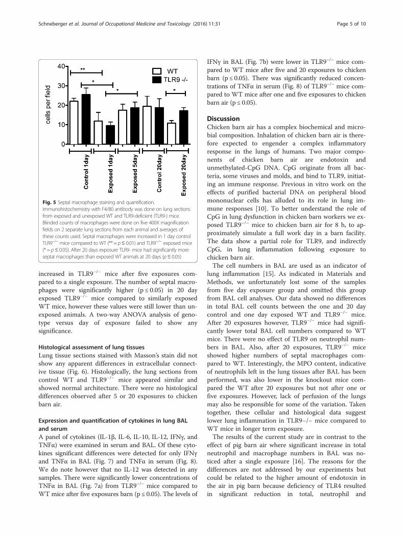
BAL total cell numbersThere were no differences in total BAL cell numbers be-tween one day or 20 day control TLR9−/− and WT ani-mals or those exposed for one day to chicken barn air(Fig. 1). The total BAL cell numbers were significantlylower in TLR9−/− mice compared to WT mice after 20exposures (p ≤ 0.05).
Fig. 1 BAL cell counts. BAL from exposed and unexposed WTand TLR9-deficient (TLR9-) mice (n = 6) was spun briefly at 400g, supernatant removed, and cells resuspended to 100 μl forcounting with hemocytometer. BAL cell numbers were lower inTLR9−/− mice compared to WT mice after 20exposures (* = p ≤ 0.05)
Schneberger et al. Journal of Occupational Medicine and Toxicology (2016) 11:31 Page 3 of 10

Neutrophil recruitment into BAL and lung tissuesThere were no effects of number of exposures and thestrain on the numbers of neutrophils in BAL (Fig. 2).We used an MPO assay as a surrogate for neutrophilstrapped in the lung tissues that are not amenable to lav-age. MPO assay did not reveal any differences in neutro-phil recruitment in lung tissues between the WT andTLR9−/− control mice as well as those exposed once orfive times to barn air (Fig. 3). However, after 20 expo-sures there were reduced MPO concentrations (p ≤ 0.05)in lung homogenates from TLR9−/− animals comparedto similarly exposed WT mice. This reduction was notsignificantly lower than the unexposed control though.
Macrophage recruitment into BAL and lung tissuesIn addition to neutrophils, macrophages are importantplayers in promoting or resolving lung inflammation.There were no differences in macrophage BAL numbersfrom normal one day WT and TLR9−/− mice (Fig. 4).However, TLR9−/− mice had more macrophages in BALcompared to WT mice after single exposure (p ≤ 0.05).Interestingly, the BAL macrophage numbers were lowerin 20 day exposed TLR9−/− mice compared to 20 daywild type exposed animals (p ≤ 0.05).To determine the number of tissue macrophages in
lavaged lungs, we stained lung sections with macrophageF4/80 antibody. Single exposure to barn air induced asignificant decrease in the number of septal macro-phages in TLR9−/− and wild-type mice compared to un-exposed mice at the same time point (Fig. 5, p ≤ 0.05and p ≤ 0.01). Septal macrophages were significantly
Fig. 2 Neutrophil cell counts. BAL from exposed and unexposed WTand TLR9-deficient (TLR9-) mice (n = 6) was spun briefly at 400 g,supernatant removed, and cells resuspended and stained with ahemacolor kit. Neutrophils were counted and numbers converted toabsolute numbers. No significant differences were seen in neutrophilcounts between any groups
Fig. 3 Myeloperoxidase activity assay for lung neutrophilquantitation. TMB substrate was added to protein extracts of wholelung tissue from exposed and unexposed WT and TLR9-deficient(TLR9-) mice and read at a450 after 2 min (n = 6). Activity wasassessed by comparison to a standard curve. A reduction was seenin 20 day exposed TLR9- mice compared to WT mice exposed forthe same time (* = p≤ 0.05)
Fig. 4 Macrophage cell counts. BAL from exposed and unexposedWT and TLR9-deficient (TLR9-) mice (n = 6) was spun briefly at 400 g,supernatant removed, and cells resuspended and stained with ahemacolor kit. Macrophages were counted and numbers convertedto absolute numbers. Macrophage numbers were higher in 1 dayexposed TLR9−/− animals compared to WT mice (* = p ≤ 0.05). Asimilar increase was seen in 20 day control TLR9−/− mice comparedto WT mice exposed for the same time (* = p≤ 0.05)
Schneberger et al. Journal of Occupational Medicine and Toxicology (2016) 11:31 Page 4 of 10
increased in TLR9−/− mice after five exposures com-pared to a single exposure. The number of septal macro-phages were significantly higher (p ≤ 0.05) in 20 dayexposed TLR9−/− mice compared to similarly exposedWT mice, however these values were still lower than un-exposed animals. A two-way ANOVA analysis of geno-type versus day of exposure failed to show anysignificance.
Histological assessment of lung tissuesLung tissue sections stained with Masson’s stain did notshow any apparent differences in extracellular connect-ive tissue (Fig. 6). Histologically, the lung sections fromcontrol WT and TLR9−/− mice appeared similar andshowed normal architecture. There were no histologicaldifferences observed after 5 or 20 exposures to chickenbarn air.
Expression and quantification of cytokines in lung BALand serumA panel of cytokines (IL-1β, IL-6, IL-10, IL-12, IFNγ, andTNFα) were examined in serum and BAL. Of these cyto-kines significant differences were detected for only IFNγand TNFα in BAL (Fig. 7) and TNFα in serum (Fig. 8).We do note however that no IL-12 was detected in anysamples. There were significantly lower concentrations ofTNFα in BAL (Fig. 7a) from TLR9−/− mice compared toWT mice after five exposures barn (p ≤ 0.05). The levels of
IFNγ in BAL (Fig. 7b) were lower in TLR9−/− mice com-pared to WT mice after five and 20 exposures to chickenbarn (p ≤ 0.05). There was significantly reduced concen-trations of TNFα in serum (Fig. 8) of TLR9−/− mice com-pared to WT mice after one and five exposures to chickenbarn air (p ≤ 0.05).
DiscussionChicken barn air has a complex biochemical and micro-bial composition. Inhalation of chicken barn air is there-fore expected to engender a complex inflammatoryresponse in the lungs of humans. Two major compo-nents of chicken barn air are endotoxin andunmethylated-CpG DNA. CpG originate from all bac-teria, some viruses and molds, and bind to TLR9, initiat-ing an immune response. Previous in vitro work on theeffects of purified bacterial DNA on peripheral bloodmononuclear cells has alluded to its role in lung im-mune responses [10]. To better understand the role ofCpG in lung dysfunction in chicken barn workers we ex-posed TLR9−/− mice to chicken barn air for 8 h, to ap-proximately simulate a full work day in a barn facility.The data show a partial role for TLR9, and indirectlyCpG, in lung inflammation following exposure tochicken barn air.The cell numbers in BAL are used as an indicator of
lung inflammation [15]. As indicated in Materials andMethods, we unfortunately lost some of the samplesfrom five day exposure group and omitted this groupfrom BAL cell analyses. Our data showed no differencesin total BAL cell counts between the one and 20 daycontrol and one day exposed WT and TLR9−/− mice.After 20 exposures however, TLR9−/− mice had signifi-cantly lower total BAL cell numbers compared to WTmice. There were no effect of TLR9 on neutrophil num-bers in BAL. Also, after 20 exposures, TLR9−/− miceshowed higher numbers of septal macrophages com-pared to WT. Interestingly, the MPO content, indicativeof neutrophils left in the lung tissues after BAL has beenperformed, was also lower in the knockout mice com-pared the WT after 20 exposures but not after one orfive exposures. However, lack of perfusion of the lungsmay also be responsible for some of the variation. Takentogether, these cellular and histological data suggestlower lung inflammation in TLR9−/− mice compared toWT mice in longer term exposure.The results of the current study are in contrast to the
effect of pig barn air where significant increase in totalneutrophil and macrophage numbers in BAL was no-ticed after a single exposure [16]. The reasons for thedifferences are not addressed by our experiments butcould be related to the higher amount of endotoxin inthe air in pig barn because deficiency of TLR4 resultedin significant reduction in total, neutrophil and
Fig. 5 Septal macrophage staining and quantification.Immunohistochemistry with F4/80 antibody was done on lung sectionsfrom exposed and unexposed WT and TLR9-deficient (TLR9-) mice.Blinded counts of macrophages were done on five 400X magnificationfields on 2 separate lung sections from each animal and averages ofthese counts used. Septal macrophages were increased in 1 day controlTLR9−/− mice compared to WT (** = p≤ 0.01) and TLR9−/− exposed mice(* = p≤ 0.05). After 20 days exposure TLR9- mice had significantly moreseptal macrophages than exposed WT animals at 20 days (p≤ 0.05)
Schneberger et al. Journal of Occupational Medicine and Toxicology (2016) 11:31 Page 5 of 10

macrophage BAL numbers compared to the WT animalsexposed to pig barn air [5, 7]. There are also likely differ-ences in the total exposure doses found in each barnwhich could also account for these changes. Second, thedifferences could be related to the microbial content inchicken and pig barns as even the WT animals didn'tshow acute inflammation as experienced after exposureto pig barn air. The migration of inflammatory cells intoalveolar spaces is regulated through development of achemotactic gradient produced through production ofchemokines such as IL-8, MIP-1 and MCP-1 by airwayepithelial cells and alveolar macrophages stimulated byinhaled molecules such as endotoxins [17]. The main-tenance in the number of macrophages which resolveand modulate inflammation in the BAL of TLR9−/− micecompared to WT mice following one exposure suggestsa modulatory role for TLR9. The early increase in
macrophages without any effect on neutrophils or totalcells in the BAL in TLR9−/− mice is interesting whencombined with fewer tissue neutrophils but comparabletissue macrophages after 20 exposures to chicken barnair. Although neutrophils migrate during the early phaseof acute lung inflammation, their migration may con-tinue over a longer period of time in chronic inflamma-tion stimulated through repeated exposures in diseasessuch as COPD [18, 19]. The data suggest that deficiencyof TLR9 dampens lung inflammation induced by expos-ure to chicken barn air.The expression of inflammation including recruitment
of neutrophils and macrophages is regulated through ad-hesion proteins and inflammatory mediators [20, 21]. Tounderstand the role of inflammatory mediators, we ex-amined the expression of IL-1β, IL-6, IL-10, IL-12, IFNγ,and TNFα in BAL and serum of mice in our study.
Fig. 6 Histopathology of lung samples. Histological assessment shows normal histology in unexposed WT (a) and unexposed mutant (b). Oneday exposed WT (c) and TLR9−/−animals (d). WT mice exposed for five days (e) showed little difference to the 5 day exposed mutant mice (f).Lung sections from both WT (g) and mutant mice (h) exposed for 20 days showed normal histology. Bar = 100 micron
Schneberger et al. Journal of Occupational Medicine and Toxicology (2016) 11:31 Page 6 of 10

These cytokines are produced following ligation ofbacterial DNA by TLR9 [22, 23]. We did not findany differences in control or exposed WT or TLR9−/− mice for IL-1β, IL-6, IL-10, and IL-12. Thiswould suggest that while the barn may be an envir-onment known to induce inflammation in the lung,changes in many individual cytokines may not besignificantly elevated, or that responses are too vari-able. While IFNγ was reduced in BAL from TLR9−/−
mice after 5 and 20 exposures, TNFα was reducedonly after 5 exposures. The levels of IFNγ were
lower in the serum of TLR9−/− mice compared toWT mice exposed once or five times to chickenbarn air. Although there were differences in IFNγand TNFα in BAL fluid between WT and TLR9−/−
mice after five exposures, there were no differencesin the numbers of cells recruited into alveoli afterone or five exposures. There were however moreseptal macrophages in TLR9−/− mice compared toWT mice following five exposures. Because macro-phages play a role in resolution of inflammation[21], reduced levels of TNFα along with increasednumbers of macrophages may indicate a pro-inflammatory role for TLR9 in lung inflammation in-duced following exposure to chicken barn air. Be-cause TNFα promotes expression of adhesionmolecules [24], reduced levels of TNFα may havealso contributed to reduced migration of neutrophilsin lungs of TLR9−/− mice compared to WT mice fol-lowing 20 exposures to chicken barn air. However,there were more septal macrophages present at thesame time. TNFα produced by activated airway epi-thelial cells and alveolar macrophages is an import-ant regulator of lung inflammation as well [24].Because of the role of TLR9 signaling in inductionof pulmonary IFNγ expression [25], the reducedlevels of IFNγ in the knockout mice is understand-able. However, the effects of reduced expression ofIFNγ on the inflammation phenotype in our experi-ments are not apparent. The role of IFNγ as amodulator of Th2 immune response is somethingthat we need to address in future experiments. It isknown that IFNγ is reduced in many cases ofasthma [26] and in cases of endotoxin exposure [27].This is typically associated with a more Th2 cytokineprofile. Pig and cattle farm workers had higher
Fig. 8 Bio-plex ELISA of mouse serum. Serum was collected bycollecting blood via cardiac puncture and then briefly centrifugingto remove cells from exposed and unexposed WT and TLR9−/− mice(n = 6). ELISA was done with antibodies to IL-1β, IL-6, IL-10, IL-12,IFNγ, and TNFα. IFNγ was significantly reduced after 1 and 5 daysexposure in TLR9−/− mice compared to WT (*** = p≤ 0.01)
Fig. 7 Bio-plex ELISA of mouse bronchoalveolar lavage. BAL was collected by washing lungs 3 times with cold HEPES buffer from exposed andunexposed WT and TLR9−/− mice (n = 6). Samples were centrifuged briefly to remove cells and fluid decanted. ELISA was done with antibodies toIL-1β, IL-6, IL-10, IL-12, IFNγ, and TNFα. TNFα (a) was significantly reduced after 5 days exposure in TLR9−/− mice compared to WT (* = p≤ 0.06).IFNγ (b) was significantly reduced after 5 and 20 days exposure in TLR9−/− mice compared to WT (** = p≤ 0.05)
Schneberger et al. Journal of Occupational Medicine and Toxicology (2016) 11:31 Page 7 of 10
incidence of asthma compared to other farm workers[28] which has been attributed to higher endotoxinlevels.The lack of pronounced differences in lung inflamma-
tion in WT mice compared to TLR9−/− mice may be dueto impaired expression of TLR9 in murine alveolar mac-rophages [29]. Because alveolar macrophages play crit-ical roles in lung inflammation induced followinginhalation of microbes or their products [30–32], im-paired TLR9 expression may explain lack of distinct dif-ferences in inflammation between WT and TLR9−/−
mice. While recognition of endotoxin is still intact inboth mouse strains, the differences in lung inflammationare rather subtle. This however does not inform us ofthe reasons for lack of differences between WT controland one or five day exposed mice. This issue needs to beaddressed through comparison of lung inflammation inmice in which alveolar macrophages are depleted priorto their exposure to barn air.One possible explanation for both BAL and septal
macrophage results is that the septal macrophages exertan anti-inflammatory effect on the lung, leading to fewerBAL cells. There is evidence for this in a recent studythat showed that signaling through TLR4 and TLR9 inseptal macrophages induced expression of cytokinessuch as IL-10 and generally induced an anti-inflammatory response [33]. Yet another possibility isthat migration of macrophages into the alveolar spacerequires a transition through the alveolar septa [34]. Ifthis is the case then a reduction in macrophages at onetime point in the septa being mirrored by an increase inthe BAL could be a reflection of the increased move-ment of these cells into the alveolar space as appears tohappen in our experiments. This however still raises thequestion of the reason for such an increased migrationof macrophages.The response of cellular influx in and out of the lung
with TLR9 inhibition is quite intriguing and somewhatperplexing. We suspect that within the first day of ex-posure lack of TLR9 signaling reduces the rate of out-migration of alveolar macrophage to tissue and lymphnodes [35]. The general reduction in exposed groupinterstitial macrophages which are known to secreteanti-inflammatory cytokines to the same stimuli [33]suggest migration in and out of BAL may be more likelyat this point.By day 20 however TLR9−/− animals are seeing in-
creases in these interstitial macrophage populations, sug-gesting a less permissive migration scenario. At thispoint we see this reflected in reduced total BAL cells,BAL macrophages, and tissue neutrophils. As such, wewould hope to do more work on the effects of dusts andTLR9 signaling in interstitial macrophages to better de-termine their role in lung cellular migration.
Lung responses to bacterial DNA will depend on theamount of inhaled DNA. Attempts to purify DNA frombarn dust from a previous experiment typically producedaround 1 μg of DNA from a filter kept in a similar barnfor 8 h (unpublished observations). Of this a portion ofrecovered DNA will be methylated eukaryotic DNA, fur-ther reducing the amount of stimulatory DNA. There-fore, the predicted exposure to bacterial DNA in thebarn is probably quite low, certainly in comparison towhat is used in in vitro trials [10]. However, as has beenmentioned, dust and endotoxin levels in this facility werelower compared to a wider survey [3]. We would thuspredict a greater effect of bacterial DNA in many facil-ities compared to what is shown here. Although somedifferences between WT and TLR9−/− mice were noticedafter single exposure, the divergence in immune re-sponses between mouse populations at 5 days or latermay indicate a requirement for exposure to a sufficientdose of stimulatory DNA that the mice do not see in asingle day. Indeed, as Roy and colleagues found, 10 μg ofbarn dust induced detectable responses [10], whichwould be approximately the dose encountered by themice after 5 exposures. This accumulated effect thoughwould have to result from continued exposure, not anaccumulation of DNA within the tissue, as other studieshave shown that internalized DNA is rapidly degraded[36]. What this suggests is that a low unmethylatedDNA dose (perhaps 1 μg/day) may cause changes tolung cytokine secretion, especially in the context ofstimulation or challenge by other pro-inflammatory mol-ecules or organisms (in this case endotoxins and/or pro-teoglycans and particulate dust). Another considerationis what happens the CpG DNA in absence of TLR9 re-ceptor? To what degree could other potential sensingmechanisms be altered by CpG DNA not binding toTLR9?We do note that a study like this has a number of lim-
itations that must be recognized. First, dust and barnconditions are likely to differ between specific facilities,and moreso between facilities such as swine and chickenfacilities, though there is clear evidence of worker lungproblems in both as discussed earlier. Second, this studywas limited to infiltration of cells and cytokine expres-sion in the lung, possibly ignoring other potentialmarkers of lung inflammation. Even in the cases of ex-posure there was not a significant increase of inflamma-tory cells and cytokines above controls animals of thesame strain, so there was not a clear indication on in-flammation in mice over the course of the experiment.This having been said however, there were clear changesin cytokine expression, macrophage numbers, and MPOconcentration in the lungs of mice, with most of thesechanges being present after a longer term exposure tobarn air.
Schneberger et al. Journal of Occupational Medicine and Toxicology (2016) 11:31 Page 8 of 10
ConclusionsIn conclusion, while we saw little change to BAL cell popu-lations and cytokines in mice that had been exposed tochicken barn air for 1, 5, or 20 days, there were subtlechanges in TLR9−/− animals. In TLR9−/− animals we de-tected changes to total BAL cells, and alveolar macrophagenumbers by 1 day exposure, and still at 20 days. Septalmacrophages were also increased in the TLR9−/− animalsby 20 days exposure, in a pattern opposite to that seen inalveolar macrophages. There were also reductions in TNFαand IFNγ in the TLR9−/− knockout mice. These resultssuggest that TLR9 plays a role in the innate immune re-sponse to chicken barn air exposure that enhances indica-tors of inflammation (TNFα, IFNγ, MPO) at later timepoints (5 day or 20 day exposures). However, the full effectsof TLR9 may be more complex as shown by increased BALcell numbers and alveolar macrophages after single day ex-posure, and increased septal macrophages in these sameanimals at 20 days.
AbbreviationsBAL, bronchoalveolar lavage; LPS, lipopolysaccharide; MPO, myeloperoxidase;TLR, toll-like receptor
AcknowledgementsWe would like to thank Amberlea Farms Ltd. of Saskatoon, SK Canada forallowing access to their facilities for this study.
FundingThe work was supported by a Discovery Grant from the Natural Sciences andEngineering Research Council of Canada to Baljit Singh. David Schnebergerwas supported by a PHARE Graduate Scholarship from the CanadianInstitutes for Health Research and a Founding Chairs Fellowship from theCanadian Centre for Health and Safety in Agriculture, University ofSaskatchewan.
Authors’ contributionsDS conceived of the study, analyzed data, and carried out most of theexperiments and writing of the manuscript. GA and SC assisted with carryingout of experiments. BS assisted with experiment design, writing of paper,and guidance of the project. All authors read and approved the finalmanuscript.
Competing interestsThe authors declare they have no competing interests.
Consent for publicationNot applicable.
Ethics approval and consent to participateThe experimental protocols were approved by the University ofSaskatchewan Committee on Animal Care Assurance and all experimentsconducted according to guidelines of the Canadian Council on Animal Care.
Received: 2 December 2015 Accepted: 22 June 2016
References1. Donham KJ, Cumro D, Reynolds SJ, Merchant JA. Dose–response
relationships between occupational aerosol exposures and cross-shiftdeclines of lung function in poultry workers: recommendations for exposurelimits. J Occup Environ Med. 2000;42(3):260–9.
2. Kirychuk SP, Senthilselvan A, Dosman JA, Juorio V, Feddes JJR, Willson P,et al. Respiratory symptoms and lung function in poultry confinementworkers in Western Canada. Can Respir J. 2003;10(7):375–80.
3. Kirychuk SP, Dosman JA, Reynolds SJ, Willson P, Senthilselvan A, Feddes JJ,et al. Total dust and endotoxin in poultry operations: comparison betweencage and floor housing and respiratory effects in workers. J Occup EnvironMed. 2006;48(7):741–8.
4. Just N, Duchaine C, Singh B. An aerobiological perspective of dust in cage-housed and floor-housed poultry operations. J Occup Med Toxicol. 2009;4:13.
5. Senthilselvan A, Zhang Y, Dosman JA, Barber EM, Holfeld LE, Kirychuk SP,et al. Positive human health effects of dust suppression with canola oil inswine barns. Am J Respir Crit Care Med. 1997;156(2 Pt 1):410–7.
6. Smit LAM, Heederik D, Doekes G, Krop EJM, Rijkers GT, Wouters IM. Ex vivocytokine release reflects sensitivity to occupational endotoxin exposure. EurRespir J. 2009;34(4):795–802.
7. Charavaryamath C, Juneau V, Suri SS, Janardhan KS, Townsend H, Singh B. Roleof toll-like receptor 4 in lung inflammation following exposure to swine barnair. Exp Lung Res. 2008;34(1):19–35.
8. Zejda JE, Barber E, Dosman JA, Olenchock SA, McDuffie HH, Rhodes C, et al.Respiratory health status in swine producers relates to endotoxin exposurein the presence of low dust levels. J Occup Med. 1994;36(1):49–56.
9. Medzhitov R, Janeway Jr C. Innate immune recognition: mechanisms andpathways. Immunol Rev. 2000;173:89–97.
10. Roy SR, Schiltz AM, Marotta A, Shen Y, Liu AH. Bacterial DNA in house andfarm barn dust. J Allergy Clin Immunol. 2003;112(3):571–8.
11. Schneberger D, Caldwell S, Kanthan R, Singh B. Expression of Toll-likereceptor 9 in mouse and human lungs. J Anat. 2013;222(5):495–503.
12. Schneberger D, Caldwell S, Suri SS, Singh B. Expression of toll-like receptor 9in horse lungs. Anat Rec (Hoboken). 2009;292(7):1068–77.
13. Schneberger D, Lewis D, Caldwell S, Singh B. Expression of toll-like receptor9 in lungs of pigs, dogs and cattle. Int J Exp Pathol. 2011;92(1):1–7.
14. Dosman JA, Fukushima Y, Senthilselvan A, Kirychuk SP, Lawson JA, Pahwa P,et al. Respiratory response to endotoxin and dust predicts evidence ofinflammatory response in volunteers in a swine barn. Am J Ind Med. 2006;49(9):761–6.
15. Balbi B, Pignatti P, Corradi M, Baiardi P, Bianchi L, Brunetti G, et al.Bronchoalveolar lavage, sputum and exhaled clinically relevant inflammatorymarkers: Values in healthy adults. Eur Respir J. 2007;30(4):769–81.
16. Charavaryamath C, Janardhan KS, Townsend HG, Willson P, Singh B. Multipleexposures to swine barn air induce lung inflammation and airway hyper-responsiveness. Respir Res. 2005;6:50-62.
17. Tam A, Wadsworth S, Dorscheid D, Man SFP, Sin DD. The airway epithelium:More than just a structural barrier. Ther Adv Respir Dis. 2011;5(4):255–73.
18. Stockley RA. Neutrophils and the pathogenesis of COPD. Chest. 2002;121(5 SUPPL):151S–5S.
19. Gane J, Stockley R. Mechanisms of neutrophil transmigration across thevascular endothelium in COPD. Thorax. 2012;67(6):553–61.
20. Borregaard N. Neutrophils, from Marrow to Microbes. Immunity. 2010;33(5):657–70.
21. Murray PJ, Wynn TA. Protective and pathogenic functions of macrophagesubsets. Nat Rev Immunol. 2011;11(11):723–37.
22. Parilla NW, Hughes VS, Lierl KM, Wong HR, Page K. CpG DNA modulatesinterleukin Iβ-induced interleukin-8 expression in human bronchial epithelial(16HBE14o-) cells. Respir Res. 2006;7:84-92.
23. Chen L, Arora M, Yarlagadda M, Oriss TB, Krishnamoorthy N, Ray A, et al.Distinct responses of lung and spleen dendritic cells to the TLR9 agonistCpG oligodeoxynucleotide. J Immunol. 2006;177(4):2373–83.
24. Lauterbach M, O'Donnell P, Asano K, Mayadas TN. Role of TNF priming andadhesion molecules in neutrophil recruitment to intravascular immunecomplexes. J Leukocyte Biol. 2008;83(6):1423–30.
25. Zhou R, Norton JE, Zhang N, Dean DA. Electroporation-mediated transfer ofplasmids to the lung results in reduced TLR9 signaling and inflammation.Gene Ther. 2007;14(9):775–80.
26. Kumar RK, Webb DC, Herbert C, Foster PS. Interferon-γ as a possible targetin chronic asthma. Inflamm Allergy Drug Targets. 2006;5(4):253–6.
27. Alexis NE, Lay JC, Almond M, Peden DB. Inhalation of low-dose endotoxinfavors local TH2 response and primes airway phagocytes in vivo. J AllergyClin Immunol. 2004;114(6):1325–31.
28. Eduard W, Douwes J, Omenaas E, Heederik D. Do farming exposures causeor prevent asthma? Results from a study of adult Norwegian farmers.Thorax. 2004;59(5):381–6.
29. Suzuki K, Suda T, Naito T, Ide K, Chida K, Nakamura H. Impaired toll-likereceptor 9 expression in alveolar macrophages with no sensitivity to CpGDNA. Am J Respir Crit Care Med. 2005;171(7):707–13.
Schneberger et al. Journal of Occupational Medicine and Toxicology (2016) 11:31 Page 9 of 10
30. Palmberg L, Larsson B, Malmberg P, Larsson K. Induction of IL-8 productionin human alveolar macrophages and human bronchial epithelial cells invitro by swine dust. Thorax. 1998;53(4):260–4.
31. Koay MA, Gao X, Washington MK, Parman KS, Sadikot RT, Blackwell TS, et al.Macrophages are necessary for maximal nuclear factor-kB activation inresponse to endotoxin. Am J Resp Cell Mol Biol. 2002;26(5):572–8.
32. Zhao M, Fernandez LG, Doctor A, Sharma AK, Zarbock A, Tribble CG, et al.Alveolar macrophage activation is a key initiation signal for acute lungischemia-reperfusion injury. Am J Physiol Lung Cell Mol Physiol. 2006;291(5):L1018–26.
33. Hoppstadter J, Diesel B, Zarbock R, Breinig T, Monz D, Koch M, et al.Differential cell reaction upon Toll-like receptor 4 and 9 activation in humanalveolar and lung interstitial macrophages. Respir Res. 2010;11:124.
34. Landsman L, Jung S. Lung macrophages serve as obligatory intermediatebetween blood monocytes and alveolar macrophages. J Immunol. 2007;179(6):3488–94.
35. Thepen T, Claassen E, Hoeben K, Breve J, Kraal G. Migration of alveolarmacrophages from alveolar space to paracortical T cell area of the draininglymph node. Adv Exp Med Biol. 1993;329:305–10.
36. Kawabata K, Takakura Y, Hashida M. The fate of plasmid DNA afterintravenous injection in mice: involvement of scavenger receptors in itshepatic uptake. Pharm Res. 1995;12(6):825–30.
• We accept pre-submission inquiries
• Our selector tool helps you to find the most relevant journal
• We provide round the clock customer support
• Convenient online submission
• Thorough peer review
• Inclusion in PubMed and all major indexing services
• Maximum visibility for your research
Submit your manuscript atwww.biomedcentral.com/submit
Submit your next manuscript to BioMed Central and we will help you at every step:
Schneberger et al. Journal of Occupational Medicine and Toxicology (2016) 11:31 Page 10 of 10