tls-erg leukemia fusion protein inhibits rna splicing mediated by serine-arginine proteins
DESCRIPTION
biolTRANSCRIPT
MOLECULAR AND CELLULAR BIOLOGY,0270-7306/00/$04.0010
May 2000, p. 3345–3354 Vol. 20, No. 10
Copyright © 2000, American Society for Microbiology. All Rights Reserved.
TLS-ERG Leukemia Fusion Protein Inhibits RNA SplicingMediated by Serine-Arginine Proteins
LIU YANG,1,2 LISA J. EMBREE,1,2 AND DENNIS D. HICKSTEIN1,2,3*
Medical Research Service, VA Puget Sound Health Care System, Seattle, Washington 98108,1 andDivisions of Oncology2 and Molecular Medicine,3 University of Washington
School of Medicine, Seattle, Washington 98195
Received 25 October 1999/Returned for modification 1 December 1999/Accepted 16 February 2000
The translocation liposarcoma (TLS) gene is fused to the ETS-related gene (ERG) in human myeloidleukemia, resulting in the generation of a TLS-ERG protein. We demonstrate that both TLS and the TLS-ERGleukemia fusion protein bind to RNA polymerase II through the TLS N-terminal domain, which is retained inthe fusion protein; however, TLS recruits members of the serine-arginine (SR) family of splicing factorsthrough its C-terminal domain, whereas the TLS-ERG fusion protein lacks the ability to recruit SR proteinsdue to replacement of the C-terminal domain by the fusion partner ERG. In transient-transfection assays, theTLS-ERG fusion protein inhibits E1A pre-mRNA splicing mediated by these TLS-associated SR proteins(TASR), and stable expression of the TLS-ERG fusion protein in K562 cells alters the splicing profile of CD44mRNA. These results suggest that TLS fusion proteins may lead to cellular abnormalities by interfering withthe splicing of important cellular regulators.
The TLS (also called FUS) gene was originally identified atthe site of the t(12;16) chromosomal translocation in malignantliposarcomas, where it is fused to the CHOP (c/EBP homolo-gous protein) gene (12, 36). In human myeloid leukemias withthe t(16;21) chromosomal translocation, the TLS gene is fusedto the ERG (ETS-related gene) gene (23). In both instances,the resultant TLS-CHOP and TLS-ERG fusion proteins gen-erated by these translocations retain the N-terminal domain ofTLS; however, in both fusion proteins the C-terminal domainof TLS is replaced by the DNA-binding domain from thecorresponding transcription factor. The oncogenic potentialsof both the TLS-CHOP and the TLS-ERG fusion proteinshave been confirmed in transformation assays using mouse celllines (24, 51) and normal human hematopoietic cells (32),respectively.
The role of TLS in normal cellular function and the mech-anisms whereby TLS fusion proteins lead to transformationremain unclear. TLS belongs to a family of closely relatedproteins, including Ewing’s sarcoma protein, EWS (13), andTATA-binding protein-associated factor, TAFII68 (3). BothEWS and TAFII68 interact with components of the RNA poly-merase II (Pol II) complex (3, 4, 34), thus implicating TLS intranscriptional activation. The N-terminal domains of the TLSfamily of proteins are rich in glutamine, serine, and tyrosine,which are amino acid residues commonly found in transcrip-tional activation domains. Since TLS fusion proteins acquiretheir C-terminal region from the DNA-binding domains of thecorresponding transcription factors, TLS fusion proteins arethought to lead to transformation by transcriptional activationof target genes (33). Moreover, TLS fusion proteins have beenshown to transactivate reporter genes in transient-transfectionassays (35, 51). However, mutagenesis studies have failed todemonstrate a correlation between the ability to transactivateand the ability to transform (26, 28).
The lack of a correlation between transactivation and trans-
formation with TLS fusion proteins suggested that TLS fusionproteins might transform cells through the disruption of im-portant cellular processes other than transcription. In this re-gard, there is considerable circumstantial evidence that TLSparticipates in RNA processing. The C-terminal domain ofTLS contains two sequence motifs, ribonucleoprotein consen-sus sequence (RNP-CS) and arginine-glycine-glycine repeats(RGG), which are signatures of RNA-binding proteins (6).TLS binds to RNA and shuttles between the nucleus and thecytoplasm (52). When overexpressed in erythroid cells, TLSinduces the preferential use of the most distal 59 splice sitesduring E1A pre-mRNA splicing (19). TLS also associates withsplicing factors ribonucleoprotein (RNP) A1 (51) and SF1(49).
In this study, we demonstrate that TLS and its fusion proteinTLS-ERG interact with RNA Pol II through the N-terminaldomain of TLS, the domain preserved in the fusion protein.However, wild-type TLS recruits members of the serine-argi-nine family of splicing factors through the C-terminal domainof TLS, and the TLS-ERG fusion protein lacks this ability dueto replacement of its C-terminal domain by the fusion partner.In the E1A pre-mRNA splicing assay, the TLS-ERG fusionprotein interferes with E1A pre-mRNA splicing mediated byTLS-associated SR proteins in a dominant-negative manner.These results suggest that TLS protein functions as a dockingmolecule in the recruitment of SR splicing factors and that theTLS-ERG fusion protein inhibits this function of TLS.
MATERIALS AND METHODS
Two-hybrid screen and cDNA cloning. A yeast two-hybrid-screen was per-formed as previously described (47), using the C-terminal domain of TLS as thebait (Fig. 1). To obtain full-length mouse TASR-2 cDNA, the TASR-2 insertidentified from the yeast two-hybrid screen was used as a probe in the hybrid-ization screening of a Uni-ZAP phage cDNA library derived from murine EMLcells with erythroid-myeloid-lymphoid potentials (42). pBluescript phagemidcontaining the full-length TASR-2 cDNA was prepared after in vivo excisionfrom the Uni-ZAP XR vector and was used in sequencing reactions with dyeterminators (Applied Biosystems). Human TASR-2 cDNA was obtained fromK562 leukemia cells by reverse transcription-PCR (RT-PCR) using primersdesigned on the basis of the mouse TASR-2 sequence.
In vitro translation. The TNT coupled reticulocyte lysate system (Promega)was used in the in vitro translation of TASR-2 cDNA. The DNA template was a
* Corresponding author. Mailing address: Department of Medicine/Oncology, University of Washington School of Medicine, 1660 S. Co-lumbian Way, GMR 151, Seattle, WA 98108. Phone: (206) 764-2705.Fax: (206) 764-2827. E-mail: [email protected].
3345
pBluescript phagemid containing the full-length mouse TASR-2 cDNA alongwith the empty pBluescript vector as a negative control. The [35S]methionine-labeled protein was prepared as specified by the manufacturer and separated bysodium dodecyl sulfate-polyacrylamide gel electrophoresis (SDS-PAGE) on a12% polyacrylamide gel.
Plasmid construction. The cDNAs for TLS, TLS-ERG, and ERG were clonedinto the EcoRI-SmaI sites of the pSG5-FL vector for transfection and expressionof these proteins with the Flag epitope at the N-terminal end. Plasmid pSG5-FL-TLS-NTD contains a DNA insert corresponding to amino acids 1 to 290 ofthe TLS sequence, whereas pSG5-FL-TLS-CTD contains a DNA insert corre-sponding to amino acids 357 to 525 of the TLS sequence. Myc-tagged expressionplasmids pCS2-MT-TASR-1 and pCS2-MT-TASR-2 were generated by in-framecloning of full-length TASR cDNAs into the EcoRI-StuI sites of pCS2-MTvector. For the in vivo splicing assay, TASR cDNAs were inserted into the pMHvector (Boehringer Mannheim) to generate pMH-TASR-1 and pMH-TASR-2with the influenza virus hemagglutinin (HA) epitope tagged at the C-terminalends of TASR proteins. Reporter plasmid pCS3-MT-E1A was a kind gift from F.Moreau-Gachelin (19), and reporter plasmid pCS3-MT-E1A-9S was constructedby cloning the cDNA for the 9S E1A splicing isoform into the EcoRI-XbaI sitesof pCS3-MT vector.
Immunoprecipitation and Western blotting. For expression of Flag- or Myc-tagged proteins, 10 mg of the pSG5-Flag-expression construct and 10 mg of thepCS2-Myc-expression construct were introduced into 3 3 106 COS-7 cells byelectroporation. At 48 h after electroporation, the cells were lysed with 0.6 ml oflysis buffer A (10 mM Tris-HCl [pH 7.4], 2.5 mM MgCl2, 100 mM NaCl, 0.5%Triton X-100). Prior to cell lysis, 30 ml of D8 polyclonal rabbit anti-Flag antibody(Santa Cruz Biotechnology), 3 ml of 9E10 monoclonal mouse anti-Myc antibody(Sigma), or 10 ml of 8WG16 monoclonal mouse anti-RNA Pol II antibody(Research Diagnostics, Inc.) was incubated with 30 ml of protein A/G agarose(Santa Cruz Biotechnology) for 50 min at 4°C in 0.3 ml of buffer A, and theantibody-protein A/G-agarose complex was then incubated with 0.2 ml of freshcell lysate for 20 min at 4°C with gentle rocking. After the samples were washedwith RIPA buffer four times, 50 ml of SDS-PAGE sample buffer was added to theagarose beads. The samples were heated at 100°C for 3 min, 20 ml of the samplewas separated by SDS-PAGE in a 10% polyacrylamide gel, and the proteins weredetected with M2 monoclonal mouse anti-Flag antibody (Sigma) or the 9E10monoclonal mouse anti-Myc antibody as described in Results. Protein bandswere visualized using the enhanced chemiluminescence Western blotting analysissystem (Amersham).
E1A pre-mRNA splicing assay. For in vivo splicing of E1A pre-mRNA, 2 mgof pCS3-MT-E1A and 2 mg of pMH-TASR plus 6 mg of pSG5-Flag constructwere mixed with 60 ml of N-[1-(2,3-dioleoxy)propyl]-N,N,N-trimethylammonium(DOTAP; Boeringer Mannheim). The total amount of DNA for each 60-mmdish was kept at 5 mg by the addition of the corresponding empty vector. TheDNA-DOTAP mixture was then added to two duplicate dishes with 65% con-fluent HeLa cells in 4 ml of Dulbecco modified Eagle medium containing 1%fetal bovine serum. After 7 h of incubation with the DNA-DOTAP mixture, thecells were replenished with Dulbecco modified Eagle medium containing 10%fetal bovine serum and further incubated for 40 h. The cells from one dish werethen lysed with 0.25 ml of RIPA buffer for Western blot analysis, while the cellsfrom the other dish were used for RNA isolation with an RNeasy column(Qiagen). Total RNA from transfected HeLa cells in each 60-mm dish was elutedwith 50 ml of H2O from the RNeasy column.
For RT-PCR amplification of various E1A isoforms, 10 ml of total HeLa RNAwas used for overnight hybridization to 10 pmol of T7 primer. After RT withSuperScript H2 reverse transcriptase (GIBCO-BRL), the RNA was degraded byRNase H in a 40-ml reaction mixture. A 1.5-ml volume of the reaction mixturewas then used as the DNA templates for PCR amplification of various E1Asplicing isoforms. The forward primer was 59GAGCTTGGGCGACCTCA39(RR67), and the reverse primer was 59AATACGACTCACTATAG39 (T7). Thereactions were carried out in 50 ml containing 10 pmol of each primer, 0.2 mM
each deoxynucleoside triphosphate, 1.5 mM MgCl2, 13 Taq buffer, and 2.5 U ofPlatinum Taq polymerase (GIBCO-BRL). PCR was performed by a 2-min in-cubation at 94°C followed by 35 cycles of 1 min at 94°C, 1 min at 55°C, and 1 minat 72°C, with a final extension for 7 min at 72°C. To eliminate mismatchesbetween different E1A isoforms, 3 ml of 0.1 M EDTA (pH 8.0) was added to each50 ml of PCR products and the annealing was carried out at 80°C for 10 h. ThePCR products were then separated on a 1.5% agarose gel, denatured withNaOH, transferred to a nylon membrane, and detected with a 32P-labeled E1AcDNA probe.
For the RNase protection assay, 10 ml of the total RNA was hybridized to1.5 3 106 cpm of 32P-labeled RNA probe antisense to the E1A genomic se-quence (covering bases 499 to 1316 of the E1A gene). After overnight hybrid-ization, excessive RNA probe was digested with a mixture of RNase A plusRNase T1 supplied with the RNase protection assay system (PharMingen). Theprotected antisense E1A RNA fragments were isolated as specified by the manu-facturer and were separated on a 6% QuickPoint precast denaturing gel (Novex).
CD44 splicing assay. For the generation of retroviral constructs that expressFlag-tagged proteins, Flag-TLS, Flag-TLS-ERG, and Flag-ERG cDNA cassetteswere cloned into the HpaI-BamHI sites of retroviral vector LXSN. The retroviralparticles were first packaged in PG13 cells and then transduced into K562 cells.After selection with G418 at a concentration of 1 mg/ml of medium, the G418-resistant cells were collected for Western blotting analysis and RNA isolation.
For RT-PCR analysis of CD44 splicing, 10 mg of total RNA from K562 cellswas hybridized overnight to 10 pmol of oligo(dT) adapter primer 59GGCCACGCGTCGACTAGTACTTTTTTTTTTTTTTTTT39. After RT by SuperScriptH2 reverse transcriptase, the total RNA was degraded by RNase H in a 40-mlreaction mixture. A 1.5-ml sample of the RT mixture was then used as the DNAtemplates for PCR amplification of various CD44 splicing isoforms with thePlatinum Taq DNA polymerase. To detect subtle changes of CD44 alternativesplicing after expression of the Flag-TLS-ERG cassette in K562 cells, two pairsof CD44 primers were used in the PCR. The first primer pair consisted of59TCCCAGTATGACACATATTGC39 (P1) and 59CAGCTGTCCCTGTTGTCGAA39 (V6D), and the second primer pair consisted of 59GTACGTCTTCAAATACCATC39 (V3U) and 59CAAGATGATCAGCCATTCTGG39 (P2). PCRamplification was performed by a 2-min incubation at 94°C followed by 35 cyclesof 1 min at 94°C, 1 min at 55°C, and 1 min at 72°C, with a final extension for 7min at 72°C. To eliminate mismatches between different CD44 splicing isoforms,3 ml of 0.1 M EDTA (pH 8.0) was added to each 50 ml of PCR products and theannealing was carried out at 80°C for 10 h. The PCR products were thenseparated on a 1.5% agarose gel, denatured with NaOH, transferred to a nylonmembrane, and detected with a 32P-labeled CD44H cDNA probe spanningconstitutive exons 3, 4, 5, 16, 17, and 18 (see Fig. 7B).
RESULTS
TLS N-terminal domain mediates interactions with RNAPol II. The structural features of TLS, TLS-ERG, ERG, TLS-NTD (N-terminal domain), and TLS-CTD (C-terminal do-main) are shown in the schematic (Fig. 1). The N-terminaldomain of TLS is rich in glutamine, serine, and tyrosine resi-dues and has been suggested to function as a transcriptionalactivation domain (25, 35, 51). In addition, EWS and TAFII68,two proteins highly homologous to TLS, interact with compo-nents of the RNA Pol II complex (3, 4, 34). These observationssuggested that TLS might physically interact with RNA Pol II.
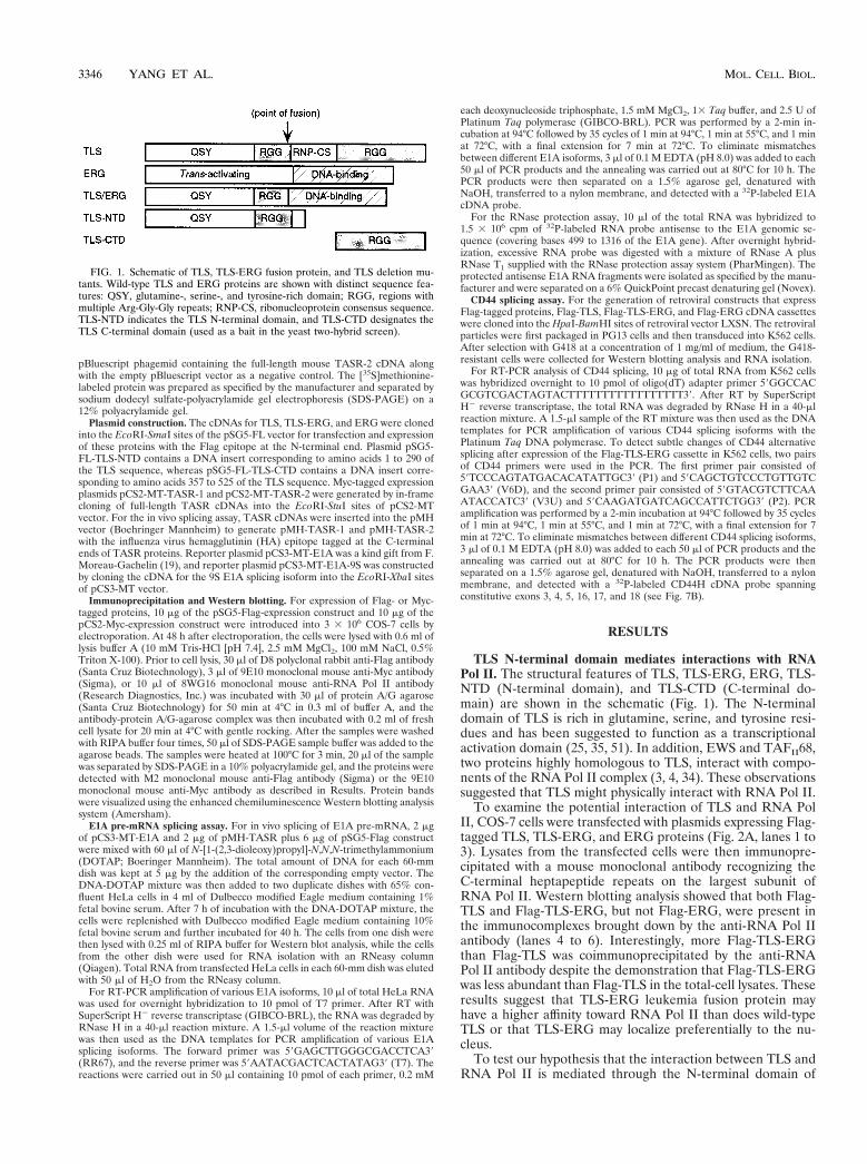
To examine the potential interaction of TLS and RNA PolII, COS-7 cells were transfected with plasmids expressing Flag-tagged TLS, TLS-ERG, and ERG proteins (Fig. 2A, lanes 1 to3). Lysates from the transfected cells were then immunopre-cipitated with a mouse monoclonal antibody recognizing theC-terminal heptapeptide repeats on the largest subunit ofRNA Pol II. Western blotting analysis showed that both Flag-TLS and Flag-TLS-ERG, but not Flag-ERG, were present inthe immunocomplexes brought down by the anti-RNA Pol IIantibody (lanes 4 to 6). Interestingly, more Flag-TLS-ERGthan Flag-TLS was coimmunoprecipitated by the anti-RNAPol II antibody despite the demonstration that Flag-TLS-ERGwas less abundant than Flag-TLS in the total-cell lysates. Theseresults suggest that TLS-ERG leukemia fusion protein mayhave a higher affinity toward RNA Pol II than does wild-typeTLS or that TLS-ERG may localize preferentially to the nu-cleus.
To test our hypothesis that the interaction between TLS andRNA Pol II is mediated through the N-terminal domain of
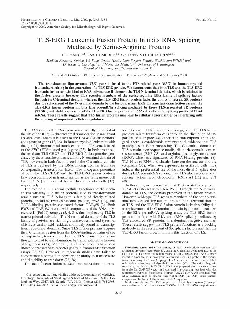
FIG. 1. Schematic of TLS, TLS-ERG fusion protein, and TLS deletion mu-tants. Wild-type TLS and ERG proteins are shown with distinct sequence fea-tures: QSY, glutamine-, serine-, and tyrosine-rich domain; RGG, regions withmultiple Arg-Gly-Gly repeats; RNP-CS, ribonucleoprotein consensus sequence.TLS-NTD indicates the TLS N-terminal domain, and TLS-CTD designates theTLS C-terminal domain (used as a bait in the yeast two-hybrid screen).
3346 YANG ET AL. MOL. CELL. BIOL.
TLS, two additional plasmids expressing the Flag-tagged N-terminal domain of TLS (Flag-TLS-NTD) or the Flag-taggedC-terminal domain of TLS (Flag-TLS-CTD) were constructed(Fig. 1A). After transfection, both Flag-TLS-NTD and Flag-TLS-CTD were expressed in COS-7 cells and migrated fasterthan Flag-TLS on SDS-PAGE (Fig. 2B, lanes 7 to 9). Along
with Flag-TLS, Flag-TLS-NTD was detectable in the anti-RNA Pol II immunocomplex (lanes 10 and 11) whereas Flag-TLS-CTD was absent from the immunocomplex (lane 12).These results suggested that the N-terminal domain of wild-type TLS protein interacts with RNA Pol II and that this abilityis retained by the TLS-ERG fusion protein.
FIG. 2. Association of TLS, TLS-ERG, and TLS-NTD with RNA Pol II. (A) COS-7 cells were transfected with Flag-tagged TLS, TLS-ERG, or ERG expressionplasmid, and the cell lysates were probed with the M2 anti-Flag antibody to confirm protein expression (lanes 1 to 3). The same lysates were immunoprecipitated witha mouse monoclonal 8WG16 antibody (Research Diagnostics, Inc.) recognizing the C-terminal heptapeptide repeat on the largest subunit of RNA Pol II (lanes 4 to6), and the immunoprecipitates were blotted with an M2 anti-Flag antibody or a rabbit polyclonal anti-RNA Pol II antibody (Santa Cruz Biotechnology). (B) COS-7cells were transfected with Flag-tagged TLS deletion constructs, and the lysates were probed with the M2 anti-Flag antibody to confirm protein expression (lanes 7 to9). The same lysates were immunoprecipitated with the 8WG16 anti-RNA Pol II antibody and blotted with an anti-Flag antibody or an anti-RNA Pol II antibody (lanes10 to 12). (C) HeLa cell lysate (lane 13) was immunoprecipitated (IP) with the 8WG16 anti-RNA Pol II antibody (lane 14) or a mouse immunoglobulin G (IgG) (lane15) and then blotted with a rabbit anti-TLS antibody and a rabbit anti-Pol II antibody.
VOL. 20, 2000 INTERFERENCE IN RNA SPLICING BY TLS-ERG FUSION PROTEIN 3347
To confirm that endogenous TLS associates with endoge-nous RNA Pol II, we carried out coimmunoprecipitation ex-periments with the same anti-RNA Pol II antibody using hu-man cell lysates. In these experiments, we used a rabbit anti-TLS antibody (a kind gift from F. Moreau-Gachelin) thatrecognizes the TLS N-terminal region to detect endogenousTLS. We found that TLS is abundantly expressed in a varietyof human cell lines including HeLa (Fig. 2C, lane 13). Endog-enous TLS was also present in the anti-Pol II immunocomplex(lane 14) but not in the immunocomplex with the mouse IgG(lane 15), indicating that endogenous TLS associates withRNA Pol II.
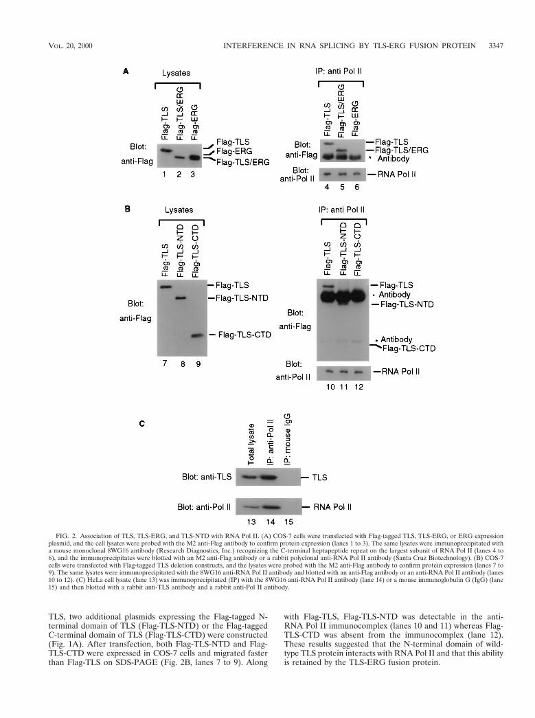
TASR-2 is a serine-arginine splicing factor. We recentlydemonstrated through yeast two-hybrid screening that TLSinteracts with a well-known SR protein SC35 and a novel SRprotein termed TASR-1 (47). To obtain a more complete pic-ture of the proteins interacting with TLS, we carried out asecond yeast two-hybrid screen of a mouse hematopoieticcDNA library using the C-terminal domain of TLS as the bait(Fig. 1A). We isolated a cDNA encoding a second novel TLS-associated SR protein termed TASR-2. Mouse TASR-2 cDNA(GenBank accession number AF060490) is 2.7 kb long andencodes a protein of 262 amino acids with a calculated molec-ular mass of 31 kDa (Fig. 3A). The cDNA encoding humanTASR-2 (GenBank accession number AF067730) was isolatedfrom K562 cells by RT-PCR. Mouse and human TASR-2 areidentical at the amino acid level, suggesting that TASR-2 isevolutionarily conserved. Similar to SC35 and TASR-1, TASR-2 also has typical RNP2 and RPN1 motifs in its N-terminalregion and multiple serine-arginine repeats in its C-terminalregion (Fig. 3B) that are characteristic of prototype SR pro-teins. Even though the calculated molecular mass of TASR-2 is31 kDa, in vitro-translated TASR-2 protein migrated as a bandof 37 kDa (Fig. 3C, lane 1) when analyzed by SDS-PAGE. Thisdiscrepancy between the calculated molecular size and thatdetermined by SDS-PAGE is typical of SR proteins such asSC35 and SF2/ASF (16) and results from phosphorylation ofthe serine residues and perhaps regions of alternating charge.
To confirm that TASR-2 functions as a splicing factor, itseffect on the alternative splicing of an E1A reporter gene wastested. The alternative splicing of E1A pre-mRNA leads to thegeneration of five different splicing isoforms designated 13S,12S, 11S, 10S, and 9S mRNA (see Fig. 5A). For these studiesof E1A pre-mRNA splicing, HeLa cells were transfected withthe pCS3-MT-E1A reporter containing the E1A genomic se-quence, and the alternative splicing of the E1A pre-mRNA wasexamined by RT-PCR. In HeLa cells the dominant splicingproducts of E1A pre-mRNA are 13S and 12S isoforms (Fig.3D, lane 5). TASR-1 promotes multiple splice site selectionsleading to 11S, 10S, and 9S isoforms (lane 6) whereas coex-pression of TASR-2 promotes 59 splice site selection, leadingprimarily to the 9S isoform (lane 7). Due to the presence ofresidual plasmid DNA in the total RNA, the E1A genomicsequence could be amplified by PCR (lane 4).
The TLS C-terminal domain mediates interaction with SRproteins. We have previously shown through coimmunopre-cipitation that TLS interacts with SC35 and TASR-1 (47).Since the C-terminal domain of TLS is replaced by the DNA-binding domain of ERG in the TLS-ERG leukemia fusionprotein, we investigated the interaction between TLS and TLS-ERG and the TASR-1 and TASR-2 proteins. Plasmids ex-pressing Flag-tagged TLS, TLS-ERG, or ERG were cotrans-fected into COS-7 cells with plasmids expressing Myc-taggedTASR-1 or TASR-2, and lysates from the cotransfected cellswere used for immunoprecipitation. In immunoprecipitatesbrought down using an anti-Flag antibody, Myc-TASR-1 and
Myc-TASR-2 were found to associate with the wild-type Flag-TLS (Fig. 4A, lanes 1 and 4) but not with the Flag-TLS-ERGleukemia fusion protein or wild-type Flag-ERG (lanes 2, 3,5, and 6). In the reciprocal immunoprecipitation, only Flag-TLS was coimmunoprecipitated with Myc-TASR-1 and Myc-TASR-2 using an anti-Myc antibody (Fig. 4B, lanes 7 and 10)whereas Flag-TLS-ERG and Flag-ERG were not immunopre-cipitated (lanes 8, 9, 11, and 12). Since Flag-TLS and Flag-TLS-ERG differ only in their C-terminal domains, these resultssuggested that the TLS C-terminal domain was responsible for
FIG. 3. Amino acid sequence and characterization of TASR-2. (A) The pre-dicted amino acid sequence of TASR-2 protein is shown. Mouse TASR-2 (Gen-Bank accession number AF060490) and human TASR-2 (GenBank accessionnumber AF067730) are identical at the amino acid level. RNP2 and RNP1consensus sequences are underlined. Arg-Ser or Ser-Arg dipeptide repeats areshown in bold type. (B) Schematic comparison of SC35, TASR-1, and TASR-2.The RNP consensus sequences are shown in gray boxes. The RS domains are inhatched boxes. (C) In vitro translation reaction products with pBluescript-TASR-2 as the template (lane 1) are separated by SDS-PAGE on a 12% poly-acrylamide gel along with protein molecular markers (lane 2) and control reac-tion products with the pBluescript empty vector (lane 3). The position of theTASR-2 protein is indicated by an arrow. (D) A 2.5-mg sample of pCS3-MT-E1Areporter plasmid was cotransfected with 2.5 mg of pCR3 empty vector (lanes 4and 5) or with 2.5 mg of pCR3-TASR-1 (lane 6) and 2.5 mg of pCS3-TASR-2(lane 7) expression plasmid into HeLa cells. RT-PCR was carried out as de-scribed in Materials and Methods, and the E1A splicing products were confirmedby hybridization to a 32P-labeled E1A DNA probe. Lane 4 contains controlRT-PCR products from a reaction carried out in the absence of reverse tran-scriptase to show the presence of residual pCS3-MT-E1A template DNA in thetotal RNA.
3348 YANG ET AL. MOL. CELL. BIOL.
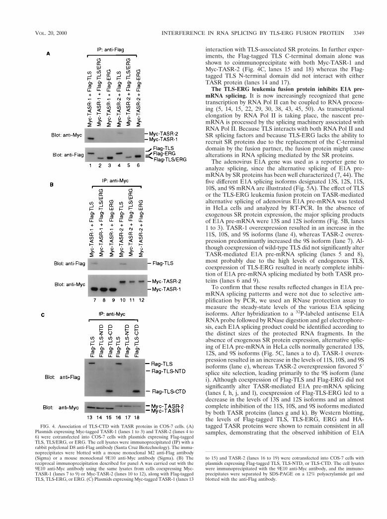
interaction with TLS-associated SR proteins. In further exper-iments, the Flag-tagged TLS C-terminal domain alone wasshown to coimmunoprecipitate with both Myc-TASR-1 andMyc-TASR-2 (Fig. 4C, lanes 15 and 18) whereas the Flag-tagged TLS N-terminal domain did not interact with eitherTASR protein (lanes 14 and 17).
The TLS-ERG leukemia fusion protein inhibits E1A pre-mRNA splicing. It is now increasingly recognized that genetranscription by RNA Pol II can be coupled to RNA process-ing (5, 14, 15, 22, 29, 30, 38, 43, 45, 50). As transcriptionalelongation by RNA Pol II is taking place, the nascent pre-mRNA is processed by the splicing machinery associated withRNA Pol II. Because TLS interacts with both RNA Pol II andSR splicing factors and because TLS-ERG lacks the ability torecruit SR proteins due to the replacement of the C-terminaldomain by the fusion partner, the fusion protein might causealterations in RNA splicing mediated by the SR proteins.
The adenovirus E1A gene was used as a reporter gene toanalyze splicing, since the alternative splicing of E1A pre-mRNA by SR proteins has been well characterized (7, 44). Thefive different E1A splicing isoforms designated 13S, 12S, 11S,10S, and 9S mRNA are illustrated (Fig. 5A). The effect of TLSor the TLS-ERG leukemia fusion protein on TASR-mediatedalternative splicing of adenovirus E1A pre-mRNA was testedin HeLa cells and analyzed by RT-PCR. In the absence ofexogenous SR protein expression, the major splicing productsof E1A pre-mRNA were 13S and 12S isoforms (Fig. 5B, lanes1 to 3). TASR-1 overexpression resulted in an increase in the11S, 10S, and 9S isoforms (lane 4), whereas TASR-2 overex-pression predominantly increased the 9S isoform (lane 7). Al-though coexpression of wild-type TLS did not significantly alterTASR-mediated E1A pre-mRNA splicing (lanes 5 and 8),most probably due to the high levels of endogenous TLS,coexpression of TLS-ERG resulted in nearly complete inhibi-tion of E1A pre-mRNA splicing mediated by both TASR pro-teins (lanes 6 and 9).
To confirm that these results reflected changes in E1A pre-mRNA splicing patterns and were not due to selective am-plification by PCR, we used an RNase protection assay tomeasure the steady-state levels of the various E1A splicingisoforms. After hybridization to a 32P-labeled antisense E1ARNA probe followed by RNase digestion and gel electrophore-sis, each E1A splicing product could be identified according tothe distinct sizes of the protected RNA fragments. In theabsence of exogenous SR protein expression, alternative splic-ing of E1A pre-mRNA in HeLa cells normally generated 13S,12S, and 9S isoforms (Fig. 5C, lanes a to d). TASR-1 overex-pression resulted in an increase in the levels of 11S, 10S, and 9Sisoforms (lane e), whereas TASR-2 overexpression favored 59splice site selection, leading primarily to the 9S isoform (lanei). Although coexpression of Flag-TLS and Flag-ERG did notsignificantly alter TASR-mediated E1A pre-mRNA splicing(lanes f, h, j, and l), coexpression of Flag-TLS-ERG led to adecrease in the levels of 13S and 12S isoforms and an almostcomplete inhibition of the 11S, 10S, and 9S isoforms mediatedby both TASR proteins (lanes g and k). By Western blotting,the levels of Flag-tagged TLS, TLS-ERG, ERG and HA-tagged TASR proteins were shown to remain consistent in allsamples, demonstrating that the observed inhibition of E1A
FIG. 4. Association of TLS-CTD with TASR proteins in COS-7 cells. (A)Plasmids expressing Myc-tagged TASR-1 (lanes 1 to 3) and TASR-2 (lanes 4 to6) were cotransfected into COS-7 cells with plasmids expressing Flag-taggedTLS, TLS/ERG, or ERG. The cell lysates were immunoprecipitated (IP) with arabbit polyclonal D8 anti-Flag antibody (Santa Cruz Biotechnology). The immu-noprecipitates were blotted with a mouse monoclonal M2 anti-Flag antibody(Sigma) or a mouse monoclonal 9E10 anti-Myc antibody (Sigma). (B) Thereciprocal immunoprecipitation described for panel A was carried out with the9E10 anti-Myc antibody using the same lysates from cells coexpressing Myc-TASR-1 (lanes 7 to 9) or Myc-TASR-2 (lanes 10 to 12), along with Flag-taggedTLS, TLS-ERG, or ERG. (C) Plasmids expressing Myc-tagged TASR-1 (lanes 13
to 15) and TASR-2 (lanes 16 to 19) were cotransfected into COS-7 cells withplasmids expressing Flag-tagged TLS, TLS-NTD, or TLS-CTD. The cell lysateswere immunoprecipitated with the 9E10 anti-Myc antibody, and the immuno-precipitates were separated by SDS-PAGE on a 12% polyacrylamide gel andblotted with the anti-Flag antibody.
VOL. 20, 2000 INTERFERENCE IN RNA SPLICING BY TLS-ERG FUSION PROTEIN 3349
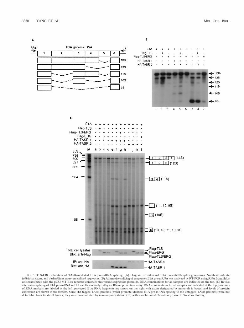
FIG. 5. TLS-ERG inhibition of TASR-mediated E1A pre-mRNA splicing. (A) Diagram of individual E1A pre-mRNA splicing isoforms. Numbers indicateindividual exons, and dashed lines represent spliced sequences. (B) Alternative splicing of exogenous E1A pre-mRNA was analyzed by RT-PCR using RNA from HeLacells transfected with the pCS3-MT-E1A reporter construct plus various expression plasmids. DNA combinations for all samples are indicated on the top. (C) In vivoalternative splicing of E1A pre-mRNA in HeLa cells was analyzed by an RNase protection assay. DNA combinations for all samples are indicated at the top, positionsof RNA markers are labeled at the left, protected E1A RNA fragments are shown on the right with exons designated by numerals in boxes, and levels of proteinexpression are shown at the bottom. Since HA-tagged TASR proteins (which promote identical E1A pre-mRNA splicing to the untagged TASR proteins) were notdetectable from total-cell lysates, they were concentrated by immunoprecipitation (IP) with a rabbit anti-HA antibody prior to Western blotting.
3350 YANG ET AL. MOL. CELL. BIOL.
splicing was not due to variation in transfection or decrease inTASR expression caused by the TLS-ERG fusion protein. Un-spliced E1A pre-mRNA was not detected by RNase protec-tion, consistent with previous reports (7, 44) indicating thatunprocessed E1A pre-mRNA is labile under the in vivo exper-imental conditions. The SR splicing factor SC35 was not as-sessed in this study, because endogenous SC35 is abundant inHeLa cells and overexpression of SC35 has minimal effect onE1A pre-mRNA splicing (44, 47).
To rule out the possibility that the observed TLS-ERG in-hibition of TASR-mediated E1A pre-mRNA splicing was dueto repression of transcription from the pCS3-MT-E1A re-porter or preferential destabilization of the 11S, 10S, and 9SE1A splicing isoforms, we constructed an additional reporterplasmid, pCS3-MT-E1A-9S, which was generated from thesame vector as pCS3-MT-E1A but contains a cDNA insertcorresponding to the 9S E1A splicing isoform. When this con-struct was analyzed under the same conditions, the resultant 9SE1A mRNA levels were not decreased by coexpression ofTLS-ERG or altered by TASR-1 or TASR-2 (data not shown).These results indicated that TLS-ERG did not suppress tran-scription from the pCS3-MT-E1A vector or selectively desta-bilize the 9S E1A splicing isoform.
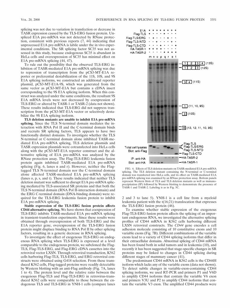
TLS deletion mutants are unable to inhibit E1A pre-mRNAsplicing. Since the TLS N-terminal domain mediates the in-teraction with RNA Pol II and the C-terminal domain bindsand recruits SR splicing factors, TLS appears to have twofunctionally distinct domains. To investigate whether the TLSN-terminal or C-terminal domain alone inhibited TASR-me-diated E1A pre-mRNA splicing, TLS deletion plasmids andTASR expression plasmids were cotransfected into HeLa cellsalong with the pCS3-MT-E1A reporter construct and the al-ternative splicing of E1A pre-mRNA was analyzed by theRNase protection assay. The Flag-TLS-ERG leukemia fusionprotein again inhibited TASR-mediated E1A pre-mRNAsplicing (Fig. 6, lanes n and r). However, neither the Flag-tagged TLS N-terminal domain nor the C-terminal domainalone affected TASR-mediated E1A pre-mRNA splicing(lanes o, p, s, and t). These results indicated that neither TLSdeletion mutant was sufficient to disrupt E1A pre-mRNA splic-ing mediated by TLS-associated SR proteins and that both theTLS N-terminal domain (RNA Pol II-interaction domain) andthe ERG C-terminal domain (DNA-binding domain) were re-quired for the TLS-ERG leukemia fusion protein to inhibitE1A pre-mRNA splicing.
Stable expression of the TLS-ERG fusion protein affectsCD44 alternative splicing. We have shown that coexpression ofTLS-ERG inhibits TASR-mediated E1A pre-mRNA splicingin transient-transfection experiments. Since these results wereobtained through overexpression of both TLS-ERG and theE1A reporter gene, overexpression of the TLS-ERG fusionprotein might displace binding to RNA Pol II by other splicingfactors, resulting in a generic decrease in RNA splicing.
To investigate the effect of exogenous TLS-ERG on endog-enous RNA splicing when TLS-ERG is expressed at a levelcomparable to the endogenous protein, we subcloned the Flag-TLS, Flag-TLS-ERG, and Flag-ERG cDNA cassettes into theretroviral vector LXSN. Through retroviral transduction, K562cells harboring Flag-TLS, TLS-ERG, and ERG retroviral con-structs were obtained using G418 selection. From these trans-duced K562 cells, Flag-tagged proteins were readily detectableby Western blotting with an anti-Flag antibody (Fig. 7A, lanes1 to 4). The protein level and the relative ratio between theexogenous Flag-TLS and Flag-TLS-ERG in retrovirus-trans-duced K562 cells were comparable to those between the en-dogenous TLS and TLS-ERG in YNH-1 cells (compare lanes
2 and 3 to lane 5). YNH-1 is a cell line from a myeloidleukemia patient with the t(16;21) translocation that expressesthe TLS-ERG fusion protein (46).
To examine whether stable expression of the exogenousFlag-TLS-ERG fusion protein affects the splicing of an impor-tant endogenous RNA, we investigated the alternative splicingprofiles of CD44 mRNA in K562 cells harboring differentLXSN retroviral constructs. The CD44 gene encodes a celladhesion molecule consisting of 10 constitutive exons and 10variable exons (Fig. 7B). Different combinations of the variableexons lead to a variety of CD44 splicing isoforms that differ intheir extracellular domains. Abnormal splicing of CD44 mRNAhas been found both in solid tumors and in leukemia (10), andrecently it has been suggested that stage-specific changes in SRproteins may be linked to changes in CD44 splicing duringdifferent stages of mammary cancer (41).
The predominant CD44 mRNA in K562 cells is the CD44Hisoform which lacks any of the variable exons (data not shown).To detect subtle changes in variable-exon-containing CD44splicing isoforms, we used RT-PCR and primers P1 and V6Dto amplify CD44 isoforms that contain the variable V6 exonand primers V3U and P2 to amplify CD44 isoforms that con-tain the variable V3 exon. The amplified CD44 products were
FIG. 6. Effects of TLS deletion mutants on TASR-mediated E1A pre-mRNAsplicing. The TLS deletion mutant containing the N-terminal or C-terminaldomain was transfected into HeLa cells, and its effect on TASR-mediated E1Apre-mRNA splicing was examined by an RNase protection assay. Bottom panelsrepresent Western blots of TLS and TLS deletion mutants as well as immuno-precipitation (IP) followed by Western blotting to demonstrate the presence ofTASR-1 and TASR-2. Labeling is as in Fig. 5C.
VOL. 20, 2000 INTERFERENCE IN RNA SPLICING BY TLS-ERG FUSION PROTEIN 3351
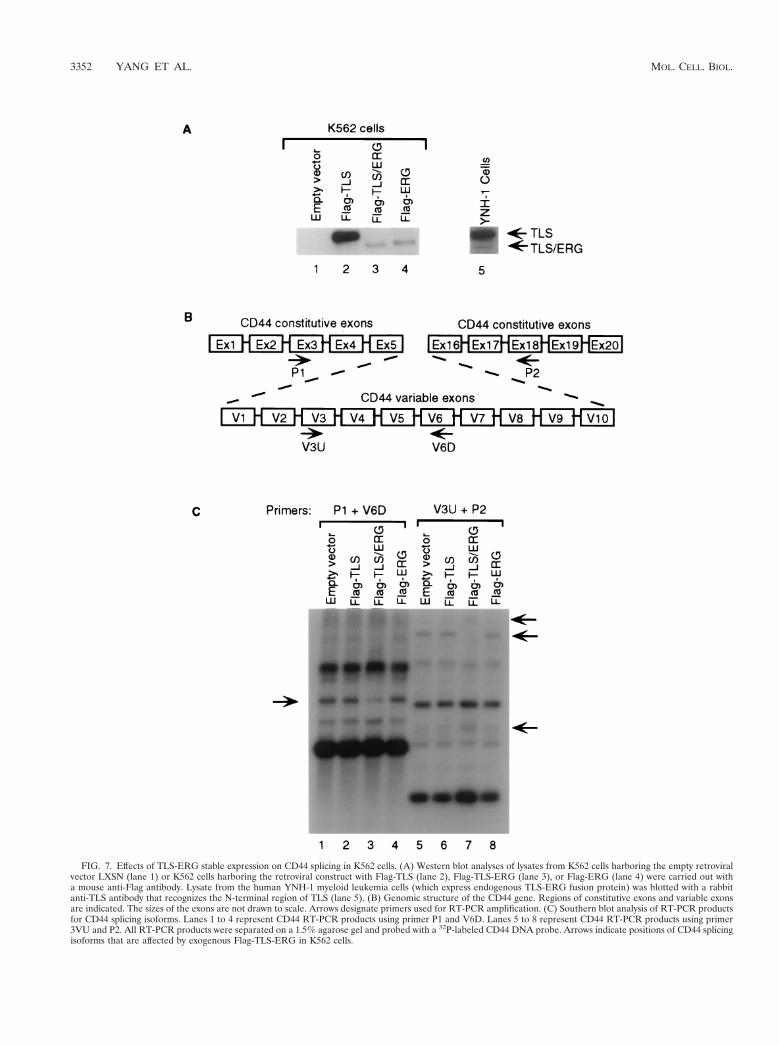
FIG. 7. Effects of TLS-ERG stable expression on CD44 splicing in K562 cells. (A) Western blot analyses of lysates from K562 cells harboring the empty retroviralvector LXSN (lane 1) or K562 cells harboring the retroviral construct with Flag-TLS (lane 2), Flag-TLS-ERG (lane 3), or Flag-ERG (lane 4) were carried out witha mouse anti-Flag antibody. Lysate from the human YNH-1 myeloid leukemia cells (which express endogenous TLS-ERG fusion protein) was blotted with a rabbitanti-TLS antibody that recognizes the N-terminal region of TLS (lane 5). (B) Genomic structure of the CD44 gene. Regions of constitutive exons and variable exonsare indicated. The sizes of the exons are not drawn to scale. Arrows designate primers used for RT-PCR amplification. (C) Southern blot analysis of RT-PCR productsfor CD44 splicing isoforms. Lanes 1 to 4 represent CD44 RT-PCR products using primer P1 and V6D. Lanes 5 to 8 represent CD44 RT-PCR products using primer3VU and P2. All RT-PCR products were separated on a 1.5% agarose gel and probed with a 32P-labeled CD44 DNA probe. Arrows indicate positions of CD44 splicingisoforms that are affected by exogenous Flag-TLS-ERG in K562 cells.
3352 YANG ET AL. MOL. CELL. BIOL.
then separated by gel electrophoresis and probed with a 32P-labeled CD44 DNA probe. As shown in Fig. 7C, expression ofthe Flag-TLS or Flag-ERG protein in K562 cells did not alterthe profile of CD44 isoforms containing the V6 exon (comparelane 1 with lanes 2 and 4) or the profile of CD44 isoformscontaining the V3 exon (compare lane 5 with lanes 6 and 8).However, expression of the Flag-TLS-ERG fusion protein inK562 cells changed the profile of endogenous CD44 isoformscontaining the V6 exon (compare lane 1 with lane 3) and theV3 exon (compare lane 5 with lane 7).
DISCUSSION
These studies indicate that TLS interacts with RNA Pol IIthrough the N-terminal domain of TLS and interacts withmembers of the SR family of splicing factors through the C-terminal domain of TLS. In contrast, the TLS-ERG fusionprotein binds RNA Pol II but is unable to bind SR proteins,due to replacement of its C-terminal domain by the fusionpartner. The TLS-ERG fusion protein not only lacks the abilityto bridge the binding of RNA Pol II to SR proteins but alsoinhibits E1A pre-mRNA splicing mediated by these SR pro-teins in transfected HeLa cells. When Flag-TLS-ERG was in-troduced through retroviral transduction into K562 cells andexpressed at a level comparable to the endogenous protein, itchanged the splicing profile of the endogenous CD44, a celladhesion molecule whose abnormal splicing is associated withcellular transformation and tumor metastasis (10).
RNA splicing, a critical step in gene expression, is increas-ingly recognized as a cotranscriptional event (reviewed in ref-erences 31 and 40). Experimental evidence now indicates thattranscription, capping, and polyadenylation are also intimatelylinked by RNA Pol II through its association with RNA-pro-cessing factors (9, 21). Among different components of thetranscriptional machinery, the C-terminal domain of the larg-est subunit of RNA Pol II complex is thought to be especiallycritical for recruitment of RNA-processing factors (29, 30).Even though a novel set of C-terminal domain associated SR-like proteins directly interact via their C-terminal domain-interacting domains with RNA Pol II (11, 48), prototypical SRproteins including SC35 and ASF/SF2 lack such C-terminaldomain-interacting domains. At present, the way in whichthese SR proteins associate with RNA Pol II remains unclear.Recently, a novel transcription coactivator, p52, was found tointeract specifically with ASF/SF2 and was suggested to act asan adapter to coordinate transcription and splicing (17). Basedon our observation that TLS interacts with both RNA Pol IIand SR proteins, we propose that TLS functions as a dockingmolecule by recruiting SR splicing factors to RNA Pol II, thuscoupling gene transcription with RNA splicing.
The TLS gene is ubiquitously expressed, suggesting that TLSmay be essential for cell function (1). In myeloid leukemia cellswith the t(16;21) translocation and in liposarcoma cells withthe t(12;16) translocation, only one TLS allele is interrupted bychromosomal translocation while the remaining TLS allele isintact (12, 36, 46). This indicates that the TLS fusion proteinfunctions in a dominant-negative manner.
The TLS-ERG fusion protein not only inhibits TASR-me-diated E1A pre-mRNA splicing, leading to 11S, 10S, and 9Sisoforms, but also decreases the expression of the 13S and 12SE1A splicing isoforms. The generation of 13S and 12S isoformsmay be mediated by additional splicing factors present in HeLacells, which in turn implies that the splicing pathway via TLSpotentially includes other splicing factors. Based on the obser-vation that expression of the TLS-ERG leukemia fusion pro-tein, despite the presence of endogenous TLS protein, is suf-
ficient to inhibit E1A pre-mRNA splicing in HeLa cells and toalter CD44 splicing in K562 cells, the TLS-ERG fusion proteinappears to function in a dominant-negative manner to inter-fere with RNA splicing. Our finding that neither the N-termi-nal nor the C-terminal deletion mutant of TLS was sufficient toblock TASR-mediated E1A splicing suggests that inclusion ofthe ERG DNA-binding domain is required for the dominant-negative function of the TLS-ERG leukemia fusion protein,possibly by conferring a higher affinity of the fusion protein forRNA Pol II and/or by localizing the fusion protein to thenucleus.
One can envisage at least two potential mechanisms wherebythe TLS-ERG fusion protein alters CD44 splicing. In the firstscenario, if splicing into a specific CD44 isoform requires dock-ing by TLS, the TLS-ERG fusion protein might block thispathway, leading to degradation of the unfinished CD44 pre-mRNA. In the second scenario, if one CD44 splicing pathwayis blocked and alternative splicing pathways exist, the inhibi-tion of the first pathway by the TLS-ERG fusion protein mightpush the splicing of CD44 pre-mRNA through alternativeroutes, thus increasing the chance of aberrant splicing.
SR proteins appear to possess the capacity to alter geneexpression by influencing RNA splicing. The important regu-latory roles of SR proteins in cellular processes have beendemonstrated by their functions in Drosophila developmentand sex determination (2, 20, 37), their involvement in T-cellactivation (39), and their close association with cell cycle con-trol (27). Even though transcriptional deregulation by onco-genic fusion proteins has attracted much of the attention, ab-errant RNA splicing is also frequently detected in cancer cellsand is associated with cellular transformation (8, 18) and tu-mor metastasis (10). The mechanism underlying aberrant splic-ing has yet to be elucidated. This study links a specific chro-mosomal translocation, and the resultant leukemia fusionprotein, to disruption of RNA splicing. Although our observa-tion is made from cells expressing exogenous Flag-TLS-ERGfusion protein and further experiments with cancer cells har-boring TLS translocations are needed, these results suggestthat TLS fusion proteins may lead to abnormal splicing ofgenes critical for cell growth and differentiation.
Ewing’s sarcoma protein EWS and TATA-binding protein-associated factor TAFII68 share sequence homology with TLS,and it is likely that both EWS and TAFII68 also interact withSR splicing factors. The potential disruption of coupling be-tween transcription and RNA splicing by EWS fusion proteinsespecially deserves further investigation. In addition to fusionwith the Fli-1 protein in 90% of cases of Ewing’s sarcoma,EWS is involved in chromosomal translocations with a varietyof transcription factors including ERG, ETV1, E1A-F, FEV,ATF-1, WT1, and TEC1 (4). In all of these fusions involvingEWS, the N-terminal domain of EWS is retained. EWS fusionproteins may thus function in a manner analogous to the TLSfusion proteins by interfering with the recruitment of RNA-processing factors such as the SR proteins.
ACKNOWLEDGMENTS
We thank R. S. Morrison, M. B. Roth, and Y.-C. Yang for helpfuldiscussions; T. R. Bauer, Jr., S. Collins, and B. Kwiatkowski for criticalreading of the manuscript; and S. L. Danner and S. M. Stray for DNAsequencing.
REFERENCES
1. Aman, P., I. Panagopoulos, C. Lassen, T. Fioretos, M. Mencinger, H. Tor-esson, M. Hoglund, A. Forster, T. H. Rabbits, D. Ron, N. Mandahl, and F.Mitelman. 1996. Expression patterns of the human sarcoma-associated genesFUS and EWS and genomic structure of FUS. Genomics 37:1–8.
2. Amrein, H., M. L. Hedley, and T. Maniatis. 1994. The role of specific
VOL. 20, 2000 INTERFERENCE IN RNA SPLICING BY TLS-ERG FUSION PROTEIN 3353
protein-RNA and protein-protein interactions in positive and negative con-trol of pre-mRNA splicing by Transformer-2. Cell 76:735–746.
3. Bertolotti, A., Y. Lutz, D. J. Heard, P. Chambon, and L. Tora. 1996.hTAFII68, a novel RNA/ss DNA-binding protein with homology to thepro-oncoprotein TLS/FUS and EWS is associated with both TFIID andRNA polymerase II. EMBO J. 15:5022–5031.
4. Bertolotti, A., T. Melot, J. Acker, M. Vigneron, O. Delattre, and L. Tora.1998. EWS, but not EWS–FLI-1, is associated with both TFIID and RNApolymerase II: interactions between two members of the TET family, EWSand hTAFII68, and subunits of TFIID and RNA polymerase II complexes.Mol. Cell. Biol. 18:1489–1497.
5. Beyer, A. L., and Y. N. Osheim. 1988. Splice site selection, rate of splicing,and alternative splicing on nascent transcripts. Genes Dev. 2:754–765.
6. Burd, C. G., and G. Dreyfuss. 1994. Conserved structures and diversity offunctions of RNA-binding proteins. Science 265:615–621.
7. Caceres, J. F., S. Stamm, D. M. Helfman, and A. R. Krainer. 1994. Regu-lation of alternative splicing in vivo by overexpression of antagonistic splicingfactors. Science 265:1706–1709.
8. Carstens, R. P., J. V. Eaton, H. R. Krigman, P. J. Walther, and M. A.Garcia-Blanco. 1997. Alternative splicing of fibroblast growth factor receptor2 (FGF-R2) in human prostate cancer. Oncogene 15:3059–3065.
9. Cho, E. J., T. Takagi, C. R. Moore, and S. Buratowski. 1997. mRNA cappingenzyme is recruited to the transcription complex by phosphorylation of theRNA polymerase II carboxy-terminal domain. Genes Dev. 11:3319–3326.
10. Cooper, D. L., and G. J. Dougherty. 1995. To metastasize or not? Selectionof CD44 splice sites. Nat. Med. 1:635–637.
11. Corden, J. L., and M. Patturajan. 1997. A CTD function linking transcrip-tion to splicing. Trends Biochem. Sci. 22:413–416.
12. Crozat, A., P. Aman, N. Mandahl, and D. Ron. 1993. Fusion of CHOP to anovel RNA-binding protein in human myxoid liposarcoma. Nature 363:640–644.
13. Delattre, O., J. Zucman, B. Plougastel, C. Desmaze, T. Melot, M. Peter, H.Kovar, I. Houbert, P. DeJong, G. Rouleau, A. Aurias, and G. Thomas. 1992.Gene fusion with an ETS DNA-binding domain caused by chromosometranslocation in human tumors. Nature 359:162–165.
14. Du, L., and S. L. Warren. 1997. A functional interaction between the car-boxy-terminal domain of RNA polymerase II and pre-mRNA splicing. J. CellBiol. 136:5–18.
15. Fakan, S., G. Leser, and T. E. Martin. 1986. Immunoelectron microscopevisualization of nuclear ribonucleoprotein antigens within spread transcrip-tion complexes. J. Cell Biol. 103:1153–1157.
16. Fu, X. D., and T. Maniatis. 1992. Isolation of a complementary DNA thatencodes the mammalian splicing factor SC35. Science 256:535–538.
17. Ge, H., Y. Si, and A. P. Wolffe. 1998. A novel transcriptional coactivator, p52,functionally interacts with the essential splicing factor ASF/SF2. Mol. Cell2:751–759.
18. Ge, K., J. DuHadaway, W. Du, M. Herlyn, U. Rodeck, and G. C. Prendergast.1999. Mechanism for elimination of a tumor suppressor: aberrant splicing ofa brain-specific exon causes loss of function of Bin1 in melanoma. Proc. Natl.Acad. Sci. USA 96:9689–9694.
19. Hallier, M., A. Lerga, S. Barnache, A. Tavitian, and F. Moreau-Gachelin.1998. The transcription factor Spi-1/PU.1 interacts with the potential splicingfactor TLS. J. Biol. Chem. 273:4838–4842.
20. Heinrichs, V., and B. S. Baker. 1997. In vivo analysis of the functionaldomains of the Drosophila splicing regulator RBP1. Proc. Natl. Acad. Sci.USA 94:115–120.
21. Hirose Y., and J. L. Manley. 1998. RNA polymerase II is an essential mRNApolyadenylation factor. Nature 395:93–96.
22. Huang, S., and D. L. Spector. 1996. Intron-dependent recruitment of pre-mRNA splicing factors to sites of transcription. J. Cell Biol. 133:719–732.
23. Ichikawa, H., K. Shimizu, R. Katsu, and M. Ohki. 1994. An RNA-bindingprotein gene, TLS/FUS, is fused to ERG in human myeloid leukemia witht(16;21) chromosomal translocation. Cancer Res. 54:2865–2868.
24. Ichikawa, H., K. Shimizu, R. Katsu, and M. Ohki. 1999. Dual transformingactivities of the FUS (TLS)-ERG leukemia fusion protein conferred by twoN-terminal domains of FUS (TLS). Mol. Cell. Biol. 19:7639–7650.
25. Immanuel, D., H. Zinszner, and D. Ron. 1995. Association of SARFH(sarcoma-associated RNA-binding fly homolog) with regions of chromatintranscribed by RNA polymerase II. Mol. Cell. Biol. 15:4562–4571.
26. Jaishankar, S., J. Zhang, M. F. Roussel, and S. J. Baker. 1999. Transformingactivity of EWS/Fli is not strictly dependent upon DNA-binding activity.Oncogene 18:5592–5597.
27. Jumaa, H., J. L. Guenet, and P. J. Nielsen. 1997. Regulated expression andRNA processing of transcripts from the Srp20 splicing factor gene during thecell cycle. Mol. Cell. Biol. 17:3116–3124.
28. Lessnick, S. L., B. S. Braun, C. T. Denny, and W. A. May. 1995. Multipledomains mediate transformation by the Ewing’s sarcoma EWS/FLI-1 fusiongene. Oncogene 10:423–431.
29. McCracken, S., N. Fong, K. Yankulov, S. Ballantyne, G. Pan, J. Greenblatt,S. D. Patterson, M. Wickens, and D. Bentley. 1997. The C-terminal domainof RNA polymerase II couples mRNA processing to transcription. Nature385:357–361.
30. Mortillaro, M. J., B. J. Blencowe, X. Wei, H. Nakayasu, L. Du, S. L. Warren,P. A. Sharp, and R. Berezney. 1996. A hyperphosphorylated form of the largesubunit of RNA polymerase II is associated with splicing complexes and thenuclear matrix. Proc. Natl. Acad. Sci. USA 93:8253–8257.
31. Neugebauer, K. M., and M. B. Roth. 1997. Transcription units as RNAprocessing units. Genes Dev. 11:3279–3285.
32. Pereira, D. S., C. Dorrell, C. Y. Ito, O. I. Gan, B. Murdoch, V. N. Rao, J. P.Zou, E. S. Reddy, and J. E. Dick. 1998. Retroviral transduction of TLS-ERGinitiates a leukemogenic program in normal human hematopoietic cells.Proc. Natl. Acad. Sci. USA 95:8239–8244.
33. Perrotti, D., S. Bonatti, R. Trotta, R. Martinez, T. Skorski, P. Salomoni, E.Grassilli, R. V. Lozzo, D. R. Cooper, and B. Calabretta. 1998. TLS/FUS, apro-oncogene involved in multiple chromosomal translocations, is a novelregulator of BCR/ABL-mediated leukemogenesis. EMBO J. 17:4442–4455.
34. Petermann, R., B. M. Mossier, D. NT. Aryee, V. Khazak, E. A. Golemis, andH. Kovar. 1998. Oncogenic EWS-Fli1 interacts with hsRPB7, a subunit ofhuman RNA polymerase II. Oncogene 17:603–610.
35. Prasad, D. D., M. Ouchida, L. Lee, V. N. Rao, and E. S. Reddy. 1994.TLS/FUS fusion domain of TLS/FUS-erg chimeric protein resulting fromthe t(16;21) chromosomal translocation in human myeloid leukemia func-tions as a transcriptional activation domain. Oncogene 9:3717–3729.
36. Rabbitts, T. H., A. Forster, R. Larson, and P. Nathan. 1993. Fusion of thedominant negative transcription regulator CHOP with a novel gene FUS bytranslocation t(12;16) in malignant liposarcoma. Nat. Genet. 4:175–180.
37. Ring, H. Z., and J. T. Lis. 1994. The SR protein B52/SRp55 is essential forDrosophila development. Mol. Cell. Biol. 14:7499–7506.
38. Sass, H., and T. Pederson. 1984. Transcription-dependent localization of U1and U2 small nuclear ribonucleoproteins at major sites of gene activity inpolytene chromosomes. J. Mol. Biol. 180:911–926.
39. Screaton, G. R., J. F. Caceres, A. Mayeda, M. V. Bell, M. Plebanski, D. G.Jackson, J. I. Bell, and A. R. Krainer. 1995. Identification and characteriza-tion of three members of the human SR family of pre-mRNA splicingfactors. EMBO J. 14:4336–4349.
40. Steinmetz, E. J. 1997. Pre-mRNA processing and the CTD of RNA poly-merase II: the tail that wags the dog? Cell 89:491–494.
41. Stickeler, E., F. Kittrell, D. Medina, and S. M. Berget. 1999. Stage-specificchanges in SR splicing factors and alternative splicing in mammary tumor-igenesis. Oncogene 18:3574–3582.
42. Tsai, S., S. Bartelmez, E. Sitnicka, and S. Collins. 1994. Lymphohemato-poietic progenitors immortalized by a retroviral vector harboring a domi-nant-negative retinoic acid receptor can recapitulate lymphoid, myeloid, anderythroid development. Genes Dev. 8:2831–2841.
43. Vincent, M., P. Lauriault, M.-F. Dubois, S. Lavoie, O. Bensaude, and B.Chabot. 1996. The nuclear matrix protein p255 is a highly phosphorylatedform of RNA polymerase II largest subunit which associates with spliceo-somes. Nucleic Acids Res. 24:4649–4652.
44. Wang, J., and J. L. Manley. 1995. Overexpression of the SR proteins ASF/SF2 and SC35 influences alternative splicing in vivo in diverse ways. RNA1:335–346.
45. Xing, Y., C. V. Johnson, P. R. Dobner, and J. B. Lawrence. 1993. Higher levelorganization of individual gene transcription and RNA splicing. Science259:1326–1330.
46. Yamamoto, K., H. Hamaguchi, K. Nagata, M. Kobayashi, F. Tanimoto, andM. Taniwaki. 1997. Establishment of a novel human acute myeloblasticleukemia cell line (YNH-1) with t(16;21), t(1;16) and 12q13 translocations.Leukemia 11:599–608.
47. Yang, L., L. J. Embree, S. Tsai, and D. D. Hickstein. 1998. Oncoprotein TLSinteracts with serine-arginine proteins involved in RNA splicing. J. Biol.Chem. 273:27761–27764.
48. Yuryev, A., M. Patturajan, Y. Litingtung, R. V. Joshi, C. Gentile, M. Gebara,and J. L. Corden. 1996. The C-terminal domain of the largest subunit ofRNA polymerase II interacts with a novel set of serine/arginine-rich pro-teins. Proc. Natl. Acad. Sci. USA 93:6975–6980.
49. Zhang, D., A. J. Paley, and G. Childs. 1998. The transcriptional repressorZFM1 interacts with and modulates the ability of EWS to activate transcrip-tion. J. Biol. Chem. 273:18086–18091.
50. Zhang, G., K. L. Taneja, R. H. Singer, and M. R. Green. 1994. Localizationof pre-mRNA splicing in mammalian nuclei. Nature 372:809–812.
51. Zinszner, H., R. Albalat, and D. Ron. 1994. A novel effector domain from theRNA-binding protein TLS or EWS is required for oncogenic transformationby CHOP. Genes Dev. 8:2513–2526.
52. Zinszner, H., J. Sok, D. Immanuel, Y. Yin, and D. Ron. 1997. TLS (FUS)binds RNA in vivo and engages in nucleo-cytoplasmic shuttling. J. Cell Sci.110:1741–1750.
3354 YANG ET AL. MOL. CELL. BIOL.